28
Patho PictoTrans Part 2 Klim &Wilson

8/14/2019 OS 215 Patho Power Point Reviewer Part 2
http://slidepdf.com/reader/full/os-215-patho-power-point-reviewer-part-2 1/28
Patho PictoTrans
Part 2
Klim &Wilson

8/14/2019 OS 215 Patho Power Point Reviewer Part 2
http://slidepdf.com/reader/full/os-215-patho-power-point-reviewer-part-2 2/28
Pathology of the Uterine
Corpus

8/14/2019 OS 215 Patho Power Point Reviewer Part 2
http://slidepdf.com/reader/full/os-215-patho-power-point-reviewer-part-2 3/28
III. Germ CellTumors:
Yolk Sac Tumor
Endodermal Sinus Tumor•Hyaline globules (arrows) of varying sizes are present inendodermal sinus tumors.• These globules may bemistaken for red blood cells butthey vary in size and have adiffering tinctorial quality thanred blood cells (see left size of field).• These globules areaccumulations of alpha-1-antitrypsin.
Perivascular Schiller-Duval
bodies are the most distinctivefeature.

8/14/2019 OS 215 Patho Power Point Reviewer Part 2
http://slidepdf.com/reader/full/os-215-patho-power-point-reviewer-part-2 4/28
IV. Sex CordStromal Tumors:Granulosa CellTumor
This is a granulosa cell tumor of ovary with a variegated cutsurface. These tumors are derivedfrom the ovarian stroma and oftenhave a component of thecoma.
They are often hormonally activeand can produce large amounts of estrogen such that the patientmay initially present withbleeding from endometrialhyperplasia.
Granulosa Cell Tumor of Ovary(40X)• The red arrows highlight a
typical feature of granulosa cellneoplasms.• The small cyst like space issimilar to the Call-Exner bodiesnormally seen in granulosa cellsaround a follicle.• The homogeneity of the nuclei
which are generally round to ovalcan be appreciated.• The c to lasm is eosino hilic.

8/14/2019 OS 215 Patho Power Point Reviewer Part 2
http://slidepdf.com/reader/full/os-215-patho-power-point-reviewer-part-2 5/28
IV. Sex CordStromal Tumors:Thecoma
Solid sheets of large roundcells with vacuolatedcytoplasm containing lipid.
These are luteinized theca cellsa.k.a. yellow cells.
8/14/2019 OS 215 Patho Power Point Reviewer Part 2
http://slidepdf.com/reader/full/os-215-patho-power-point-reviewer-part-2 6/28
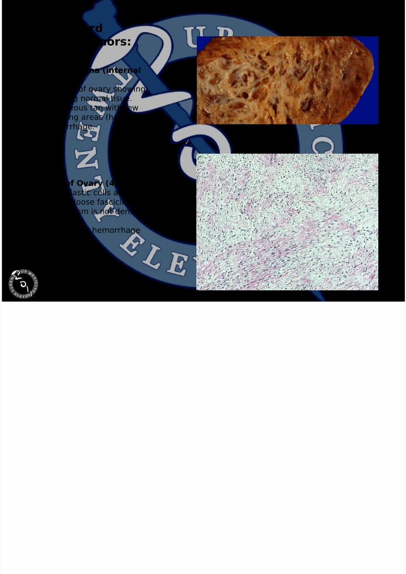
IV. Sex CordStromal Tumors:Fibroma
Ovarian Fibroma (internalview only)•Cut surface of ovary showingno remaining normal tisue.•Homogeneous tan with fewdegenerating areas (holes).•No hemorrhage.
Fibroma of Ovary (4X)• The neoplastic cells arearranged in loose fascicles.
• The neoplasm is not denselycellular.• No necrosis or hemorrhagecan be seen.

8/14/2019 OS 215 Patho Power Point Reviewer Part 2
http://slidepdf.com/reader/full/os-215-patho-power-point-reviewer-part-2 7/28
IV. Sex CordStromal Tumors:Fibrothecoma
Here are bilateral benignovarian tumors. These provedto be fibrothecomas. Thethecoma component of theneoplasm gives the tumor ayellowish cast because of thelipid content and can alsoproduce estrogen. These aretumors that arise from theovarian stroma. They arebilateral in only about 10% of cases. A right-sidedhydrothorax in association withthis tumor is known as Meig'ssyndrome.
Ovarian Fibrothecoma (cutsurface)•No recognizable normalovary.• Yellow tinge due tocholesterol in theca cells.
•Little hemorrhage or necrosis.

8/14/2019 OS 215 Patho Power Point Reviewer Part 2
http://slidepdf.com/reader/full/os-215-patho-power-point-reviewer-part-2 8/28
IV. Sex CordStromal Tumors:Sertoli-Leydig CellTumor
Sertoli-Leydig Cell Tumor,Poorly DifferentiatedSheets of tumor cells with amicrocystic pattern. Lowpower.
Sertoli-Leydig Cell Tumor, WellDifferentiatedWell-formed tubules lined bySertoli cells. High power.

8/14/2019 OS 215 Patho Power Point Reviewer Part 2
http://slidepdf.com/reader/full/os-215-patho-power-point-reviewer-part-2 9/28
V. MetastaticOvarian Tumors:Krukenberg Tumor
Metastatic tumors to ovary areuncommon, but there is one situationin which a metastaticadenocarcinoma to ovary appears asa large mass and resembles aprimary tumor: a so-called"Krukenberg" tumor of ovary whichhas a signet ring histologic patternand usually is metastatic from aprimary in gastrointestinal tract.Seen here extending out of the pelvisat autopsy is a large right ovarianmass. Metastases are also present inthe lower right portion of liver.
Krukenberg Tumor.Signet ring cells. High power.

8/14/2019 OS 215 Patho Power Point Reviewer Part 2
http://slidepdf.com/reader/full/os-215-patho-power-point-reviewer-part-2 10/28
Pathology of the Fallopian
Tube

8/14/2019 OS 215 Patho Power Point Reviewer Part 2
http://slidepdf.com/reader/full/os-215-patho-power-point-reviewer-part-2 11/28
I. Salpingitis: Acute Pyosalpinx
Pyosalpinx• The arrow points to theremnant of recognizable tubalepithelium.• The fallopian tubearchitecture is obscured by thedense acute inflammation.
Pyosalpinx (High Power)• This high power view showsmarked acute inflammation of the tubal epithelium.• The arrows point to epithelialcells.• Neutrophils infiltratebetween and into epithelialcells as well as the laminapropria.

8/14/2019 OS 215 Patho Power Point Reviewer Part 2
http://slidepdf.com/reader/full/os-215-patho-power-point-reviewer-part-2 12/28
I. Salpingitis: Acute Hydrosalpinx
Hydrosalpinx• The arrow points to thepreserved tubal epithelium.• Beneath the epithelium theabnormal lymphocytic infiltrateand increased fibrousconnective tissue is seen.
Hydrosalpinx• The blue arrow points tonormal ciliated epithelium.• The cells with clear spacesaround them in the epitheliumare lymphcytes.• Lymphocytes are alsopresent in the submucosa.

8/14/2019 OS 215 Patho Power Point Reviewer Part 2
http://slidepdf.com/reader/full/os-215-patho-power-point-reviewer-part-2 13/28
I. Salpingitis:Chronic
Lymphocytes and plasma cellsin the tubal wall. Tubal wallsare fused and there is luminalobstruction.

8/14/2019 OS 215 Patho Power Point Reviewer Part 2
http://slidepdf.com/reader/full/os-215-patho-power-point-reviewer-part-2 14/28
I. Salpingitis:Granulomatous
During the early stages manygranulomas are present. As thedisease progresses fibrosisdevelops and the number of granulomas decrease. Caseousabscess may develop.Calcification is common.Scarring and distortion of the
tubes lead to infertility.
8/14/2019 OS 215 Patho Power Point Reviewer Part 2
http://slidepdf.com/reader/full/os-215-patho-power-point-reviewer-part-2 15/28
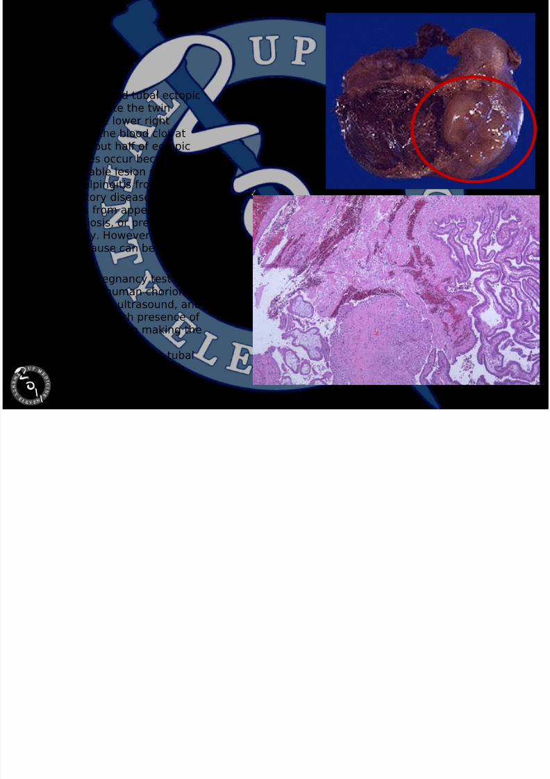
II. EctopicPregnancy
This is a ruptured tubal ectopicpregnancy. Note the twinfetuses at the lower rightadjacent to the blood clot atthe left. About half of ectopicpregnancies occur because of an identifiable lesion such aschronic salpingitis from pelvic
inflammatory disease oradhesions from appendicitis,endometriosis, or previouslaparotomy. However, in half of cases no cause can be found.
A positive pregnancy test
(presence of human chorionicgonadotropin), ultrasound, andculdocentesis with presence of blood are helpful in making thediagnosis of ectopicpregnancy. Seen here is tubalepithelium at the right, with
rupture site and chorionic villiat the lower left.

8/14/2019 OS 215 Patho Power Point Reviewer Part 2
http://slidepdf.com/reader/full/os-215-patho-power-point-reviewer-part-2 16/28
III. Cysts andTumors: BenignNeoplasms Endometriosis
Take note of the ectopicendometrial type glands withinthe fallopian tubes. The foldedstructures at the right is thetubal mucosa surrounding thelumen.
Low Power
High Power

8/14/2019 OS 215 Patho Power Point Reviewer Part 2
http://slidepdf.com/reader/full/os-215-patho-power-point-reviewer-part-2 17/28
III. Cysts and Tumors:Malignant Neoplasms(PrimaryAdenocarcinoma)
Adenocarcinoma of thefallopian tube associated withuterine leiomyomata.
Endometrioid Adenocarcinomaof the Fallopian Tube.
This tumor resembles itsendometrial counterpart. Lowpower.

8/14/2019 OS 215 Patho Power Point Reviewer Part 2
http://slidepdf.com/reader/full/os-215-patho-power-point-reviewer-part-2 18/28
III. Cysts andTumors: BenignNeoplasms Leiomyoma
Spindle-shaped smooth musclecells.
Low Power
Did you know?Leiomyomata is the plural form of leiomyoma

8/14/2019 OS 215 Patho Power Point Reviewer Part 2
http://slidepdf.com/reader/full/os-215-patho-power-point-reviewer-part-2 19/28
.Tumors: MalignantNeoplasms(MetastaticTumors)
Carcinosarcoma (MalignantMullerian Mixed Tumor).
This tumor is characterized byan intimate admixture of carcinomatous andsarcomatous components.Note the markedly atypicalglandular and stromal
components. Low power.

8/14/2019 OS 215 Patho Power Point Reviewer Part 2
http://slidepdf.com/reader/full/os-215-patho-power-point-reviewer-part-2 20/28
Sources:
• http://www.uchsc.edu/pathology/gyn-atlas/atlasft.htm
• http://library.med.utah.edu/WebPath/FEMH
TML/FEMIDX.html• http://pathweb.uchc.edu/
• http://library.med.utah.edu
• www.gfmer.ch/

8/14/2019 OS 215 Patho Power Point Reviewer Part 2
http://slidepdf.com/reader/full/os-215-patho-power-point-reviewer-part-2 21/28
Gross Pathology
Specimens

8/14/2019 OS 215 Patho Power Point Reviewer Part 2
http://slidepdf.com/reader/full/os-215-patho-power-point-reviewer-part-2 22/28
I. Adenomyosis
The uterus is abnormally thick. Why? There are discolorations (“blood lakes”,
indicated by black arrows) indicating thepresence of endometrial stroma andglands.Adenomyosis has a different pathogenesiscompared to endometriosis.
Endometrial Glands still follow normalmenstrual cycle: when patientmenstruates bleeding inside
formation of blood lakes.
When conducting surgery, different sets of instruments for the different layers toprevent implantation in other sites.
Treatment options:
1.) Remove the uterus2.) Stop menstruation (BCP)
Before treating using Birth Control Pills(BCP):
•History of Cancer: Estrogen andProgesterone specific
•Get a sample of the endometrium(atypia): might hasten the development
(The blood lakes are less appreciated because of
discoloration due to treatment with formalin.)

8/14/2019 OS 215 Patho Power Point Reviewer Part 2
http://slidepdf.com/reader/full/os-215-patho-power-point-reviewer-part-2 23/28
II. SquamousCervical CA LC Non-keratinizing
• Fungating mass in thecervical area
• Most important Factor: TumorStaging
• ↑size: ↑ mortality/morbidity
• Stage 3&4: chemo/radio
• Stage 1&2: Radical Surgery
• If prognosis is poor minimalsurgery just to alleviate thesymptoms
• PTs die of renal failure: massobstruct the posterior wall obstruct the ureters uremia
8/14/2019 OS 215 Patho Power Point Reviewer Part 2
http://slidepdf.com/reader/full/os-215-patho-power-point-reviewer-part-2 24/28
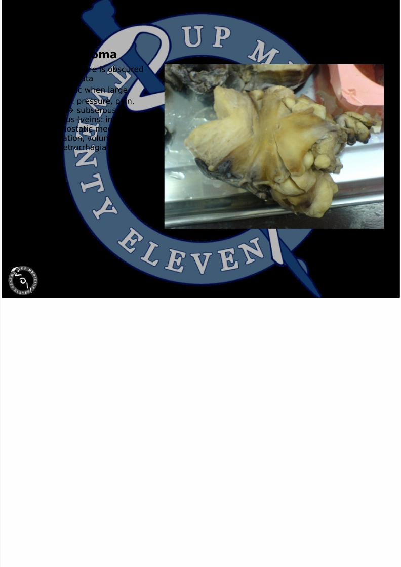
III. Leiomyoma
• Uterine structure is obscuredby leiomyomata
• Symptomatic when large
• Symptoms: pressure, pain,bleeding subserous,submucous (veins: interferewith hemostatic mechanism
↑ duration, volume(menometrorrhagia anemia)

8/14/2019 OS 215 Patho Power Point Reviewer Part 2
http://slidepdf.com/reader/full/os-215-patho-power-point-reviewer-part-2 25/28
IV. Endometrioticcyst
• ↑ Blood Lake (blood oceansaccording to sir) voluminous gets biggerand bigger Dark Chocolateappearance
• Some are as big as sportsballs
• Can be mistakenradiologically as neoplastic
• Solid: Malignant
• Cystic: Benign

8/14/2019 OS 215 Patho Power Point Reviewer Part 2
http://slidepdf.com/reader/full/os-215-patho-power-point-reviewer-part-2 26/28
V. Serous OvarianTumor
• Abalone-like• In-between tumor: LMP (Low
malignancy potential) borderline tumor at ovary
• Does not infiltrate
• Metastasizes but very slowcompared to malignant
tumors• Treated as a malignant tumor

8/14/2019 OS 215 Patho Power Point Reviewer Part 2
http://slidepdf.com/reader/full/os-215-patho-power-point-reviewer-part-2 27/28
VI. MalignantOvarian Tumor
• Firm consistency

8/14/2019 OS 215 Patho Power Point Reviewer Part 2
http://slidepdf.com/reader/full/os-215-patho-power-point-reviewer-part-2 28/28
VII. Teratoma
• Cystic benign• Contains hair, sebum(“pomada” according to sir),molars, yellow areas(fat/solidified oil: at normalbody temp. liquid but sincein room temp solidified lard)
• Immature tissue: malignant Immature teratoma
• Treatment: chemotherapyafter surgery


