48
Danika Lehmann BSc MSc BN RN March 19, 2011
| Date post: | 09-Mar-2016 |
| Category: |
Documents |
| Upload: | champion-equine-sciences |
| View: | 218 times |
| Download: | 1 times |

Danika Lehmann BSc MSc BN RN
March 19, 2011

Joint anatomy & function
Pathophysiology of osteoarthritis
Treatment & management of osteoathritis
Specific examples

Articular cartilage Subchondral bone Synovial membrane Synovial fluid Joint capsule Ligaments
Intrasynovial Extrasynovial

Connective tissue forming glassy-smooth covering on surface of synovial joint
Only a few millimeters thick
Load transmission & friction reduction
Consists of cells suspended in matrix
Devoid vascular, lymphatic, & neural connections

Chondrocytes (cells) Production & maintenance of cartilage Synthesis of collagens, proteoglycans, and other
macromolecules
Extracellular matrix (gelatinous fluid) Products of chondrocytes Water

Zonal organization from surface to depth:
Articular Cartilage and Osteoarthritis (1992).

Negatively charged matrix = swelling pressure
Resistance to compression during load
Tissue deformation & expulsion of water lead to greater resistance to further deformation
Equilibrium: external loading force = internal swelling pressure
Buckwalter Articular Cartilage & Knee Joint Function 1990 Maroudas J Orthop Res 1987

Connective tissue forming rigid skeletal frame
Cortical (compact)
Cancellous (spongy or trabecular)
Trabeculae aligned to resists stress/strain
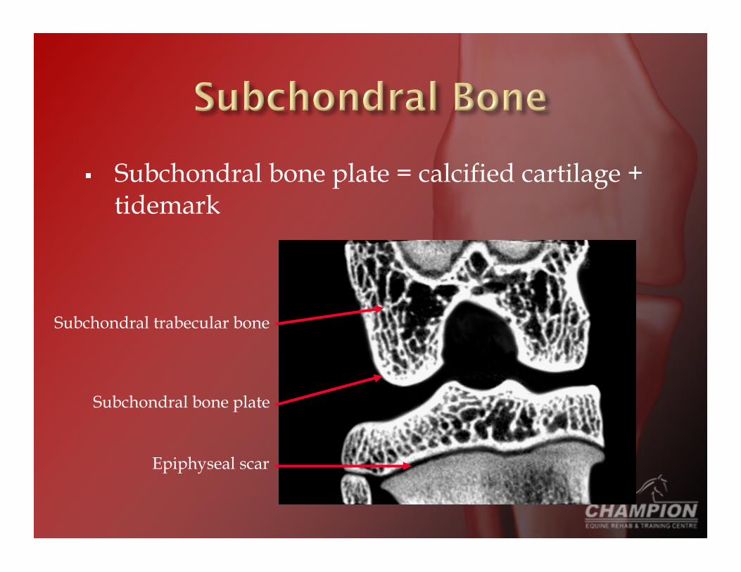
Subchondral bone plate = calcified cartilage + tidemark
Subchondral bone plate
Subchondral trabecular bone
Epiphyseal scar
Cells: Osteoblasts – producers Osteocytes – communicators Osteoclasts – destructors Periosteal cells – vascular/nerve supply
90-95% collagen fibers
Ground substance

Major mineral reservoir of calcium & phosphate
Hydroxyapatite = crystalline salt for structural rigidity
Magnesium, sodium, potassium, carbonate
Vigorita Orthopaedic Pathology 1999 Guyton Textbook of Medical Physiology 1996
Collagen fibers tensile strength
Crystalline salts compressional strength
Strength correlated with: Trabecular architecture Bone mineral density Crystal number Crystal size Crystal distribution
Landis Bone 1995 Buckwalter Instr Course Lect 1996

Concept of bone plasticity
Bone deposition = resorption
Physiological importance: 1. Bone adjusts strength in proportion to stress 2. Rearrangement of shape for proper support 3. Brittle, weak bone replaced with new
Rubin Rheum Dis Clin North Am 1988

“Synovium”
1 - 4 cells thick lining joint capsule
Produces synovial fluid to lubricate joint & reduce friction
Rheumatic disease responsible for deterioration of articular cartilage, subchondral bone, and synovium
Destruction and failure of synovial joints
Sangha J Rheumatol Suppl 2 2000

“…a group of overlapping diseases, which may have different aetiologies but with similar biologic, morphologic, and clinical outcomes. The disease process not only affects the articular cartilage, but involves the entire joint, including the subchondral bone, ligaments, capsule, synovial membrane, and periarticular muslces.”
American Academy of Orthopaedic Surgeons 1994


Aetiology largely unknown
Theories: 1. Biomechanical
Joint fracture Ligament damage Joint laxity Injury/trauma Obesity
2. Biochemical Water Proteoglycan type, size, aggregation
Brandt J Rheumatol 1986
Stages:
1. Disruption of cartilage matrix
2. Response to tissue damage
3. Failure of repair process
Mankin N Engl J Med 1974 Cohen J Orthop Sports Phys Ther 1998

Stage 1: Disruption of cartilage matrix Water retention Decrease cartilage resilience & stiffness Fibrillation (“cut velvet”)
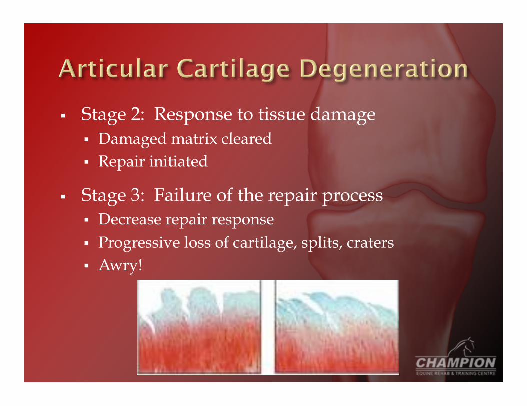
Stage 2: Response to tissue damage Damaged matrix cleared Repair initiated
Stage 3: Failure of the repair process Decrease repair response Progressive loss of cartilage, splits, craters Awry!

Full thickness cartilage loss 12wks post-trauma (a) femur, (b) tibia, (c) trochlea.
MRI of stifle joint showing progressive cartilage loss post-trauma.

Failure of cartilage repair process ??
Damage or death of chondrocytes from mechanical stress & lack of protection by functional matrix?
Synthesis & accumulation of matrix molecules that inhibit repair process?
Buckwalter Articular Cartilage & Knee Joint Function 1990

Cartilage changes cannot be dissociated from subchondral bone changes
Highly debated whether bone changes precede, are concurrent with, or succeed articular cartilage changes
Dequeker Microsc Res Tech 1997 Radin Clin Orthop 1986

Hypermetabolic state: Increased bone mineral density (sclerosis) Formation of microcysts Osteophytes
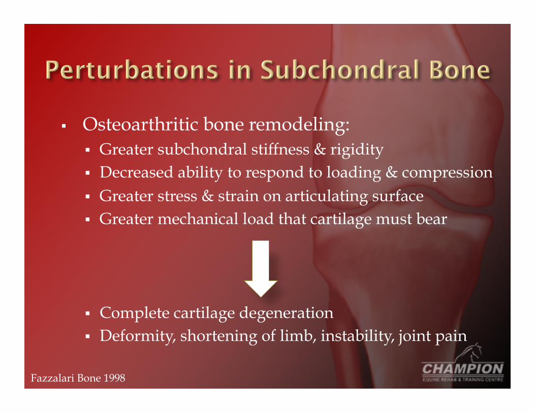
Osteoarthritic bone remodeling: Greater subchondral stiffness & rigidity Decreased ability to respond to loading & compression Greater stress & strain on articulating surface Greater mechanical load that cartilage must bear
Complete cartilage degeneration Deformity, shortening of limb, instability, joint pain
Fazzalari Bone 1998

Clinical history
X-rays
Arthroscopy
Joint pain & tenderness
Crepitus, limitation in movement
Occasional effusion (excess fluid) with variable degrees of inflammation
No systemic effects
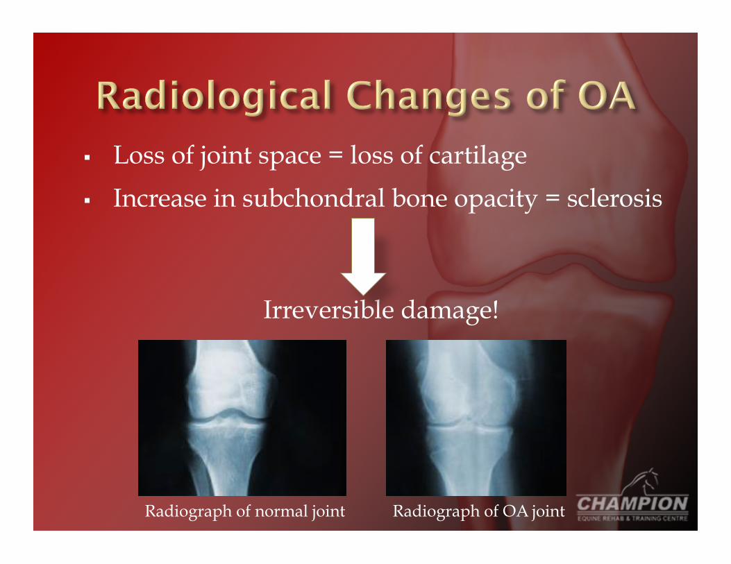
Loss of joint space = loss of cartilage
Increase in subchondral bone opacity = sclerosis
Irreversible damage!
Radiograph of normal joint Radiograph of OA joint

Joint space
Sclerosis & loss of joint space
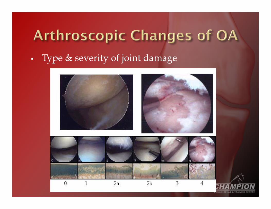
Type & severity of joint damage
Many drugs to ease pain, discomfort, swelling, & manage inflammation: Steroids NSAIDS DMSO Glucosamine Hyaluronate PSGAGs
* These drugs DO NOT stop or reverse the osteoarthritic process *
“Corticosteroids” or “glucosteroids” E.g., dexamethasone, methylprednisolone,
tramcinolone acetate Often via intra-articular injection Manage pain & inflammation
Side Effects: Laminitis Further damage by stopping/delaying anabolic
process?
Labens Vet Rec 2007

“NSAIDs” E.g., phenylbutazone, naproxen, flunixin
meglumine, diclofenac Various routes of administration Manage pain & inflammation
Side effects: Stomach ulcers, bleeding, slowing of healing process

“DMSO” Topical drug to control inflammation & swelling Organic solvent has ability to carry other drugs
through skin
Side effects: Cataracts in dogs…long-term use in horses? Organic solvent - wear rubber or latex gloves
Precursor in building connective tissue E.g., glucosamine sulfate or hydrochloride,
Cosequin (glucosamine + chondroitin) Oral supplement; sulfate easiest to absorb &
essential for connective tissue Efficacy?
Side effects: ?
Pearson Equine Vet J 2009

E.g., Legend® Intravenous or intra-articular injection Manage inflammation & increase quality of
synovial fluid
Side effects: IV – depression, lethargy, fever IA – lameness, joint effusion, joint or injection site
swelling, joint pain, severe inflammatory reaction, joint sepsis
Frisbie Am J Vet Res 2009, Palmieri Acta Biomed 2010, Mouzopoulos Minerva Med 2011, Kuemmerle Vet Comp Orthop Traumatol 2006

“PSGAGs” or “GAGs” E.g., Adequan® Often multiple intramuscular injections Manage inflammation, synovial fluid effusion,
synovial membrane vascularity/fibrosis
Side effects: ?
Frisbie Am J Vet Res 2009, Gaustad Equine Vet J 1995, Todhunter Vet Surg 1993

Exercise Maintains or improves strength of peri-articular
structures (muscles, ligaments, tendons) Decreases bone loss resulting from inactivity Controls joint swelling, stiffness & pain Improves joint ROM Replenishes synovial fluid Enhances energy & stamina Decreases anxiety Improves mood & well-being Promotes state of relaxation
Minor Rheum Dis Clin North Am 1999
Goals of an exercise program:
Preserve or restore ROM & flexibility around joint
Increase muscle strength & endurance
Increase aerobic conditioning to improve health, mood & decrease health risks associated with sedentary lifestyle
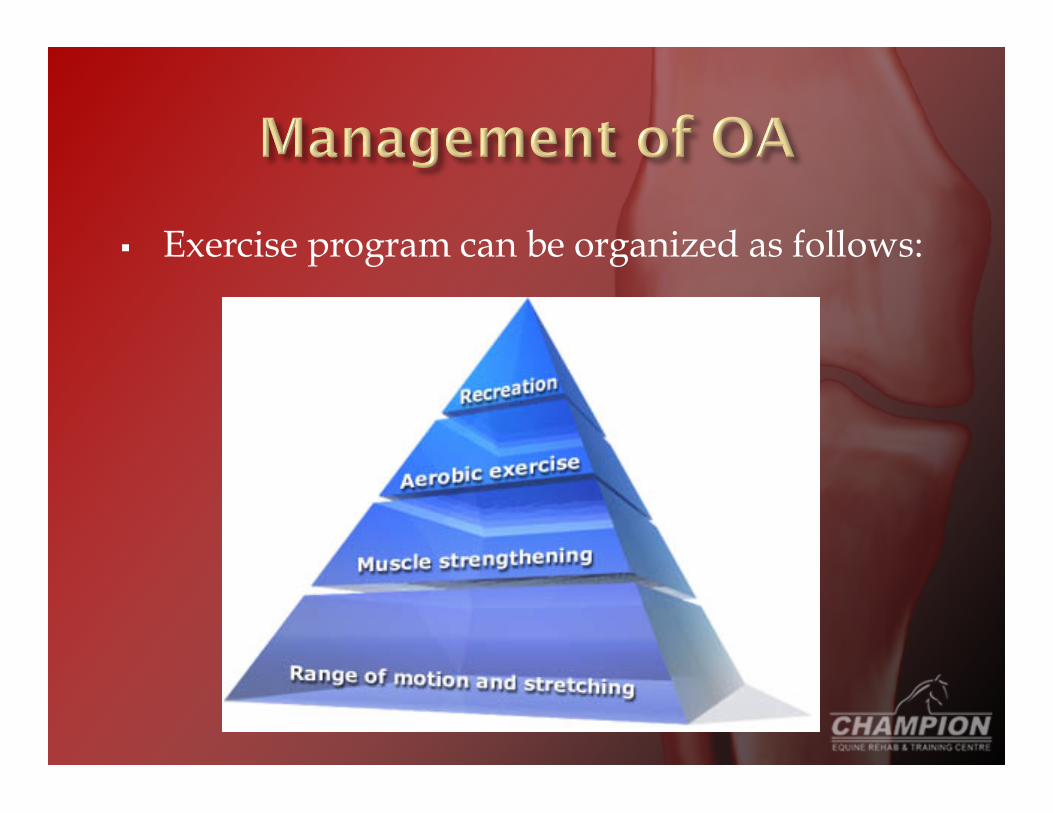
Exercise program can be organized as follows:
Serous athritis
Septic arthritis
Bone spavin
Osselets
Omarthritis
“Acute synovitis” Swollen, tender, fluid-filled joint Joint stress or injury Does not necessarily progress to OA
Tx: Rest & application of cold or hot/cold therapy Veterinary recommended medications Corrective shoeing

“Infectious arthritis” Bacterial infection of the joint Often causes irreversible damage, progressing
to OA
Tx: Immediate veterinary attention Broad-spectrum antibiotics, joint aspiration & C&S

“Jack spavin”, osteoarthritis of the hock joint Caused by hard use or poor conformation Pain concentrated to inside of hock May begin as a cold lameness
Tx: Discontinue stressful activity Medication to manage pain & inflammation Corrective shoeing Surgery (in advanced cases)

Osteoarthritis of the fetlock Chronic stress injury from repeated concussion “Green” = acute synovitis “True” = osteoarthritis
Tx: Green – same as acute synovitis True – symptomatic relief
Osteoarthritis of the shoulder (usually 2° to #) Shoulder joint lameness, swelling, tenderness Nerve block, x-rays, arthroscopy helpful for Dx
Tx: Veterinary attention Surgery & medications


