55
OSTEOGENESIS IMPERFECTA & THE EFFICACY/ SAFETY OF INTRAVENOUS ZOLEDRONIC ACID TREATMENT OF PEDIATRIC OSTEOPOROSIS Prof. Abdulmoein Al-Agha, FRCPCH (UK) [email protected]

OSTEOGENESIS IMPERFECTA & THE EFFICACY/
SAFETY OF INTRAVENOUS ZOLEDRONIC ACID
TREATMENT OF PEDIATRIC OSTEOPOROSIS
Prof. Abdulmoein Al-Agha, FRCPCH (UK) [email protected]

OBJECTIVES
Basics of Bone physiology
What happens in osteoporosis!!
Different causes of Pediatric Osteoporosis
Presentation ,diagnosis & management of patients with OI
Importance of making the right diagnosis
Local published Data on usage & Safety of Zoledronic acid therapy in cases of Pediatric Osteoporosis over 13- year follow up

MECHANISM OF BONE FORMATION
AND BREAKDOWN
Key words
Osteoblasts : cells that synthesize bone matrix
Osteoid : Unmineralized bone matrix
Osteoclasts: cells that resorb bone.
Modeling : formation of bone
Remodeling: breakdown and renewal of bone.
Calcidiol : 25 (OH)2 D
Calcitriol : 1, 25 (OH)2 D

COMPONENTS OF BONE Calcified matrix (90%)
composed of collagen fibers (type-1),
Glycosaaminoglycan containing spindle shaped crystals of hydroxyapatite
Mineral Element
Crystals of Calcium and Phosphate are arranged either amorphously or as Hydroxyapatite Ca10 (PO4)6 (OH)2 on or within the collagen fibers.
Na, Zn, Mg, Cu and fluoride
Non Collagenous Components (Proteins)
Osteocalcin: protein produced by the Osteoblasts
α2 HS- glycoprotein: produced by the liver and absorbed by the bone matrix
Amino Acids: about one fourth of amino acids present in collagen are proline and hydroxproline.


Peak bone mass: accrued during adolescence

BONE TURNOVER CYCLE – HORMONAL BALANCE
ENABLES APPROPRIATE ACTIVITY OF
OSTEOBLASTS
VS OSTEOCLASTS
Bone Resorption
Bone Formation
GH IGF-1 DHEA Androgens
Estrogen PTH Cortisol

OSTEOPOROSIS
preventable disease no cure new interest in childhood and adolescence as critical years for bone acquisition

NORMAL BONE
Female, age 30 years

Osteoporosis
Female, age 88 years

DEFINITION OF “OSTEOPOROSIS” IN
CHILDREN
No WHO definitions in children and teens
Concern for low bone mass
• BMD Z-score by DXA < -2.0 SD
• Slightly low if Z-score between -1.0 and -2.0
“Diagnosis of osteoporosis in children and adolescents should NOT be made on the basis of BMD alone.”
Int’l Soc Clinical Densitometry 2007

OSTEOPOROSIS:
CLINICAL MANIFESTATIONS
Early osteoporosis is asymptomatic
As skeletal integrity declines, fractures occur,
often with minimal trauma
Vertebral compression fractures are most
common, hip and wrist fractures also are major
problems
End stage disease associated with marked
dorsal kyphosis

Source Undetermined

DETERMINANTS OF BONE MASS
Extrinsic
Diet
Body mass/habitus
Hormonal milieu
Illnesses
Exercise
Lifestyle choices
Intrinsic
Gender
Family History
Ethnicity
Genetic factors

GENETIC FACTORS
Striking patterns within families
Candidate genes:
•Vitamin D receptor
•Estrogen receptor
• IGF-I receptor
•TGF-
•Alleles involved in collagen synthesis :COL -1

AT-RISK CHILDREN AND ADOLESCENTS
*Obesity
*Poor diet/little sun exposure
Anorexia nervosa/chronic amenorrhea/delayed puberty
Turner syndrome
Growth hormone deficiency
Medications: glucocorticoids, anticonvulsants, depot medroxyprogesterone, GnRH agonists
Gastrointestinal disease (IBD)
Cerebral palsy/neuromuscular diseases
Rheumatologic diseases: SLE, JRA, dermatomyositis
Cystic fibrosis
Celiac disease
Renal failure
Diabetes mellitus
Hemoglobinopathies (sickle cell, thalassemia) + hemophilia
Immobilized patients
HIV
Hyperprolactinemia

OSTEOGENESIS IMPERFECTA
Manifest itself with 1 or more of the following findings:
Blue sclerae
Triangular facies
Macrocephaly
Hearing loss
Defective dentition
Barrel chest
Scoliosis
Limb deformities
Fractures
Joint laxity
Growth retardation
Constipation and sweating

OSTEOGENESIS IMPERFECTA
Pathologic changes seen in all tissues in which type 1 collagen is an important constituent (e.g., bone, ligament, dentin, and sclera)
Basic defect : qualitative or quantitative reduction in type 1 collagen
Mutations in genes encoding type 1 collagen affect the coding of 1 of the 2 genes
Mutations are either genetically inherited or new
Inherited mutations : recurrence risk in subsequent pregnancies of 50% if a parent is affected
New mutations unpredictable recurrence risk


OSTEOGENESIS IMPERFECTA
Incidence : 1 case for every 20,000 live births
Equally common in males and females
Described in every human population in which
skeletal dysplasias have been studied
No predilection for a particular race
Family history , but most cases due to new
mutations
Commonly present with fractures after minor
trauma

OSTEOGENESIS
IMPERFECTA
Clinical presentation depends on phenotype
Sillence classificatiom : 4 types on basis of clinical and radiologic features
Dentinogenesis imperfecta denoted as subtype B, whereas OI without dentinogenesis imperfecta is denoted as subtype A

TYPES OF OI
8 types described so far
OI types range from a mild form with no
deformity, normal stature and few fractures to a
form that is lethal during the perinatal period (prior
to and after birth).
Medical problems a person will depend on the
type of OI
OI varies greatly from person to person, even
among people with the same type of OI, even
within the same family

TYPE I
mildest and most common form
50% of the total OI population
mild bone fragility
relatively few fractures
minimal limb deformities
child might not fracture until he or she is learning to walk
Some children have few obvious signs of OI or fractures
while others experience multiple fractures of the long
bones, compression fractures of the vertebrae, and chronic
pain.
Appear healthy yet need to accommodate for bone
fragility


COMPLICATIONS
Repeated respiratory
infections
Basilar impression
caused by a large head,
which causes brainstem
compression
Cerebral hemorrhage
caused by birth trauma
High risk for
complications of
anesthesia

DIFFERENTIAL DIAGNOSES
Achondroplasia
Menkes Kinky Hair Disease
Hereditary Rickets
Thanatophoric Dysplasia
Jeune dystrophy
Camptomelic dysplasia
Chondrodysplasia punctata
Chondroectodermal dysplasia (Ellis–van Creveld syndrome)
Non- accidental injury
Hypophosphatasia


DIAGNOSTIC WORK-UP
Rule-out systemic
disease
Consider insidious
celiac disease
25-hydroxyvitamin D
PTH
Calcium, phosphorus,
magnesium
Other:
• Ceruloplasmin, copper, IGF-I,
DHEAS
Bone age
Urinary calcium/creatinine
(spot/24 h)
If amenorrhea: thyroid
function, FSH, prolactin

MEASUREMENT OF SKELETAL STATUS
Bone density
Dual energy x-ray
absorptiometry (DXA) – 2D
Quantitative ultrasound
(QUS)
Quantitative CT – 3D
(including pQCT)
High-resolution pQCT
(XtremeCT)
Peripheral vs. axial (central)
measurements
Bone quality
High-resolution MRI
Micro-CT (from biopsy
specimens)
Hip structural analysis
(bone geometry)
Fracture rates

DXA SCANNER – OPEN CONFIGURATION
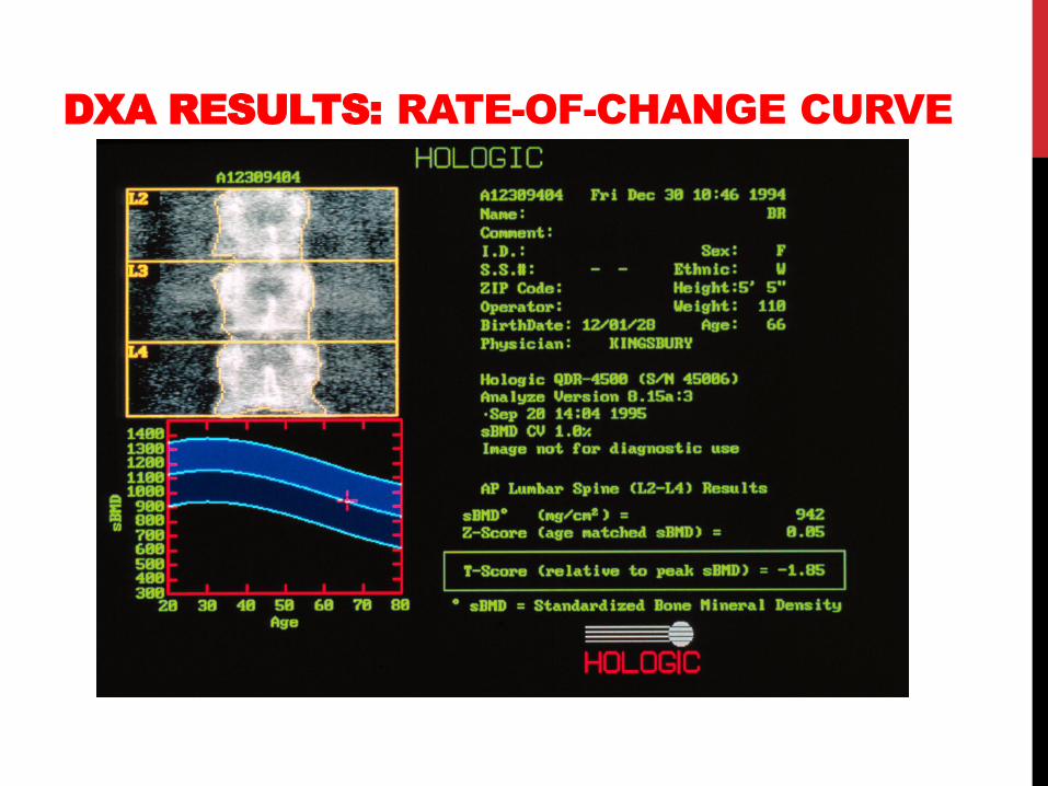
DXA RESULTS: RATE-OF-CHANGE CURVE

RADIAL AND TIBIAL MEASUREMENTS
Peripheral QCT
Quantitative Ultrasound
OSTEOPOROSIS TREATMENT WITH
ZOLEDRONIC ACID IN PEDIATRIC
POPULATION AT A UNIVERSITY
HOSPITAL IN WESTERN SAUDI
ARABIA: A 13-YEAR EXPERIENCE
ABDULMOEIN E. AL-AGHA, FRCPCH,
RAHAF S. HAYATALHAZMI, MBBS.
SAUDI MED J 2015; VOL. 36 (11): 1312-1318


Up to date, many studies have investigated bisphosphonate
treatment primarily with the use of Pamidronate in many
bone related diseases
As to the ZA treatment of pediatric osteoporosis, there are
no much published data on long-term use, safety and
efficacy
There were no Saudi local, Arabian, or even internationally
published data on a large study number of children
receiving ZA (when our study conducted)
In our study, we aimed to review a 13-year experience
with primary and secondary causes of osteoporosis, as
well as the efficacy, and safety of intravenous ZA as the
treatment of choice in our pediatric population at the
King Abdulaziz University Hospital (KAUH),Jeddah,
Kingdom of Saudi Arabia (KSA)

METHODS
A retrospective observational study Patients population:
131 patients aged 6 weeks to 18 years with primary and
secondary osteoporosis followed up at the Pediatric Endocrine
Outpatient Clinic at KAUH, Jeddah, KSA between January 2002
and January 2015.
Osteoporosis (131)
Primary (72) Secondary (59)
Hematological diseases 9.9%
Gastrointestinal diseases 9.9%
Endocrine diseases 7.6%
Renal diseases 6.1%
Immobilization 6.1%
Drug induced 4.5%

Data
Data were obtained from direct interview of
patients and/or their parents
All laboratory results were obtained from
the KAUH electronic Phoenix system
Informed verbal consent was acquired from
all patients and/ or their parents prior to the
start of therapy

Inclusion criteria:
- Patients with confirmed diagnosis of osteoporosis based on:
- clinical or biochemical high bone turnover markers of C-
terminal telopeptide [CTX], and osteocalcin levels
- and/ or a z-score ≤-2.0 SD on a bone densitometry DXA scan
were included in the study.
- Patients with a normal bone profile
- calcium, phosphate, and alkaline phosphatase (as well as
normal total vitamin D , parathyroid hormone levels before the
start of treatment
Exclusion criteria:
- mineral metabolism disturbances, major data insufficiency,
and a creatinine clearance rate < 30-35 mL/ min
METHODS

Treatment administration:
Intravenous ZA was used as the treatment of choice in both groups
throughout study
Was administered intravenously at a dose of 0.05 mg/kg maximum
dose to be given is 2 mg/infusion, in neonates and infants, the dose
was 0.025 mg/kg
The first 5 infusions were given once every 3 months, then once
every 6 months, depending on the clinical and biochemical marker
response
All patients were admitted to the general ward for 2 days to receive
their first infusion to enable close monitoring of the acute
complications that might occur
Subsequent ZA infusions were given during day care unit admissions
OVER 30-60 minutes duration
METHODS

Precautions for the treatment:
- Acute complications of the first dose were:
- fever, myalgia, hypocalcemia, flu-like symptoms, and bone pain
- To prevent hypocalcemia, all patients were given a continuous intravenous calcium infusion of 200-400 mg/kg/day
- ibuprofen 10 mg/kg was administered 3-4 times per day to minimize the fever and myalgia that were frequently observed in patients after the first infusion
- All patients were advised to maintain sufficient oral calcium intake consisting of a daily dose of 1200 mg together with a daily dose of prophylactic vitamin D (400-800 IU)
METHODS

• Serum calcium, vitamin D, phosphate, PTH, & alkaline phosphatase levels were measured before the start of treatment
• Levels of both bone markers serum osteocalcin and CTX were measured at baseline, before treatment, and then every 3-6 months throughout the treatment period
• Creatinine level was calculated at baseline, and at each treatment visit before the ZA infusion
LABORATORY ASSESSMENTS

- Data from our institute’s system were obtained only for
57/131 (31.6%) patients who had their BMD measured
before treatment, and 18/57 (13.7%) underwent the
measurements after starting their treatment course for
comparison
- In all patients, total body and lumbar spine BMD
measurement Z scores were adjusted for age, gender,
puberty, and body size as appropriate. Low BMD was
defined as a BMD z-score ≤ -2.0 SD
- Z score of -1.0 to -2.0 SD was defined as osteopenia
- Eventually, due to lack of sufficient follow-up DXA
measurement data, BMD measurements were excluded
from the subsequent statistical analysis
RADIOLOGICAL ASSESSMENT


The fracture rate / year was calculated by dividing
the number of fractured bones prior to the start of
treatment by the number of years from the first
fracture to the first dose
For those in which treatment started before one
year of age, fracture rate was calculated by
dividing the number of fractures by the duration in
months, and then multiplying it by 12
Quality of life (QOL) was also considered and
defined as normal, or below normal compared with
that in children their age and according to post-
treatment improvement
FRACTURE ASSESSMENT


Acute & chronic adverse events were observed and monitored throughout the study
Acute side effects were reported after the first ZA infusion:
fever, hypocalcemia, decreased intake, bone pain, myalgia, and flu-like symptoms
Nephrocalcinosis was assessed by renal ultrasound at baseline before the start of treatment, and then annually
The renal profile at baseline and after each infusion cycle was checked for any complications
ADVERSE EVENTS

RESULTS

RESULTS
Fractures and bone deformity:
- A bone deformity was present in 43/72 (59.7%)
patients before the start of treatment
- Fractures were the initial presentation in 53/72
(73.6%) patients, which represents more than 2-
thirds of the group’s subjects
- The mean number of fractures before treatment
was 4.86 ± 8.10, which significantly decreased
after treatment to 1.47 ± 5.10 ( p=0.000)

Quality of life (QOL)
- 40/72 (55.6%) patients were assessed as
having below normal QOL prior to treatment
- After treatment with ZA infusion, 38/40
(95%) patients reported improved QOL and
2/40 (5%) patients reported no change
- A 2-tailed paired-sample t-test revealed a
significant subjective improvement in QOL
(t(52) = 6.385, p=0.001) with a confidence
interval (CI) of 95%


• Acute-phase reaction, including fever, hypocalcemia, flu-like symptoms, decreased intake, and bone pain usually occurs in most children with the initiation of intravenous or oral agents.
• patients with primary osteoporosis:
- Using a paired t-test, we found a statistically significant improvement in pain frequency after ZA treatment (t (17) = 4.994, p=0.000, 95% CI).
• patients with secondary osteoporosis:
- Using the same paired t-test, evidence proved a statistically significant improvement in pain frequency post-treatment in the secondary group (t(18) = 4.53, p=0.000, 95% CI)
CLINICAL SAFETY

• Another acute adverse event of ZA infusion was the decrease
in calcium level
• patients with primary osteoporosis:
- Mean pre-treatment calcium level in group one was 2.296 ±
0.18 and post- treatment calcium level was 2.149 ± 0.129
• patients with secondary osteoporosis:
- mean pre-treatment calcium level was 2.22 ± 0.17 and mean
post-treatment calcium level was 2.01 ± 0.25
• This decrease in calcium level was observed during the first
ZA infusion, while no chronic events were reported throughout
our 13-year experience with ZA
CLINICAL SAFETY

To summarizes our 13-year experience using ZA therapy in a
pediatric population with osteoporosis at KAUH, Jeddah, KSA
This study is considered the first reported long-term
observational clinical trial of a Middle-Eastern pediatric
population.
The main goals of pharmacological therapy in osteoporosis,
including decreasing the fracture rate, decreasing bone pain,
increasing mobility, increasing independence, and decreasing bone
turnover marker levels were achieved, the results of this study
prove that cyclic intravenous ZA is an efficient treatment for children
and adolescents with osteoporosis.
In our patient cohort, clinical symptoms improved dramatically after
the start of ZA treatment. Fractures and bone pain were the 2
dominant presenting symptoms in our population. We had an
encouraging result regarding pain relief and a reduction in fractures
after ZA treatment
SUMMARY


