Osteogenic potential of a chalcone in a critical-size defect in rat calvaria bone Xana Raquel Ortolan a , Bruna Proiss Fenner a , Telmo José Mezadri b , David Rivero Tames c , Rogério Corrêa d , Fátima de Campos Buzzi d, * a Postgraduate Program in Pharmaceutical Sciences, Laboratory of Histology, University of Vale do Itajaí e UNIVALI, Itajaí, Santa Catarina, Brazil b Department of Cell and Tissue Biology, Laboratory of Histology, University of Vale do Itajaí (UNIVALI), Itajaí, Santa Catarina, Brazil c Department of Sciences/Histology, Laboratory of Histology, University of Vale do Itajaí (UNIVALI), Itajaí, Santa Catarina, Brazil d Department of Chemistry, Chemical-Pharmaceutical Investigations Center (NIQFAR)/CCS, University of Vale do Itajaí (UNIVALI), Itajaí, Santa Catarina, Brazil article info Article history: Paper received 17 December 2012 Accepted 31 July 2013 Keywords: Chalcone Critical-size Osteogenesis abstract Background: This study describes the bone formation stimulated by the application of a type of chalcone to critical-size defects in rat calvarial bone. Material and methods: Sixty female Wistar rats were divided into 6 groups of 10 animals per group: control (no treatment), vehicle (vaseline) and the chalcone (1-phenyl-3-(4-chlorophenyl)-2-propen-1- one) suspended in vaseline at 10%. A critical-size defect of 5 mm was prepared using a trephine in the calvarial bone, after which the treatment was applied, in a single dose, according to the experimental group. The samples were evaluated macroscopically using ImageJ software, and histologically 30 and 45 days after surgery. Results: At 30 days after surgery, there was significant bone formation (p < 0.05) in the groups treated with chalcone, compared with the other groups. Many active osteoblasts were observed adjacent to the borders of the newly formed bone tissue. 45 days after surgery in the chalcone group, the surgical defects showed complete bone closure. Conclusion: The results of this study suggest that chalcone has significant potential to induce the for- mation of new bone. Ó 2013 European Association for Cranio-Maxillo-Facial Surgery. Published by Elsevier Ltd. All rights reserved. 1. Introduction Diseases such as osteoporosis, traumatic injuries, orthopaedic surgery, and resection of primary tumours can result in bone de- fects, which become critical defect wounds when there is no spontaneous repair, and which require replacement materials to fill the injured tissue (Mauney et al., 2005). Some widely-used techniques used to treat musculoskeletal diseases are autologous, allogeneic or xenotransplants. However, these methods require rigorous control of the transmission of in- fectious agents (Laurencin and El-Amin, 2008; Heneghan and McCabe, 2009; Putzier et al., 2009; Bayat et al., 2010; Lee et al., 2012; Zwitser et al., 2012). Tissue engineering has developed the use of extracellular matrices, as support for organ function, allowing cell proliferation, migration and differentiation in the process of bone regeneration (Elsalanty and Genecov, 2009; Chan et al., 2009; Tuzlakoglu and Reis, 2009; Mieszawska and Kaplan, 2010; Lee et al., 2012; Stockmann et al., 2012); and also the development of an intelli- gent matrix, i.e. without the addition of molecules from the TGF- beta family, which can initiate the cascade of cellular differentia- tion to produce new bone (Srouji et al., 2005; Ripamonti, 2010). Many investigations of active principles derived from medicinal plants have been reported in relation to the repair of non- mineralized tissue (Sasidharan et al., 2010; Mezadri et al., 2012; Manoj and Murugan, 2012). However, little is known about their use in the repair of bone tissue. Chalcones, molecules that are found abundantly in several plant species, have shown intense pharmaceutical activity, including antinociceptive (Campos-Buzzi et al., 2006), antifungals (Konduru * Corresponding author. Núcleo de Investigações Químico-Farmacêuticas (NIQ- FAR)/CCS, Universidade do Vale do Itajaí (UNIVALI), Caixa Postal 360, CEP 88302- 202, Itajaí, Santa Catarina, Brazil. Tel.: þ55 47 3341 7855; fax: þ55 47 3341 7601. E-mail address: [email protected](F. de Campos Buzzi). Contents lists available at ScienceDirect Journal of Cranio-Maxillo-Facial Surgery journal homepage: www.jcmfs.com 1010-5182/$ e see front matter Ó 2013 European Association for Cranio-Maxillo-Facial Surgery. Published by Elsevier Ltd. All rights reserved. http://dx.doi.org/10.1016/j.jcms.2013.07.020 Journal of Cranio-Maxillo-Facial Surgery xxx (2013) 1e5 Please cite this article in press as: Ortolan XR, et al., Osteogenic potential of a chalcone in a critical-size defect in rat calvaria bone, Journal of Cranio-Maxillo-Facial Surgery (2013), http://dx.doi.org/10.1016/j.jcms.2013.07.020
Transcript
lable at ScienceDirect
Journal of Cranio-Maxillo-Facial Surgery xxx (2013) 1e5
Contents lists avai
Journal of Cranio-Maxillo-Facial Surgery
journal homepage: www.jcmfs.com
Osteogenic potential of a chalcone in a critical-size defect in ratcalvaria bone
Xana Raquel Ortolan a, Bruna Proiss Fenner a, Telmo José Mezadri b, David Rivero Tames c,Rogério Corrêa d, Fátima de Campos Buzzi d,*a Postgraduate Program in Pharmaceutical Sciences, Laboratory of Histology, University of Vale do Itajaí e UNIVALI, Itajaí, Santa Catarina, BrazilbDepartment of Cell and Tissue Biology, Laboratory of Histology, University of Vale do Itajaí (UNIVALI), Itajaí, Santa Catarina, BrazilcDepartment of Sciences/Histology, Laboratory of Histology, University of Vale do Itajaí (UNIVALI), Itajaí, Santa Catarina, BrazildDepartment of Chemistry, Chemical-Pharmaceutical Investigations Center (NIQFAR)/CCS, University of Vale do Itajaí (UNIVALI), Itajaí,Santa Catarina, Brazil
a r t i c l e i n f o
Article history:Paper received 17 December 2012Accepted 31 July 2013
Keywords:ChalconeCritical-sizeOsteogenesis
* Corresponding author. Núcleo de Investigações QFAR)/CCS, Universidade do Vale do Itajaí (UNIVALI), C202, Itajaí, Santa Catarina, Brazil. Tel.: þ55 47 3341 78
1010-5182/$ e see front matter � 2013 European Asshttp://dx.doi.org/10.1016/j.jcms.2013.07.020
Please cite this article in press as: Ortolan XCranio-Maxillo-Facial Surgery (2013), http:/
a b s t r a c t
Background: This study describes the bone formation stimulated by the application of a type of chalconeto critical-size defects in rat calvarial bone.Material and methods: Sixty female Wistar rats were divided into 6 groups of 10 animals per group:control (no treatment), vehicle (vaseline) and the chalcone (1-phenyl-3-(4-chlorophenyl)-2-propen-1-one) suspended in vaseline at 10%. A critical-size defect of 5 mm was prepared using a trephine in thecalvarial bone, after which the treatment was applied, in a single dose, according to the experimentalgroup. The samples were evaluated macroscopically using ImageJ software, and histologically 30 and 45days after surgery.Results: At 30 days after surgery, there was significant bone formation (p < 0.05) in the groups treatedwith chalcone, compared with the other groups. Many active osteoblasts were observed adjacent to theborders of the newly formed bone tissue. 45 days after surgery in the chalcone group, the surgical defectsshowed complete bone closure.Conclusion: The results of this study suggest that chalcone has significant potential to induce the for-mation of new bone.
� 2013 European Association for Cranio-Maxillo-Facial Surgery. Published by Elsevier Ltd. All rightsreserved.
1. Introduction
Diseases such as osteoporosis, traumatic injuries, orthopaedicsurgery, and resection of primary tumours can result in bone de-fects, which become critical defect wounds when there is nospontaneous repair, and which require replacement materials to fillthe injured tissue (Mauney et al., 2005).
Some widely-used techniques used to treat musculoskeletaldiseases are autologous, allogeneic or xenotransplants. However,these methods require rigorous control of the transmission of in-fectious agents (Laurencin and El-Amin, 2008; Heneghan andMcCabe, 2009; Putzier et al., 2009; Bayat et al., 2010; Lee et al.,2012; Zwitser et al., 2012).
R, et al., Osteogenic potentia/dx.doi.org/10.1016/j.jcms.201
Tissue engineering has developed the use of extracellularmatrices, as support for organ function, allowing cell proliferation,migration and differentiation in the process of bone regeneration(Elsalanty and Genecov, 2009; Chan et al., 2009; Tuzlakoglu andReis, 2009; Mieszawska and Kaplan, 2010; Lee et al., 2012;Stockmann et al., 2012); and also the development of an intelli-gent matrix, i.e. without the addition of molecules from the TGF-beta family, which can initiate the cascade of cellular differentia-tion to produce new bone (Srouji et al., 2005; Ripamonti, 2010).
Many investigations of active principles derived from medicinalplants have been reported in relation to the repair of non-mineralized tissue (Sasidharan et al., 2010; Mezadri et al., 2012;Manoj and Murugan, 2012). However, little is known about theiruse in the repair of bone tissue.
Chalcones, molecules that are found abundantly in several plantspecies, have shown intense pharmaceutical activity, includingantinociceptive (Campos-Buzzi et al., 2006), antifungals (Konduru
Surgery. Published by Elsevier Ltd. All rights reserved.
l of a chalcone in a critical-size defect in rat calvaria bone, Journal of3.07.020
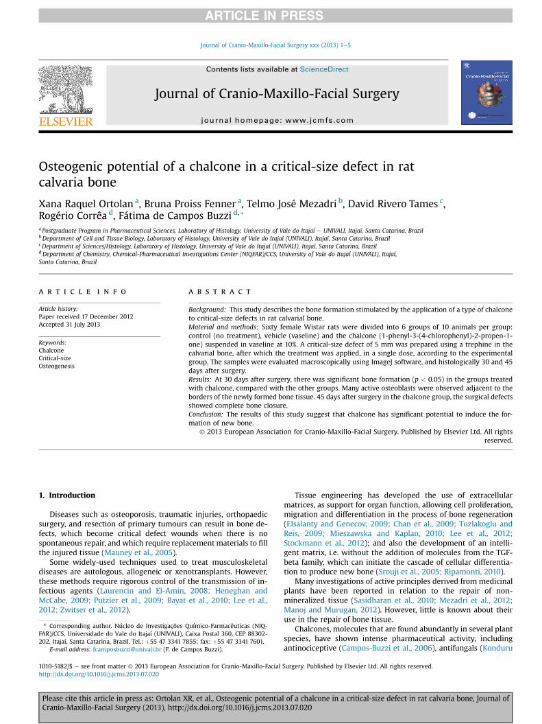
Fig. 1. Chemical structure of synthetic chalcone 1-phenyl-3-(4-chlorophenyl)-2-propen-1-one.
X.R. Ortolan et al. / Journal of Cranio-Maxillo-Facial Surgery xxx (2013) 1e52
et al., 2013), antimicrobial (Nowakowska et al., 2008) and anti-inflammatory effects (Tran et al., 2009).
Studies have reported synthetic chalcones exhibiting promisingproperties in the inhibition of inflammatory enzymes and have alsoshown inhibitory action on the activation of mast cells, neutrophils,and macrophages, important mediators of inflammation, inter-fering in the formation of bone tissue (Oliveira et al., 2010).
Recently, Tames et al. (2010), based on reports of Corrêa et al.(2008), Batovska and Todorova (2010), Han et al. (2010); Vogelet al. (2010) describing the biological and pharmacological prop-erties of chalcones, have suggested inducing bone repair using thechalcone 1-phenyl-3-(4-chlorophenyl)-2-propen-1-one in acritical-size defect model in rats.
Carpeggiani et al. (2010) also found potential to stimulate repairof the pulpedentin complex in a model to study pulp capping in ratmolars, using the same molecule.
Continuing the studies of Tames et al. (2010), this study aims toevaluate the osteogenic potential of the synthetic chalcone 1-phenyl-3-(4-chlorophenyl)-2-propen-1-one at 30 and 45 days af-ter treatment.
2. Material and methods
The chalcone 1-phenyl-3-(4-chlorophenyl)-2-propen-1-one(Fig. 1) was synthesized by an adaptation of the general methodof ClaiseneSchmidt condensation (Corrêa et al., 2001; Campos-Buzzi et al., 2007). The method uses an equimolar mixture of 4-chlorobenzaldehyde and acetophenone dissolved in water andethanol, in the presence of 10% sodium hydroxide. The solutionwasstirred mechanically for 24 h. In order to confirm the end of thereaction, formation of the product was analyzed by thin layerchromatography. The product was percolated and washed severaltimes with cold distilled water, until the wash waters presented aneutral pH value. The isolated product was dried under reducedpressure in the presence of phosphorus pentoxide. All the solidsobtained were purified by recrystallization with ethanol 95% andcharacterized by their melting point using a Microquimica APF-300apparatus (Microquímica, Florianopolis, SC, Brazil). Microanalysis
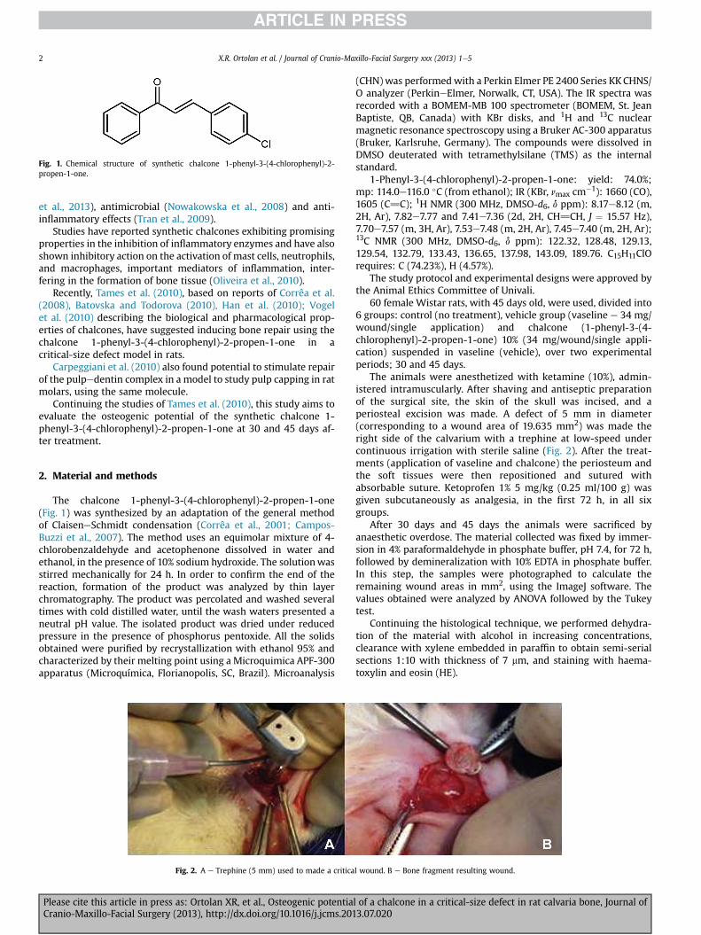
Fig. 2. A e Trephine (5 mm) used to made a critica
Please cite this article in press as: Ortolan XR, et al., Osteogenic potentiaCranio-Maxillo-Facial Surgery (2013), http://dx.doi.org/10.1016/j.jcms.201
(CHN) was performedwith a Perkin Elmer PE 2400 Series KKCHNS/O analyzer (PerkineElmer, Norwalk, CT, USA). The IR spectra wasrecorded with a BOMEM-MB 100 spectrometer (BOMEM, St. JeanBaptiste, QB, Canada) with KBr disks, and 1H and 13C nuclearmagnetic resonance spectroscopy using a Bruker AC-300 apparatus(Bruker, Karlsruhe, Germany). The compounds were dissolved inDMSO deuterated with tetramethylsilane (TMS) as the internalstandard.
The study protocol and experimental designs were approved bythe Animal Ethics Committee of Univali.
60 female Wistar rats, with 45 days old, were used, divided into6 groups: control (no treatment), vehicle group (vaseline e 34 mg/wound/single application) and chalcone (1-phenyl-3-(4-chlorophenyl)-2-propen-1-one) 10% (34 mg/wound/single appli-cation) suspended in vaseline (vehicle), over two experimentalperiods; 30 and 45 days.
The animals were anesthetized with ketamine (10%), admin-istered intramuscularly. After shaving and antiseptic preparationof the surgical site, the skin of the skull was incised, and aperiosteal excision was made. A defect of 5 mm in diameter(corresponding to a wound area of 19.635 mm2) was made theright side of the calvarium with a trephine at low-speed undercontinuous irrigation with sterile saline (Fig. 2). After the treat-ments (application of vaseline and chalcone) the periosteum andthe soft tissues were then repositioned and sutured withabsorbable suture. Ketoprofen 1% 5 mg/kg (0.25 ml/100 g) wasgiven subcutaneously as analgesia, in the first 72 h, in all sixgroups.
After 30 days and 45 days the animals were sacrificed byanaesthetic overdose. The material collected was fixed by immer-sion in 4% paraformaldehyde in phosphate buffer, pH 7.4, for 72 h,followed by demineralization with 10% EDTA in phosphate buffer.In this step, the samples were photographed to calculate theremaining wound areas in mm2, using the ImageJ software. Thevalues obtained were analyzed by ANOVA followed by the Tukeytest.
Continuing the histological technique, we performed dehydra-tion of the material with alcohol in increasing concentrations,clearance with xylene embedded in paraffin to obtain semi-serialsections 1:10 with thickness of 7 mm, and staining with haema-toxylin and eosin (HE).
l wound. B e Bone fragment resulting wound.
l of a chalcone in a critical-size defect in rat calvaria bone, Journal of3.07.020
Table 1Reduction in wound area (mm2) at 30 and 45 post-treatment.
Average wound areas
Control Vehicle Chalcone
30 days 18.26 � 1.08 17.92 � 1.17 7.82 � 3,2445 days 16.27 � 2.43 14.58 � 1.39 0.0
Shows the means � standard deviation of the wound areas in mm2. Notice that themean values for the critical wounds in the control and vehicle groups are signifi-cantly higher than those of the experimental groups (at 30 and 45 days).
X.R. Ortolan et al. / Journal of Cranio-Maxillo-Facial Surgery xxx (2013) 1e5 3
3. Results
3.1. Quantitative analysis
Thirty days after surgery, themean remainingwound area in thecontrol group was 18.26 mm2 and 17.92 mm2 in the vehicle group.The animals treated with the chalcone molecule showed a meanremainingwound area of 7.82mm2 (Table 1 and Fig. 3), a significantresult compared to the control and vehicle groups (p < 0.05).
The groups sacrificed at 45 days after treatment showed a meanremaining wound area of 16.27 mm2 in the control group and14.58 mm2 in the vehicle group, representing a significant differ-ence comparedwith the experimental group (p< 0.05) (Table 1 andFig. 4).
3.2. Qualitative analysis
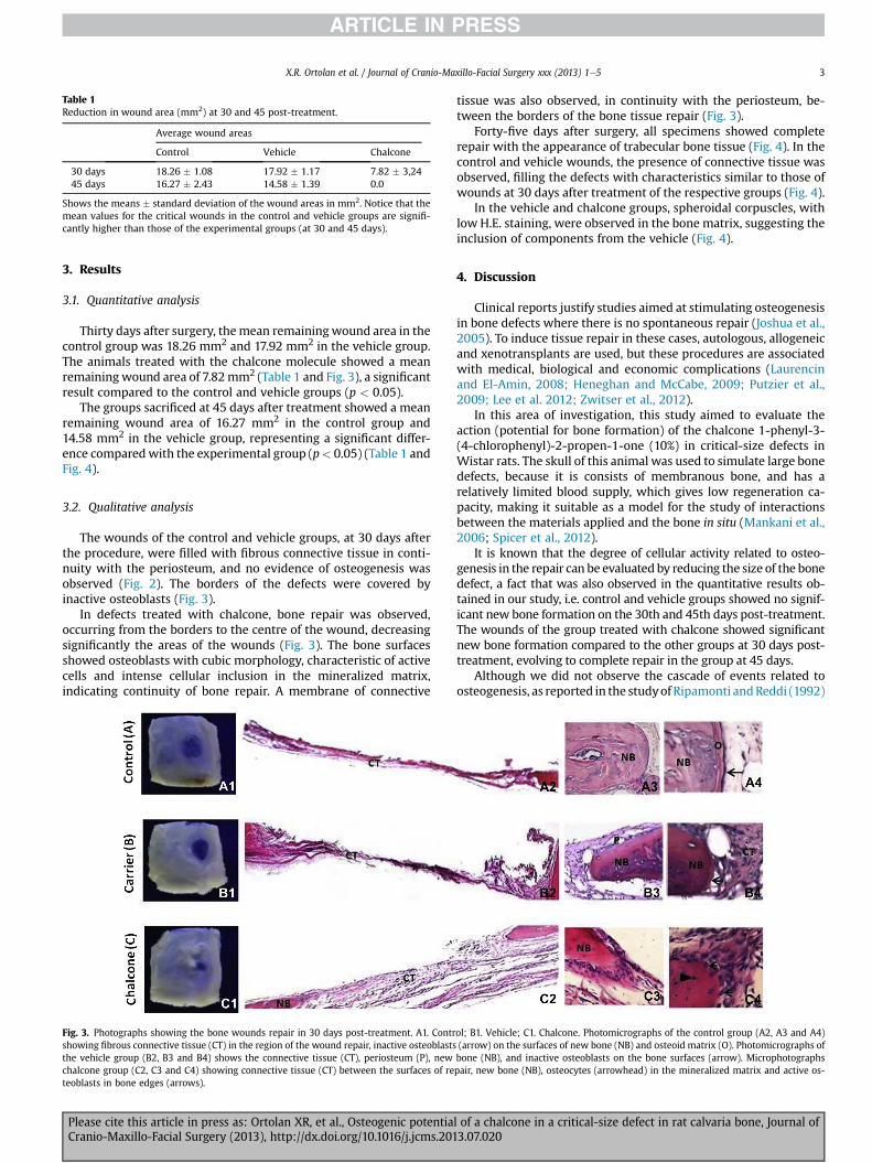
The wounds of the control and vehicle groups, at 30 days afterthe procedure, were filled with fibrous connective tissue in conti-nuity with the periosteum, and no evidence of osteogenesis wasobserved (Fig. 2). The borders of the defects were covered byinactive osteoblasts (Fig. 3).
In defects treated with chalcone, bone repair was observed,occurring from the borders to the centre of the wound, decreasingsignificantly the areas of the wounds (Fig. 3). The bone surfacesshowed osteoblasts with cubic morphology, characteristic of activecells and intense cellular inclusion in the mineralized matrix,indicating continuity of bone repair. A membrane of connective
Fig. 3. Photographs showing the bone wounds repair in 30 days post-treatment. A1. Contrshowing fibrous connective tissue (CT) in the region of the wound repair, inactive osteoblaststhe vehicle group (B2, B3 and B4) shows the connective tissue (CT), periosteum (P), newchalcone group (C2, C3 and C4) showing connective tissue (CT) between the surfaces of reteoblasts in bone edges (arrows).
Please cite this article in press as: Ortolan XR, et al., Osteogenic potentiaCranio-Maxillo-Facial Surgery (2013), http://dx.doi.org/10.1016/j.jcms.201
tissue was also observed, in continuity with the periosteum, be-tween the borders of the bone tissue repair (Fig. 3).
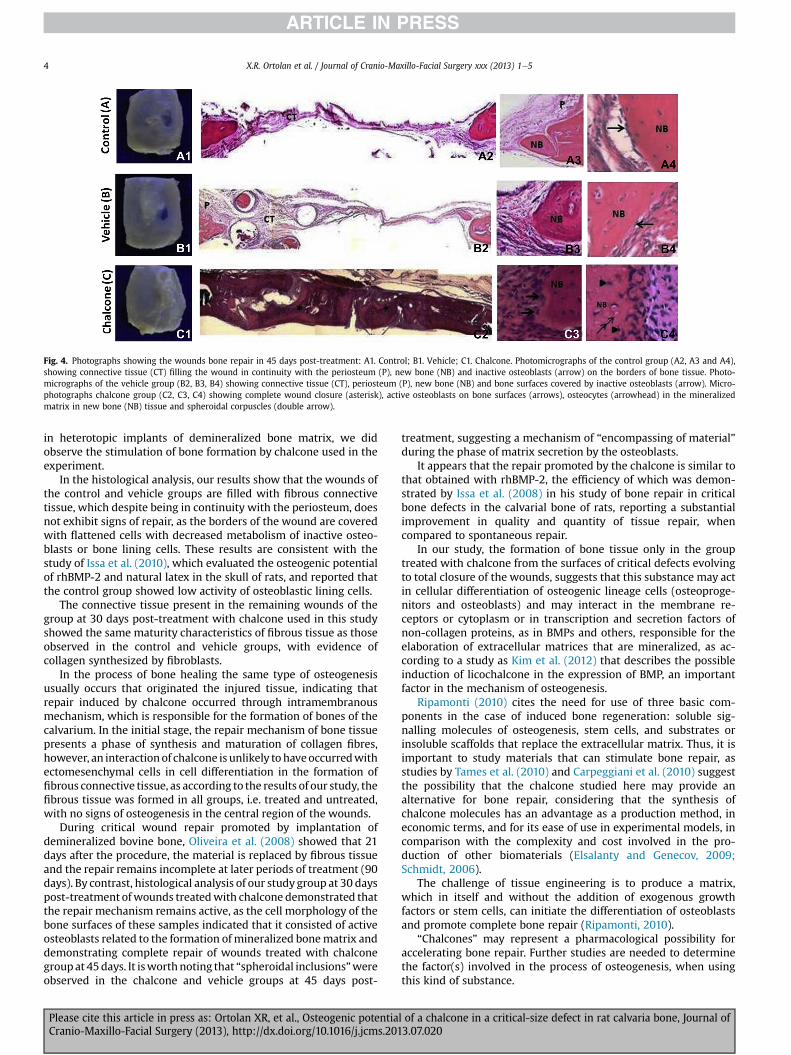
Forty-five days after surgery, all specimens showed completerepair with the appearance of trabecular bone tissue (Fig. 4). In thecontrol and vehicle wounds, the presence of connective tissue wasobserved, filling the defects with characteristics similar to those ofwounds at 30 days after treatment of the respective groups (Fig. 4).
In the vehicle and chalcone groups, spheroidal corpuscles, withlow H.E. staining, were observed in the bone matrix, suggesting theinclusion of components from the vehicle (Fig. 4).
4. Discussion
Clinical reports justify studies aimed at stimulating osteogenesisin bone defects where there is no spontaneous repair (Joshua et al.,2005). To induce tissue repair in these cases, autologous, allogeneicand xenotransplants are used, but these procedures are associatedwith medical, biological and economic complications (Laurencinand El-Amin, 2008; Heneghan and McCabe, 2009; Putzier et al.,2009; Lee et al. 2012; Zwitser et al., 2012).
In this area of investigation, this study aimed to evaluate theaction (potential for bone formation) of the chalcone 1-phenyl-3-(4-chlorophenyl)-2-propen-1-one (10%) in critical-size defects inWistar rats. The skull of this animal was used to simulate large bonedefects, because it is consists of membranous bone, and has arelatively limited blood supply, which gives low regeneration ca-pacity, making it suitable as a model for the study of interactionsbetween the materials applied and the bone in situ (Mankani et al.,2006; Spicer et al., 2012).
It is known that the degree of cellular activity related to osteo-genesis in the repair can be evaluated by reducing the size of the bonedefect, a fact that was also observed in the quantitative results ob-tained in our study, i.e. control and vehicle groups showed no signif-icant new bone formation on the 30th and 45th days post-treatment.The wounds of the group treated with chalcone showed significantnew bone formation compared to the other groups at 30 days post-treatment, evolving to complete repair in the group at 45 days.
Although we did not observe the cascade of events related toosteogenesis, as reported in the studyofRipamonti andReddi (1992)
ol; B1. Vehicle; C1. Chalcone. Photomicrographs of the control group (A2, A3 and A4)(arrow) on the surfaces of new bone (NB) and osteoid matrix (O). Photomicrographs ofbone (NB), and inactive osteoblasts on the bone surfaces (arrow). Microphotographspair, new bone (NB), osteocytes (arrowhead) in the mineralized matrix and active os-
l of a chalcone in a critical-size defect in rat calvaria bone, Journal of3.07.020
Fig. 4. Photographs showing the wounds bone repair in 45 days post-treatment: A1. Control; B1. Vehicle; C1. Chalcone. Photomicrographs of the control group (A2, A3 and A4),showing connective tissue (CT) filling the wound in continuity with the periosteum (P), new bone (NB) and inactive osteoblasts (arrow) on the borders of bone tissue. Photo-micrographs of the vehicle group (B2, B3, B4) showing connective tissue (CT), periosteum (P), new bone (NB) and bone surfaces covered by inactive osteoblasts (arrow). Micro-photographs chalcone group (C2, C3, C4) showing complete wound closure (asterisk), active osteoblasts on bone surfaces (arrows), osteocytes (arrowhead) in the mineralizedmatrix in new bone (NB) tissue and spheroidal corpuscles (double arrow).
X.R. Ortolan et al. / Journal of Cranio-Maxillo-Facial Surgery xxx (2013) 1e54
in heterotopic implants of demineralized bone matrix, we didobserve the stimulation of bone formation by chalcone used in theexperiment.
In the histological analysis, our results show that the wounds ofthe control and vehicle groups are filled with fibrous connectivetissue, which despite being in continuity with the periosteum, doesnot exhibit signs of repair, as the borders of the wound are coveredwith flattened cells with decreased metabolism of inactive osteo-blasts or bone lining cells. These results are consistent with thestudy of Issa et al. (2010), which evaluated the osteogenic potentialof rhBMP-2 and natural latex in the skull of rats, and reported thatthe control group showed low activity of osteoblastic lining cells.
The connective tissue present in the remaining wounds of thegroup at 30 days post-treatment with chalcone used in this studyshowed the same maturity characteristics of fibrous tissue as thoseobserved in the control and vehicle groups, with evidence ofcollagen synthesized by fibroblasts.
In the process of bone healing the same type of osteogenesisusually occurs that originated the injured tissue, indicating thatrepair induced by chalcone occurred through intramembranousmechanism, which is responsible for the formation of bones of thecalvarium. In the initial stage, the repair mechanism of bone tissuepresents a phase of synthesis and maturation of collagen fibres,however, an interactionof chalcone is unlikely tohaveoccurredwithectomesenchymal cells in cell differentiation in the formation offibrous connective tissue, as according to the results of our study, thefibrous tissue was formed in all groups, i.e. treated and untreated,with no signs of osteogenesis in the central region of the wounds.
During critical wound repair promoted by implantation ofdemineralized bovine bone, Oliveira et al. (2008) showed that 21days after the procedure, the material is replaced by fibrous tissueand the repair remains incomplete at later periods of treatment (90days). By contrast, histological analysis of our study group at 30 dayspost-treatment ofwounds treatedwith chalcone demonstrated thatthe repair mechanism remains active, as the cell morphology of thebone surfaces of these samples indicated that it consisted of activeosteoblasts related to the formation of mineralized bonematrix anddemonstrating complete repair of wounds treated with chalconegroupat 45days. It isworthnoting that “spheroidal inclusions”wereobserved in the chalcone and vehicle groups at 45 days post-
Please cite this article in press as: Ortolan XR, et al., Osteogenic potentiaCranio-Maxillo-Facial Surgery (2013), http://dx.doi.org/10.1016/j.jcms.201
treatment, suggesting a mechanism of “encompassing of material”during the phase of matrix secretion by the osteoblasts.
It appears that the repair promoted by the chalcone is similar tothat obtained with rhBMP-2, the efficiency of which was demon-strated by Issa et al. (2008) in his study of bone repair in criticalbone defects in the calvarial bone of rats, reporting a substantialimprovement in quality and quantity of tissue repair, whencompared to spontaneous repair.
In our study, the formation of bone tissue only in the grouptreated with chalcone from the surfaces of critical defects evolvingto total closure of the wounds, suggests that this substance may actin cellular differentiation of osteogenic lineage cells (osteoproge-nitors and osteoblasts) and may interact in the membrane re-ceptors or cytoplasm or in transcription and secretion factors ofnon-collagen proteins, as in BMPs and others, responsible for theelaboration of extracellular matrices that are mineralized, as ac-cording to a study as Kim et al. (2012) that describes the possibleinduction of licochalcone in the expression of BMP, an importantfactor in the mechanism of osteogenesis.
Ripamonti (2010) cites the need for use of three basic com-ponents in the case of induced bone regeneration: soluble sig-nalling molecules of osteogenesis, stem cells, and substrates orinsoluble scaffolds that replace the extracellular matrix. Thus, it isimportant to study materials that can stimulate bone repair, asstudies by Tames et al. (2010) and Carpeggiani et al. (2010) suggestthe possibility that the chalcone studied here may provide analternative for bone repair, considering that the synthesis ofchalcone molecules has an advantage as a production method, ineconomic terms, and for its ease of use in experimental models, incomparison with the complexity and cost involved in the pro-duction of other biomaterials (Elsalanty and Genecov, 2009;Schmidt, 2006).
The challenge of tissue engineering is to produce a matrix,which in itself and without the addition of exogenous growthfactors or stem cells, can initiate the differentiation of osteoblastsand promote complete bone repair (Ripamonti, 2010).
“Chalcones” may represent a pharmacological possibility foraccelerating bone repair. Further studies are needed to determinethe factor(s) involved in the process of osteogenesis, when usingthis kind of substance.
l of a chalcone in a critical-size defect in rat calvaria bone, Journal of3.07.020
X.R. Ortolan et al. / Journal of Cranio-Maxillo-Facial Surgery xxx (2013) 1e5 5
5. Conclusions
The results of this study indicate that the synthetic chalcone 1-phenyl-3-(4-chlorophenyl)-2-propen-1-one shows stimulating ac-tion on bone tissue formation, from the surfaces of the criticalwound; promoting complete repair in the group of animals at 45days post-treatment.
References
Batovska DI, Todorova IT: Trends in utilization of the pharmacological potential ofchalcones. Curr Clin Pharmacol 5: 1e29, 2010
Bayat M, Momen-Heravi F, Marjani M, Motahhary P: A comparison of bonereconstruction following application of bone matrix gelatin and autogenousbone grafts to alveolar defects: an animal study. J Craniomaxillofac Surg 38:288e292, 2010
Campos-Buzzi F, Campos JP, Tonini PP, Corrêa R, Yunes RA, Boeck P, et al: Anti-nociceptive effects of synthetic chalcones obtained from xanthoxyline. Archivder Pharmazie 339: 361e365, 2006
Carpeggiani MHLAF, Corrêa R, Tames DR, Mezadri TJ, Campos Buzzi F, Ortolan XR:Avaliação do potencial indutor de uma chalcona sintética na formação dedentina terciária de reparo em polpas de molares de ratos. Braz Oral Res 24:162e183, 2010
Chan WD, Perinpanayagam H, Goldberg HA, Hunter GK, Dixon SJ, Santos Jr GC, et al:Tissue engineering scaffolds for the regeneration of craniofacial bone. J CanDent Assoc 75: 373e377, 2009
Corrêa R, Campos-Buzzi F, Cechinel Filho V, Nunes RJ: Antinociceptive activity andpreliminary structure activity relationship of chalcone-like compounds. Zeits-chrift für Naturforschung C 63: 830e836, 2008
Corrêa R, Pereira MA, Buffon D, dos Santos L, Cechinel Filho V, Santos AR, et al:Antinociceptive properties of chalcones. Structure activity relationships. ArchPharm 10: 332e334, 2001
Elsalanty ME, Genecov DG: Bone grafts in craniofacial surgery. CraniomaxillofacTrauma Reconstruct 2: 125e134, 2009
Han H, Zhao Y, Hao Y, Cuthbertson T, Hartman RF, Rose SD: Cell cycle arrest andapoptosis induction by an anticancer chalcone epoxide. Arch Pharm 343: 429e439, 2010
Heneghan HM, McCabe JP: Use of autologous bone graft in anterior cervicaldecompression: morbidity & quality of life analysis. BMC Musculoskelet Disord10: 158e165, 2009
Issa JP, Defino HL, Netto JC, Volpon JB, Regalo SC, Iyomasa MM, et al: Evaluation ofrhbmp-2 and natural latex as potential osteogenic proteins in critical size de-fects by histomorphometric methods. Anat Rec 293(5): 794e801, 2010
Issa JP, do Nascimento C, Iyomasa MM, Siéssere S, Regalo SC, Defino HL, et al: Bonehealing process in critical-sized defects by rhbmp-2 using poloxamer gel andcollagen sponge as carriers. Micron 39(1): 17e24, 2008
Joshua RM, Volloch V, Kaplan DL: Role of adult mesenchymal stem cells in bonetissue engineering applications. Current Status and Future Prospects. Tissue Eng11(5e6): 787e802, 2005
Kim SN, Bae SJ, Kwak HB, Min YK, Jung SH, Kim CH, et al: In vivo and in vivoosteogenic activity of licochalcone A. Amino Acids 42: 1455e1465, 2012
Konduru NK, Dey S, Sajid M, Owais M, Ahmed N: Synthesis and antibacterial andantifungal evaluation of some chalcone based sulfones and bisulfones. Eur JMed Chem 59: 23e30, 2013
Laurencin CT, El-Amin SF: Xenotransplantation in orthopaedic surgery. J Am AcadOrthop Surg 16(1): 4e8, 2008
Please cite this article in press as: Ortolan XR, et al., Osteogenic potentiaCranio-Maxillo-Facial Surgery (2013), http://dx.doi.org/10.1016/j.jcms.201
Lee JS, Park WY, Cha JK, Jung UW, Kim CS, Lee YK, et al: Periodontal tissue reactionto customized nano-hydroxyapatite block scaffold in one-wall intrabonydefect: a histologic study in dogs. J Periodontal Implant Sci 42(2): 50e58,2012
Mankani MH, Kuznetsov SA, Wolfe RM, Marshall GW, Robey PG: In vivo bone for-mation by human bone marrow stromal cells: reconstruction of the mousecalvarium and mandible. Stem Cells 24(9): 2140e2149, 2006
Manoj GS, Murugan K: Wound healing activity of methanolic and aqueous extractsof Plagiochila beddomei Steph. thallus in rat model. Indian J Exp Biol 50(8): 551e558, 2012
Mauney JR, Volloch V, Kaplan DL: Role of adult mesenchymal stem cells in bonetissue engineering applications: current status and future prospects. Tissue Eng11(5e6): 787e802, 2005
Mezadri TJ, Tames DR, Reis RKT, Ortolan XR:Morphophysiological evaluation of a kindof propolis in tissue repair in diabetic rats. Lat Am J Pharm 31: 777e781, 2012
Nowakowska Z, Kedzia B, Schroeder G: Synthesis physicochemical properties andantimicrobial evaluation of new (E) chalcones. Eur J Med Chem 43(4): 707e713,2008
Oliveira PS, Da Silva NVP, Vasconcelos A, Stefanello FM: Avaliação da ação anti-oxidante de derivados sintéticos das chalconas. XIX CIC, XII ENPOS, 2010
OliveiraRC, Oliveira FH, Cestari TM, Taga R, Granjeiro JM:Morphometric evaluation ofthe repair of critical-size defects using demineralized bovine bone and autoge-nous bone grafts in rat calvaria. Clin Oral Implants Res 19: 749e754, 2008
Putzier M, Strube P, Funk JF, Gross F, Mönig H, Perka C, et al: Allogenic versusautologous cancellous bone in lumbar segmental spondylodesis: a randomizedprospective study. Eur Spine J 18: 687e695, 2009
Ripamonti U, Reddi AH: Growth and morphogenetic factors in bone induction: roleof osteogenin and related bone morphogenetic proteins in craniofacial andperiodontal bone repair. Crit Rev Oral Biol Med 3(1e2): 1e14, 1992
Ripamonti U: Soluble and insoluble signals sculpt osteogenesis in angiogenesis.World J Biol Chem 26: 109e132, 2010
Sasidharan S, Nilawatyi R, Xavier R, Latha LY, Amala R: Wound healing potential ofElaeis guineensis Jacq leaves in an infected albino rat model. Molecules 30:3186e3199, 2010
Schmidt C: Futuro da odontologia impregnado com BMP. Innov Implant J BiomatEsthet 1(2): 19e24, 2006
Spicer PP, Kretlow JD, Young S, Jansen JA, Kasper K, Mikos AG: Evaluation of boneregeneration using the rat critical size calvaria defect. Nat Protoc 7: 1918e1929,2012
Srouji S, Rachmiel A, Blumenfeld I, Livne E: Mandibular defect repair by TGF-b andIGF-1 released from a biodegradable osteoconductive hydrogel.J Craniomaxillofac Surg 33: 79e84, 2005
Stockmann P, Park J, von Wilmowsky C, Nkenke E, Felszeghy E, Dehner JF, et al:Guided bone regeneration in pig calvarial bone defects using autologousmesenchymal stem/progenitor cells e a comparison of different tissue sources.J Craniomaxillo Fac Surg 40(4): 310e320, 2012
Tames SFA, Nascimento L, Mezadri TJ, Corrêa R, Campos Buzzi F, Ortolan XR: Estudo(in vivo) do potencial osteogênico de uma chalcona em ferida crítica de calotacraniana de ratos. Braz Oral Res 24: 128e149, 2010
Tran TD, Park H, Kim HP, Ecker GF, Thai Km: Inhibitory activity of prostaglandin E2production by the synthetic 2-hydroxychalcone analogues. Synthesis and SARstudy. Bioorg Med Chem Lett 29: 1650e1653, 2009
Tuzlakoglu K, Reis RL: Biodegradable polymeric fiber structures in tissue engi-neering. Tissue Eng Part B Rev 15: 17e27, 2009
Vogel S, Barbic M, Jurgenliemk G, Heilmann J: Synthesis, cytotoxicity, anti-oxidative and anti-inflammatory activity of chalcones and influence of A-ring modifications on the pharmacological effect. Eur J Med Chem 45: 2206e2213, 2010
Zwitser EW, Jiya TU, Licher HG, van Royen BJ: Design and management of an or-thopaedic bone bank in the Netherlands. Cell Tissue Bank 13(1): 63e69, 2012
l of a chalcone in a critical-size defect in rat calvaria bone, Journal of3.07.020