74
Osteology – Anatomical Orientations, Planes, Directions

Osteology – Anatomical Orientations, Planes, Directions
Osteology – Anatomical Orientations, Planes, Directions
• In order to be able to describe orientation of skeleton and dental structures and their component parts in 3 dimensions, uniformity of description is necessary

Osteology – Anatomical Orientations, Planes, Directions
Reference Positions:
• 1. Standard Anatomical Position of
Skeleton – for skeleton as a whole
• 2. Frankfort Horizontal (FH) - for skulls

Osteology – Orientations:Standard Anatomical Position
• Standing
• Feet together pointing forward
• Palms forward (no bones crossed), arms at
sides
• Looking forward
• No matter what position a bone or skeleton is
found in, surfaces are referred to as if individual
is standing erect in standard anatomical position

Standard Anatomical Position
-standing-feet together-palms forward-looking forward

From Gosling et al
Standard Anatomical Position
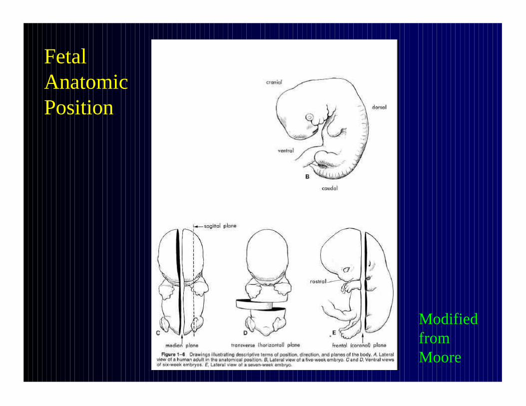
Modified from Moore
Fetal Anatomic Position

Osteology – Orientations:Frankfort Horizontal for Skulls
• Named for city of 1884 convention
• Plane defined by 3 points: right and left porion (at top of auditory meatus), and left orbitale (bottom of left orbit)
• 6 standard viewing perspectives: all perpendicular or parallel to FH (norma occipitalis – from behind, frontalis – from in front, basalis – from base, lateralis – from right and left sides, verticalis –from above)
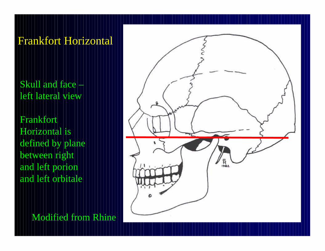
Frankfort Horizontal
Modified from Rhine
Skull and face –left lateral view
Frankfort Horizontal is defined by plane between right and left porion and left orbitale

Osteology – Planes of Reference
1. Sagittal (midsagittal, median, midline)
2. Coronal (frontal) plane
3. Transverse (horizontal) plane
4. Oblique plane

Planes of Reference - Sagittal
• Sagittal = midline = median = midsagittal
• Divides body into right and left halves
• Any planar slice paralleling this plane is parasagittal
• Is a vertical anteroposterior plane
• Parallel to sagittal suture of skull

Planes of Reference - Sagittal
Posterior
Anterior
Right Left
Schematic human –seen from above
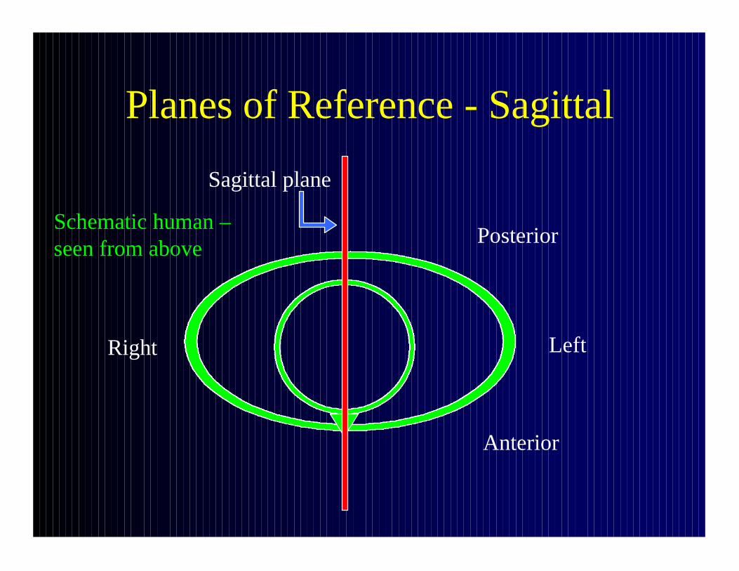
Planes of Reference - Sagittal
Posterior
Anterior
Sagittal plane
Right Left
Schematic human –seen from above

Planes of Reference - Sagittal
Posterior
Anterior
Sagittal plane
Parasagittal planes
Right Left
Schematic human –seen from above

Planes of Reference – Sagittal
Right Left
Body is divided into left and right halves

Planes of Reference - Coronal
• Coronal = frontal
• Divides body into anterior and posterior halves
• At right angle to sagittal plane
• Vertical side-to-side plane
• Approximately parallel to coronal suture

Planes of Reference – Coronal (Frontal)
Posterior
Anterior
Coronalplane
Schematic human –seen from above

Planes of Reference - Transverse
• Transverse = horizontal
• Slices through body at any height
• Perpendicular to sagittal and coronal planes
• In case of an organ or other structure, a transverse or cross section is at right angles to long axis of that organ or structure

Planes of Reference – Transverse (Horizontal)
Transverse plane slices through body at any height and is perpendicular to sagittal and coronal planes

Planes of Reference – Long Axis and Transverse Axis of Organ
Long axis
Transverse axes
Planes of Reference - Oblique
• May lie at any other angle
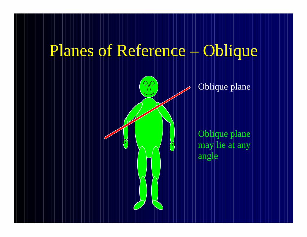
Planes of Reference – Oblique
Oblique plane may lie at any angle
Oblique plane

General Skeletal Terminology
• Cranial skeleton: skull (= cranium + mandible)
• Post-cranial skeleton: remainder of skeleton (axial and appendicular skeleton)
• Axial skeleton: bones of trunk, including ribs, vertebrae, sternum, pelvis
• Appendicular skeleton: bones of limbs, including shoulder girdle
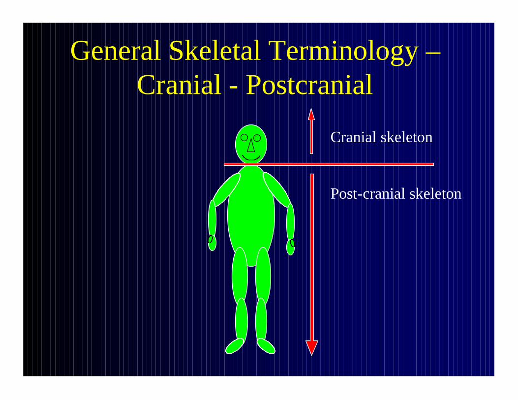
General Skeletal Terminology –Cranial - Postcranial
Cranial skeleton
Post-cranial skeleton

General Skeletal Terminology –Axial - Appendicular
Axial skeleton = skeleton of the trunk
Appendicular skeleton = skeleton of the limbs
Terms of Relationship
• “Three pairs of relative terms can express relationship of any given structure to another” (Grant)
• Superior-Inferior (up-down)
• Anterior-Posterior (front-back)
• Medial-Lateral (to and away from midline)

Terms of Relationship:Superior - Inferior
• Superior: toward head end of body, hominid or quadruped; “cranial” and “cephalic” are synonymous for hominids and quadrupeds
• Inferior: opposite of superior; away from head in hominids; “caudal” refers to tail end of quadruped, but can be used to describe fetal orientation, e.g. craniocaudal length of fetus, craniocaudal growth of CNS
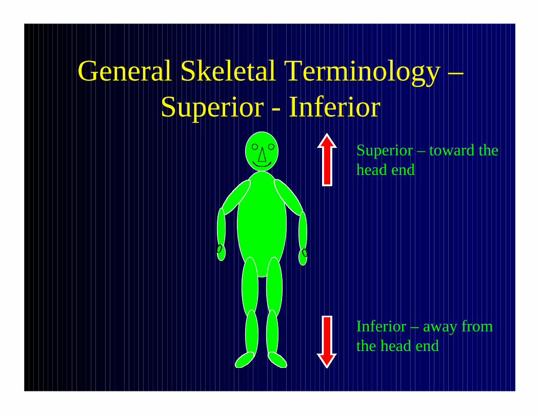
General Skeletal Terminology –Superior - Inferior
Superior – toward the head end
Inferior – away from the head end
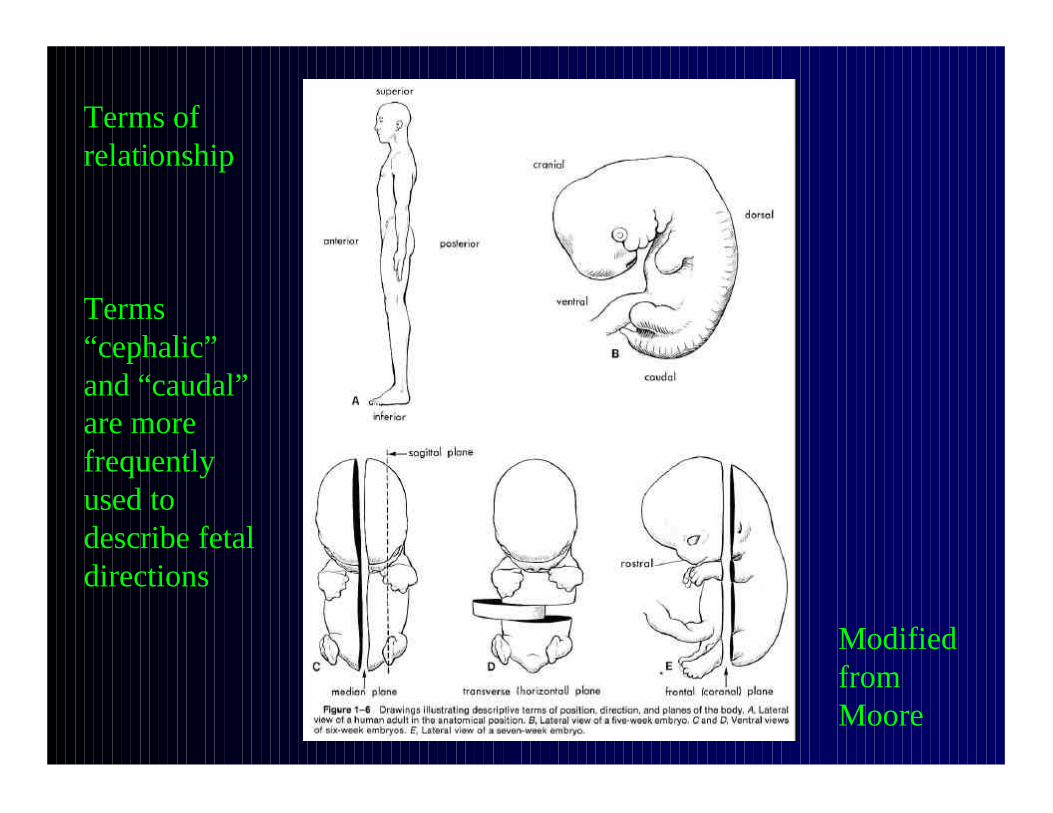
Terms of relationship
Terms “cephalic” and “caudal” are more frequently used to describe fetal directions
Modified from Moore

Terms of Relationship:Superior - Inferior
• “Superior” and “inferior” are usually used to describe direction in bones of axial skeleton
• “Proximal” and “distal” are usually used to describe direction in bones of appendicularskeleton, i.e. proximal meaning nearer to the axis of the body and distal meaning further from the axis of the body
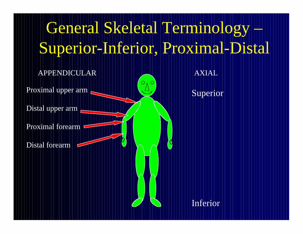
General Skeletal Terminology –Superior-Inferior, Proximal-Distal
Superior
Inferior
Proximal upper arm
Distal upper arm
Proximal forearm
Distal forearm
APPENDICULAR AXIAL

Terms of Relationship:Anterior - Posterior
• Anterior: toward front of body; “ventral” can be used homologously for hominids and quadrupeds; “palmar” or “volar” for palms of hands, “plantar” for soles of feet
• Posterior: toward back of body, opposite of anterior; “dorsal” can be used homologously for both hominids and quadrupeds; by convention “dorsum”is top of foot or back of hand

General Skeletal Terminology -Anterior
Superior
Inferior
AnteriorAnterior

General Skeletal Terminology –Posterior
Posterior Posterior
Superior
Inferior
Terms of Relationship:Medial - Lateral
• Medial: toward midline (median plane of body)
• Lateral: opposite of midline; away from midline
• May be used to describe axial and appendicular elements of skeleton

General Skeletal Terminology –Medial - Lateral
Medial –toward the midline
Lateral –away from the midline

Terms of Comparison
• Describe relationships of structures between species, as well as within species
Terms of Comparison:Proximal - Distal
• Proximal: end of a bone nearest the axial skeleton; used mainly to describe direction in limb bones, i.e. to describe direction in bones of appendicular skeleton
• Distal: opposite of proximal; farthest from axial skeleton
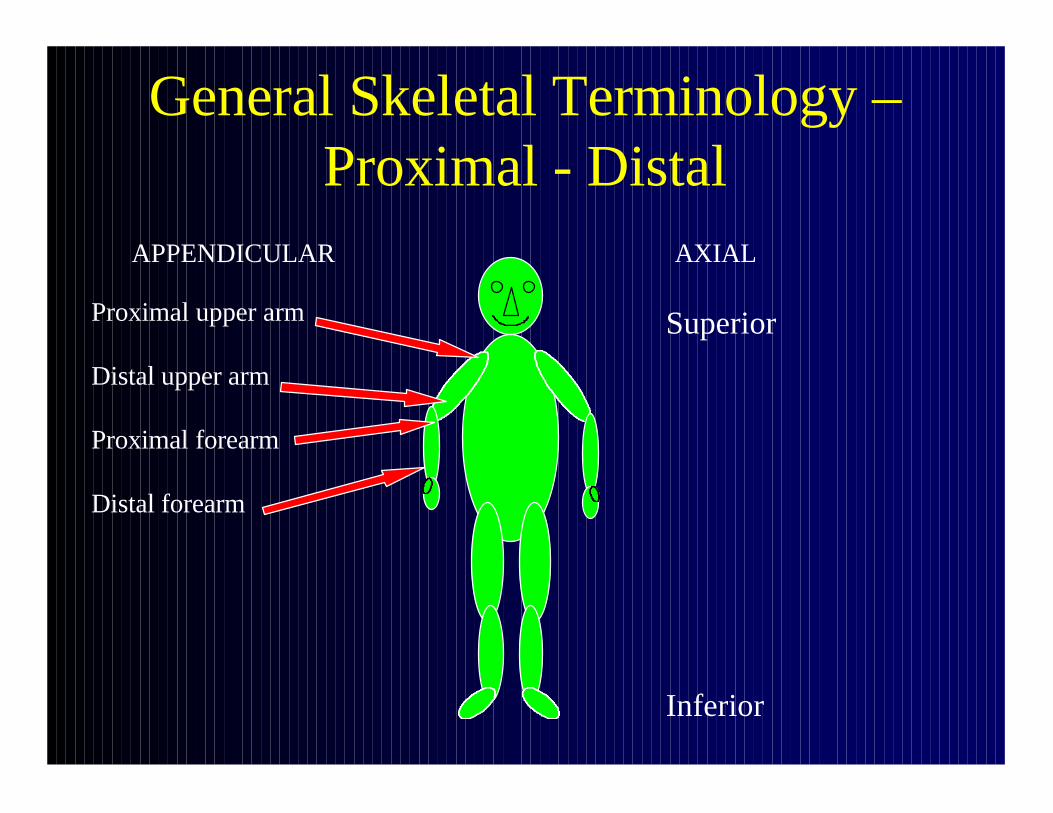
General Skeletal Terminology –Proximal - Distal
Superior
Inferior
Proximal upper arm
Distal upper arm
Proximal forearm
Distal forearm
APPENDICULAR AXIAL

Terms of Comparison:Ulnar - Radial
• Ulnar: near the little finger side; used for forearm, hand; corresponds to medial
• Radial: near the thumb side; used for forearm, hand; corresponds to lateral

General Skeletal Terminology –Radial - Ulnar
Radial –near the thumb side
Ulnar -near the little finger side

Terms of Comparison:Tibial - Fibular
• Tibial: near the tibial or shinbone side; used for lower leg, foot; corresponds to medial
• Fibular: near the fibular side; used for lower leg, foot; corresponds to lateral

General Skeletal Terminology –Tibial - Fibular
Fibular –near the fibular side
Tibial –near the shinbone side

Terms of Comparison:External - Internal
• External: outer
• Internal: inner

Terms of Comparison –Internal - External
From Grant From Grant

Terms of Comparison:Endocranial - Ectocranial
• Endocranial: inner surface of cranial vault
• Ectocranial: outer surface of cranial vault

Terms of Comparison –Endocranial - Ectocranial
Ectocranial – outer surface
Endocranial – inner surface
From Grant

Terms of Comparison:Superficial - Deep
• Superficial: close to surface
• Deep: opposite of superficial; far from surface

From Grant
Terms of Comparison –Superficial - Deep
From Grant

Terms of Comparison:Subcutaneous
• Just below skin
Terms of Comparison:Ipsilateral - Contralateral
• Ipsilateral: same side of body
• Contralateral: opposite side of body
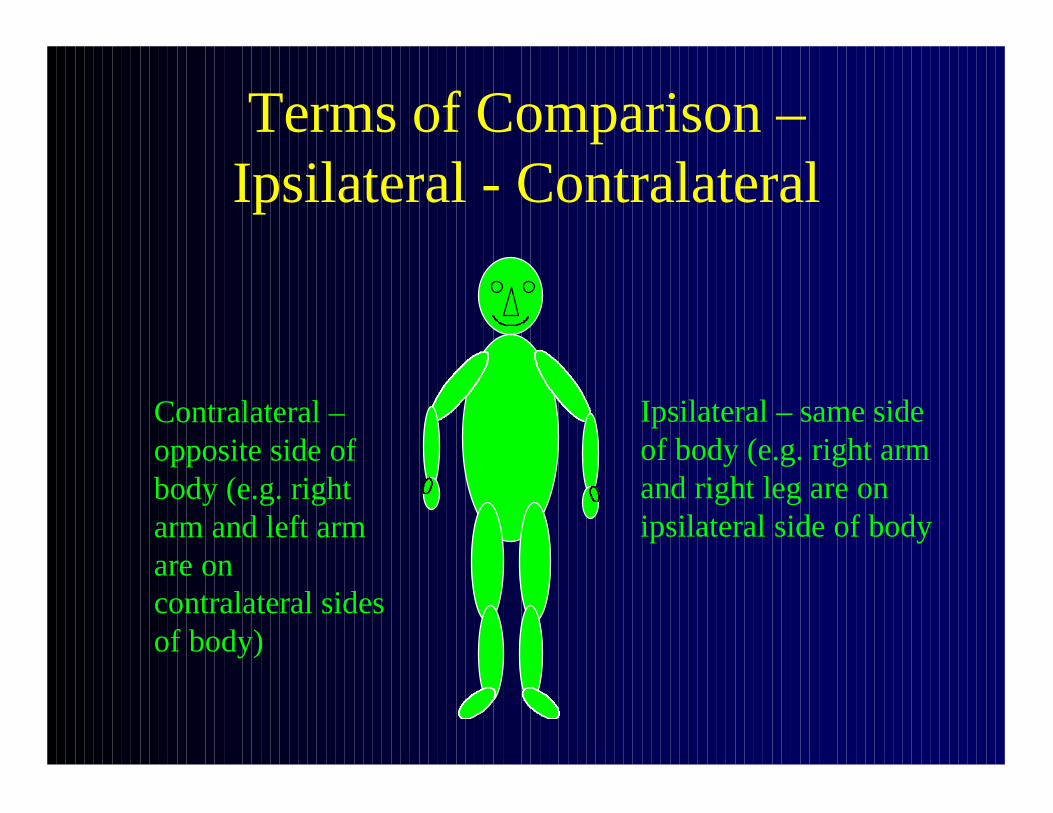
Terms of Comparison –Ipsilateral - Contralateral
Ipsilateral – same side of body (e.g. right arm and right leg are on ipsilateral side of body
Contralateral –opposite side of body (e.g. right arm and left arm are on contralateral sides of body)

Attachment of Muscles
• Proximal end: origin
• Distal end: insertion

Dental Terminology
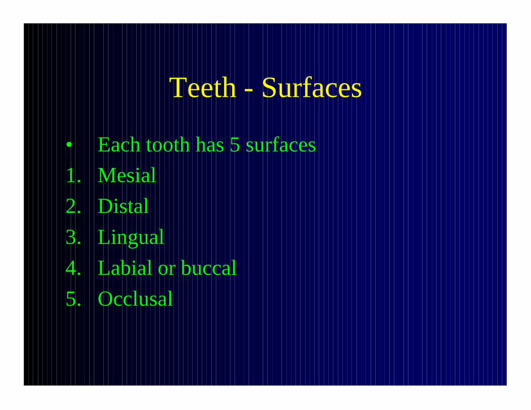
Teeth - Surfaces
• Each tooth has 5 surfaces
1. Mesial
2. Distal
3. Lingual
4. Labial or buccal
5. Occlusal

Dental Terminology:Mesial – Distal
• Mesial: toward point on midline where central incisors contact each other (medial surfaces of front teeth and anterior surfaces of side teeth are mesial (proximal))
• Distal: opposite of mesial

Dental Terminology – Mesial - Distal
Mesial: toward the midline
Distal: opposite of mesial

Dental Terminology:Lingual – Labial - Buccal
• Lingual: toward the tongue; opposite of labial or buccal
• Labial (toward the lip): opposite of lingual; used for incisors and canines (i.e. anterior surfaces of front teeth)
• Buccal (toward the cheek): opposite of lingual; used for premolars, molars (i.e. lateral surfaces of side teeth)
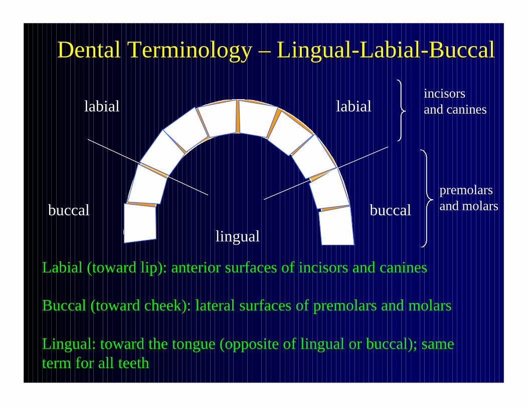
Dental Terminology – Lingual-Labial-Buccal
Labial (toward lip): anterior surfaces of incisors and canines
Buccal (toward cheek): lateral surfaces of premolars and molars
Lingual: toward the tongue (opposite of lingual or buccal); sameterm for all teeth
labial labial
buccal buccal
lingual
incisorsand canines
premolars and molars

Dental Terminology – Occlusal Surfaces
• Occlusal surfaces are biting surfaces, i.e. where teeth of upper (maxillary) and lower (mandibular) jaws meet are all “occlusal” surfaces (an occlusal surfaces faces the opposite dental arch)
• Occlusal = masticatory• Occlusal or biting surfaces of incisors =
incisal surfaces

Dental Terminology – Occlusal Surfaces
Occlusal surface: biting surface, masticatory surface; where maxillary and mandibular surfaces meet

Modified from Rhine
Dental Terminology –Occlusal Surfaces
Occlusal surface: biting surface, masticatory surface; where maxillary and mandibular surfaces meet

Dental Terminology -Contact Surfaces
• Proximal (mesial) and distal surfaces of adjacent teeth are contact surfaces
• Exceptions: distal surfaces of last molars
• Proximal (mesial) and distal surfaces of adjacent teeth rub against one another with chewing motion, producing areas of wear – these areas may match on adjacent teeth – may be useful in helping to establish dental identification

Dental Terminology:Interproximal
• In contact with adjacent teeth in the samejaw

Modified from Grant
Dental Terminology –Interproximal Regions and Contact Surfaces
Interproximal regions areindicated by arrows

Dental Terminology – Interproximal Contact Surfaces
Modified from Brothwell
Wear facets on mesial and distal surfaces are indicative of an interproximal articulation with adjacent teeth
Dental Terminology:Tooth Axes
• Mesiodistal: axis from mesial to distal surface
• Buccolingual or labiolingual: axis from buccal or labial to lingual surface
Teeth – Measurements -Mesiodistal Diameter
• Maximum diameter between mesial and distal contact points

Teeth – measurements – schematic – crown view
labial or buccal
lingual
Mesiodistal diameter –maximum diameter between mesial and distal contact points
Teeth – Measurements –Buccolingual Diameter
• Maximum diameter at right angles to mesiodistal diameter

Teeth – measurements – schematic – crown view
labial or buccal
lingual
Buccolingual diameter –maximum diameter at right angles to mesiodistal diameter

Teeth – Measurements – Crown Module
• A measurement of “relative crown mass”
• Average of mesiodistal and buccolingual diameters

Teeth – measurements – schematic – crown view
labial or buccal
lingual
Crown module –a measurement of relative crown mass
Crown module = (mesiodistal diameter + buccolingual diameter) /2

END


