Godefroit P., Codrea V. & Weishampel D. B. 2009. — Osteology of Zalmoxes shqiperorum (Dinosauria, Ornithopoda), based on new specimens from the Upper Cretaceous of Nalat,-Vad (Romania). Geodiversitas 31 (3) : 525-553.
ABSTRACTNălaţ-Vad is a new fossil locality discovered in 2002 in the Sănpetru Formation (Maastrichtian, Late Cretaceous) of the Haţeg Basin (Transylvania, Romania). Th is site has, among others, yielded the most complete skeleton that can be referred to the ornithopods dinosaur Zalmoxes shqiperorum Weishampel, Jianu, Cziki & Norman, 2003, but also isolated elements belonging to both juveniles and adult individuals. Th is material provides new information about the anatomy of Z. shqiperorum, and about the inter- and intraspecifi c variability within Zal-moxes. Zalmoxes robustus (Nopsca, 1902) and Z. shqiperorum were apparently sympatric species in Transylvania by latest Cretaceous time. Th e co-existence in the same locality of two closely-related species is not an isolated case among ornithopod dinosaurs.
Pascal GODEFROITDépartement de Paléontologie,
Institut royal des Sciences naturelles de Belgique,29 rue Vautier, B-1000 Bruxelles (Belgium)
Osteology of Zalmoxes shqiperorum(Dinosauria, Ornithopoda), based on new specimens from the Upper Cretaceousof Nalat,-Vad (Romania)
526 GEODIVERSITAS • 2009 • 31 (3)
Godefroit P. et al.
MOTS CLÉSDinosauria,
Ornithopoda,Zalmoxes shqiperorum,
Crétacé supérieur,bassin de Haţeg,
sympatrie.
RÉSUMÉOstéologie de Zalmoxes shqiperorum (Dinosauria, Ornithopoda), d’après de nou-veaux spécimens du Crétacé supérieur de Nălaţ-Vad (Roumanie).Nălaţ-Vad est un nouveau site fossilifère découvert en 2002 dans la Formation de Sănpetru (Maastrichtien, Crétacé supérieur) du bassin de Haţeg (Transylvanie, Roumanie). Ce site a notamment livré le squelette le plus complet à ce jour pou-vant être attribué au dinosaure ornithopode Zalmoxes shqiperorum Weishampel, Jianu, Cziki & Norman, 2003, mais également des ossements isolés appartenant à des individus juvéniles et adultes. Ce matériel apporte de nouvelles informations sur l’anatomie de Z. shqiperorum, mais également sur la variabilité inter- et intra-spécifi que au sein même du genre Zalmoxes. Zalmoxes robustus (Nopsca, 1902) et Z. shqiperorum étaient apparemment des espèces sympatriques en Transylvanie au Crétacé supérieur. La coexistence dans une même localité de deux espèces proches n’est pas un cas isolé chez les dinosaures ornithopodes.
INTRODUCTION
Although Rhabdodontidae probably comprises the most abundant dinosaurs from the Upper Cretaceous of Europe, these medium-sized ornitho-pods remained poorly understood until recently. Rhabdodon priscus Matheron, 1869 was described from fragmentary material discovered in the lower Maastrichtian of La Nerthe (Bouches-du-Rhône, France). Additional material from diff erent locali-ties in southern France, referred to as R. priscus, was described later by Lapparent (1947), Garcia et al. (1999), Pincemaille-Quillévéré (2002), and Pincemaille-Quillévéré et al. (2006). Buff etaut & Le Loeuff (1991) described Rhabdodon septimani-cus from a dentary discovered in the Campano-Maastrichtian of Montmouliers (Hérault, France). Rhabdodon Matheron, 1869 specimens have also been described in diff erent Campano-Maastrichtian localities from northern Spain (Pereda-Suberbiola & Sanz 1999). Bunzel (1871) described Iguanodon suessi from the Upper Cretaceous of Niederöster-reich (Austria). Seeley (1881) subsequently created the genus Mochlodon to include this species. In the beginning of the 20th century, Nopcsa described important ornithopod material from the Upper Cretaceous of the Haţeg Basin in Transylvania. Part of this material was referred to Mochlodon suessi Seeley, 1881 and another part to a new species
Mochlodon robustum Nopcsa, 1902 (Nopcsa 1902, 1904). He later suggested that Mochlodon might be identical to Rhabdodon and that sexual dimorphism might be responsible for the observed diff erences between the two forms (Nopcsa 1915, 1929). Ösi (2004) reported rhabdodontid fossils in the Upper Cretaceous of Hungary. Weishampel et al. (2003) recently reviewed the rhabdodontid mate-rial from Transylvania, and referred it to as a new genus, Zalmoxes. Th ey distinguished two species: Z. robustus (Nopcsa, 1902), known from abun-dant skull and postcranial material, and the more poorly known species Z. shqiperorum Weishampel, Jianu, Cziki & Norman, 2003. Both species are known from several localities in the Haţeg Basin, but also from the Vintu de Jos area (Fig. 1). Th e holotype of Z. shqiperorum (BMNH R4900) is a single incomplete adult individual. Much of the referred material pertains to a partially associated skeleton of a juvenile, which includes most of the pelvis and associated dorsal, sacral and caudal ver-tebrae (FGGUB R1087-1133 and R1355-1357). From the skull of Z. shqiperorum, only the dentary is known to date. Both the fore- and hindlimbs were also incompletely known. Sachs & Hornung (2006) referred specimens discovered in Austria to Zalmoxes.
New material that can be referred to as Z. shqipero-rum was discovered in 2002 at Nălaţ-Vad, a new
527
Osteology of Zalmoxes shqiperorum (Dinosauria, Ornithopoda) from Romania
GEODIVERSITAS • 2009 • 31 (3)
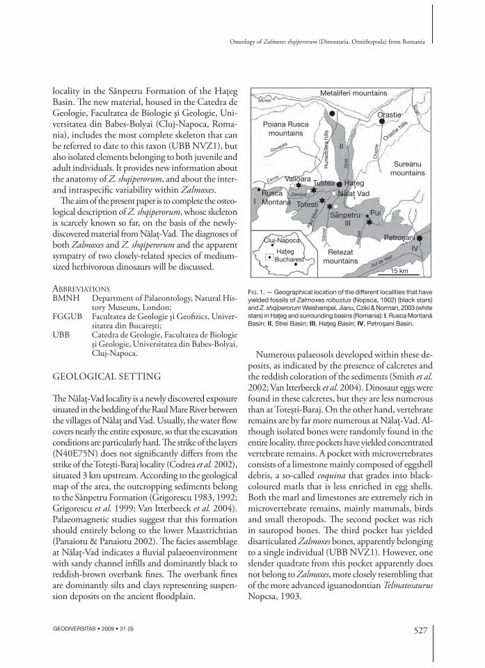
Metaliferi mountains
Sureanumountains
Petrosani
Retezatmountains
Poiana Ruscamountains
I
II
III
IV
RuscaMontana
Hun
edoa
ra h
ills
Orastie
HategValioara Tustea
Totesti
Nalat Vad
Sanpetru
Sib
isel
Densus
Govajdia
Cerna
Râu
l Mar
e
Bar
bat
Cugir
Jiul de Vest
Str
ei
Ora
stie
Mures
Pui
Cluj-Napoca
HategBucharest
Orastie hills
15 km
‚˘
˘
˘
‚
‚
‚
FIG. 1. — Geographical location of the different localities that have yielded fossils of Zalmoxes robustus (Nopsca, 1902) (black stars) and Z. shqiperorum Weishampel, Jianu, Cziki & Norman, 2003 (white stars) in Haţeg and surrounding basins (Romania): I, Rusca Montană Basin; II, Strei Basin; III, Haţeg Basin; IV, Petroşani Basin.
locality in the Sănpetru Formation of the Haţeg Basin. Th e new material, housed in the Catedra de Geologie, Facultatea de Biologie şi Geologie, Uni-versitatea din Babes-Bolyai (Cluj-Napoca, Roma-nia), includes the most complete skeleton that can be referred to date to this taxon (UBB NVZ1), but also isolated elements belonging to both juvenile and adult individuals. It provides new information about the anatomy of Z. shqiperorum, and about the inter- and intraspecifi c variability within Zalmoxes.
Th e aim of the present paper is to complete the osteo-logical description of Z. shqiperorum, whose skeleton is scarcely known so far, on the basis of the newly-discovered material from Nălaţ-Vad. Th e diagnoses of both Zalmoxes and Z. shqiperorum and the apparent sympatry of two closely-related species of medium-sized herbivorous dinosaurs will be discussed.
ABBREVIATIONSBMNH Department of Palaeontology, Natural His-
tory Museum, London;FGGUB Facultatea de Geologie şi Geofi zics, Univer-
sitatea din Bucareşti;UBB Catedra de Geologie, Facultatea de Biologie
şi Geologie, Universitatea din Babes-Bolyai, Cluj-Napoca.
GEOLOGICAL SETTING
Th e Nălaţ-Vad locality is a newly discovered exposure situated in the bedding of the Raul Mare River between the villages of Nălaţ and Vad. Usually, the water fl ow covers nearly the entire exposure, so that the excavation conditions are particularly hard. Th e strike of the layers (N40E75N) does not signifi cantly diff ers from the strike of the Toteşti-Baraj locality (Codrea et al. 2002), situated 3 km upstream. According to the geological map of the area, the outcropping sediments belong to the Sănpetru Formation (Grigorescu 1983, 1992; Grigorescu et al. 1999; Van Itterbeeck et al. 2004). Palaeomagnetic studies suggest that this formation should entirely belong to the lower Maastrichtian (Panaiotu & Panaiotu 2002). Th e facies assemblage at Nălaţ-Vad indicates a fl uvial palaeoenvironment with sandy channel infi lls and dominantly black to reddish-brown overbank fi nes. Th e overbank fi nes are dominantly silts and clays representing suspen-sion deposits on the ancient fl oodplain.
Numerous palaeosols developed within these de-posits, as indicated by the presence of calcretes and the reddish coloration of the sediments (Smith et al. 2002; Van Itterbeeck et al. 2004). Dinosaur eggs were found in these calcretes, but they are less numerous than at Toteşti-Baraj. On the other hand, vertebrate remains are by far more numerous at Nălaţ-Vad. Al-though isolated bones were randomly found in the entire locality, three pockets have yielded concentrated vertebrate remains. A pocket with microvertebrates consists of a limestone mainly composed of eggshell debris, a so-called coquina that grades into black-coloured marls that is less enriched in egg shells. Both the marl and limestones are extremely rich in micro vertebrate remains, mainly mammals, birds and small theropods. Th e second pocket was rich in sauropod bones. Th e third pocket has yielded disarticulated Zalmoxes bones, apparently belonging to a single individual (UBB NVZ1). However, one slender quadrate from this pocket apparently does not belong to Zalmoxes, more closely resembling that of the more advanced iguanodontian Telmatosaurus Nopcsa, 1903.
528 GEODIVERSITAS • 2009 • 31 (3)
Godefroit P. et al.
SYSTEMATIC PALAEONTOLOGY
Order ORNITHISCHIA Seeley, 1887Suborder ORNITHOPODA Marsh, 1881
Genus Zalmoxes Weishampel, Jianu, Cziki & Norman, 2003
TYPE SPECIES. — Zalmoxes robustus (Nopcsa, 1902) by original designation.
EMENDED DIAGNOSIS. — Unambiguous synapomorphies of the genus include the following characters: reduced external mandibular fenestra positioned anteriorly along the upper border of the surangular; well-developed supra-acetabular process on the ilium; absence of an obturator process on the ischium; arched ischial shaft.
Th e following characters may be synapomorphic for Z. ro-bustus and Z. shqiperorum, but their presence in Rhabdodon remains unknown (see discussion): extensive, complex squa-mose suture between quadratojugal and jugal; post-temporal foramen transmitted through the body of the squamosal; curved shelf on the lateral surface of the postorbital; lateral splaying of the quadrate; deep predentary; long, dorsoven-trally-narrow, twisted preacetabular process.
Th e following characters cannot be observed either in Z. shqiperorum or in the diff erent species of Rhabdodon. Th erefore, subsequent discoveries may shift their dis-tribution either inclusively (as synapomorphies for the Rhabdodontidae clade) or exclusively (as autapomorphies for Z. robustus): absence of the scar for m. adductor man-dibulae externus superfi cialis on the squamosal; large, disc-shaped quadratojugal; frontal with complex transverse sutural surface that extensively overlaps the parietal; high lateroventral processes on the predentary.
HOLOTYPE. — BMNH R.4900, left dentary, sacrum, right scapula, right coracoid, partial ?left ilium, partial right ilium, right ischium, left distal ischium and left femur.
EMENDED DIAGNOSIS. — Species of Zalmoxes charac-terised by the following autapomorphies: occipital con-dyle not separated from the sphenooccipital tubercles (= basal tubera) by a distinct neck; dentary with an
angular buccal emargination that forms a horizontal platform extending for the full length of the dentition behind and medial to the coronoid process; scapular blade narrow, strap-like proximally, expanding sharply posterodistally; expanded region of the scapula adja-cent to the coracoid suture; acromial process forming a prominent fl ange; deltopectoral crest of the humerus particularly prominent, extending over the proximal half of the humerus; iliac peduncle of the ischium parti-cularly developed; distal end of the ischium forming a boot-like expansion.
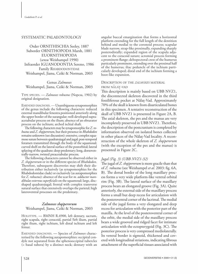
DESCRIPTION OF THE ZALMOXES MATERIAL
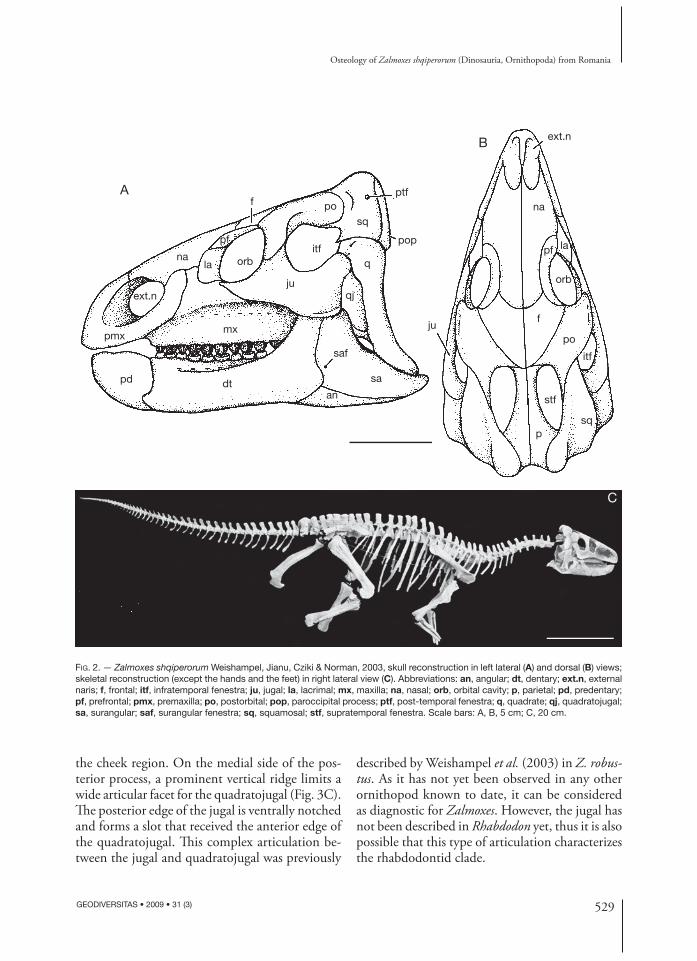
FROM NALAT,-VAD Th is description is mainly based on UBB NVZ1, the disconnected skeleton discovered in the third fossiliferous pocket at Nălaţ-Vad. Approximately 70% of the skull is known from disarticulated bones in this specimen. A tentative reconstruction of the skull of UBB NVZ1 is presented in Figure 2A, B. Th e axial skeleton, the pes and the manus are very incompletely preserved in UBB NVZ1. Th erefore, the description of the postcranium is completed by information observed on isolated bones collected in other places of the Nălaţ-Vad locality. A recon-struction of the whole skeleton of Z. shqiperorum (with the exception of the pes and the manus) is presented in Figure 2C.
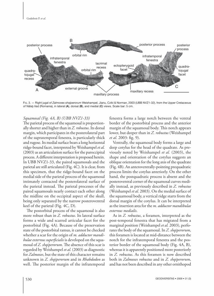
Jugal (Fig. 3) (UBB NVZ1-32)Th e jugal of Z. shqiperorum is more gracile than that of Z. robustus (see Weishampel et al. 2003: fi g. 6A, B). Th e dorsal border of the long maxillary proc-ess forms a very wide platform-like ventral orbital rim (Fig. 3B). Th e lateral surface of the maxillary process bears an elongated groove (Fig. 3A). Quite anteriorly, the external side of the maxillary process forms a small but deep recess for articulation with the posteroventral corner of the lacrimal. Th e medial side of the jugal forms a very elongated and deep recess for articulation with the posterior part of the maxilla. At the level of the posteroventral corner of the orbit, the medial side of the maxillary process bears a wide grooved and ridged facet for intimate articulation with the ectopterygoid (Fig. 3C). Th e posterior process is very compressed mediolaterally. Its ventral border is sigmoid, thickened and cov-ered with longitudinal striations, indicating fi brous attachment of the superfi cial tissues associated with
529
Osteology of Zalmoxes shqiperorum (Dinosauria, Ornithopoda) from Romania
GEODIVERSITAS • 2009 • 31 (3)
A
B
na
ext.n
pmx
pd dt
mx
la orb
f
pfitf
posq
ptf
pop
q
ext.n
na
orb
pf la
f
po
itf
sq
stf
p
ju
qjju
saf
an
sa
C
FIG. 2. — Zalmoxes shqiperorum Weishampel, Jianu, Cziki & Norman, 2003, skull reconstruction in left lateral (A) and dorsal (B) views; skeletal reconstruction (except the hands and the feet) in right lateral view (C). Abbreviations: an, angular; dt, dentary; ext.n, external naris; f, frontal; itf, infratemporal fenestra; ju, jugal; la, lacrimal; mx, maxilla; na, nasal; orb, orbital cavity; p, parietal; pd, predentary; pf, prefrontal; pmx, premaxilla; po, postorbital; pop, paroccipital process; ptf, post-temporal fenestra; q, quadrate; qj, quadratojugal; sa, surangular; saf, surangular fenestra; sq, squamosal; stf, supratemporal fenestra. Scale bars: A, B, 5 cm; C, 20 cm.
the cheek region. On the medial side of the pos-terior process, a prominent vertical ridge limits a wide articular facet for the quadratojugal (Fig. 3C). Th e posterior edge of the jugal is ventrally notched and forms a slot that received the anterior edge of the quadratojugal. Th is complex articulation be-tween the jugal and quadratojugal was previously
described by Weishampel et al. (2003) in Z. robus-tus. As it has not yet been observed in any other ornithopod known to date, it can be considered as diagnostic for Zalmoxes. However, the jugal has not been described in Rhabdodon yet, thus it is also possible that this type of articulation characterizes the rhabdodontid clade.
530 GEODIVERSITAS • 2009 • 31 (3)
Godefroit P. et al.
AC
Bposterior process
external groove
lacrimalrecess ectopterygoid
facet
maxillary processmaxillary recess
quadra-tojugalfacet
quadra-tojugalrecess
posteriorprocess
posterior process
infratemporalfenestrainfratemporal
fenestra
orbital rim
orbital rim
FIG. 3. — Right jugal of Zalmoxes shqiperorum Weishampel, Jianu, Cziki & Norman, 2003 (UBB NVZ1-32), from the Upper Cretaceous of Nălaţ-Vad (Romania), in lateral (A), dorsal (B), and medial (C) views. Scale bar: 5 cm.
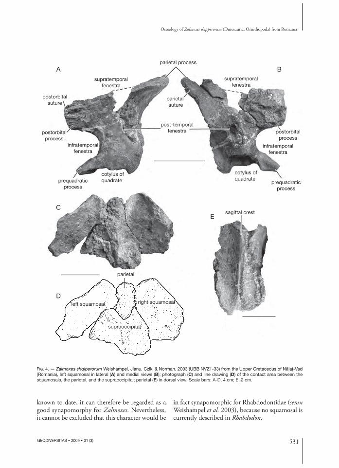
Squamosal (Fig. 4A, B) (UBB NVZ1-33)Th e parietal process of the squamosal is proportion-ally shorter and higher than in Z. robustus. Its dorsal margin, which participates in the posterolateral part of the supratemporal fenestra, is particularly thick and rugose. Its medial surface bears a long horizontal ridge-bound facet, interpreted by Weishampel et al. (2003) as an articulation surface for the paroccipital process. A diff erent interpretation is proposed herein. In UBB NVZ1-33, the paired squamosals and the parietal are still articulated (Fig. 4C). It is clear, from this specimen, that the ridge-bound facet on the medial side of the parietal process of the squamosal intimately contacted the posterolateral surface of the parietal instead. Th e parietal processes of the paired squamosals nearly contact each other along the midline on the occipital aspect of the skull, being only separated by the narrow posteroventral keel of the parietal (Fig. 4C, D).
Th e postorbital process of the squamosal is also more robust than in Z. robustus. Its lateral surface forms a wide and scarred articular facet for the postorbital (Fig. 4A). Because of the preservation state of the postorbital ramus, it cannot be checked whether a scar for the origin of m. adductor mandi-bulae externus superfi cialis is developed on the squa-mosal of Z. shqiperorum. Th e absence of this scar is regarded by Weishampel et al. (2003) as diagnostic for Zalmoxes, but the state of this character remains unknown in Z. shqiperorum and in Rhabdodon as well. Th e posterior margin of the infratemporal
fenestra forms a large notch between the ventral border of the postorbital process and the anterior margin of the squamosal body. Th is notch appears lower, but deeper than in Z. robustus (Weishampel et al. 2003: fi g. 9).
Ventrally, the squamosal body forms a large and deep cotylus for the head of the quadrate. As pre-viously noted by Weishampel et al. (2003), the shape and orientation of the cotylus suggests an oblique orientation for the long axis of the quadrate (Fig. 4B). An anteroventrally-pointing prequadratic process limits the cotylus anteriorly. On the other hand, the postquadratic process is absent and the posteroventral corner of the squamosal curves medi-ally instead, as previously described in Z. robustus (Weishampel et al. 2003). On the medial surface of the squamosal body, a vertical ridge starts from the dorsal margin of the cotylus. It can be interpreted as the insertion area for the m. adductor mandibulae externus medialis.
As in Z. robustus, a foramen, interpreted as the post-temporal fenestra that has migrated from a marginal position (Weishampel et al. 2003), perfo-rates the body of the squamosal. In Z. shqiperorum, this foramen is located at mid-distance between the notch for the infratemporal fenestra and the pos-terior border of the squamosal body (Fig. 4A, B), whereas it is apparently positioned more posteriorly in Z. robustus. As this foramen is now described both in Zalmoxes robustus and in Z. shqiperorum, and has not been described in any other ornithopod
531
Osteology of Zalmoxes shqiperorum (Dinosauria, Ornithopoda) from Romania
GEODIVERSITAS • 2009 • 31 (3)
A
CE
D
Bparietal process
parietal suture
supratemporalfenestra
infratemporalfenestra
infratemporalfenestra
supratemporalfenestra
post-temporalfenestra
postorbitalsuture
postorbitalprocess
prequadraticprocess
prequadraticprocess
cotylus ofquadrate
cotylus ofquadrate
parietal
left squamosal right squamosal
sagittal crest
supraoccipital
postorbitalprocess
FIG. 4. — Zalmoxes shqiperorum Weishampel, Jianu, Cziki & Norman, 2003 (UBB NVZ1-33) from the Upper Cretaceous of Nălaţ-Vad (Romania), left squamosal in lateral (A) and medial views (B); photograph (C) and line drawing (D) of the contact area between the squamosals, the parietal, and the supraoccipital; parietal (E) in dorsal view. Scale bars: A-D, 4 cm; E, 2 cm.
known to date, it can therefore be regarded as a good synapomorphy for Zalmoxes. Nevertheless, it cannot be excluded that this character would be
in fact synapomorphic for Rhabdodontidae (sensu Weishampel et al. 2003), because no squamosal is currently described in Rhabdodon.
532 GEODIVERSITAS • 2009 • 31 (3)
Godefroit P. et al.
A B
C D E F
head
squamosal suture
quadratojugal suture
jugal wing jugal wing
head
lateralcondyle
pterygoidwing
medialcondyle
medialcondyle
pterygoidwing
lateral condyle
FIG. 5. — Zalmoxes shqiperorum Weishampel, Jianu, Cziki & Norman, 2003 from the Upper Cretaceous of Nălaţ-Vad (Romania), dor-sal part of right quadrate (UBB NVZ1-39) in lateral (A) and medial (B) views; dorsal part of left quadrate (UBB NVZ1-40) in lateral (C), posterior (D), medial (E), and anterior (F) views. Scale bar: 5 cm.
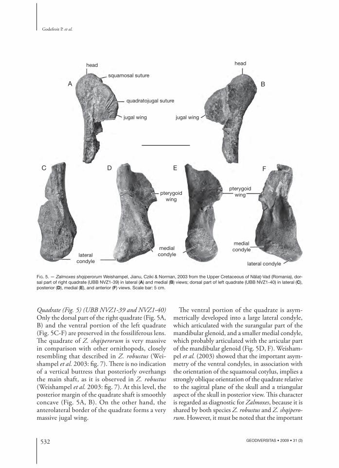
Quadrate (Fig. 5) (UBB NVZ1-39 and NVZ1-40) Only the dorsal part of the right quadrate (Fig. 5A, B) and the ventral portion of the left quadrate (Fig. 5C-F) are preserved in the fossiliferous lens. Th e quadrate of Z. shqiperorum is very massive in comparison with other ornithopods, closely resembling that described in Z. robustus (Wei-shampel et al. 2003: fi g. 7). Th ere is no indication of a vertical buttress that posteriorly overhangs the main shaft, as it is observed in Z. robustus (Weishampel et al. 2003: fi g. 7). At this level, the posterior margin of the quadrate shaft is smoothly concave (Fig. 5A, B). On the other hand, the anterolateral border of the quadrate forms a very massive jugal wing.
Th e ventral portion of the quadrate is asym-metrically developed into a large lateral condyle, which articulated with the surangular part of the mandibular glenoid, and a smaller medial condyle, which probably articulated with the articular part of the mandibular glenoid (Fig. 5D, F). Weisham-pel et al. (2003) showed that the important asym-metry of the ventral condyles, in association with the orientation of the squamosal cotylus, implies a strongly oblique orientation of the quadrate relative to the sagittal plane of the skull and a triangular aspect of the skull in posterior view. Th is character is regarded as diagnostic for Zalmoxes, because it is shared by both species Z. robustus and Z. shqipero-rum. However, it must be noted that the important
533
Osteology of Zalmoxes shqiperorum (Dinosauria, Ornithopoda) from Romania
GEODIVERSITAS • 2009 • 31 (3)
asymmetry of the distal quadrate condyles also characterises the more derived hadrosaurid clade (Weishampel et al. 1993; Godefroit et al. 1998). Although it is incompletely preserved, the pterygoid wing seems massive.
Parietal (Fig. 4E) (UBB NVZ1-33)Only the posterior part of the parietal, which inti-mately contacts the medial side of the squamosals, is preserved in UBB NVZ1-33. As previously observed in Z. robustus (Weishampel et al. 2003), the parietal appears particularly narrow and lozenge-shaped in cross-section. Its dorsal surface bears a high and sharp sagittal crest, much better developed than in Z. robustus (Weishampel et al. 2003: fi g. 10C). Th e height of the sagittal crest progressively lessens posteriorly, as it slightly bifurcates.
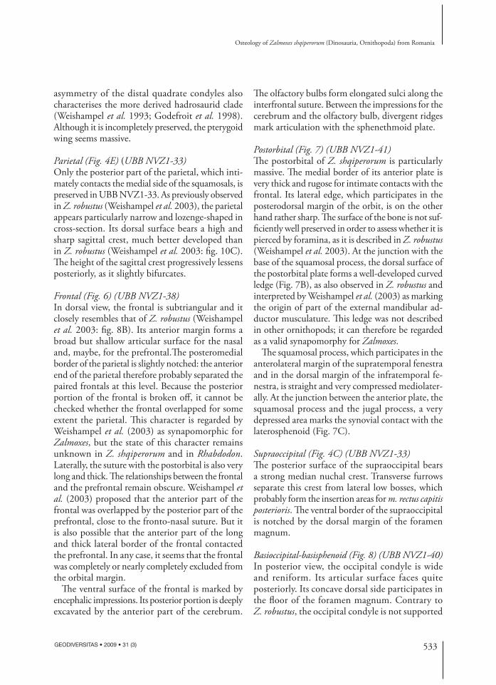
Frontal (Fig. 6) (UBB NVZ1-38)In dorsal view, the frontal is subtriangular and it closely resembles that of Z. robustus (Weishampel et al. 2003: fi g. 8B). Its anterior margin forms a broad but shallow articular surface for the nasal and, maybe, for the prefrontal.Th e posteromedial border of the parietal is slightly notched: the anterior end of the parietal therefore probably separated the paired frontals at this level. Because the posterior portion of the frontal is broken off , it cannot be checked whether the frontal overlapped for some extent the parietal. Th is character is regarded by Weishampel et al. (2003) as synapomorphic for Zalmoxes, but the state of this character remains unknown in Z. shqiperorum and in Rhabdodon. Laterally, the suture with the postorbital is also very long and thick. Th e relationships between the frontal and the prefrontal remain obscure. Weishampel et al. (2003) proposed that the anterior part of the frontal was overlapped by the posterior part of the prefrontal, close to the fronto-nasal suture. But it is also possible that the anterior part of the long and thick lateral border of the frontal contacted the prefrontal. In any case, it seems that the frontal was completely or nearly completely excluded from the orbital margin.
Th e ventral surface of the frontal is marked by encephalic impressions. Its posterior portion is deeply excavated by the anterior part of the cerebrum.
Th e olfactory bulbs form elongated sulci along the inter frontal suture. Between the impressions for the cerebrum and the olfactory bulb, divergent ridges mark articulation with the sphenethmoid plate.
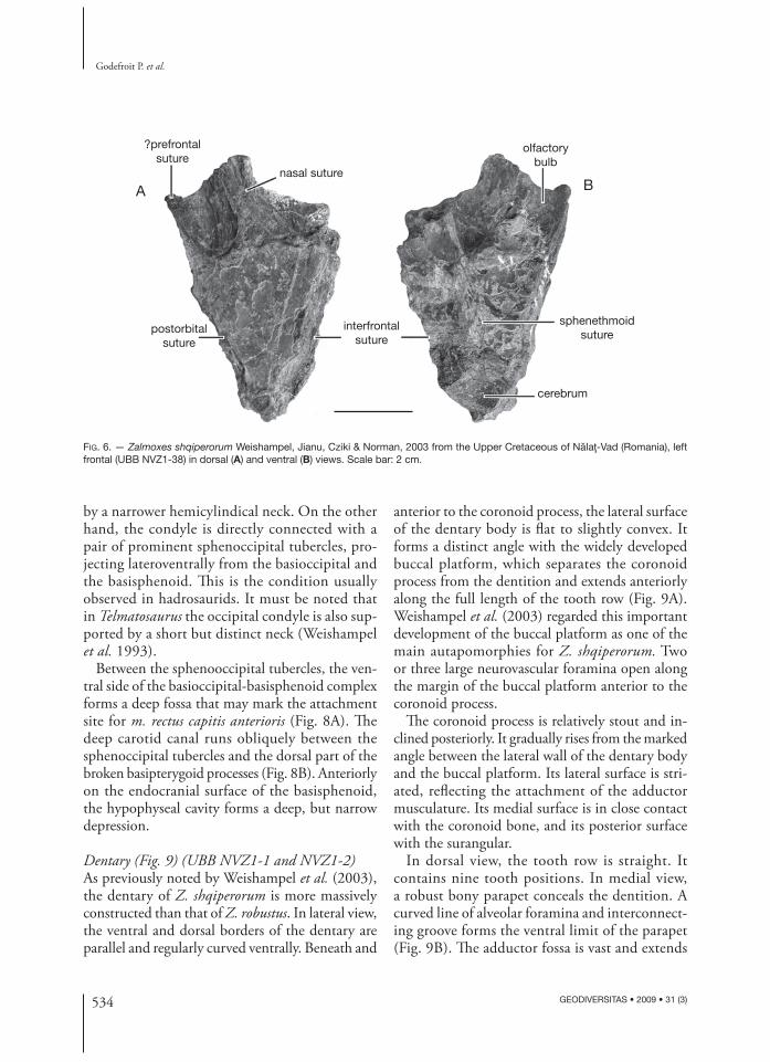
Postorbital (Fig. 7) (UBB NVZ1-41)Th e postorbital of Z. shqiperorum is particularly massive. Th e medial border of its anterior plate is very thick and rugose for intimate contacts with the frontal. Its lateral edge, which participates in the posterodorsal margin of the orbit, is on the other hand rather sharp. Th e surface of the bone is not suf-fi ciently well preserved in order to assess whether it is pierced by foramina, as it is described in Z. robustus (Weishampel et al. 2003). At the junction with the base of the squamosal process, the dorsal surface of the postorbital plate forms a well-developed curved ledge (Fig. 7B), as also observed in Z. robustus and interpreted by Weishampel et al. (2003) as marking the origin of part of the external mandibular ad-ductor musculature. Th is ledge was not described in other ornithopods; it can therefore be regarded as a valid synapomorphy for Zalmoxes.
Th e squamosal process, which participates in the anterolateral margin of the supratemporal fenestra and in the dorsal margin of the infratemporal fe-nestra, is straight and very compressed mediolater-ally. At the junction between the anterior plate, the squamosal process and the jugal process, a very depressed area marks the synovial contact with the laterosphenoid (Fig. 7C).
Supraoccipital (Fig. 4C) (UBB NVZ1-33)Th e posterior surface of the supraoccipital bears a strong median nuchal crest. Transverse furrows separate this crest from lateral low bosses, which probably form the insertion areas for m. rectus capitis posterioris. Th e ventral border of the supraoccipital is notched by the dorsal margin of the foramen magnum.
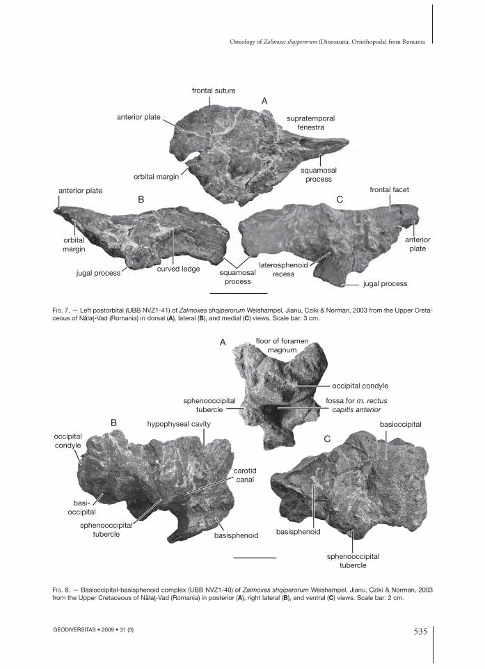
Basioccipital-basisphenoid (Fig. 8) (UBB NVZ1-40)In posterior view, the occipital condyle is wide and reniform. Its articular surface faces quite posteriorly. Its concave dorsal side participates in the fl oor of the foramen magnum. Contrary to Z. robustus, the occipital condyle is not supported
534 GEODIVERSITAS • 2009 • 31 (3)
Godefroit P. et al.
FIG. 6. — Zalmoxes shqiperorum Weishampel, Jianu, Cziki & Norman, 2003 from the Upper Cretaceous of Nălaţ-Vad (Romania), left frontal (UBB NVZ1-38) in dorsal (A) and ventral (B) views. Scale bar: 2 cm.
A B
?prefrontalsuture
nasal suture
interfrontalsuture
postorbitalsuture
sphenethmoidsuture
cerebrum
olfactorybulb
by a narrower hemicylindical neck. On the other hand, the condyle is directly connected with a pair of prominent sphenoccipital tubercles, pro-jecting lateroventrally from the basioccipital and the basisphenoid. Th is is the condition usually observed in hadrosaurids. It must be noted that in Telmatosaurus the occipital condyle is also sup-ported by a short but distinct neck (Weishampel et al. 1993).
Between the sphenooccipital tubercles, the ven-tral side of the basioccipital-basisphenoid complex forms a deep fossa that may mark the attachment site for m. rectus capitis anterioris (Fig. 8A). Th e deep carotid canal runs obliquely between the sphenoccipital tubercles and the dorsal part of the broken basipterygoid processes (Fig. 8B). Anteriorly on the endocranial surface of the basisphenoid, the hypophyseal cavity forms a deep, but narrow depression.
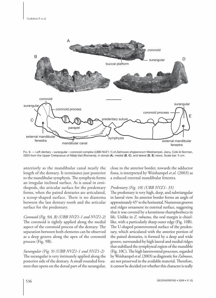
Dentary (Fig. 9) (UBB NVZ1-1 and NVZ1-2)As previously noted by Weishampel et al. (2003), the dentary of Z. shqiperorum is more massively constructed than that of Z. robustus. In lateral view, the ventral and dorsal borders of the dentary are parallel and regularly curved ventrally. Beneath and
anterior to the coronoid process, the lateral surface of the dentary body is fl at to slightly convex. It forms a distinct angle with the widely developed buccal platform, which separates the coronoid process from the dentition and extends anteriorly along the full length of the tooth row (Fig. 9A). Weishampel et al. (2003) regarded this important development of the buccal platform as one of the main autapomorphies for Z. shqiperorum. Two or three large neurovascular foramina open along the margin of the buccal platform anterior to the coronoid process.
Th e coronoid process is relatively stout and in-clined posteriorly. It gradually rises from the marked angle between the lateral wall of the dentary body and the buccal platform. Its lateral surface is stri-ated, refl ecting the attachment of the adductor musculature. Its medial surface is in close contact with the coronoid bone, and its posterior surface with the surangular.
In dorsal view, the tooth row is straight. It contains nine tooth positions. In medial view, a robust bony parapet conceals the dentition. A curved line of alveolar foramina and interconnect-ing groove forms the ventral limit of the parapet (Fig. 9B). Th e adductor fossa is vast and extends
535
Osteology of Zalmoxes shqiperorum (Dinosauria, Ornithopoda) from Romania
GEODIVERSITAS • 2009 • 31 (3)
FIG. 7. — Left postorbital (UBB NVZ1-41) of Zalmoxes shqiperorum Weishampel, Jianu, Cziki & Norman, 2003 from the Upper Creta-ceous of Nălaţ-Vad (Romania) in dorsal (A), lateral (B), and medial (C) views. Scale bar: 3 cm.
FIG. 8. — Basioccipital-basisphenoid complex (UBB NVZ1-40) of Zalmoxes shqiperorum Weishampel, Jianu, Cziki & Norman, 2003 from the Upper Cretaceous of Nălaţ-Vad (Romania) in posterior (A), right lateral (B), and ventral (C) views. Scale bar: 2 cm.
A
B C
frontal suture
anterior plate
anterior plate
anteriorplate
orbital margin
orbitalmargin
squamosalprocess
supratemporalfenestra
squamosalprocess
laterosphenoidrecess
frontal facet
curved ledgejugal processjugal process
C
A
B
occipital condyle
fossa for m. rectuscapitis anterior
basioccipital
sphenooccipitaltubercle
basisphenoidbasisphenoid
carotidcanal
hypophyseal cavity
occipitalcondyle
basi-occipital
sphenooccipitaltubercle
sphenooccipitaltubercle
floor of foramenmagnum
536 GEODIVERSITAS • 2009 • 31 (3)
Godefroit P. et al.
A
B D
CE
buccal platformsurangular
coronoid
symphysis
predentary suture
parapet
mandibular canal
external mandibularfenestra external mandibular
fenestra
surangular surangular
coronoid
coronoid processcoronoid process
FIG. 9. — Left dentary – surangular – coronoid complex (UBB NVZ1-1) of Zalmoxes shqiperorum Weishampel, Jianu, Cziki & Norman, 2003 from the Upper Cretaceous of Nălaţ-Vad (Romania), in dorsal (A), medial (B, C), and lateral (D, E) views. Scale bar: 5 cm.
anteriorly as the mandibular canal nearly the length of the dentary. It terminates just posterior to the mandibular symphysis. Th e symphysis forms an irregular inclined surface. As is usual in orni-thopods, the articular surface for the predentary forms, when the paired dentaries are articulated, a scoop-shaped surface. Th ere is no diastema between the last dentary tooth and the articular surface for the predentary.
Coronoid (Fig. 9A, B) (UBB NVZ1-1 and NVZ1-2)Th e coronoid is tightly applied along the medial aspect of the coronoid process of the dentary. Th e separation between both elements can be observed as a deep groove along the apex of the coronoid process (Fig. 9B).
Surangular (Fig. 9) (UBB NVZ1-1 and NVZ1-2)Th e surangular is very intimately applied along the posterior side of the dentary. A small rounded fora-men that opens on the dorsal part of the surangular,
close to the anterior border, towards the adductor fossa, is interpreted by Weishampel et al. (2003) as a reduced external mandibular fenestra.
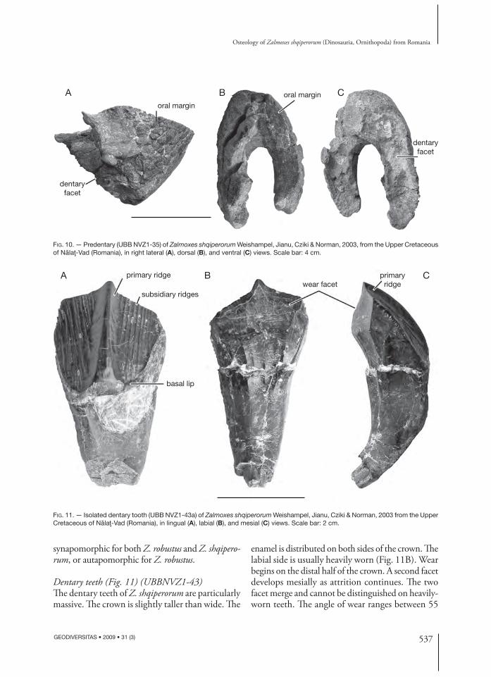
Predentary (Fig. 10) (UBB NVZ1- 35)Th e predentary is very high, deep, and subtriangular in lateral view. Its anterior border forms an angle of approximately 45° to the horizontal. Numerous grooves and ridges ornament its external surface, suggesting that it was covered by a keratinous rhamphotheca in life. Unlike in Z. robustus, the oral margin is chisel-like, with a particularly sharp outer edge (Fig. 10B). Th e U-shaped posteroventral surface of the preden-tary, which articulated with the anterior portion of the paired dentaries, is formed by a deep and wide groove, surrounded by high lateral and medial ridges that stabilized the symphyseal region of the mandible (Fig. 10C). Th e high lateroventral processes, regarded by Weishampel et al. (2003) as diagnostic for Zalmoxes, are not preserved in the available material. Th erefore, it cannot be decided yet whether this character is really
537
Osteology of Zalmoxes shqiperorum (Dinosauria, Ornithopoda) from Romania
GEODIVERSITAS • 2009 • 31 (3)
FIG. 10. — Predentary (UBB NVZ1-35) of Zalmoxes shqiperorum Weishampel, Jianu, Cziki & Norman, 2003, from the Upper Cretaceous of Nălaţ-Vad (Romania), in right lateral (A), dorsal (B), and ventral (C) views. Scale bar: 4 cm.
A B Coral margin
dentaryfacet
oral margin
dentaryfacet
A B Cprimaryridgewear facet
basal lip
subsidiary ridges
primary ridge
FIG. 11. — Isolated dentary tooth (UBB NVZ1-43a) of Zalmoxes shqiperorum Weishampel, Jianu, Cziki & Norman, 2003 from the Upper Cretaceous of Nălaţ-Vad (Romania), in lingual (A), labial (B), and mesial (C) views. Scale bar: 2 cm.
synapomorphic for both Z. robustus and Z. shqipero-rum, or autapomorphic for Z. robustus.
Dentary teeth (Fig. 11) (UBBNVZ1-43)Th e dentary teeth of Z. shqiperorum are particularly massive. Th e crown is slightly taller than wide. Th e
enamel is distributed on both sides of the crown. Th e labial side is usually heavily worn (Fig. 11B). Wear begins on the distal half of the crown. A second facet develops mesially as attrition continues. Th e two facet merge and cannot be distinguished on heavily-worn teeth. Th e angle of wear ranges between 55
538 GEODIVERSITAS • 2009 • 31 (3)
Godefroit P. et al.
FIG. 12. — Dorsal vertebra (UBB NVZ1-5) of Zalmoxes shqiperorum Weishampel, Jianu, Cziki & Norman, 2003 from the Upper Creta-ceous of Nălaţ-Vad (Romania), in anterior (A), left lateral (B), and posterior (C) views. Scale bar: 4 cm.
B
AC
neural spine
transverse process
transverseprocess
prezygapophysis
para-pophysis
parapophysisdiapophysis
parapophysis post-zygapophysis
neural spine
and 75° to the horizontal. Th e lingual side of the crown is characterized by a strong primary ridge, slightly mesial to the midline of the crown (contra Weishampel et al. 2003). From eight to 13 slightly divergent vertical subsidiary ridges cover either side of the median ridge (Fig. 11A). Th e mesial edge of the crown bears tiny crenulations. Th ere is no real cingulum, but a thin enamelled lip marks the base of the distal part of the crown. Th e root is some-what higher than the crown and regularly curved labially. Grooves on either side of the root and at the base of the crown indicate that the teeth were closely packed (Fig. 11C).
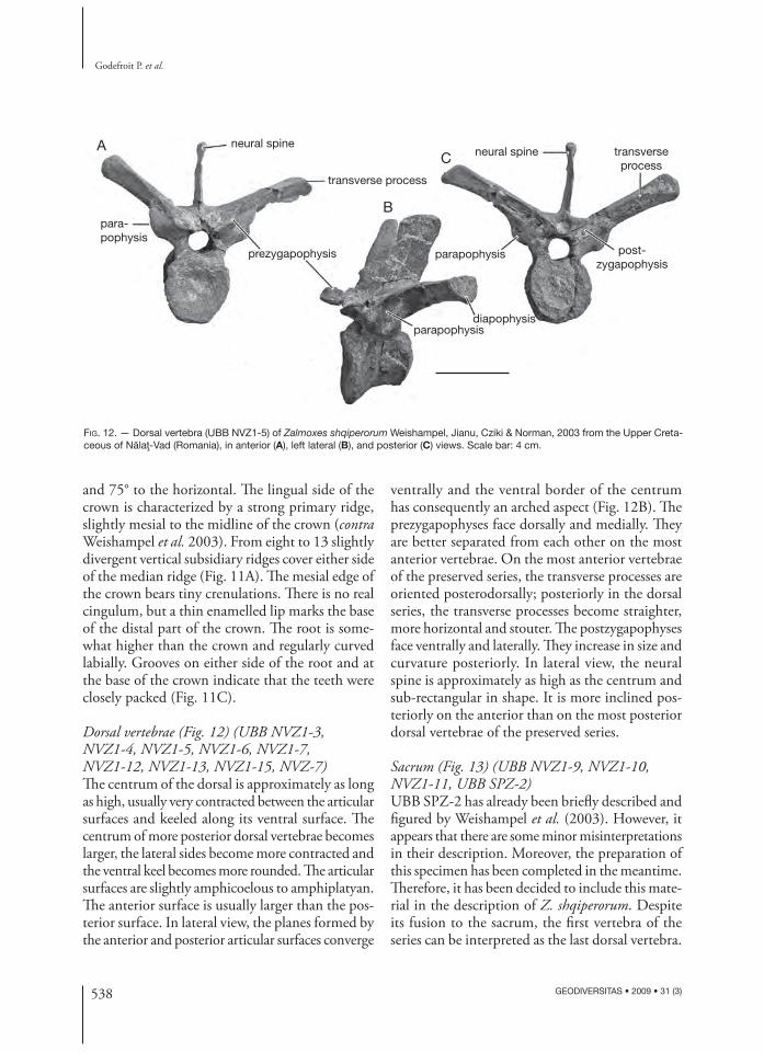
Dorsal vertebrae (Fig. 12) (UBB NVZ1-3, NVZ1-4 , NVZ1-5, NVZ1-6, NVZ1-7, NVZ1-12 , NVZ1-13, NVZ1-15, NVZ-7)Th e centrum of the dorsal is approximately as long as high, usually very contracted between the articular surfaces and keeled along its ventral surface. Th e centrum of more posterior dorsal vertebrae becomes larger, the lateral sides become more contracted and the ventral keel becomes more rounded. Th e articular surfaces are slightly amphicoelous to amphiplatyan. Th e anterior surface is usually larger than the pos-terior surface. In lateral view, the planes formed by the anterior and posterior articular surfaces converge
ventrally and the ventral border of the centrum has consequently an arched aspect (Fig. 12B). Th e prezygapophyses face dorsally and medially. Th ey are better separated from each other on the most anterior vertebrae. On the most anterior vertebrae of the preserved series, the transverse processes are oriented posterodorsally; posteriorly in the dorsal series, the transverse processes become straighter, more horizontal and stouter. Th e postzygapophyses face ventrally and laterally. Th ey increase in size and curvature posteriorly. In lateral view, the neural spine is approximately as high as the centrum and sub-rectangular in shape. It is more inclined pos-teriorly on the anterior than on the most posterior dorsal vertebrae of the preserved series.
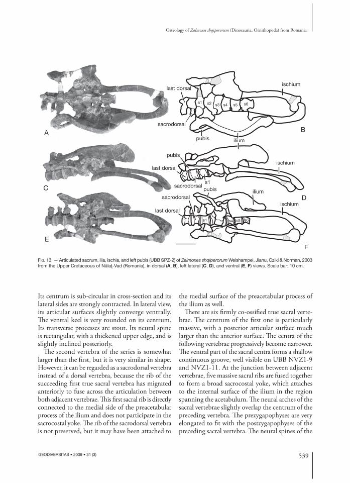
Sacrum (Fig. 13) (UBB NVZ1-9, NVZ1-10, NVZ1-11, UBB SPZ-2)UBB SPZ-2 has already been briefl y described and fi gured by Weishampel et al. (2003). However, it appears that there are some minor misinterpretations in their description. Moreover, the preparation of this specimen has been completed in the meantime. Th erefore, it has been decided to include this mate-rial in the description of Z. shqiperorum. Despite its fusion to the sacrum, the fi rst vertebra of the series can be interpreted as the last dorsal vertebra.
539
Osteology of Zalmoxes shqiperorum (Dinosauria, Ornithopoda) from Romania
GEODIVERSITAS • 2009 • 31 (3)
A
C
E
B
D
F
ischium
iliumpubis
last dorsal
sacrodorsal
ischium
ischium
iliumpubis
sacrodorsal
s1
s1
s1 s2 s4s3
s4
s5
s5
s6
s6
sacrodorsal
last dorsal
last dorsal
pubis
FIG. 13. — Articulated sacrum, ilia, ischia, and left pubis (UBB SPZ-2) of Zalmoxes shqiperorum Weishampel, Jianu, Cziki & Norman, 2003 from the Upper Cretaceous of Nălaţ-Vad (Romania), in dorsal (A, B), left lateral (C, D), and ventral (E, F) views. Scale bar: 10 cm.
Its centrum is sub-circular in cross-section and its lateral sides are strongly contracted. In lateral view, its articular surfaces slightly converge ventrally. Th e ventral keel is very rounded on its centrum. Its transverse processes are stout. Its neural spine is rectangular, with a thickened upper edge, and is slightly inclined posteriorly.
Th e second vertebra of the series is somewhat larger than the fi rst, but it is very similar in shape. However, it can be regarded as a sacrodorsal vertebra instead of a dorsal vertebra, because the rib of the succeeding fi rst true sacral vertebra has migrated anteriorly to fuse across the articulation between both adjacent vertebrae. Th is fi rst sacral rib is directly connected to the medial side of the preacetabular process of the ilium and does not participate in the sacrocostal yoke. Th e rib of the sacrodorsal vertebra is not preserved, but it may have been attached to
the medial surface of the preacetabular process of the ilium as well.
Th ere are six fi rmly co-ossifi ed true sacral verte-brae. Th e centrum of the fi rst one is particularly massive, with a posterior articular surface much larger than the anterior surface. Th e centra of the following vertebrae progressively become narrower. Th e ventral part of the sacral centra forms a shallow continuous groove, well visible on UBB NVZ1-9 and NVZ1-11. At the junction between adjacent vertebrae, fi ve massive sacral ribs are fused together to form a broad sacrocostal yoke, which attaches to the internal surface of the ilium in the region spanning the acetabulum. Th e neural arches of the sacral vertebrae slightly overlap the centrum of the preceding vertebra. Th e prezygapophyses are very elongated to fi t with the postzygapophyses of the preceding sacral vertebra. Th e neural spines of the
540 GEODIVERSITAS • 2009 • 31 (3)
Godefroit P. et al.
FIG. 14. — Caudal vertebra (UBB NVZ1-14) of Zalmoxes shqiperorum Weishampel, Jianu, Cziki & Norman, 2003 from the Upper Cre-taceous of Nălaţ-Vad (Romania), in anterior (A), dorsal (B), and left lateral (C) views. Scale bar: 4 cm.
Aprezygapophysis
postzygapophysis
prezygapophysis
caudal rib
B
C
sacrals are not well preserved, but they were low, rectangular in lateral view, and their thickened upper edges were at least partially fused together.
Caudal vertebrae (Fig. 14) (UBB NVZ1-8, NVZ1-14, NVZ-8, NVZ-15, NVZ-16)Th e articular surfaces of the caudal vertebrae of Z. shqiperorum are slightly amphicoelous and nearly as wide as high. In lateral view, the proximal and distal articular surfaces are sub-parallel and inclined posteriorly. Th e distal articular surface is higher than the proximal surface and the ventral margin, between both articular surfaces, is strongly concave. Th e haemapophyseal facets are more distinct on the ventral surface of UBB NVZ1-8 than in the more proximal vertebrae. A pair of longitudinal ridges joins the haemapophyseal facets and the ventral surface of the centrum looks consequently furrowed. Th e slender caudal ribs are fused to the sides of the centra along the neurocentral suture. Th e pre- and postzygapophyseal processes become more elongated as the size of the neural arch diminishes and as the centrum becomes proportionally more elongated, and the zygapophyseal surfaces become nearly vertical.
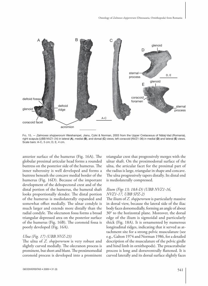
Scapula (Fig. 15A-C) (UBB NVZ1-21, NVZ1-24, NVZ2-2)As previously described by Weishampel et al. (2003), the scapula of Z. shqiperorum is markedly diff erent
from that of Z. robustus. Th e shaft of the scapular blade is very narrow and strap-like. Its posterodorsal end sharply expands posteriorly. Th e ventral end of the scapula is also much expanded. Th e anteroventral acromial region is fl ared and its lateral surface bears a strong deltoid ridge, which limits a vast deltoid fossa (Fig. 15A). Both the sutural surface for the coracoid and the glenoid part of the scapula are thick, moderately concave and very rugose.
Coracoid (Fig. 15D, E) (UBB NVZ1)Th e coracoid is characterized by its unusually long and prominent posterior sternal process, as previ-ously described by Weishampel et al. (2003). Th e glenoid facet is reniform and concave at its centre. Th e glenoid facet forms a prominent lip on the lat-eral surface of the coracoid. Th e coracoid foramen perforates the thickest part of the bone, beneath the scapulocoracoid suture.
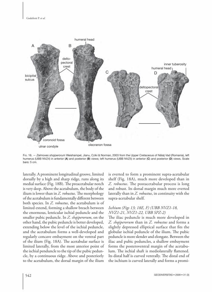
Humerus (Fig. 16) (UBB NVZ2-3, NVZ2-4, NVZ-5)Th e humerus of Z. shqiperorum diff ers from that of Z. robustus by the important development of its deltopectoral crest, which extends over the proximal end of the humerus. Th e edge of the deltopecto-ral crest is regularly concave and turned medially, and its distal tip is very salient. Th e deltopectoral crest therefore limits a wide bicipital sulcus on the
541
Osteology of Zalmoxes shqiperorum (Dinosauria, Ornithopoda) from Romania
GEODIVERSITAS • 2009 • 31 (3)
FIG. 15. — Zalmoxes shqiperorum Weishampel, Jianu, Cziki & Norman, 2003 from the Upper Cretaceous of Nălaţ-Vad (Romania), right scapula (UBB NVZ1-24) in lateral (A), medial (B), and dorsal (C) views; left coracoid (NVZ1-36) in medial (D) and lateral (E) views. Scale bars: A-C, 5 cm; D, E, 4 cm.
A B C
D
E
deltoid fossa
deltoid ridgeglenoid
glenoid
glenoid
coracoid facet
coracoid foramen
acromion
A-C
D, Esternalprocess
sternalprocess
anterior surface of the humerus (Fig. 16A). Th e globular proximal articular head forms a rounded buttress on the posterior side of the humerus. Th e inner tuberosity is well developed and forms a buttress beneath the concave medial border of the humerus (Fig. 16D). Because of the important development of the deltopectoral crest and of the distal portion of the humerus, the humeral shaft looks proportionally slender. Th e distal portion of the humerus is mediolaterally expanded and somewhat off set medially. Th e ulnar condyle is much larger and extends more distally than the radial condyle. Th e olecranon fossa forms a broad triangular depressed area on the posterior surface of the humerus (Fig. 16B). Th e coronoid fossa is poorly developed (Fig. 16A).
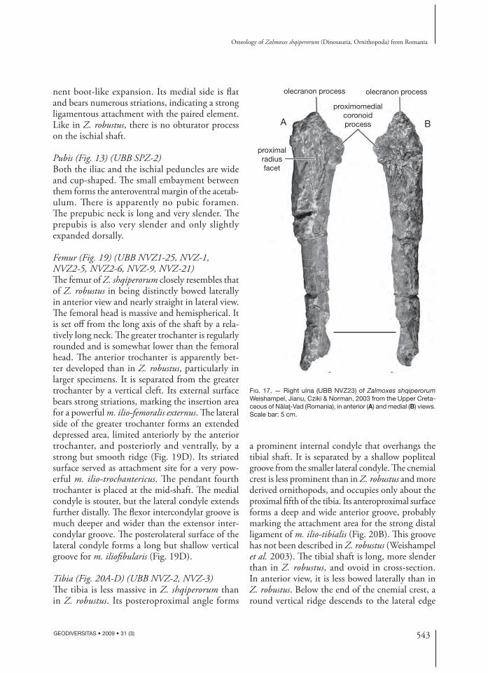
Ulna (Fig. 17) (UBB NVZ-23)Th e ulna of Z. shqiperorum is very robust and slightly curved medially. Th e olecranon process is prominent, but short and blunt. Th e proximo medial coronoid process is developed into a prominent
triangular crest that progressively merges with the ulnar shaft. On the proximodorsal surface of the ulna, the articular facet for the proximal part of the radius is large, triangular in shape and concave. Th e ulna progressively tapers distally. Its distal end is mediolaterally compressed.
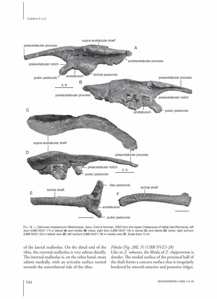
Ilium (Figs 13; 18A-D) (UBB NVZ1-16, NVZ1-17, UBB SPZ-2)Th e ilium of Z. shqiperorum is particularly massive in dorsal view, because the lateral side of the iliac body faces dorsomedially, forming an angle of about 30° to the horizontal plane. Moreover, the dorsal edge of the ilium is sigmoidal and particularly thick (Fig. 18A). It is ornamented by numerous longitudinal ridges, indicating that it served as at-tachment site for a strong pelvic musculature (see e.g., Galton 1974 and Norman 1986, for a detailed description of the musculature of the pelvic girdle and hind limb in ornithopods). Th e preacetabular process is long and dorsoventrally fl attened. It is curved laterally and its dorsal surface slightly faces
542 GEODIVERSITAS • 2009 • 31 (3)
Godefroit P. et al.
A B
C Dbicipitalsulcus
ulnar condyle
coronoid fossa
olecranon fossa
humeral head
humeral headinner tuberosity
delto-pectoral
crest
deltopectoralcrest
FIG. 16. — Zalmoxes shqiperorum Weishampel, Jianu, Cziki & Norman, 2003 from the Upper Cretaceous of Nălaţ-Vad (Romania), left humerus (UBB NVZ4) in anterior (A) and posterior (B) views; left humerus (UBB NVZ5) in anterior (C) and posterior (D) views. Scale bars: 5 cm.
laterally. A prominent longitudinal groove, limited dorsally by a high and sharp ridge, runs along its medial surface (Fig. 18B). Th e preacetabular notch is very deep. Above the acetabulum, the body of the ilium is lower than in Z. robustus. Th e morphology of the acetabulum is fundamentally diff erent between both species. In Z. robustus, the acetabulum is of limited extend, forming a shallow breach between the enormous, lenticular ischial peduncle and the smaller pubic peduncle. In Z. shqiperorum, on the other hand, the pubic peduncle is better developed, extending below the level of the ischial peduncle, and the acetabulum forms a well-developed and regularly concave embayment on the ventral part of the ilium (Fig. 18A). Th e acetabular surface is limited laterally, from the most anterior point of the ischial penduncle to the tip of the pubic pedun-cle, by a continuous ridge. Above and posteriorly to the acetabulum, the dorsal margin of the ilium
is everted to form a prominent supra-acetabular shelf (Fig. 18A), much more developed than in Z. robustus. Th e postacetabular process is long and robust. Its dorsal margin much more everted laterally than in Z. robustus, in continuity with the supra-acetabular shelf.
Ischium (Figs 13; 18E, F) (UBB NVZ1-18, NVZ1-21, NVZ1-22, UBB SPZ-2)Th e iliac peduncle is much more developed in Z. shqiperorum than in Z. robustus and forms a slightly depressed elliptical surface that fi ts the globular ischial peduncle of the ilium. Th e pubic peduncle is more slender and elongate. Between the iliac and pubic peduncles, a shallow embayment forms the posteroventral margin of the acetabu-lum. Th e ischial shaft is mediolaterally fl attened. Its distal half is curved ventrally. Th e distal end of the ischium is curved laterally and forms a promi-
543
Osteology of Zalmoxes shqiperorum (Dinosauria, Ornithopoda) from Romania
GEODIVERSITAS • 2009 • 31 (3)
FIG. 17. — Right ulna (UBB NVZ23) of Zalmoxes shqiperorum Weishampel, Jianu, Cziki & Norman, 2003 from the Upper Creta-ceous of Nălaţ-Vad (Romania), in anterior (A) and medial (B) views. Scale bar: 5 cm.
A B
olecranon process
proximalradiusfacet
olecranon process
proximomedialcoronoidprocess
nent boot-like expansion. Its medial side is fl at and bears numerous striations, indicating a strong ligamentous attachment with the paired element. Like in Z. robustus, there is no obturator process on the ischial shaft.
Pubis (Fig. 13) (UBB SPZ-2)Both the iliac and the ischial peduncles are wide and cup-shaped. Th e small embayment between them forms the anteroventral margin of the acetab-ulum. Th ere is apparently no pubic foramen. Th e prepubic neck is long and very slender. Th e prepubis is also very slender and only slightly expanded dorsally.
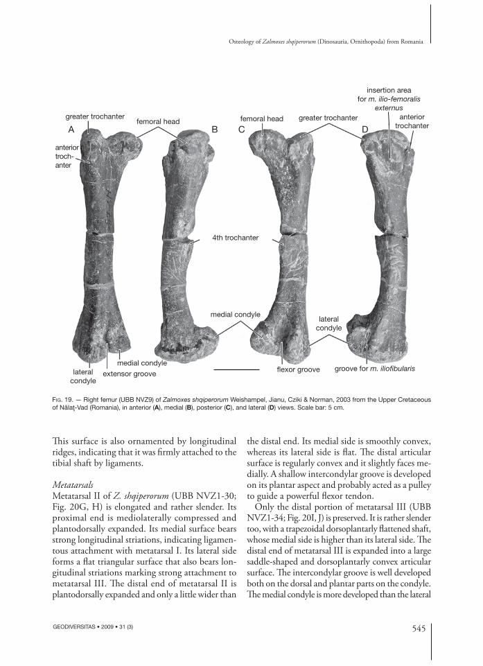
Femur (Fig. 19) (UBB NVZ1-25, NVZ-1, NVZ2-5, NVZ2-6, NVZ-9, NVZ-21)Th e femur of Z. shqiperorum closely resembles that of Z. robustus in being distinctly bowed laterally in anterior view and nearly straight in lateral view. Th e femoral head is massive and hemispherical. It is set off from the long axis of the shaft by a rela-tively long neck. Th e greater trochanter is regularly rounded and is somewhat lower than the femoral head. Th e anterior trochanter is apparently bet-ter developed than in Z. robustus, particularly in larger specimens. It is separated from the greater trochanter by a vertical cleft. Its external surface bears strong striations, marking the insertion area for a powerful m. ilio-femoralis externus. Th e lateral side of the greater trochanter forms an extended depressed area, limited anteriorly by the anterior trochanter, and posteriorly and ventrally, by a strong but smooth ridge (Fig. 19D). Its striated surface served as attachment site for a very pow-erful m. ilio-trochantericus. Th e pendant fourth trochanter is placed at the mid-shaft. Th e medial condyle is stouter, but the lateral condyle extends further distally. Th e fl exor intercondylar groove is much deeper and wider than the extensor inter-condylar groove. Th e posterolateral surface of the lateral condyle forms a long but shallow vertical groove for m. iliofi bularis (Fig. 19D).
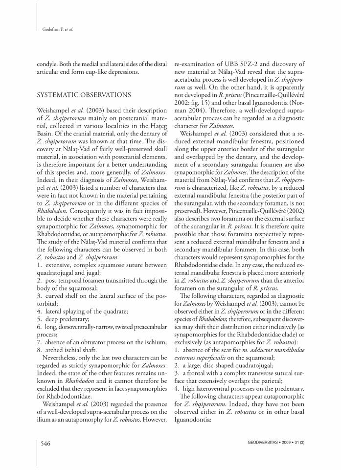
Tibia (Fig. 20A-D) (UBB NVZ-2, NVZ-3)Th e tibia is less massive in Z. shqiperorum than in Z. robustus. Its posteroproximal angle forms
a prominent internal condyle that overhangs the tibial shaft. It is separated by a shallow popliteal groove from the smaller lateral condyle. Th e cnemial crest is less prominent than in Z. robustus and more derived ornithopods, and occupies only about the proximal fi fth of the tibia. Its anteroproximal surface forms a deep and wide anterior groove, probably marking the attachment area for the strong distal ligament of m. ilio-tibialis (Fig. 20B). Th is groove has not been described in Z. robustus (Weishampel et al. 2003). Th e tibial shaft is long, more slender than in Z. robustus, and ovoid in cross-section. In anterior view, it is less bowed laterally than in Z. robustus. Below the end of the cnemial crest, a round vertical ridge descends to the lateral edge
544 GEODIVERSITAS • 2009 • 31 (3)
Godefroit P. et al.
A
B
C
D
E F
preacetabular process
preacetabular process
preacetabular process
postacetabular process
postacetabular process
supra-acetabular shelf
supra-acetabular shelf
preacetabular notch
preacetabular notch
preacetabular notch
pubic peduncle
iliac peduncleischial shaft
pubic peduncle
pubic peduncle
ischial shaft
pubic peduncle
acetabulum
acetabulum
acetabulum
ischial peduncle
A, B
C, D
FIG. 18. — Zalmoxes shqiperorum Weishampel, Jianu, Cziki & Norman, 2003 from the Upper Cretaceous of Nălaţ-Vad (Romania), left ilium (UBB NVZ1-17) in lateral (A) and medial (B) views; right ilium (UBB NVZ1-16) in dorsal (C) and lateral (D) views; right ischium (UBB NVZ1-22) in lateral view (E); left ischium (UBB NVZ1-18) in medial view (F). Scale bars: 5 cm.
of the lateral malleolus. On the distal end of the tibia, the external malleolus is very salient distally. Th e internal malleolus is, on the other hand, more salient medially, with an articular surface turned towards the anterolateral side of the tibia.
Fibula (Fig. 20E, F) (UBB NVZ1-28)Like in Z. robustus, the fi bula of Z. shqiperorum is slender. Th e medial surface of the proximal half of the shaft forms a concave surface that is irregularly bordered by smooth anterior and posterior ridges.
545
Osteology of Zalmoxes shqiperorum (Dinosauria, Ornithopoda) from Romania
GEODIVERSITAS • 2009 • 31 (3)
FIG. 19. — Right femur (UBB NVZ9) of Zalmoxes shqiperorum Weishampel, Jianu, Cziki & Norman, 2003 from the Upper Cretaceous of Nălaţ-Vad (Romania), in anterior (A), medial (B), posterior (C), and lateral (D) views. Scale bar: 5 cm.
A B C D
greater trochanter anteriortrochanter
anteriortroch-anter
greater trochanter
4th trochanter
medial condyle
medial condyle
extensor groove flexor groove
lateralcondyle
lateralcondyle
femoral headfemoral head
insertion areafor m. ilio-femoralis
externus
groove for m. iliofibularis
Th is surface is also ornamented by longitudinal ridges, indicating that it was fi rmly attached to the tibial shaft by ligaments.
Metatarsals Metatarsal II of Z. shqiperorum (UBB NVZ1-30; Fig. 20G, H) is elongated and rather slender. Its proximal end is mediolaterally compressed and plantodorsally expanded. Its medial surface bears strong longitudinal striations, indicating ligamen-tous attachment with metatarsal I. Its lateral side forms a fl at triangular surface that also bears lon-gitudinal striations marking strong attachment to metatarsal III. Th e distal end of metatarsal II is plantodorsally expanded and only a little wider than
the distal end. Its medial side is smoothly convex, whereas its lateral side is fl at. Th e distal articular surface is regularly convex and it slightly faces me-dially. A shallow intercondylar groove is developed on its plantar aspect and probably acted as a pulley to guide a powerful fl exor tendon.
Only the distal portion of metatarsal III (UBB NVZ1-34; Fig. 20I, J) is preserved. It is rather slender too, with a trapezoidal dorsoplantarly fl attened shaft, whose medial side is higher than its lateral side. Th e distal end of metatarsal III is expanded into a large saddle-shaped and dorsoplantarly convex articular surface. Th e intercondylar groove is well developed both on the dorsal and plantar parts on the condyle. Th e medial condyle is more developed than the lateral
546 GEODIVERSITAS • 2009 • 31 (3)
Godefroit P. et al.
condyle. Both the medial and lateral sides of the distal articular end form cup-like depressions.
SYSTEMATIC OBSERVATIONS
Weishampel et al. (2003) based their description of Z. shqiperorum mainly on postcranial mate-rial, collected in various localities in the Haţeg Basin. Of the cranial material, only the dentary of Z. shqiperorum was known at that time. Th e dis-covery at Nălaţ-Vad of fairly well-preserved skull material, in association with postcranial elements, is therefore important for a better understanding of this species and, more generally, of Zalmoxes. Indeed, in their diagnosis of Zalmoxes, Weisham-pel et al. (2003) listed a number of characters that were in fact not known in the material pertaining to Z. shqiperorum or in the diff erent species of Rhabdodon. Consequently it was in fact impossi-ble to decide whether these characters were really synapomorphic for Zalmoxes, synapomorphic for Rhabdodontidae, or autapomorphic for Z. robustus. Th e study of the Nălaţ-Vad material confi rms that the following characters can be observed in both Z. robustus and Z. shqiperorum:1. extensive, complex squamose suture between quadratojugal and jugal;2. post-temporal foramen transmitted through the body of the squamosal;3. curved shelf on the lateral surface of the pos-torbital;4. lateral splaying of the quadrate; 5. deep predentary;6. long, dorsoventrally-narrow, twisted pre acetabular process;7. absence of an obturator process on the ischium;8. arched ischial shaft.
Nevertheless, only the last two characters can be regarded as strictly synapomorphic for Zalmoxes. Indeed, the state of the other features remains un-known in Rhabdodon and it cannot therefore be excluded that they represent in fact synapomorphies for Rhabdodontidae.
Weishampel et al. (2003) regarded the presence of a well-developed supra-acetabular process on the ilium as an autapomorphy for Z. robustus. However,
re-examination of UBB SPZ-2 and discovery of new material at Nălaţ-Vad reveal that the supra-acetabular process is well developed in Z. shqipero-rum as well. On the other hand, it is apparently not developed in R. priscus (Pincemaille-Quillévéré 2002: fi g. 15) and other basal Iguanodontia (Nor-man 2004). Th erefore, a well-developed supra-acetabular process can be regarded as a diagnostic character for Zalmoxes.
Weishampel et al. (2003) considered that a re-duced external mandibular fenestra, positioned along the upper anterior border of the surangular and overlapped by the dentary, and the develop-ment of a secondary surangular foramen are also synapomorphic for Zalmoxes. Th e description of the material from Nălaţ-Vad confi rms that Z. shqipero-rum is characterized, like Z. robustus, by a reduced external mandibular fenestra (the posterior part of the surangular, with the secondary foramen, is not preserved). However, Pincemaille-Quillévéré (2002) also describes two foramina on the external surface of the surangular in R. priscus. It is therefore quite possible that those foramina respectively repre-sent a reduced external mandibular fenestra and a secondary mandibular foramen. In this case, both characters would represent synapomorphies for the Rhabdodontidae clade. In any case, the reduced ex-ternal mandibular fenestra is placed more anteriorly in Z. robustus and Z. shqiperorum than the anterior foramen on the surangular of R. priscus.
Th e following characters, regarded as diagnostic for Zalmoxes by Weishampel et al. (2003), cannot be observed either in Z. shqiperorum or in the diff erent species of Rhabdodon; therefore, subsequent discover-ies may shift their distribution either inclusively (as synapomorphies for the Rhabdodontidae clade) or exclusively (as autapomorphies for Z. robustus):1. absence of the scar for m. adductor mandibulae externus superfi cialis on the squamosal;2. a large, disc-shaped quadratojugal;3. a frontal with a complex transverse sutural sur-face that extensively overlaps the parietal;4. high lateroventral processes on the predentary.
Th e following characters appear autapomorphic for Z. shqiperorum. Indeed, they have not been observed either in Z. robustus or in other basal Iguanodontia:
547
Osteology of Zalmoxes shqiperorum (Dinosauria, Ornithopoda) from Romania
GEODIVERSITAS • 2009 • 31 (3)
A B
C D
E F
G
H
I
J
cnemial crest
externalmalleolus
proximaltibial facet
externalmalleolus
externalmalleolus
internalmalleolus
internalmalleolus
internalmalleolus
cnemialcrest
lateral condylepropliteal groove
anterior grooveinternal condyleinternal condyle
FIG. 20. — Zalmoxes shqiperorum Weishampel, Jianu, Cziki & Norman, 2003 from the Upper Cretaceous of Nălaţ-Vad (Romania), left tibia (UBB NVZ3) in lateral (A), anterior (B), medial (C), and posterior (D) views; ?left fi bula (UBB NVZ1-28) in lateral (E) and medial (F) views; left metatarsal II (UBB NVZ1-30) in medial (G) and lateral (H) views; ?left metatarsal III (UBB NVZ1-34) in plantar (I) and ?medial (J) views. Scale bars: A-D, 5 cm; E, F, 4 cm; G-J, 3 cm.
548 GEODIVERSITAS • 2009 • 31 (3)
Godefroit P. et al.
1. the occipital condyle is not separated from the sphenooccipital tubercles by a distinct neck;2. the dentary bears an angular buccal emargination that forms a horizontal platform extending for the full length of the dentition behind and medial to the coronoid process (Weishampel et al. 2003);3. the scapular blade is narrow and strap-like proxi-mally and expands sharply posterodistally (Wei-shampel et al. 2003);4. the region of the scapula adjacent to the cora-coid suture is expanded; acromial process forming a prominent fl ange (Weishampel et al. 2003);5. the deltopectoral crest of the humerus is par-ticularly prominent, extending over the proximal half of the humerus;6. the iliac peduncle of the ischium is particularly developed;7. the distal end of the ischium forms a boot-like expansion (Weishampel et al. 2003).Th e description of the Nălaţ-Vad material reveals that Z. shqiperorum potentially diff ers from Z. ro-bustus by a series of other characters, listed below. However, these characters cannot be adequately quantifi ed in the current state of our knowledge, because we lack more precise information about their intraspecifi c variability. Moreover, their state remains ambiguous:1. the jugal appears more gracile in Z. shqiperorum;2. the parietal process of the squamosal is appar-ently shorter, but higher;3. the infratemporal notch on the squamosal ap-pears lower, but deeper;4. the post-temporal foramen is set more ante-riorly;5. the sagittal crest is better developed;6. the dentary is more massively constructed;7. the oral margin of the predentary is apparently characterized by a sharper outer margin;8. the anterior trochanter is better developed on the femur;9. the tibia appears less massive.
SYMPATRIC SPECIES IN ORNITHOPODS
Th e observed morphological diff erences between Z. robustus and Z. shqiperorum appear, in any case,
suffi cient to support the validity of both species. Th ere is no indication that these diff erences refl ect ontogenetic variability within a single species: smaller and larger individuals are clearly represented in both groups. Th e diff erences between Z. robustus and Z. shqiperorum also appear too important to simply refl ect sexual dimorphism. As observed in lambeosaurines (Dodson 1975), in basal necera-topsians (Dodson 1976), or in ceratopsids (Lehman 1990; Sampson et al. 1997), dinosaur males and females may not be distinguished by the presence or absence of osteological characters, but instead by the diff erence in development of cranial super-structures (crests, horns, frills) at a given size. It is now usually admitted that these structures primarily functioned as signals to recognize and to compete for mate or territory. Such characters are subject to delayed growth, developing only after the onset of adult size (Dodson 1975; Sampson et al. 1997). Sexual dimorphism in the postcranial skeleton has not been well studied yet. However, Raath (1990) tentatively explained the morphological variation in the robustness of the postcranial skeleton of the basal theropod Syntarsus by sexual dimorphism: he suggested that the most robust morph might repre-sent females. In Zalmoxes, important diff erences in the skull element and in the postcranium between the two supposed species are not simple variations in the development of supracranial structures or in the robustness of the bones, but true osteological. For examples, it seems diffi cult to explain the observed diff erences in the morphology of the basioccipital, the scapula or the ischium by sexual dimorphism: these characters cannot be recognized as visual signals. Moreover, these features were apparently already developed in juveniles, before individuals have reached sexual maturity.
Figure 1 represents the geographic distribution of Z. robustus and Z. shqiperorum. Both species have been discovered together in several localities (Sănpetru, Tustea, Valioara, Vurpar) and, so far, there is no indication that they were found in dif-ferent levels. Although they clearly occupied very similar ecological niches and probably competed for the same food resources, the two species there-fore appear sympatric. As discussed below, it is not the fi rst time that two closely-related species of an
549
Osteology of Zalmoxes shqiperorum (Dinosauria, Ornithopoda) from Romania
GEODIVERSITAS • 2009 • 31 (3)
ornithopod genus are described in a same locality and are therefore regarded as sympatric.
At Bernissart (Belgium, Early Cretaceous), two or three specimens of the gracile species Iguanodon atherfi eldensis Hooley, 1925 have been discovered together with more than 20 specimens of the robust I. bernissartensis Boulenger, 1881 (Norman 1986). Paul (2007) proposed that I. bernissartensis and I. atherfi eldensis do not form a monophyletic group and that morphological diff erences are suffi cient to include the latter species into a separate genus, Mantellisaurus Paul, 2007. However, he conducted no phylogenetic analysis proving that I. bernissartensis and I. atherfi eldensis are really paraphyletic. On the other hand, in the most recent phylogenetic analysis of the group, Norman (2004: fi g. 19.22) suggested that both species may be regarded as monophyletic and, if this hypothesis is correct, it is certainly not necessary to split them into diff erent genera. In any case, it is defi nitely not proved that I. bernis-sartensis and I. atherfi eldensis really lived together in the Bernissart area. Indeed, the Bernissart fossil locality is apparently an attritional accumulation of animals dead in diff erent places and at diff erent times. During Early Cretaceous times, the Bernissart site may have been somewhat lower topographi-cally than the surrounding countryside because of subterranean dissolution, which should explain the preferential accumulation of fossils at Bernis-sart (Delmer & Van Wichelen 1980). However, these two species have also been described together in many other Early Cretaceous formations from Western Europe, suggesting that they were really sympatric in this area. Although the holotype of I. atherfi eldensis Hooley, 1825 (BMNH R5764) was collected from the Vectis Formation, most of the Iguanodon specimens collected in the south-west coast of the Isle of Wight, including partial skeletons and isolated bones of both I. atherfi eldensis and I. bernissartensis, were found in diff erent points of the underlying Wessex Formation (Martill & Naish 2001). Th e Smokejacks Pit locality (Surrey, UK) has also yielded good material belonging to both I. atherfi eldensis and I. bernissartensis (Benton & Spencer 1995). Both species were also reported from the Hastings Beds and Weald Clay of West Sussex, with I. anglicus Holl, 1829 (regarded as valid
by Norman & Weishampel [1990] and Norman [2004]), in the Tilgate Forest area, the historical place where the fi rst dinosaur specimens were dis-covered in the 19th Century (Benton & Spencer 1995; Weishampel et al. 2004). Unfortunately, because the quarries have been closed for a long time, most of the discovery sites remain uncer-tain. Th e Hastings Beds (Berriasian-Vallanginian) that outcrop along the coast east of Hastings (East Sussex, United Kingdom) are also famous for their vertebrate remains. Th ree Iguanodon species have been described from this area: I. dawsoni Lydekker, 1888 and I. fi ttoni Lydekker, 1889, from Shorden Quarry, and I. hollingtonensis Lydekker, 1889, from Hollington Quarry. Norman & Weishampel (1990) and Norman (2004) synonymised I. hollingtonensis with I. fi ttoni and accepted I. dawsoni as valid, but do not elucidate the diff erences of these poorly known taxa from the typical I. bernissartensis and I. atherfi eldensis. In France, Iguanodon specimens were found in Early Cretaceous deposits from the Saint-Dizier region (Haute-Marne). However, the two species were found in diff erent localities: I. bernissartensis at Baudonvilliers, Pont-Varin and Saint-Dizier, and I. atherfi eldensis at Wassy and Coursancelles (Martin & Buff etaut 1992; É. Buf-fetaut pers. comm. 2006). Adult and numerous juvenile remains of both I. atherfi eldensis and I. ber-nissartensis were discovered in a cavern-like deposit at a quarry near the village of Nehden in Sauerland (Germany). Contrary to the Bernissart assemblage, the Nehden fauna is dominated by I. atherfi eldensis specimens and by juveniles. According to Norman (1987) and Norman et al. (1987), the assemblage at Nehden may represent a catastrophic accumula-tion caused by a fl ash fl ood or by a herd crossing a river. However, the over-representation of juvenile specimens in a fossil assemblage is a good indicator for an attritional death profi le (Lyman 1994): the observed peaks corresponding to ages where mor-tality rates are the highest, among the very young and, to a lesser extent, the very old. Th erefore, it cannot be excluded that the Nehden assemblage represents, like Bernissart, an allochtonous accu-mulation of animals dead in diff erent places and times. Fragmentary remains identifi ed as I. bernis-sartensis and I. atherfi eldensis (“I. mantelli”) have
550 GEODIVERSITAS • 2009 • 31 (3)
Godefroit P. et al.
been described together in the Camarillas Forma-tion (Barremian) of the San Cristobal and Santa Barbara localities (Teruel Province, Spain; Sanz et al. 1984a, b). However, the described material appears poorly diagnosed and the identifi cation is mainly based on the size of the bones. Moreover, Ruiz-Omeñaca & Canudo (2004) consider that the material from Teruel Province previously re-ferred to as I. bernissartensis would in fact belong to a new iguanodontian genus. In conclusion, the distribution of the diff erent Iguanodon species clearly shows the diffi culty to decide whether the presence of two species within a same formation results from the sympatry of these species or from taphonomic processes.
Th e famous Quarry 13 in the Morrison Formation near Como (Wyoming) has yielded the holotypes of four Camptosaurus Marsh, 1885 species: C. dispar (Marsh, 1879), C. medius Marsh, 1894, C. nanus Marsh, 1894 and C. browni Gilmore, 1909. Nor-man & Weishampel (1990), and Norman (2004) considered these taxa as synonyms, but regarded C. amplus (Marsh, 1879), from the neighbouring Quarry 1A at Como Bluff , as a valid species. How-ever, this latter taxon is based on a pes, which closely resembles that of theropod dinosaurs.
Th e Dinosaur Park Formation of Alberta (Canada) has yielded at least eight valid hadrosaurid species (Ryan & Evans 2004), including two valid species of Lambeosaurus Parks, 1923 (L. lambei Parks, 1923 and L. magnicristatus Sternberg, 1935) and two species referred to Gryposaurus Lambe, 1914 (G. notabilis Lambe, 1914 and G. incurvimanus Parks, 1920). Recent data on the stratigraphic position of articulated and associated skeletons within the Dinosaur Park Formation indicate that Lambeosaurus magnicristatus has no stratigraphic overlap with L. lambei and suggests that it replaces L. lambei on a regional scale in southern Alberta (Evans & Reisz 2007). Currie & Russell (2004) also indicate that all Gryposaurus incurvimanus specimens are higher in the Dinosaur Park Formation than specimens of G. notabilis.
Two species are also currently recognized within Rhabdodon, the sister-taxon of Zalmoxes. Rhabdodon priscus is recorded in various formations and locali-ties from Aude, Hérault, Bouches-du-Rhônes, Var,
Ariège and Gard in Southern France. Rhabdodon septimanicus is currently known by a single right dentary, from the “Grès à Reptiles” formation in Montou liers (Hérault). Two species of Rhabdodon therefore apparently coexist in the “Grès à Reptiles” formation of the Saint-Chinian area in Hérault. Although R. septimanicus was doubted by some authors (Allain & Pereda-Suberbiola 2003), it was accepted by Weishampel et al. (2003) and Norman (2004). In any case, Buff etaut (2005) indicated that the abundant Rhabdodon material from the Saint-Chinian area apparently includes both a robust and a gracile form. A detailed study of Rhabdodon mate-rial is currently in progress, which should provide a fi nal answer to the question of how many species of Rhabdodon are present in the Late Cretaceous of southern France (É. Buff etaut and P. Chanthasit pers. comm. November 2006).
CONCLUSIONS
Th e new fossils discovered in the Sănpetru Formation at Nălaţ-Vad include the most complete skeleton that can be referred to date to Zalmoxes shqiperorum. Th e observed morphological diff erences between Z. robustus and Z. shqiperorum cannot be adequately explained by ontogenetic variation or sexual di-morphism. Because Z. robustus and Z. shqiperorum were discovered together in several localities, their sympatry is therefore a hypothesis that cannot be a priori rejected. Th e sympatry of closely-related species was apparently not an isolated case among ornithopod dinosaurs, even if it is not always easy to decide whether the presence of species belonging to a same genus within a same locality is the result of true sympatry, of taphonomic processes, or of imprecision in the collecting information.
AcknowledgementsWe wish to thank all the participants to the 2002 fi eld campaign at Nălaţ-Vad: Paul Dica, Cristina Fărcas, Emmanoil and Liana Săsăran, Th ierry Smith, Pierre Bultynck, Annelise Folie, Jimmy Van Itter-beeck, Suzanne Clinet, Dan Grigorescu and Zoltan Cziki. Th e fi eldwork was supported by a research
551
Osteology of Zalmoxes shqiperorum (Dinosauria, Ornithopoda) from Romania
GEODIVERSITAS • 2009 • 31 (3)
project MO/38/004 from the Belgian Federal Science Policy Offi ce. Field vehicles were kindly provided by Ford NV. We would also like to thank the reviewers for their useful comments.
REFERENCES
ALLAIN R. & PEREDA-SUBERBIOLA X. 2003. — Dinosaurs of France. Comptes Rendus Palevol 2: 27-44.
BENTON M. J. & SPENCER P. S. 1995. — Fossil Reptiles from Great Britain. Chapman and Hall, London, 386 p.
BUFFETAUT É. 2005. — Late Cretaceous vertebrates from the Saint-Chinian area (southern France): a review of previous research and an update on recent fi nds. Acta Palaeontologica Romaniae 5: 39-48.
BUFFETAUT É. & LE LOEUFF J. 1991. — Une nouvelle espèce de Rhabdodon (Dinosauria, Ornithischia) du Crétacé supérieur de l’Hérault (Sud de la France). Comptes Rendus de l’Académie des Sciences, série II, 312: 934-948.
BUNZEL E. 1871. — Die Reptilfauna der Gosau- Formation in der Neuen Welt bei Wiener-Neustadt. Abhandlungen der Kaiserlich-Königlichen Geologischen Reichsanstalt Wien 5: 1-18.
CODREA V., SMITH T., DICA P., FOLIE A., GARCIA G., GODEFROIT P. & VAN ITTERBEECK J. 2002. — Dino-saur egg nests, mammals and other vertebrates from a new Maastrichtian site of the Haţeg Basin (Romania). Comptes Rendus Palevol 1: 173-180.
CURRIE P. J. & RUSSELL D. A. 2004. — Th e geographic and stratigraphic distribution of articulated and associ-ated dinosaur remains, in CURRIE P. J. & KOPPELHUS E. B. (eds), Dinosaur Provincial Park – A Spectacular Ancient Ecosystem Revealed. Indiana University Press, Bloomington and Indianapolis: 537-569.
DELMER A. & VAN WICHELEN P. 1980. — Répertoire des puits naturels connus en terrain houiller du Hainaut. Professional Papers 5: 1-234.
DODSON P. 1975. — Taxonomic implications of rela-tive growth in lambeosaurine hadrosaurs. Systematic Zoology 24: 37-54.
DODSON P. 1976. — Quantitative aspects of relative growth and sexual dimorphism in Protoceratops. Journal of Paleontology 50: 929-940.
EVANS D. C. & REISZ R. R. 2007. — Anatomy and rela-tionships of Lambeosaurus magnicristatus, a crested hadrosaurid dinosaur (Ornithischia) from the Dino-saur Park Formation, Alberta. Journal of Vertebrate Paleontology 27: 373-393.
GALTON P. M. 1974. — Th e ornithischian dinosaur Hypsilophodon from the Wealden of the Isle of Wight. Bulletin of the British Museum (Natural History) Geo-logy 25: 1-152.
GARCIA G., PINCEMAILLE M., VIANEY-LIAUD M.,
MARANDAT B., LORENZ E., CHEYLAN G., CAPPETTA H., MICHAUX J. & SUDRE J. 1999. — Découverte du premier squelette presque complet de Rhabdodon priscus (Dinosauria, Ornithopoda) du Maastrichtien inférieur de Provence. Comptes Rendus de l’Académie des Sciences 328: 415-421.
GODEFROIT P., DONG Z.-M., BULTYNCK P., LI H. & FENG L. 1998. — New Bactrosaurus (Dinosauria: Hadrosauroidea) material from Iren Dabasu (Inner Mongolia, P. R. China). Bulletin de l’Institut royal des Sciences naturelles de Belgique, Sciences de la Terre 68 (supplement): 3-70.
GRIGORESCU D. 1983. — A stratigraphic, taphonomic and palaeoecologic approach to a “forgotten land”: the dinosaur deposits from the Haţeg Basin (Tran-sylvania, Romania). Acta Palaeontologica Polonica 28: 103-121.
GRIGORESCU D. 1992. — Nonmarine Cretaceous for-mations of Romania, in MATEER N. J. & CHEN P.-J. (eds), Aspects of Nonmarine Cretaceous Geology. China Ocean Press, Beijing: 142-164.
GRIGORESCU D., VENCZEL M., CSIKI Z. & LIMBEREA R. 1999. — New microvertebrate fossil assemblages from the uppermost Cretaceous of the Haţeg Basin (Romania). Geologie en Mijnbouw 78: 301-314.
HOLL F. 1829. — Handbuch der Petrefactenkunde. Pt.1. Verlag der Ernst’schen Buchhandlung, Quedlinburg, 232 p.
LAPPARENT A. F. DE 1947. — Les dinosauriens du Crétacé supérieur du Midi de la France. Mémoires de la Société géologique de France 56: 1-54.
LEHMAN T. M. 1990. — Th e ceratopsian subfamily Chasmosaurinae: sexual dimorphism and systemat-ics, in CARPENTER K. & CURRIE P. J. (eds), Dinosaur Systematics: Approaches and Perspectives. Cambridge University Press, Cambridge: 211-230.
LYMAN R. L. 1994. — Vertebrate Taphonomy. Cambridge University Press, Cambridge, 550 p.
MARTILL D. M. & NAISH D. 2001. — Dinosaurs of the Isle of Wight. Th e Palaeontological Association, London, 433 p.
MARTIN V. & BUFFETAUT É. 1992. — Les iguanodons (Ornithischia – Ornithopoda) du Crétacé inférieur de la region de Saint-Dizier (Haute-Marne). Revue de Paléobiologie 11: 67-96.
NOPCSA F. VON 1902. — Dinosaurierreste aus Sieben-bürgen II. (Schädelreste von Mochlodon). Mit einem Anhange: zur Phylogenie der Ornithopodiden. Denk-schriften der königlichen Akademie der Wissenschaften. Mathematisch-Naturwissenschaftlichen Klasse, Wien 72: 149-175.
NOPCSA F. VON 1904. — Dinosaurierreste aus Sieben-bürgen III. Weitere Schädelreste von Mochlodon. Denkschriften der königlichen Akademie der Wissen-schaften.Mathematisch-naturwissenschaftliche Klasse, Wien 74: 229-263.
NOPCSA F. VON 1915. — Die Dinosaurier des Sieben-
552 GEODIVERSITAS • 2009 • 31 (3)
Godefroit P. et al.
bürgischen Landesteiles Ungarns. Mitteilungen aus dem Jahrbuche der königlich ungarischen geologischen Reichsanstalt, Budapest 23: 3-24.
NOPCSA F. VON 1929. — Sexual diff erences in ornitho-podous dinosaurs. Palaeobiologica 2: 187-200.
NORMAN D. B. 1986. — On the anatomy of Iguanodon atherfi eldensis (Ornithischia: Ornithopoda). Bulletin de l’Institut royal des Sciences naturelles de Belgique, Sciences de la Terre 56: 281-372.
NORMAN D. B. 1987. — A mass-accumulation of verte-brates from the Lower Cretaceous of Nehden (Sauer-land), West Germany. Proceedings of the Royal Society of London B 230: 15-255.
NORMAN D. B. 2004. — Basal Iguanodontia, in WEI-SHAMPEL D. B., DODSON P. & OSMÓLSKA H. (eds), Th e Dinosauria, second edition. University of Cali-fornia Press, Berkeley: 413-437.
NORMAN D. B. & WEISHAMPEL D. B. 1990. — Iguano-dontidae and related Ornithopoda, in WEISHAMPEL D. B., DODSON P. & OSMÓLSKA H. (eds), Th e Dinosauria. University of California Press, Berkeley: 510-533.
NORMAN D. B., HILPERT K.-H. & HÖLDER H. 1987. — Die Wirbeltierfauna von Nehden (Sauerland), West-deutschland. Geologie und Paläontologie in Westfalen 8: 1-79.
ÖSI A. 2004. — Th e fi rst dinosaur remains from the Upper Cretaceous of Hungary (Csehbànya, Bakony Mts). Geobios 37: 749-753.
PANAIOTU C. & PANAIOTU C. 2002. — Paleomagnetic studies, in 7th European Workshop on Vertebrate Palae-ontology, Abstracts Volume and Excursions Field Guide. Sibiu, Romania: 59.
PAUL G. S. 2007. — Turning the Old into the New: a separate genus for the gracile iguanodont from the Wealden of England, in CARPENTER K. (ed.), Horns and Beaks. Indiana University Press, Bloomington and Indianapolis: 69-77.
PEREDA-SUBERBIOLA X. & SANZ J. L. 1999. — Th e ornithopod dinosaur Rhabdodon from the Upper Cretaceous of Lãno (Iberian Peninsula). Estudios del Museo de Ciencias Naturales de Alava, Número especial 14: 257-272.
PINCEMAILLE-QUILLÉVÉRÉ M. 2002. — Description d’un squelette partial de Rhabdodon priscus (Euorni-thopoda) du Crétacé Supérieur de Vitrolles (Bouches du Rhône, France). Oryctos 4: 39-70.
PINCEMAILLE-QUILLÉVÉRÉ M., BUFFETAUT É. AND QUIL-LÉVÉRÉ F. 2006. — Osteological description of the braincase of Rhabdodon (Dinosauria, Euornithopoda) and phylogenetic implications. Bulletin de la Société géologique de France 177: 97-104.
RAATH M. A. 1990. — Morphological variation in small theropods and its meaning in systematics: evidence from Syntarsus rhodesiensis, in CARPENTER K. & CUR-RIE P. J. (eds), Dinosaur Systematics: Approaches and Perspectives. Cambridge University Press, Cambridge:
91-105.RUIZ-OMEÑACA J. I. & CANUDO J. I. 2004. — Dino-
saurios ornithopódos cel Cretácico inferior de la Peninsula Ibérica. Geo-Temas 6: 63-65.
RYAN M. J. & EVANS D. C. 2004. — Ornithischian dinosaurs, in CURRIE P. J. & KOPPELHUS E. B. (eds), Dinosaur Provincial Park – A Spectacular Ancient Eco-system Revealed. Indiana University Press, Bloomington and Indianapolis: 312-348.
SACHS S. & HORNUNG J. J. 2006. — Juvenile ornitho-pod (Dinosauria: Rhabdodontidae) remains from the Upper Cretaceous (Lower Campanian, Gosau Group) of Muthmannsdorf (Lower Austria). Geobios 39: 415-425.
SAMPSON S. D., RYAN M. J. & TANKE D. H. 1997. — Craniofacial ontogeny in centrosaurine dinosaurs (Ornithischia: Ceratopsidae): taxonomic and behav-ioural implications. Zoological Journal of the Linnean Society of London 121: 293-337.
SANZ J. L., BUSCALIONI A. D., CASANOVAS M. L. & SANTAFÉ J. V. 1984a. — Th e archosaur fauna from the Upper Jurassic/Lower Cretaceous of Galve (Teruel, Spain). Th ird Symposium on Mesozoic Terrestrial Eco-systems, Tübingen: 201-207.
SANZ J. L., CASANOVAS M. L. & SANTAFÉ J. V. 1984b. — Iguanodóntidos (Reptilia, Ornithopoda) del yacimiento del Cretácico inferior de San Cristóbal (Galve, Teruel). Acta Geologica Hispanica 19: 171-176.
SEELEY H. G. 1881. — Th e reptile fauna of the Gosau Formation preserved in the Geological Museum of the University of Vienna. With a note on the geological horizon of the fossils at Neue Welt, east of Wiener Neustadt. Quarterly Journal of the Geological Society of London 37: 620-707.
SMITH T., CODREA V.A., SSRAN E., VAN ITTERBEECK J., BULTYNCK P., CSIKI Z., DICA P., FRCA C., FO-LIE A., GARCIA G. & GODEFROIT P. 2002. — A new exceptional vertebrate site from the Late Cretaceous of the Haţeg Basin (Romania). Studia Universitatis Babeş-Bolyai, Geologia Special Issue 1: 321-330.
VAN ITTERBEECK J., SSRAN E., CODREA V, SSRAN L. & BULTYNCK P. 2004. — Sedimentology of the Upper Cretaceous mammal and dinosaur bearing sites along the Raŭl Mare and Barbat Rivers, Haţeg Basin, Romania. Cretaceous Research 25: 517-530.
WEISHAMPEL D. B. 1990. — Ornithopoda, in WEI-SHAMPEL D. B., DODSON P. & OSMÓLSKA H. (eds), Th e Dinosauria. University of California Press, Ber-keley: 484-485.
WEISHAMPEL D. B., BARRETT P. M., CORIA R. A., LE LOEUFF J., XU X., ZHAO X., ASHOK S., GOMANI E. M. P. & NOTO C. R. 2004. — Dinosaur distribution, in WEISHAMPEL D. B., DODSON P. & OSMÓLSKA H. (eds), Th e Dinosauria, second edition. University of California Press, Berkeley: 517- 606.
WEISHAMPEL D. B., JIANU C.-M., CZIKI Z. & NORMAN
553
Osteology of Zalmoxes shqiperorum (Dinosauria, Ornithopoda) from Romania
GEODIVERSITAS • 2009 • 31 (3)
D. B. 2003. — Osteology and phylogeny of Zalmoxes (n.g.), an unusual ornithopod dinosaur from the latest Cretaceous of Romania. Journal of Systematic Paleontology 1: 123-143.
WEISHAMPEL D. B., NORMAN D. B. & GRIGORESCU D. 1993. — Telmatosaurus transsylvanicus from the Late Cretaceous of Romania: the most basal hadrosaurid dinosaur. Palaeontology 36: 361-385.
Submitted on 5 October 2007;accepted on 23 December 2008.