33
Osteomyelitis Osteomyelitis
| Date post: | 28-Dec-2015 |
| Category: |
Documents |
| Upload: | melina-dalton |
| View: | 271 times |
| Download: | 0 times |
OsteomyelitisOsteomyelitis
OsteomyelitisOsteomyelitis
Inflammation of bone and marrowInflammation of bone and marrow Types Types
Pyogenic osteomyelitisPyogenic osteomyelitis Tuberculous osteomyelitisTuberculous osteomyelitis
Pyogenic Pyogenic OsteomyelitiOsteomyeliti
ss
Pyogenic osteomyelitisPyogenic osteomyelitis
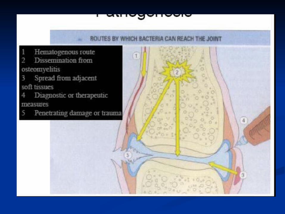
Always caused by bacteriaAlways caused by bacteria Routes of infectionRoutes of infection
Hematogenous spreadHematogenous spread Extension from a contiguous siteExtension from a contiguous site Direct implantationDirect implantation
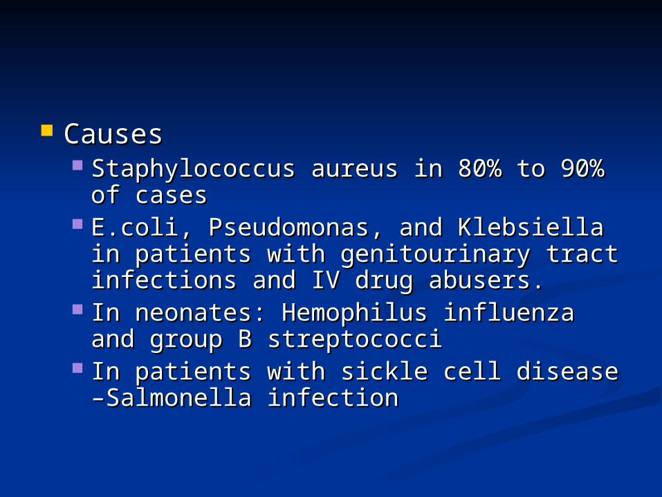
Causes Causes Staphylococcus aureus in 80% to 90% Staphylococcus aureus in 80% to 90%
of casesof cases E.coli, Pseudomonas, and Klebsiella in E.coli, Pseudomonas, and Klebsiella in
patients with genitourinary tract patients with genitourinary tract infections and IV drug abusers.infections and IV drug abusers.
In neonates: Hemophilus influenza and In neonates: Hemophilus influenza and group B streptococcigroup B streptococci
In patients with sickle cell disease –In patients with sickle cell disease –Salmonella infectionSalmonella infection
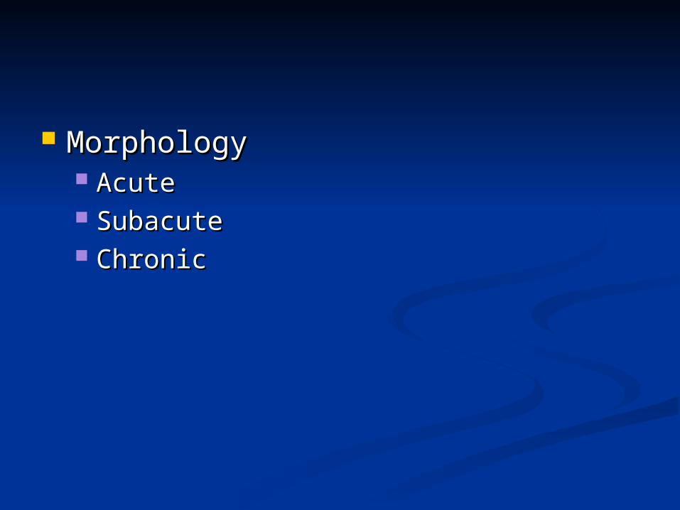
Morphology Morphology Acute Acute Subacute Subacute Chronic Chronic
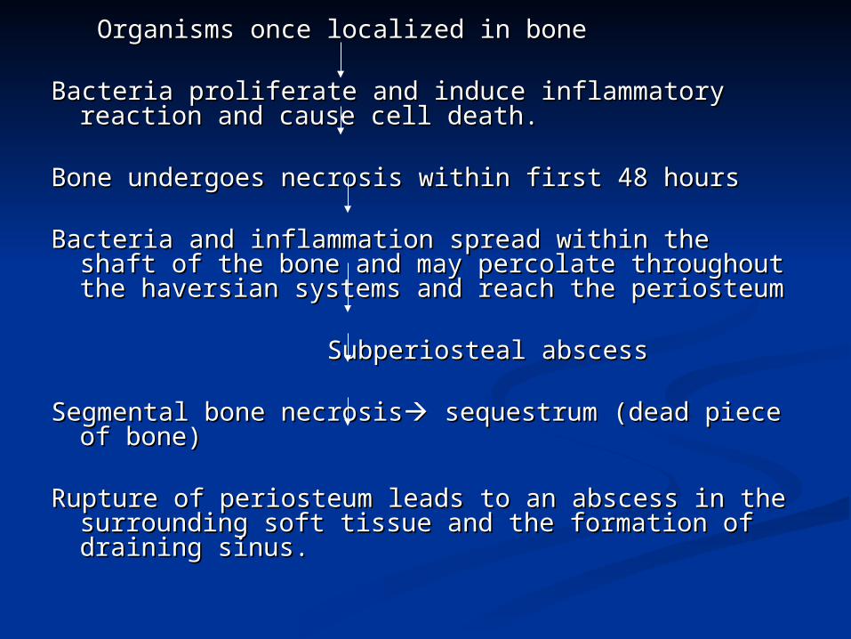
Organisms once localized in boneOrganisms once localized in bone
Bacteria proliferate and induce inflammatory reaction Bacteria proliferate and induce inflammatory reaction and cause cell death.and cause cell death.
Bone undergoes necrosis within first 48 hours Bone undergoes necrosis within first 48 hours Bacteria and inflammation spread within the shaft of Bacteria and inflammation spread within the shaft of
the bone and may percolate throughout the the bone and may percolate throughout the haversian systems and reach the periosteumhaversian systems and reach the periosteum
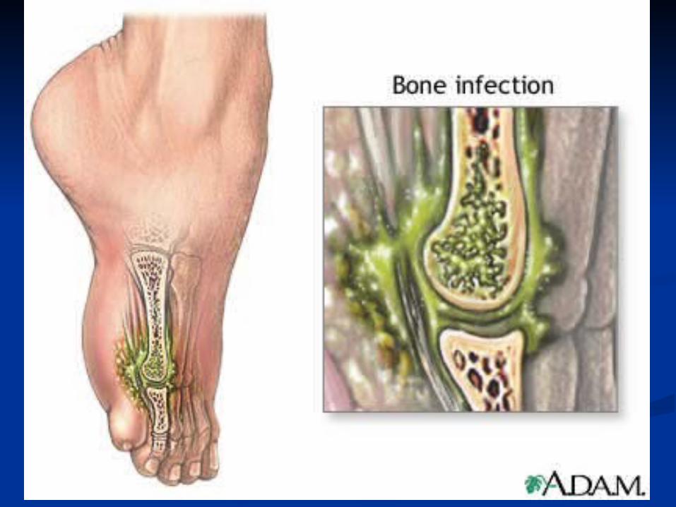
Subperiosteal abscessSubperiosteal abscess
Segmental bone necrosisSegmental bone necrosis sequestrum (dead piece of sequestrum (dead piece of bone)bone)
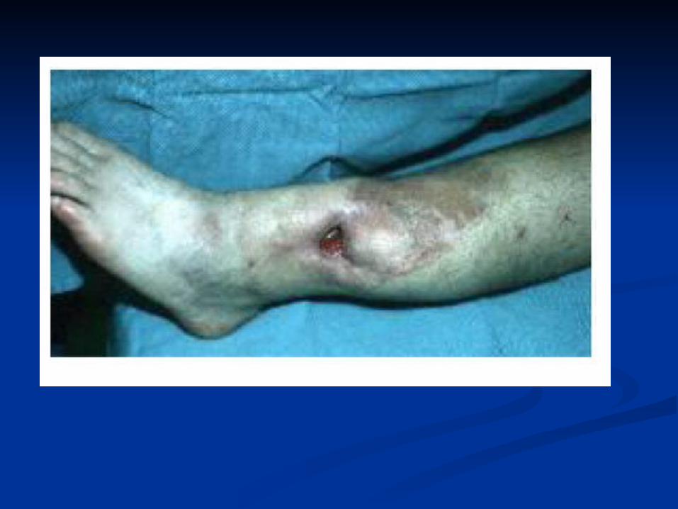
Rupture of periosteum leads to an abscess in the Rupture of periosteum leads to an abscess in the surrounding soft tissue and the formation of draining surrounding soft tissue and the formation of draining sinus.sinus.
Over time, host response developsOver time, host response develops After first week of infection chronic After first week of infection chronic
inflammatory cells become more inflammatory cells become more numerousnumerous
Cytokines from leukocytes stimulates Cytokines from leukocytes stimulates osteoclastic bone resorptionosteoclastic bone resorption ingrowth of ingrowth of fibrous tissuefibrous tissue deposition of reactive deposition of reactive bone in the peripherybone in the periphery
Reactive woven or lamellar bone which Reactive woven or lamellar bone which forms sleeve of living tissue surrounding forms sleeve of living tissue surrounding dead bone is called as involucrum.dead bone is called as involucrum.
Brodie abscess: is a small Brodie abscess: is a small intraosseous abscess that frequently intraosseous abscess that frequently involves the cortex and is walled off involves the cortex and is walled off by reactive boneby reactive bone
Sclerosing osteomyelitis of Garre: Sclerosing osteomyelitis of Garre: typically develops in jaw and is typically develops in jaw and is associated with extensive new bone associated with extensive new bone formation formation
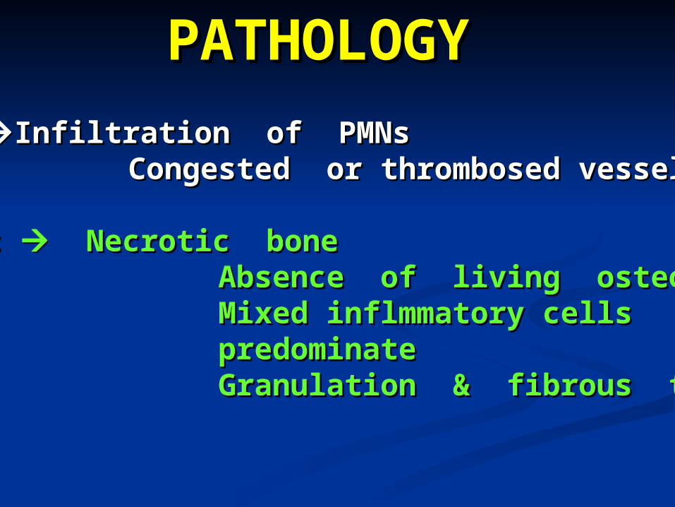
PATHOLOGYPATHOLOGY
Acute Acute Infiltration of PMNsInfiltration of PMNs Congested or thrombosed vesselsCongested or thrombosed vessels
Chronic Chronic Necrotic bone Necrotic bone Absence of living osteocyteAbsence of living osteocyte Mixed inflmmatory cells Mixed inflmmatory cells predominatepredominate Granulation & fibrous tissueGranulation & fibrous tissue
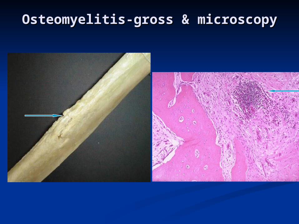
Osteomyelitis-gross & microscopyOsteomyelitis-gross & microscopy
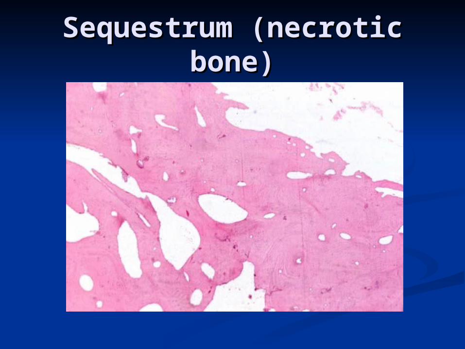
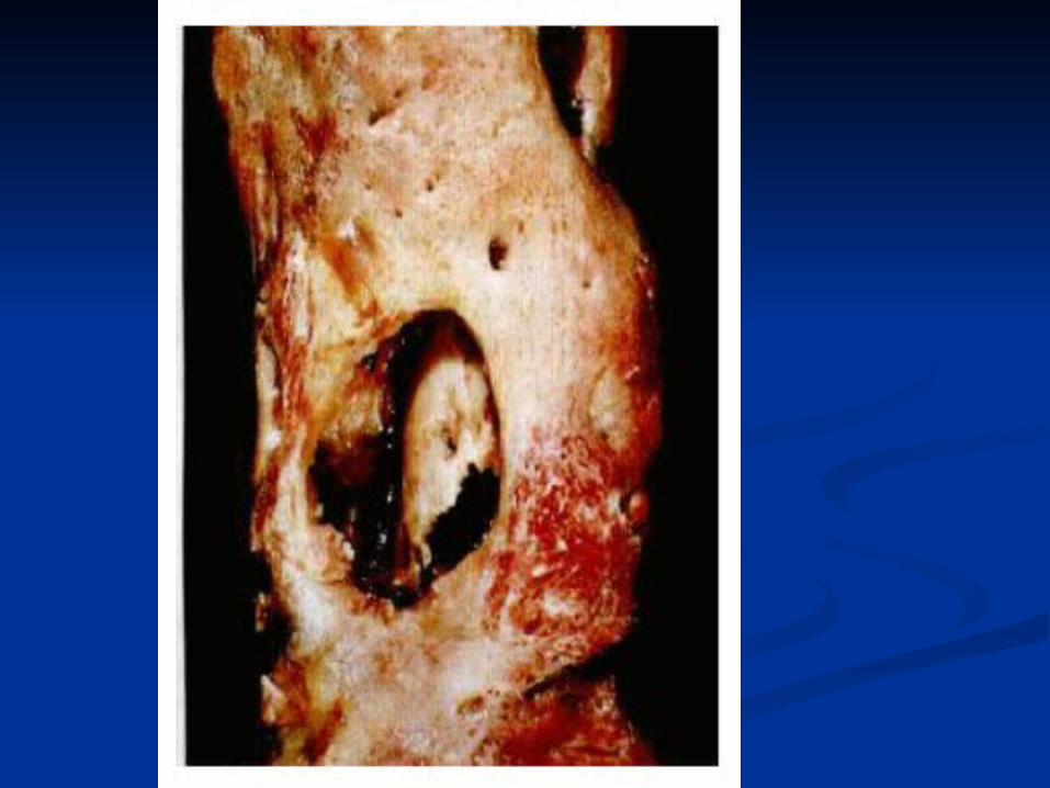
Sequestrum (necrotic Sequestrum (necrotic bone)bone)
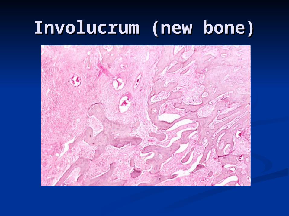
Involucrum (new bone)Involucrum (new bone)
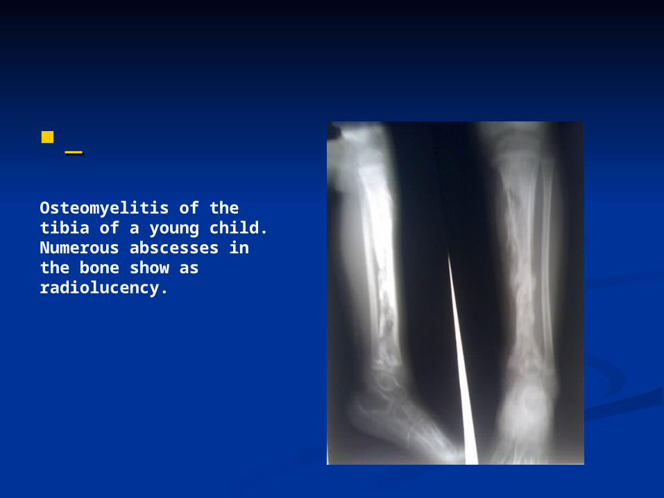
Osteomyelitis of the tibia of a young child. Numerous abscesses in the bone show as radiolucency.
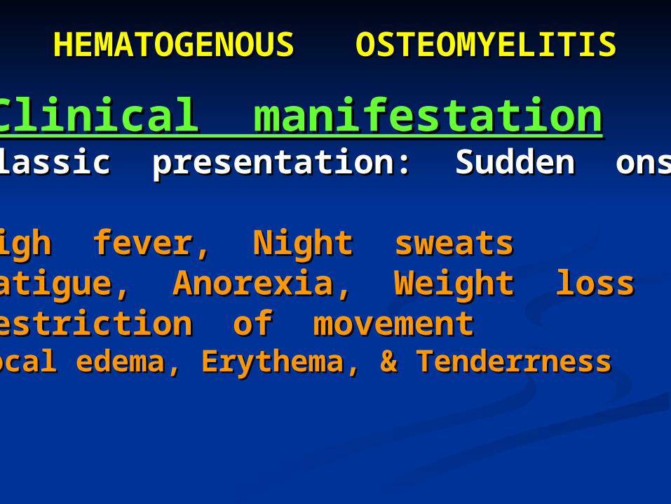
HEMATOGENOUS OSTEOMYELITISHEMATOGENOUS OSTEOMYELITIS
Clinical manifestationClinical manifestationClassic presentation: Sudden onsetClassic presentation: Sudden onset
High fever, Night sweatsHigh fever, Night sweatsFatigue, Anorexia, Weight lossFatigue, Anorexia, Weight lossRestriction of movementRestriction of movementLocal edema, Erythema, & TenderrnessLocal edema, Erythema, & Tenderrness
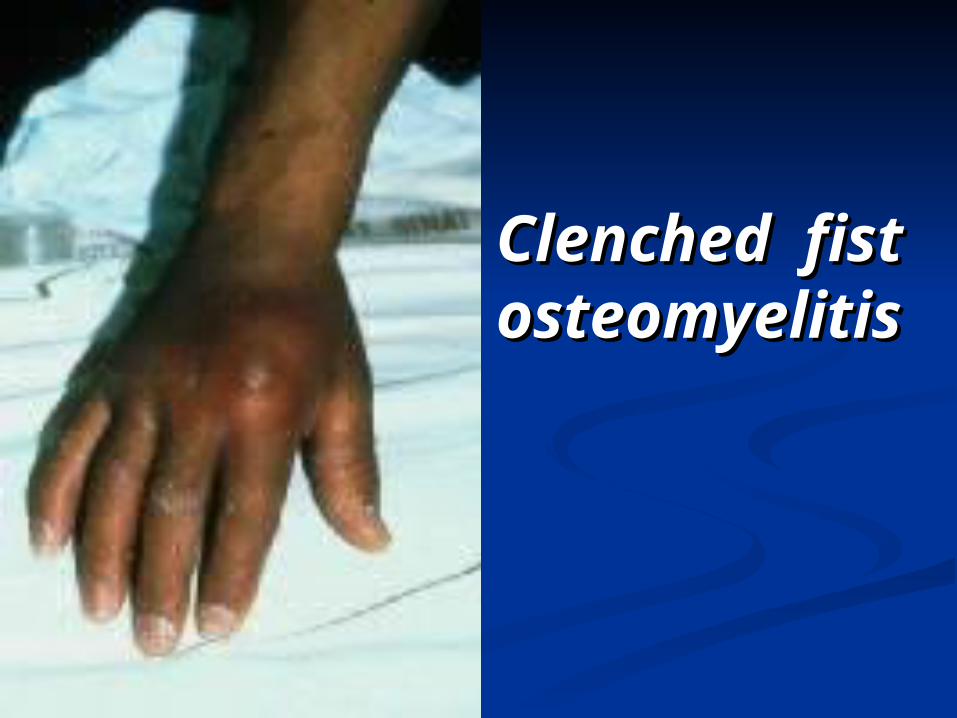
Clenched fist Clenched fist osteomyelitisosteomyelitis
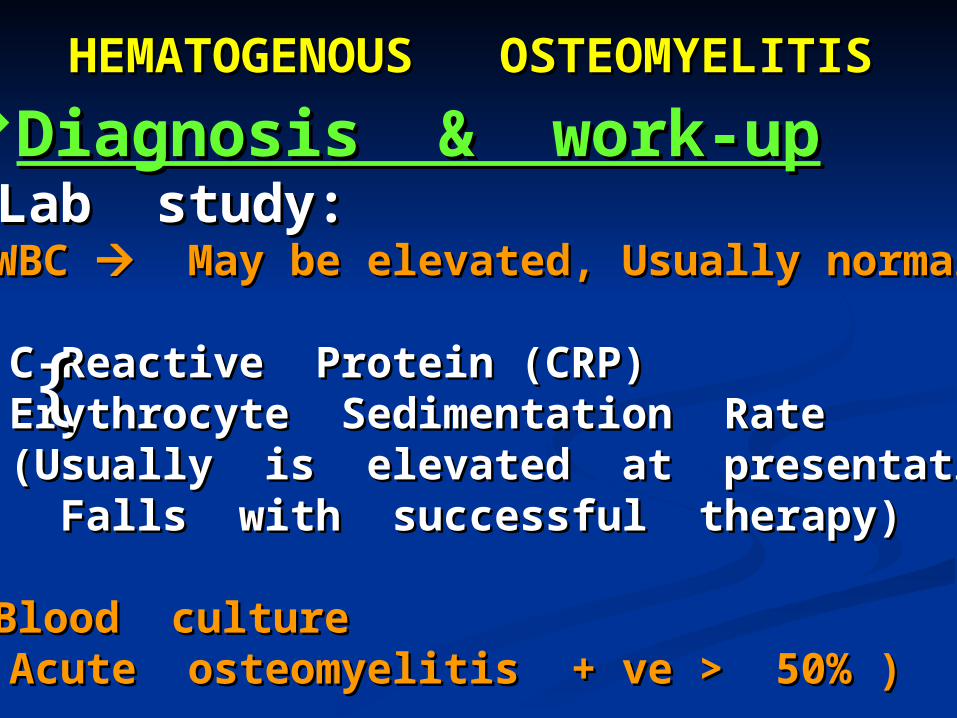
HEMATOGENOUS OSTEOMYELITISHEMATOGENOUS OSTEOMYELITIS
Diagnosis & work-upDiagnosis & work-upLab study:Lab study:
WBC WBC May be elevated, Usually normal May be elevated, Usually normal
C-Reactive Protein (CRP)C-Reactive Protein (CRP) Erythrocyte Sedimentation RateErythrocyte Sedimentation Rate (Usually is elevated at presentation(Usually is elevated at presentation Falls with successful therapy)Falls with successful therapy)
Blood cultureBlood culture( Acute osteomyelitis + ve > 50% )( Acute osteomyelitis + ve > 50% )
{{
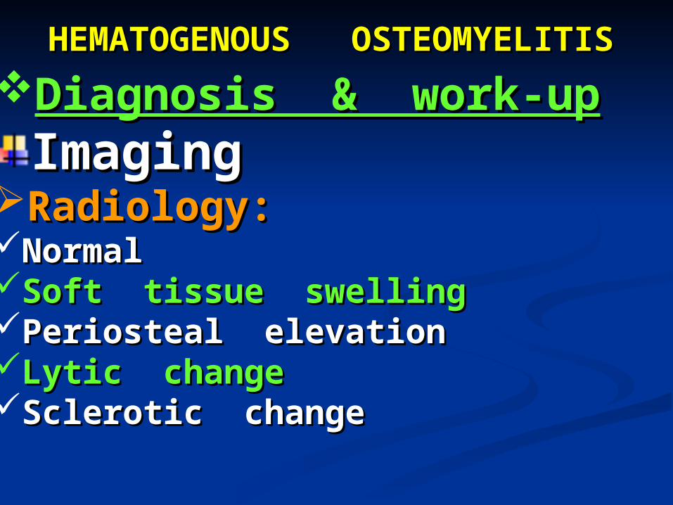
HEMATOGENOUS OSTEOMYELITISHEMATOGENOUS OSTEOMYELITIS
Diagnosis & work-upDiagnosis & work-upImagingImaging
Radiology:Radiology:NormalNormalSoft tissue swellingSoft tissue swellingPeriosteal elevationPeriosteal elevationLytic changeLytic changeSclerotic changeSclerotic change
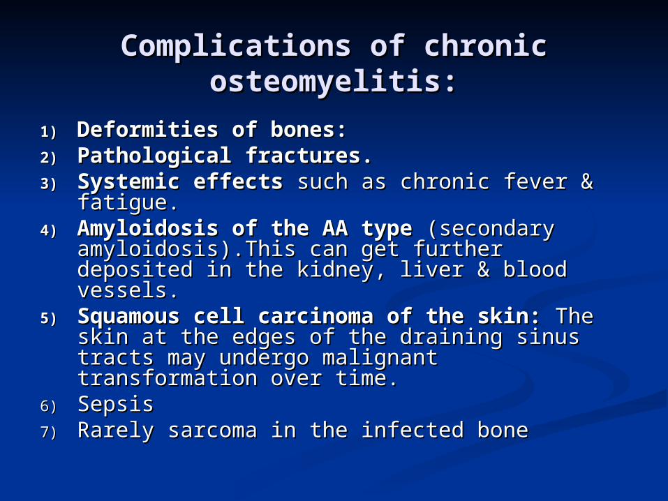
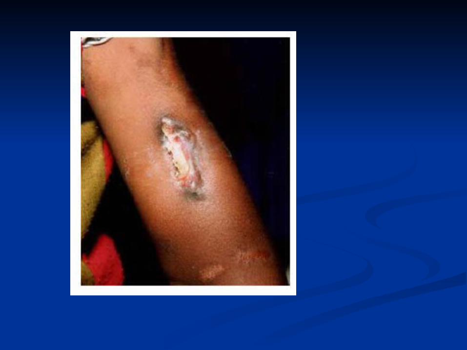
Complications of chronic Complications of chronic osteomyelitis:osteomyelitis:
1)1) Deformities of bones:Deformities of bones: 2)2) Pathological fractures.Pathological fractures.3)3) Systemic effectsSystemic effects such as chronic fever & such as chronic fever &
fatigue.fatigue.4)4) Amyloidosis of the AA typeAmyloidosis of the AA type (secondary (secondary
amyloidosis).This can get further deposited amyloidosis).This can get further deposited in the kidney, liver & blood vessels.in the kidney, liver & blood vessels.
5)5) Squamous cell carcinoma of the skin:Squamous cell carcinoma of the skin: The skin at the edges of the draining sinus The skin at the edges of the draining sinus tracts may undergo malignant tracts may undergo malignant transformation over time.transformation over time.
6)6) Sepsis Sepsis 7)7) Rarely sarcoma in the infected boneRarely sarcoma in the infected bone
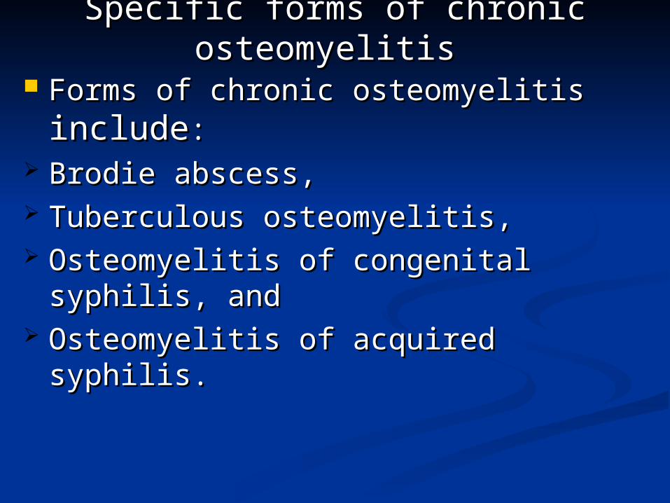
Specific forms of chronic Specific forms of chronic osteomyelitisosteomyelitis
Forms of chronic osteomyelitis Forms of chronic osteomyelitis includeinclude::
Brodie abscess, Brodie abscess, Tuberculous osteomyelitis, Tuberculous osteomyelitis, Osteomyelitis of congenital syphilis, Osteomyelitis of congenital syphilis,
and and Osteomyelitis of acquired syphilis. Osteomyelitis of acquired syphilis.
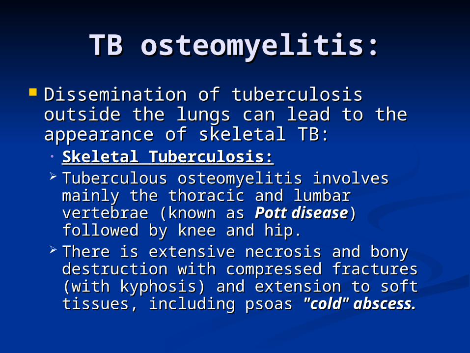
TB osteomyelitis:TB osteomyelitis:
Dissemination of tuberculosis outside Dissemination of tuberculosis outside the lungs can lead to the appearance of the lungs can lead to the appearance of skeletal TB:skeletal TB:• Skeletal Tuberculosis:Skeletal Tuberculosis: Tuberculous osteomyelitis involves mainly Tuberculous osteomyelitis involves mainly
the thoracic and lumbar vertebrae (known the thoracic and lumbar vertebrae (known as as Pott diseasePott disease) followed by knee and hip. ) followed by knee and hip.
There is extensive necrosis and bony There is extensive necrosis and bony destruction with compressed fractures destruction with compressed fractures (with kyphosis) and extension to soft (with kyphosis) and extension to soft tissues, including psoas tissues, including psoas "cold" abscess."cold" abscess.
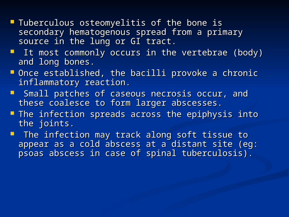
Tuberculous osteomyelitis of the bone is secondary Tuberculous osteomyelitis of the bone is secondary hematogenous spread from a primary source in the hematogenous spread from a primary source in the lung or GI tract.lung or GI tract.
It most commonly occurs in the vertebrae (body) and It most commonly occurs in the vertebrae (body) and long bones. long bones.
Once established, the bacilli provoke a chronic Once established, the bacilli provoke a chronic inflammatory reaction.inflammatory reaction.
Small patches of caseous necrosis occur, and these Small patches of caseous necrosis occur, and these coalesce to form larger abscesses. coalesce to form larger abscesses.
The infection spreads across the epiphysis into the The infection spreads across the epiphysis into the joints.joints.
The infection may track along soft tissue to appear as The infection may track along soft tissue to appear as a cold abscess at a distant site (eg: psoas abscess in a cold abscess at a distant site (eg: psoas abscess in case of spinal tuberculosis). case of spinal tuberculosis).
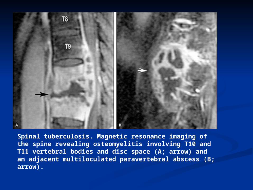
Spinal tuberculosis. Magnetic resonance imaging of the spine revealing osteomyelitis involving T10 and T11 vertebral bodies and disc space (A; arrow) and an adjacent multiloculated paravertebral abscess (B; arrow).
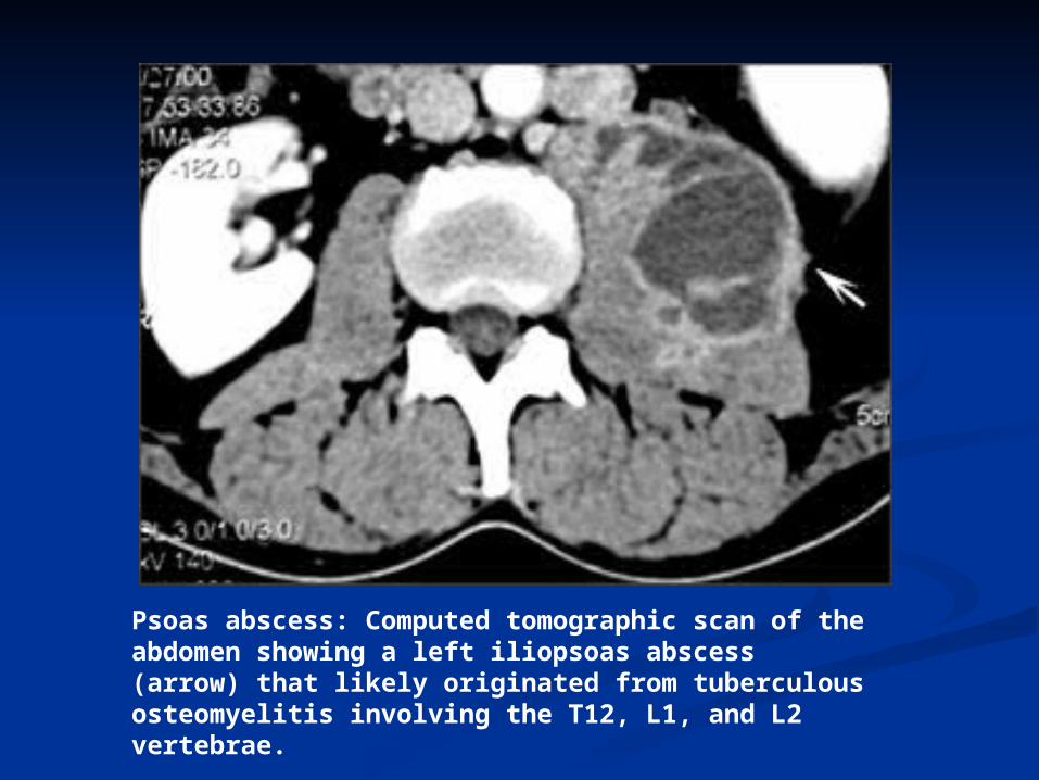
Psoas abscess: Computed tomographic scan of the abdomen showing a left iliopsoas abscess (arrow) that likely originated from tuberculous osteomyelitis involving the T12, L1, and L2 vertebrae.
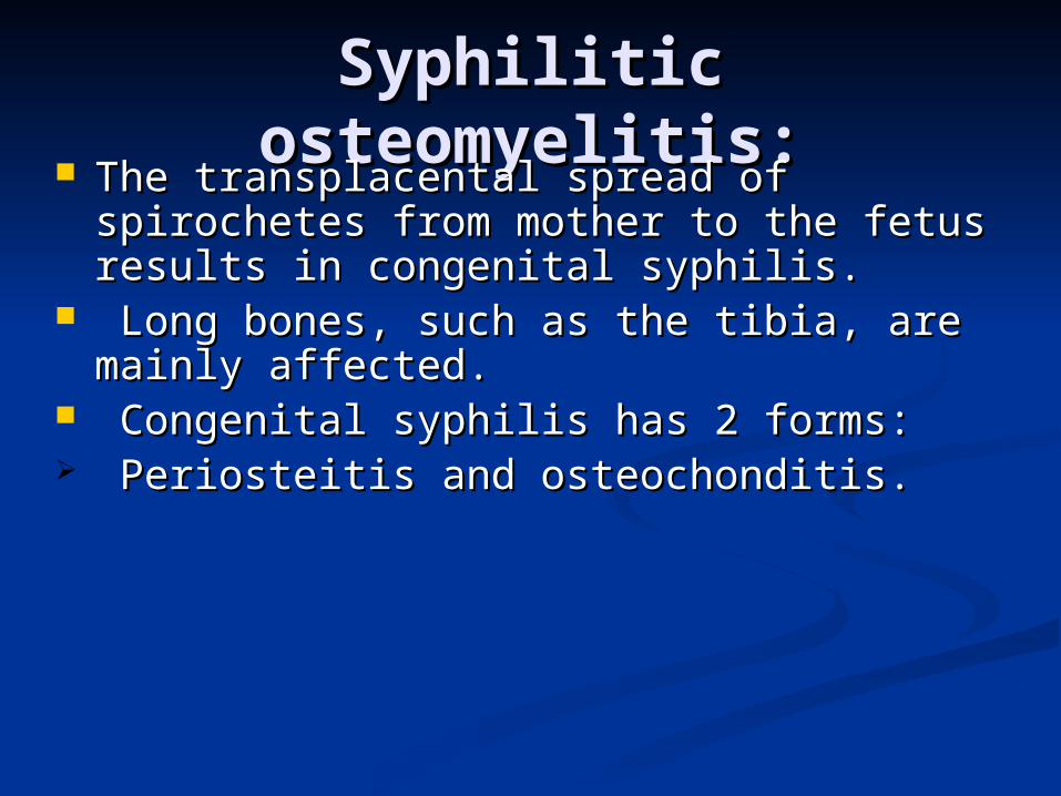
Syphilitic osteomyelitis:Syphilitic osteomyelitis: The transplacental spread of spirochetes The transplacental spread of spirochetes
from mother to the fetus results in from mother to the fetus results in congenital syphilis.congenital syphilis.
Long bones, such as the tibia, are mainly Long bones, such as the tibia, are mainly affected.affected.
Congenital syphilis has 2 forms:Congenital syphilis has 2 forms: Periosteitis and osteochonditis. Periosteitis and osteochonditis.
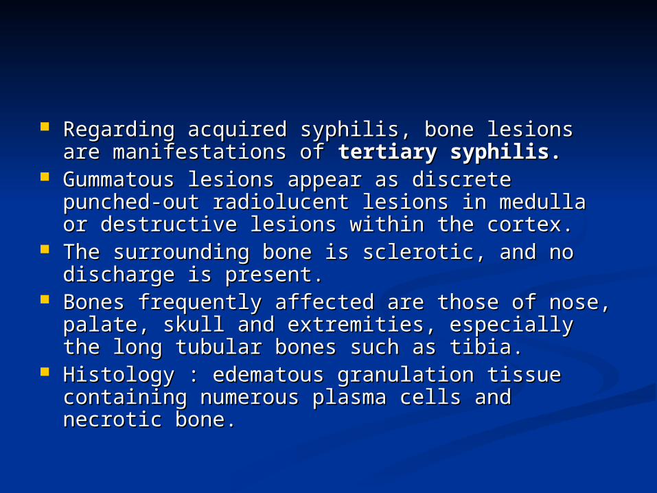
Regarding acquired syphilis, bone lesions are Regarding acquired syphilis, bone lesions are manifestations of manifestations of tertiary syphilis.tertiary syphilis.
Gummatous lesions appear as discrete punched-Gummatous lesions appear as discrete punched-out radiolucent lesions in medulla or destructive out radiolucent lesions in medulla or destructive lesions within the cortex. lesions within the cortex.
The surrounding bone is sclerotic, and no The surrounding bone is sclerotic, and no discharge is present.discharge is present.
Bones frequently affected are those of nose, Bones frequently affected are those of nose, palate, skull and extremities, especially the long palate, skull and extremities, especially the long tubular bones such as tibia. tubular bones such as tibia.
Histology : edematous granulation tissue Histology : edematous granulation tissue containing numerous plasma cells and necrotic containing numerous plasma cells and necrotic bone.bone.
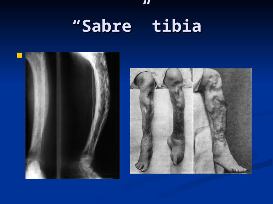
““Sabre” tibiaSabre” tibia


![Septic Arthritis of the Hip – Diagnosis and Management · 2016-08-30 · a nearby osteomyelitis or pyogenic myositis and need to be followed by an MRI [45]. Surgical Management.](https://static.documents.pub/doc/80x56/5ec42cebffe8f8129c4a7d10/septic-arthritis-of-the-hip-a-diagnosis-and-management-2016-08-30-a-nearby-osteomyelitis.jpg)


