59
OSTEOPOROSIS DR. Md Akbar Khan MS(ORTHO) Asst. Prof of Orthopaedics ACSR Govt Medical College,
| Date post: | 24-Jul-2015 |
| Category: |
Health & Medicine |
| Upload: | gousiaaks |
| View: | 28 times |
| Download: | 1 times |

OSTEOPOROSIS
DR. Md Akbar Khan MS(ORTHO) Asst. Prof of Orthopaedics ACSR Govt Medical College, Nellore

Jean Lobstein – coined the term & described its pathoanatomy.
Osteoporosis is a major public health problem, which results in substantial morbidity, mortality and high costs.
Silent disease – patients unaware of ongoing bone loss which is asymptomatic.
Fracture may be the first symptom
INTRODUCTION
Skeletal disorder characterized by low bone mass & micro-architectural deterioration of bone tissue which results in increased bone fragility and fracture susceptibility.
WHO definition – Bone density that falls 2.5 SD below the mean for young healthy adults of same race & gender
DEFINITION

Reduced bone mass Reduced mineralization Micro architectural deterioration of
bone tissue
There is Subnormal osteoid production Excessive rate of de-ossification Subnormal osteoid mineralization
CHARACTERISTIC FEATURES

CHARACTERISTIC FEATURES

Normal – BMD not more than 1 SD Osteopenia - 1 to 2.5 below SD Osteoporosis - 2.5 below SD Severe Osteoporosis – With fragility
fractures
WHO GRADING

PRIMARY OSTEOPOROSIS Type I - Postmenopausal
osteoporosis Type II - Senile osteoporosis Idiopathic - Premenopausal and
Younger
CLASSIFICATION
SECONDARY OSTEOPOROSIS Metabolic - Calcium & Vit. D deficiency Endocrine - Cushing syndrome,
Hyperparathyroidism Renal disease Gastrointestinal - IBD, Malabsorption Hereditary connective tissue diseases -
Marfan syndrome, Homocystinuria. Bone marrow infiltration - Multiple Myeloma,
lymphoma, leukemia. Drugs - Phenytoin, Corticosteroid, heparin,
lithium. Life style - Alcohol, smoking, inactivity,
immobilization, Miscellaneous - Rh. arthritis
CLASSIFICATION
Non-modifiable Peak bone mass Female sex Caucasian race Advanced age Family historyPotentially modifiable Cigarette smoking & Alcoholism Estrogen deficiency Low body weight Low calcium intake Lack of physical activity
CONTRIBUTING FACTORS

Bone formation & bone resorption - (2 Process) Osteoclast (bone resorbing cells) & Osteoblast
(bone forming cells) - (2 Type of Cells) Parathormone & Vitamin D - (2 Biomolecules) Cortical & Trabecular bones - (2 Types of
Bones) Investigations – Markers of bone formation &
resorption (2 Marker Investigations) Treatment – Drugs which enhance bone
formation & decrease resorption (2 Types of Drugs)
BALANCING ACT BETWEEN
Fragility fractures / Insufficiency fractures Outcome depends on
Bone density Severity of fall
In three most common fractures Distal radius – Fall > Density Vertebral body – Density > Fall Hip fractures – Fall & Density play equal
role
OSTEOPOROTIC FRACTURES

Risk Factors : Increased age Female gender Estrogen deficiency Inadequate calcium intake Low bone density (osteopenia) Low body weight History of fractures in adult life History of fractures in first-degree
relative Smoking and alcohol use Lack of physical activity
OSTEOPOROTIC FRACTURES

Osteoporosis is usually asymptomatic until fracture occurs.
May present as backache of varying degrees of severity
Spontaneous fracture Collapse of vertebrae Loss of height is common Thoracic kyphosis
CLINICAL MANIFESTATIONS

Osteoporosis is usually asymptomatic until fracture occurs.
May present as backache of varying degrees of severity
Spontaneous fracture Collapse of vertebrae Loss of height is common Thoracic kyphoses
CLINICAL MANIFESTATIONS

Hyperparathyroidism Paget’s disease Osteomalacia Osteogenesis imperfecta Multiple myeloma Renal Osteodystrophy Secondary tumors
DIFFERENTIAL DIAGNOSIS
Serum calcium Hyperparathyroidism / Malignancy Malnutrition / Osteomalacia
PTH Hyperparathyroidism Malignancy
PTHrP Malignancy TSH To r/o Hyperthyroidism Urinary free Cortisol Cushings disease
INVESTIGATIONS

Urine Calcium Low (<50mg/24 hrs)
Osteomalacia, Malnutrition, Malabsorption
High (300mg/24 hrs)
renal calcium leak -Males with osteoporosis
Absorptive hypercalciuria - Idiopathic
Granulomatous disease
Malignancy and diseases with bone turnover
INVESTIGATIONS

Serum & Urine immuno-electrophoresis
Multiple myeloma Urinary N – Telopeptide (NTX)
Marker of bone resorption
>40 n mol high turnover 25- hydroxy vitamin D & 1,25 hydroxy vitamin
Dlevels
Liver Disease, Renal Osteodystrophy
Monitor response to anti-osteoporotic treatments
INVESTIGATIONS
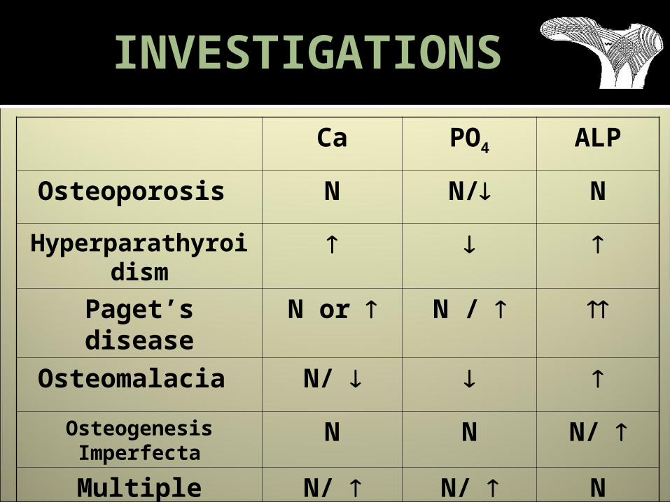
Ca PO4 ALP
Osteoporosis N N/ N
Hyperparathyroidism
Paget’s disease N or N /
Osteomalacia N/
Osteogenesis Imperfecta
N N N/
Multiple Myeloma
N/ N/ N
INVESTIGATIONS
Post menopausal osteoporosisTrabecular resorption & cortical resorption
Senile osteoporosisEndosteal resorption
HyperparathyroidismSubperiosteal resorption
RADIOLOGY

Principal tensile & compressive trabeculae on hip X Ray
Grade VI to Grade I Grade VI: Normal trabecular groups are visible Upper end of femur is occupied by cancellous bone Grade V: Both Trabeculae is accentuated Ward's triangle appears prominent Grade IV: principal tensile trabeculae are markedly reduced can be traced from lateral cortex to upper part of
femoral neck
SINGH’S INDEX

Grade III: There is break in continuity of principal
tensile trabeculae opposite greater trochanter Grade II:
Only principal compressive trabeculae stand out prominently
Remaining trabeculae have been essentially absorbed
Grade I: Principal compressive trabeculae are markedly
reduced in number and are no longer prominent Grade 6 normal Grade 3 definite osteoporosis Grade 1 is severe osteoporosis
SINGH’S INDEX

SINGH’S INDEX
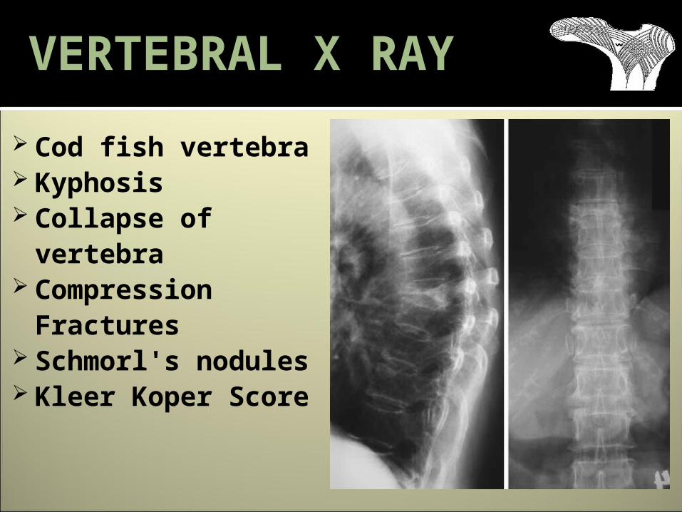
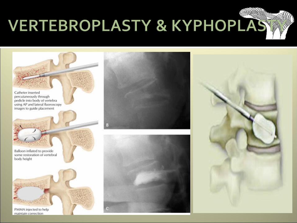
Cod fish vertebra Kyphosis Collapse of
vertebra Compression
Fractures Schmorl's
nodules Kleer Koper
Score
VERTEBRAL X RAY

Assessed from lateral view of spine – T4 – L5
Normal – Grade 0Biconcave deformity – Grade 1Wedge deformity – Grade 2Compression deformity – Grade 3
Kleer Koper Score

Reconstructive CT pictures show L 1, 2, and L3 fractures with biconcave deformities
COMPUTER TOMOGRAPHY
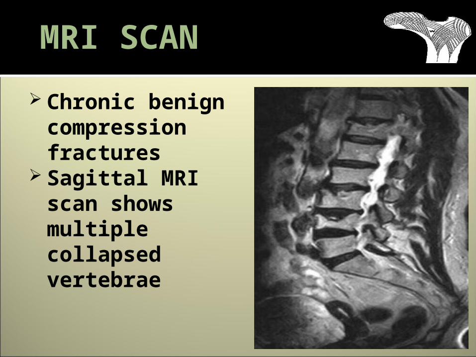
Chronic benign compression fractures
Sagittal MRI scan shows multiple collapsed vertebrae
MRI SCAN
Amount of bone matter per cubic centimeter of bone
Reported in Three terms – Gm/ mm3 , T score & Z score
Measured byDual Energy X Ray AbsortiometryQualitative UltrasoundQualitative Computer Tomography
BONE MINERAL DENSITY
Recommendation for bone density measurements:
Estrogen-deficient women at clinical risk. Individual with vertebral abnormalities - plain
film More than 3 months of steroid treatment Primary hyperparathyroidism Monitoring of drug therapy Women who have multiple risk factors Postmenopausal women who is not on
estrogen replacement. Pt. with strong Family History of osteoporosis.
All women age>65.
BONE MINERAL DENSITY

X ray photons of different energy
Sites recommended by WHO
Total proximal femurFemoral neckLumbar spineRadius with evidence
of OA / surgery at other 3 sites
DUAL ENERGY X RAY ABSORTIOMETRY

Results expressed in T & Z scores
DUAL ENERGY X RAY ABSORTIOMETRY
Emits ultrasonic waves Attenuation of waves which
predict strength of bone Measured in calcaneum At present outdated due to errors
QUALITATIVE ULTRASOUND

Mainly for spine Specifically analysis
trabecular bone Less precise than DEXA More radiation Costlier than DEXA
QUALITATIVE COMPUTER TOMOGRAPHY

Key to management is prevention. Prevention of osteoporosis is a misnomer It is actually prevention of fractures by
the time the patient already have osteoporosis
Increasing public awareness about importance and risks involved helps
Altering personal and dietary habits Regular physical activity(3-4 hrs/week) Peri-menopause & postmenopause:
calcium+ oestrogen – weight bearing exercises.
PRIMARY PREVENTION

Use handrails on stairs, Bathroom
Keep rooms free of clutter Keep floors clean but not
slippery Wear supportive, low-heeled
shoes. Don’t walk in socks; floppy
slippers Install ceiling lighting in
bedrooms Use rubber matt in
shower/tub Check posture in mirror
often
Conservative Surgical
MANAGEMENT

Calcium – 1 to 1.5 gm/day Vitamin D – 400 to 800 IU/day Weight bearing and gravity resistant
exercises Avoid alcohol, cigarette Moderate phosphate intake Prophylactic agents
Alendronate – 35mg Raloxifine – 60mg
PROPHYLAXIS

Anti resoptive class of DrugsCalcium/Vitamin D Bisphosphonates Calcitonin Selective Estrogen Receptor Modulators
(SERMS) Anabolic Drugs
Parathyroid Hormone Sodium fluoride & Strontium Renelate
Other AgentsVitamin K2-7 fortified calcitriol &
Denosumab
MEDICALTREATMENT
Mechanism of action Binds to the surface of
hydroxyapatite crystals and inhibits its resorption
First line of treatment in postmenopausal osteoporosis
Side effects Gastrointestinal intolerance Esophagitis Bone pain
BISPHOSPHONATES

Once a week (oral) Alendronate –35mg (prevention) & 70 mg treatment
Risedronate - 35mg (prevention) & 50 mg treatment
Once a month (oral) Ibandronate - 150 mg
Once In 3 months (Intravenous) Ibandronate - 3 mg / 3 ml over 15 – 30 sec
Once in a year(Intravenous)Zolendronate -5 mg / 100 ml infusion over 15–20 min
BISPHOSPHONATES

Salmon Calcitonin Nasal Spray
For postmenopausal osteoporosis
200 IU once a day intranasal, alternating nostrils
Side effects – nasal mucosal irritation SERM’S (Raloxifene)
For postmenopausal osteoporosis - 150 mg Recombinant Human PTH ( Teriparatide)
Produced genetically engineered E. Coli
Injection - 750 micrograms
Calcium & Vitamin D supplements to correct imbalance
Other Agents
Sodium Fluoride & Strontium Renelate
Increase bone mass by inhibiting osteoclasts
Stimulate osteoblasts Vitamin K2-7 fortified calcitriol &
calcium combinations Denosumab
monoclonal antibody binds with RANK Ligand
Inhibits bone resorption
Other Agents


GOALSImprove quality of lifeGive a stable fixationEarly mobilization & weight bearing
SURGICAL TREATMENT
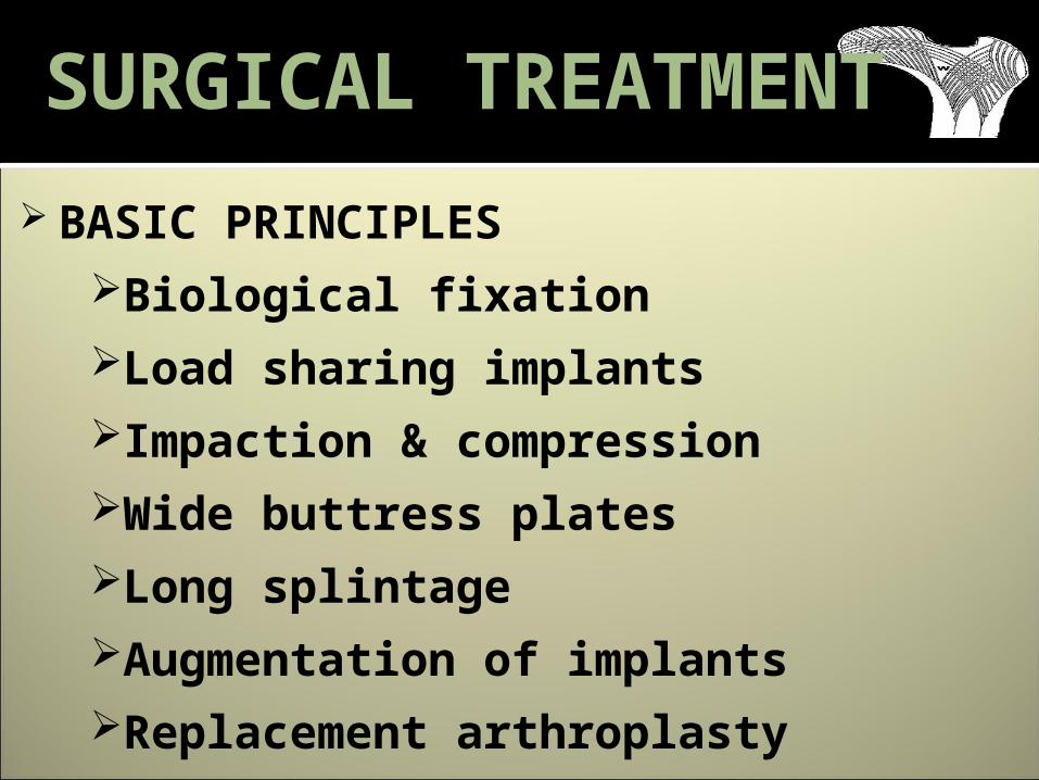
BASIC PRINCIPLESBiological fixationLoad sharing implantsImpaction & compressionWide buttress platesLong splintageAugmentation of implantsReplacement arthroplasty
SURGICAL TREATMENT

Without opening fracture site & without disturbing biomechanics
Use of longer plate with less no of screws – greater stability
BIOLOGICAL FIXATION

Interlocking nails , tension band constructs
Moved from conventional plating, DCP & LC-DCP to Interlocking nails & LCP
These bones have poor holding power of screws
Bones are like tough spring Interlocking nails & LCP locking the
screws to plates creating angular stable devices, diminishing screw holding power of bone
LOAD SHARING IMPLANTS
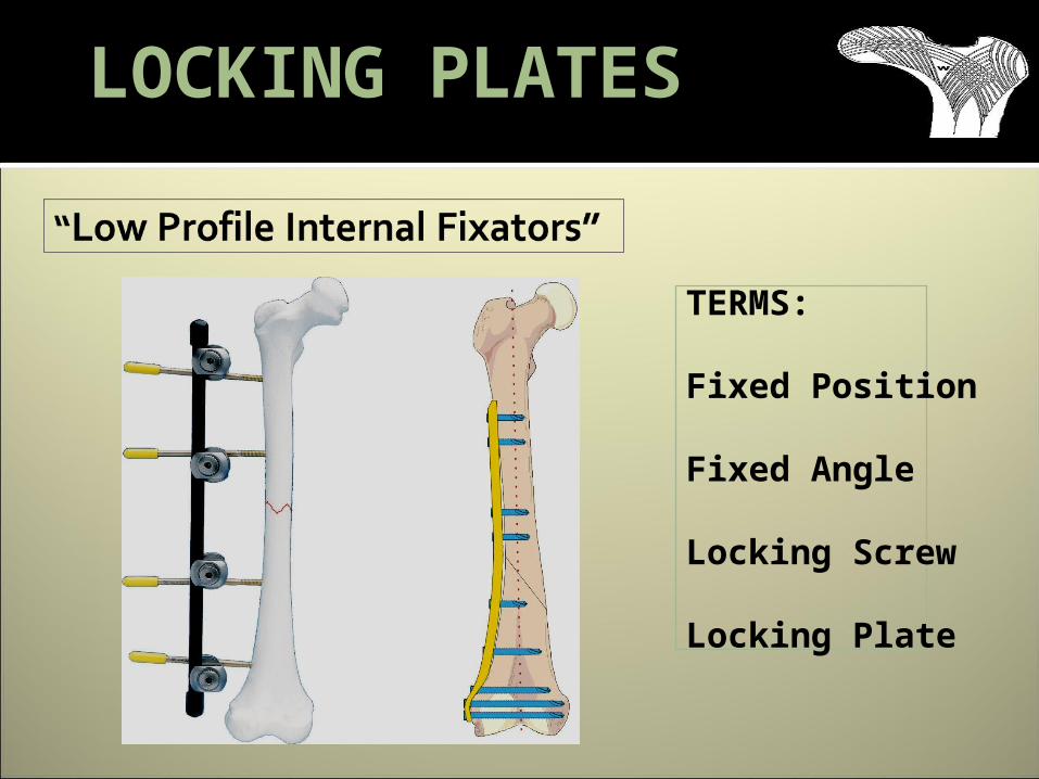
TERMS:
Fixed Position
Fixed Angle
Locking Screw
Locking Plate
LOCKING PLATES
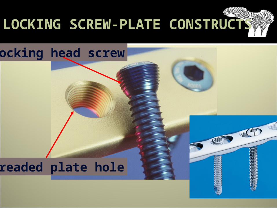
LOCKING SCREW-PLATE CONSTRUCTS
Locking head screw
Threaded plate hole

PULLOUT OF LOCKING SCREWS
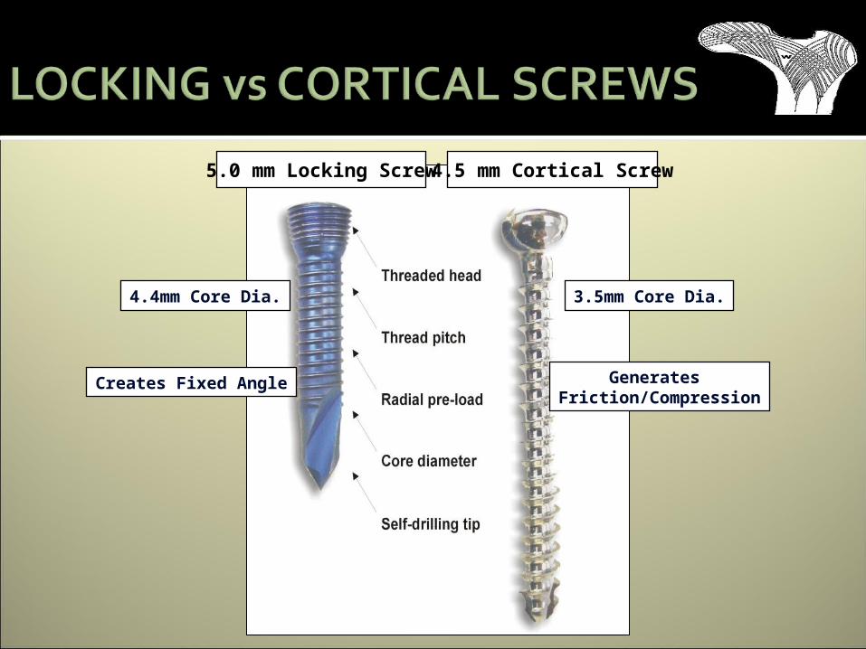
Creates Fixed Angle Generates Friction/Compression
4.4mm Core Dia. 3.5mm Core Dia.
5.0 mm Locking Screw 4.5 mm Cortical Screw


Enhances stability In comminuted fractures – controlled
impaction &compression is advisable-DHS with wt. bearing
LCP plates are used in metaphyseal fractures, upper tibial fractures, & supracondylar fractures & proximal humerus with specialized plates
IMPACTION & COMPRESSION

Bone cement Bone graft Bone subsitutes
HydroxyapatiteTricalcium phosphate HydroxyapatiteTricalcium phosphate
Biodegradable bone cementCalcium phosphate – Norian skeletal repair system
Glass isometric cement
AUGMENTATION OF IMPLANTS


If no other option
REPLACEMENT ARTHROPLASTY

Contoured plates Proximal humeral platesDistal femoral platesProximal tibial platesDistal tibial plates
LISS platesProximal tibia distal femur with zig & minimally invasive techniques
SPECIAL PLATES