Tensorial computer model of gaze--I. Oculomotor activity is expressed in non- orthogonal natural coordinates. Ostriker G, Pellionisz A, Llinás R. Neuroscience. 1985 Feb;14(2):483-500. PMID: 3873020 See Pubmed Reference at the link http://www.ncbi.nlm.nih.gov/pubmed/3873020 and searchable full .pdf file below
Transcript
Tensorial computer model of gaze--I. Oculomotor activity is expressed in non-orthogonal natural coordinates. Ostriker G, Pellionisz A, Llinás R. Neuroscience. 1985 Feb;14(2):483-500. PMID: 3873020 See Pubmed Reference at the link http://www.ncbi.nlm.nih.gov/pubmed/3873020 and searchable full .pdf file below
Neuroscience Vol. 14, No. 2, pp. 483-500, 1985 Printed in Great Britain
030&4522/85 $3.00 + 0.00 Pergamon Press Ltd
0 1985 IBRO
TENSORIAL COMPUTER MODEL OF GAZE-I. OCULOMOTOR ACTIVITY IS EXPRESSED IN
NON-ORTHOGONAL NATURAL COORDINATES
G. OSTRIKER, A. PELLIONISZ and R. LLIN~ Department of Physiology and Biophysics, New York University Medical Center,
550 First Avenue, New York 10016, U.S.A.
AIrstract-The central nervous system expresses its function in natural frames of reference. A most conspicuous feature of such frames is their non-orthogonality. Gaze stabilization and, in particular, the sensorimotor transformations performed by the vestibulo-ocular reflex, are prime examples of such general coordinate transformations between and within multidimensional non-orthogonal frames. Since such operations can be described by tensor formalisms in an abstract manner, this methodology is applied here to develop a tensorial computer model of gaze stabilization. The representation of sensorimotor transformations by a reference-frame independent method obviates the necessity to simplify the intrinsic coordinate systems either by a reduction of the dimensionality or by a presumption of orthogonality.
The frames of reference intrinsic to vestibulo-ocular reflex transformation (the vestibular semicircular canals and extraocular muscles) as well as the covariant character of the sensory input and the contravariant character of the motor output are physically obvious. A model built on these intrinsic systems of coordinates first serves to quantitate the degree of non-orthogonality in the extraocular muscle system, and thus to demonstrate both the necessity and the applicability of representing them by a formalism suitable for non-orthogonal systems, such as tensor network theory.
The actual non-orthogonality of the gaze-stabilization system can be quantitated on the basis of the difference of covariant and contravariant expressions as follows. Tensor network theory describes sensorimotor transformations by employing a covariant embedding procedure. This, however, yields a covariant intention-type motor vector. If the central nervous system were to transmit these sensory-type components directly to the extraocular muscle motor mechanism, an error-angle would occur since covariants do not physically compose the intended movement. The error in every direction of gaze would be zero only if the extraocular muscle system would constitute an orthogonal set of rotation axes. Otherwise, the error, called refraction angle, is a measure of non-orthogonality. The complexity of the quantitation of non-orthogonality is compounded by the fact that these rotation axes change with the moving eye.
Calculation of eye movements, executed both by covariant and contravariant vectors from primary and secondary eye positions, is based on the simplest assumption that the central nervous system establishes the covariant-contravariant transformation in the retinal tangent plane. The results quantitate (1) the significant degree of non-orthogonality in the oculomotor system, (2) the degree of sensitivity of non-orthogonality to the direction of eye movement and to the initial eye position.
With the degree of non-orthogonality made explicit, the errors inherent in remanent orthogonal treatments are quantitated. Presenting the differences of covariants and contravariants in the form of eye movement trajectories provides experimentally testable predictions.
The mechanism by which the CNS activates coordi- dimensional biological systems. For instance, in his nated eye movements, in response to afferent input experimental approach, Szentagothai34 measured ac- arising from the semicircular canals, has long been tivation of all extraocular muscles resulting from regarded as a multivariable sensorimotor system (see individual stimulation of vestibular canals. As an review in Ref. 22). Indeed, it was understood already example of formal account, vector analysis was ap- in the late nineteenth century’2*‘3 that the functioning plied to the multivariable action of the extraocular of this system is based on a simultaneous trans- muscles by Krewson.14 Westheimer36 adapted the formation of neuronal information from all canals to special-purpose multivariable concept and formalism all extraocular muscles. However, a limiting factor in of quartemions to eye movements. However, the developing a coherent view of this transformation above formalisms used coordinate systems that were was the lack of a unified approach allowing experi- (a) orthogonal and (b) extrinsic to the CNS. In part mental, conceptual and formal means of treating this because of the lack of cohesion among the listed multivariable sysem as a whole. experimental, conceptual and formal approaches,
Given this latter fact, two alternative strategies and because of the complexities inherent in them the were utilized to attempt a coherent description. On multidimensional approach has only been employed one hand, painstaking efforts were made to develop since in rare instances.‘6 the above three means of handling complex multi- Instead, a strategy of reducing the complex systems
483
484 G. Ostriker et al.
has prevailed. Based on the tacit assumption that the
brain may be regarded as a control mechanism, one-dimensional feedback system theory was used, in effect reducing the vestibulo-ocular reflex to a single- variable subsystem such as the loop of horizontal canal and horizontal eye muscles.26~33~37 This trend of concentrating on the dynamic aspects of single- dimensional subsystems has been highly successful experimentally, and in model interpretation, but it may be questioned whether the assumptions provid- ing the implicit basis of such reduction should be upheld at the present time. In fact, the single- dimensional approach appears to be difficult to reconcile with currently accepted data which indicate
interconnections among pathways,2.‘5 rendering such separation of horizontal and vertical directions de- batable.9,28
In general, reducibility is a characteristic feature of orthogonal (e.g. Cartesian x, y, z) multivariable sys- tems, but a decomposition of the multivariable CNS, such as the gaze-stabilization apparatus, into single dimensions along horizontal and vertical directions cannot be taken for granted. Caution is warranted in particular because of the non-orthogonality of both the extraocular motor and vestibular sensory appar- atus, and in general because the gaze stabilization involves other systems that are even less orthogonal (e.g. neck muscles). Thus, it is preferable to find a general solution for the CNS control of gaze for any system of coordinates, rather than limiting the analysis to special quasi-orthogonal solutions.
Indeed, the concept and formalism of tensor analysis was offered because this method is capable of treating CNS systems, such as gaze control or the vestibulo-ocular reflex, in their own natural multi- dimensional non-orthogonal frames of reference.23--25 The fact that the CNS expresses its function in intrinsic systems of coordinates has also been stressed in recent years, on the basis of experimental findings of such frames that are not physically obvious to the
observer.3’ Recently, proponents of the single-variable system-
analysis who maintained reference to vector analysis (e.g. to Krewson, cf. Ref. 27), although refrained from a multidimensional treatment, have turned to methods of multivariable analysis including ten- sors.9,‘o,28,30 The return of gaze analysis to multi- variable approaches is enthusiastically welcomed. However, the elevation of quantitative description from one dimension into three extrinsic coordinates goes only halfway into a general multidimensional treatment of CNS function expressed in an astound- ing multitude of intrinsic coordinates. The vestibulo- ocular reflex, for example, can only be reduced into a system that is three-dimensional throughout by artificial means. Such reduction also necessitates the bypassing of the issue of coordination in overcomplete systems, e.g. in sensorimotor mechanisms where the motor apparatus may use a higher number of dimen- sions compared to the sensory part of the system.2’ It
is expected that mutually relevant features of the dynamic analysis of single-dimensional subsystems and of multidimensional (e.g. tensorial) analyses will eventually be synthesized.
This paper attempts a general analysis in natural multidimensional frames of reference intrinsic to CNS. Tensor concept and formalism, in conjunction with customary computer modeling represents the approach to this problem, treating the complex quan- titative aspects of the gaze-stabilization system with regard to the generation of saccades adhering to Listing’s Law.
A preliminary note on this work has been pub- lished.”
The vestibulo-ocular reflex as a tensorial system
It has been suggested that the brain could be represented in a general manner as a tensorial system relating to the physical invariants of the external world via non-orthogonal vectors expressed in vari- ous natural frames of reference, intrinsic to the body, with tensorial relations among different vectorial
expressions. 23-25 A tensorial treatment appears advan- tageous, since in oblique systems of coordinates two different types of vectorial expressions (covariant and contravariant) can be assigned to any coordinate- system-invariant physical object, e.g. an eye displace- ment. The two versions can be transformed from one another via the metric tensor (see Fig. 3 in Ref. 24). In case of different sensory and motor frames, the number of vectorial variations is at least four (covar- iant and contravariant in both the sensory and motor frames). The relationships among such vectors can be expressed both in general tensorial manner24.25 and, when specifying the frames of reference, by particular vector-matrix multiplications.28
In some sensorimotor systems it is obvious that the sensory frame of reference is different from the coordinate system of the motor apparatus. A difference may exist both in the number and in the direction of the coordinate axes, and in addition neither of these intrinsic coordinate sysems may conform with a commonly presumed three- dimensional orthogonal Cartesian system.
As for the vestibulum, it has been established in all species studied that the six semicircular canals are
non-orthogonal to one another. The non-
orthogonality has been established with a high degree of precision in humans.4 It should be noted that if the human semicircular canal axes were an orthogonal set, then the angle between any two from these three axes would be 90”. In fact, these angles are of 117.76, 86.16 and 95.75”,4 and even by customary pairing of corresponding canals on the left and right side these angles are 82.8, 82.8, and 84.6”, on average 83.4”. As for other species, e.g. in guinea-pigs, the horizontal and anterior canals form an angle which falls, with 95% confidence limits, between 118.9 and 125.3”.’ Similar data for non-orthogonality are available for cats’ and rabbits.30
A
MR LR
Pig. 1. ~onstmtion of the nau-a~~~t of the axes of semicircular canals and of the extraocuhu muscle activations in human, using canal data from Blanks et &4 muscle data from Volkmann,~ modified by Hehnho1tz.‘* (A) Anterior view of the spatial arrangement of axes of the left-right paired semi&cular canals which correspond to a head which is elevated by 15”. H, left-right horizontal; RP-LA, right posterior-left anterior; RA-LP, right anterior-left posterior. (B) Anterior view of the left eye,‘which is eievated by 1 S’, turned outward by 1 So, and rolled around its visual axis to rest in a position consistent with Listing’s Law, and considered to he within an orbit (i.e. bead) which is elevated by 15O, showing the rotational axes of six extraocular muscles. IO, inferior oblique; IR, inferior reetus; LR, laterala r&us; MR, medial rectus; SO, superior obiique; SR, superior metus. (C) Combined view of the nine axes shown in (A) and (I%). Note, that the canal (yellow), agonist muscle (blue+ and antagonist muscle (red) axes form a one-ahgned set, where each and every axis points into a separate direction of the space. The clock-dial-like presentation demonstrates that ago~st-an~gonist pairs are not cohnear, moreover, &at they are not even arranged in a push-puli (e.g. 12-6 h) fashion relative to a “common” canal axis. These arrangements of axes are dependent on species, and also on the position of the eye. Without precise numerical handling of these nine distinct axis-directions (in all positions), no eye model could claim a true
quantitative character.
485
Tensorial computer model of gaze 487
Aside from the issue of non-orthogonality, it is also physically evident that the mass of endolymph within each canal is accelerated proportionally to the projection component of the head acceleration to the axis of the canal’s plane, such projections constituting by definition a covariant vector of the acceleration.
As for the motor apparatus of the two eyes, it is an at least 1Zdimensional system, and the extraocular muscles form a comparable non-orthogonal arrange- ment, although the deviation is physically less obvi- ous (see below). Since the six physical (contravariant) force-components propel the mass of the eye, each muscle acting around individual rotational axes, the vestibulo-ocular reflex may be considered the epitome of a covariant-contravariant tensorial system with different and non-orth~gon~ sensory and motor frames of reference.
A general, abstract treatment of sensorimotor schemes has been offered by the tensor approach.” In other approaches, however, for a quantitative hand- ling of specific sets of data, simplifying assumptions have been introduced that reduce the analysis to a matrix analysis which is thr~dimensional through- out.928,29 To demonstrate that the sensory and motor coordinate-axes cannot be reduced to an ima~nary three-dimensional orthogonal Cartesian system with- out introducing a significant distortion, a computer- drawn representation of the spatial arrangement of such axes is offered in Fig. 1.
Measures of the non-ort~og~naiity of the extraocular muse/e system; the refraction angle and irn~~ct of a vector
The quantitative handling of the spatial orientation of the two sets of three semicircular canals poses little technical challenge since the canals are fixed into the bony structure of the inner ear, and thus their orientation is evident. Thus, a quantitative descrip- tion can simply demonstrate the existing degree of non-orthogonality, and can plainly show the need of refraining from an arbitrary rectification of the system.
The extraocular muscle system, however, repre- sents a much more serious challenge due not only to its non-orthogonality but also to the fact that the directions of muscle pull are different with different positions of the rotation of the eye. Thus, for a quantitative handling of the complex transformations of displacements (as intended gaze shifts) into extra- ocular muscle coordinate components, a computer model had to be built.
This paper describes the first stage of the tensorial computer model of gaze, a representation of the extraocular muscle geometry, so that in sub~quent studies the rotation of the eye may be calcuiated both from covariant motor intention and contravariant motor execution signals. ” Besides using computer graphics to visualize the motor system of various species in a morphologically precise manner, this paper attempts to establish the degree of non-
orthogonality of the extraocular system where (1) the deviation from orthogonality depends on the direction of the eye movement; (2) this direction dependence of non-o~hogonality is eye-position dependent.
The twin notion of the refraction and impact of a vecto?” serves in this paper as the measure of non-orthogonality. These measures, to be extensively utilized in further developments of tensor network theory, hinge on the distinction of covariant and contravariant vectors. An important aspect of these terms is that, as shown elsewhere,‘8*22 the degree of difference or identity of co- and contravariants may be used by the CNS in generating and modifying the neuronal networks underlying gaze control.
The angle between a physically executed covariant (v,) and contravariant (0’) has been defined “refrac- tion angle”, denoted by w (where both v’ and ui are assigned to the same invariant, and thus related to one another by the metric tensor gti in the form of u’ = gV. z+). Further, i = cos(w) has been defined*O as the “impact” of the vector on the metric of g”.
The “refraction-angIe 0” and the “impact i = cos(w)“, are therefore the quantitative expressions of how closely a non-orthogonaIiy expressed vector approximates the so-called eigenvector of the metric tensor in the given frame. For example, if in a motor movement a given covariant vector is used as if it were a contravariant, it will assemble a different invariant than the one to which the covariants were originally assigned. This has been discussed for the case of cerebellar iesions,24 where the motor abnor- malities could be considered as arising from the absence of a cerebellar metric (dysmetria), which results in a movement in a wrong direction and with a wrong amplitude (ataxia).
For the eigenvector of the metric tensor, refraction should be, by definition, zero. Indeed, an eigenvector of a matrix is an input vector for which the output is of the same direction (only the amplitude may be different), i.e.
where i is the scalar eigenvalue. A method of approx- imating eigenvectors is of great theoretical signifi- cance, since the eigenvectors can be most useful in characterizing the multivariable systems found in neurobiological mechanisms.’ While in mathematics a vector is an eigenvector either precisely or not at all, biological sysems cannot be expected to operate in a mathematically exact (i.e. noise-free) manner. Thus, measures are needed which also apply as approxi- mations of high-entropy biolo~cal systems. With the new terms, a vector is precisely an eigenvector if its impact is total (i = I) but a vector may also be biolo~caily accepted as an eigenvector with only nearly total impact, e.g. with i = 0.999. The direction of the biologically acceptable eigenvector will be termed “eigen-direction”. In turn, a direction, into
488 G. Ostriker et al.
which a physically executed covariant yields the largest refraction-angle, will be called herein “refracter-direction”. Since these physical directions are the ones into which an eye movement is the least and most sensitive to a covariant-contravariant inter- change (the equivalent of an ataxia induced by cere- bellar ablation), aside their theoretical importance their establishment is also significant from the point of view of experimental verification of the theory.
Since the metric tensor in an orthogonal frame of reference is g” = 6”, the so-called Kronecker delta (implemented by a unit matrix), in such a frame every direction is an eigen-direction since the co- and contravariant vectors are identical (not only in angle, but also in amplitude). Thus, if refraction o is measured in different directions, the non- orthogonality of a system can be characterized by displaying how far the refraction-angle deviates from zero along different directions (cf. Figs 7 and 8). Importantly, for such measurements only the input-output vectors need be known, the explicit knowledge of the coordinate system is not required and a conclusion can still be drawn on the degree of its non-orthogonal character.
DESCRWHON OF THE MODEL
Selection of extrinsic reference frames
One essential premise of the present approach is the notion (not universally accepted) that the natural coordinate systems, intrinsic to the body and the CNS, cannot be assumed II priori to be limited to a Cartesian frame of reference. This fundamental fact necessitates a general vectorial formalism, such as tensor analysis, if an understanding is to be reached of how the CNS implements vector transformations within and between various general frames.
The familiar Cartesian reference frame is used in this paper only as an interface to traditional descrip- tions, such as the external representation of the physical arrangement of the extraocular muscles and retinal tangent plane and to directly compare these results with the previous models.5~6~27 However, since in tensor theory the functional analysis of CNS vectors is handled in a coordinate-system aspecific manner, the entire analysis could be carried out in natural reference frames.
Geometrical model of the eye
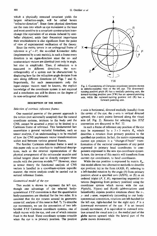
This model is shown to represent the left eye, though one advantage of the selected body- symmetrical XYZ convention is that the quantitative body-parameters are identical for the two sides. It is assumed that the eye rotates around its geometric center (cf. analysis of this issue in Ref. 7). To describe eye movements, we use the convention of two left- handed XYZ Cartesian coordinate systems, one of which moves with the eye, and one of which remains fixed in the head. These coordinate systems coincide when the eye is in primary position. The positive
X
YW
Fig. 2. Conventions of Cartesian coordinate systems shown in anterio-occipital view of the left eye. The downward- turning positive pitch (P) has a medially pointing axis, the inward-turning positive yaw (YW) has an upward-pointing axis, while the outward-turning positive roll (R) has a
forward-pointing axis.
x-axis is horizontal, directed medially (nasally) from the center of the eye; the y-axis is vertical directed upward; the z-axis points forward along the visual axis (cf. Fig. 2). Reasons for selecting this XYZ convention are discussed in Ref. 22.
In such a frame of reference any position of the eye may be represened by a 3 x 3 matrix R, which describes a rotation from primary position to the specified eye position. In fact, the matrix representing current eye position is a “change-of-basis” trans- formation of the vectorial components of any point expressed in primary head coordinates to com- ponents expressed in the new eye-coordinate system. In turn, the inverse of this matrix will transform from current eye coordinates, to head coordinates.
While the eye position is expressed by matrix R,, this model allows two alternative methods to describe eye position: (a) in the form (AXIS, 0) referring to a left-handed rotation by the angle (0) from primary position about a specified axis (AXIS), or (b) as an ordered triplet (P, Y, R), representing sequential ro- tations (beginning from primary position) using the coordinate system which moves with the eye. P(pitch), Y(yaw) and R(rol1) (abbreviations used henceforth), express positive rotations about the x-, y- and z-axes, respectively (according to our body- symmetrical convention, rotations are left-handed for the left eye, right-handed for the right eye). P is a downward movement of the eye: Y is an inward (nasal) movement; R is equivalent to outward rota- tion around the visual axis (i.e. the medial part of the globe moves upward while the lateral part of the globe moves downward).
Tensorial computer model of gaze 489
Eye movements are specified through a gener- alization of the method by which positions are de- scribed in this model. As an eye position is descibed by means of a rotation from primary position, an eye movement (from any initial eye position) may be described either in the form (AXIS, 0) or (P, Y, R). Both of these descriptions yield the rotation matrix R,,,. If initial eye position is described by marix Ri, and the eye moves to another position by means of R,,,, then the final eye position is represented by a product matrix, Rr= R,*Ri. In addition, when con- sidering a movement from eye position E, to position E2, represented by matrices R, and R2 respectively, then the product matrix R,, = R+R;’ defines the movement. This matrix can be decomposed either as (AXIS, 0), a single (left-handed) rotation about an axis defined in the eye-coordinate system in position E,, or as a triplet (P, Y, R) describing how the eye could move from position E, to E2, in three steps, about the x-, y- and z-axes that are attached to the eye.
Geometrical model of the eye muscles
With respect to the positional parameters of the extraocular muscles, this model is elaborated in three different versions, based on three different schemes. The “Robinson model“ (Rb), cf. Ref. 27, provides the most accurate geometric representation of extra- ocular muscle kinematics where the muscle paths generally follow non-great circles. These paths lie between the shortest path (great circle) and the primary-position-path of the muscle (a great circle); and the rotational axes are perpendicular to the muscle planes, therefore changing according to eye position.
The Boeder model5,6 is the basis for the two other versions of this model in which muscles are assumed to follow shortest possible paths, the great circles. With one version, henceforth called “Boeder-fixed axes model” (Bf), each muscle is considered to pull the eye about an axis which remains fixed with repsect to the head for all eye positions. In the version of “Boeder-variable axes model” (Bv) the rotational axis for a particular extraocular muscle is defined as perpendicular to the muscle plane (i.e. a particular muscle has a variable associated axis which depends upon eye position). The calculations necessary to arrive at the paths of muscles and other related parameters are given in the original publications.sT6,27
The Rb model is obviously much more soph- isticated than its predecessors, and its-acceptance is quite general. The two other models, which define extreme conditions, are carried through our analysis only in order (1) to investigate whether similar results might arise from all three models, thereby revealing some general patterns which are not sensitive to the peculiarities of these models and (2) to make the limitations of the Boeder models quantitative and thus further define their rather restricted sphere of relevance.
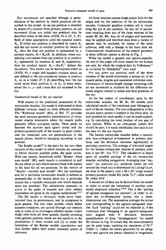
All three versions assume single points both for the origin and for the insertion of the six extraocular muscles. Computer graphics routines aid in visual- izing the eye in any position, for any set of param- eters resulting from any of the three versions of the model Bf, Bv, Rb. Any set of origins and insertions may be applied and therefore data from a variety of species may be analyzed, by means of the same software, with only a change in the basic data set. Computerized visualization of the muscle geometry in three different species is shown in Fig. 3.
While the model is general and species-aspecific, the rest of this paper will show results for the human eye only, for which the original data by Volkmann,35 as corrected by Helmholtz’* were used.
For any given eye position, each of the three versions of the model determines a unique set of six muscle paths, lengths and associated rotational axes. Calculation of muscle-length changes occurring with an eye movement is obtained by the difference be- tween lengths related to initial and final positions of the eye.
As for the system of coordinates, spun by the extraocular muscles, the Bf, Bv, Rb models yield calculated results of the rotational axes belonging to each of the six muscles. The non-orthogonality of this six-axis motor system is implicit in the above data. In each position for each model, it can be made explicit, e.g. by calculating the inner product of any pair of axis vectors. While in an orthogonal system this shou!d be zero, it is easily established that such is not the case for the eye muscles.
The human extraocular muscles define a remark- ably non-orthogonal arrangement in primary pos- ition, and the deviation increases significantly at secondary positions. The average of interaxial angles for the human extraocular muscles in primary posi- tion is not 90”, but 77.3”. This calculation is based upon all possible pairings of the six extraocular muscles, excluding antagonists. Averaging nine “sac- cadic” eye positions (i.e. positions where the roll around the visual axis is determined by Listing’s Law, see later in the paper), over a 60 x 60” range around primary position, model Rb yields 72.5”, while model Bv yields 69.8”.
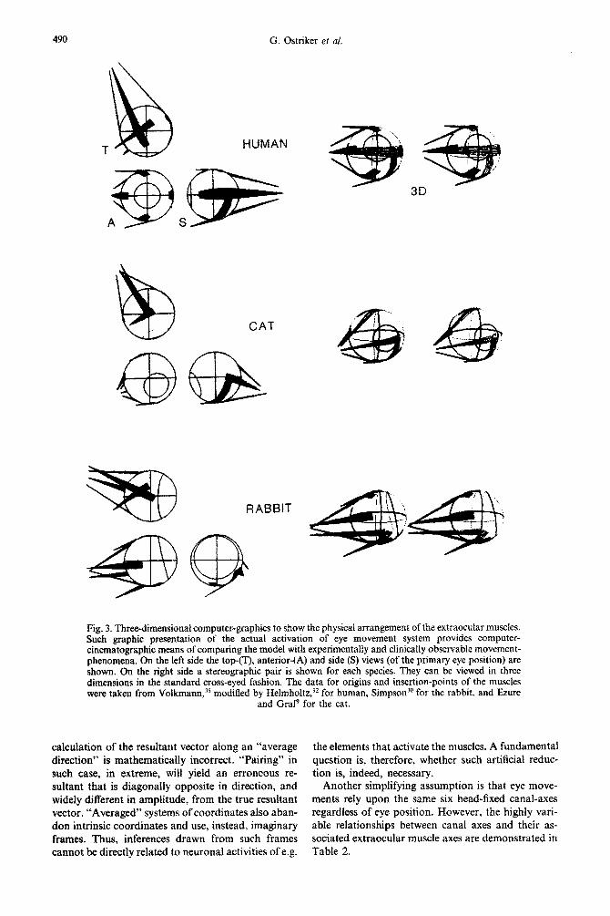
A second set of data can be derived from the model in order to reveal the limitations of another com- monly employed reduction.g.28*2g This is the “pairing of agonist-antagonist eye muscles”; in order to re- duce the six-dimensional system into a three- dimensional one. The assumption averages the actual axes corresponding to the agonist-antagonist mus- cles. Such “pairing” could not be objected to, if the rotational axes belonging to an agonist-antagonist were aligned with 0” deviation. However, quantification of these “misalignments” for model Rb (shown in Fig. 4) reveals that the non-colinearity can be quite large, reaching the maximum of 82.7” (Table 1). Unless the forces generated by an antag- onist and agonist are always identical in magnitude,
G. Ostriker er al.
CAT
Fig. 3. Thre~~ime~sionai computer-graphics to show the physical arrangement of the extraocular muscles. Such graphic pre~n~t~on of the actual activation of eye movement system provides computer- cinematographic means of comparing the model with experimentally and clinically observable movement- phenomena. On the left side the top-(T), anterior-(A) and side (S) views (of the primary eye position) are shown. On the right side a stereographic pair is shown for each species. They can be viewed in three dimensions in the standard cross-eyed fashion. The data for origins and insertion-points of the muscles were taken from Yolkmann, modified by Helmholtz,‘* for human, Simpson30 for the rabbit. and Ezure
and GraP for the cat.
calculation of the resultant vector along an “average direction” is mathematically incorrect. “Pairing” in such case, in extreme, will yield an erroneous re- sultant that is diagonally opposite in direction, and widely different in amplitude, from the true resultant vector. “Averaged” systems of coordinates also aban- don intrinsic coordinates and use, instead, imaginary frames. Thus, inferences drawn from such frames cannot be directly related to neuronal activities of e.g.
the elements that activate the muscles. A fundamental question is, therefore, whether such artificial reduc- tion is, indeed, necessary.
Another simplifying assumption is that eye move- ments rely upon the same six head-fixed canal-axes regardless of eye position. However, the highly vari- able relationships between canal axes and their as- sociated extraocular muscle axes are demonstrated in Table 2.
Tensorial computer model of gaze 491
Table 1. Misalignment of rotational axes belonging to aaonist-antagonist muscles*
Primary position Maximum Minimum Average
Model Bv Horizontal recti Vertical recti Vertical obliques
Model Rb Horizontal recti Vertical recti Vertical obliques
*All data are shown in degrees. One hundred and twenty-one “saccadic” eye positions were examined, where the positions were defined by (P, Y, R), with P and Y ranging from -30 to + 30”. in 6” increments. and R provided according to Listing’s Law.
Covariant and contravariant representation of eye movements. An oculomotor metric-type trans- formation as calculated in the retinal tangent plane
The calculation of the rotational axes belonging to the six extraocular muscles revealed the inherently non-orthogonal character of the system of coordi- nates intrinsic in the oculomotor apparatus. This fact necessitates a distinction between possible covariant- and contravariant-type vectorial expressions in this oblique motor frame. While the actual motor expres- sion that executes a displacement of the eye is con- travariant, the analysis of the mathematical covariant expression of the eye displacement in the motor frame is warranted by the following consideration, which predicts that the CNS uses such intention-type ex-
MED 24 0 24 LAT
pression within the sequence of vestibulo-ocular reflex transformations.**
A change of coordinates from sensory- (e.g. ves- tibular or retinal) to motor frame is possible by the covariant embedding procedure.2’~22~25 This trans- formation yields a covariant motor expression, which produces a different eye displacement than the con- travariant motor execution. The differences in covar- iant and contravariant forms are utilized in establish- ing the eigenvectors of the motor system.22~30
The theoretical question of how the CNS imple- ments the intention-execution (covariant to con- travariant) motor transformation is central for two basic reasons. (1) At the present time the exact procedure by which the sensory input is transformed
Fig. 4. Position dependence of the misalignment of the axes of agonist-antagonist muscles in human (model Rb) over a 60 x 60” range surrounding primary position. Key: HR, angle between axes of horizontal recti; VO, angles between axes of vertical obliques; VR, angles between axes of vertical recti. The model shows that there is no eye position where all three “pairs” of muscles were aligned. The primary position shows reasonable alignment of the horizontal recti and vertical obliques, but not of
the vertical recti muscles.
492 G. Ostriker et at.
Table 2. Misalignment of the axes of a semicircular canal and the associated extraocular muscles*
*All data are shown in degrees. One hundred and twenty- one eye positions as in Table I. Cl (canal 1) = left-right horizontal (averaged axis). C2 (canal 2) = right posterior-left anterior. C3 (canal 3) = right anterior-left posterior. Muscles (l-6) are lateral, medial, superior, inferior recti, superior, inferior obliques.
into an organized motor output in the CNS is unknown. (2) Given the overcompleteness of the motor system, e.g. generating a three-dimensional physical displacement by the activation of six mus- cles, any given covariant motor vector can be trans- formed into an infinite number of possible con- travariant expressions. Each of these infinitely numerous mat~x-transfo~atons can be imple- mented by a different metric-type operation.
While mathematically such transformation is indefinite in case of overcompleteness, there are several ways to find a solution that the CNS may employ in a particular execution. Other than a direct experimentaf investigation, two theoretical ap- proaches could be useful in this regard. One is a general analysis of the sensory to motor trans- formations in the CNS2’ which suggests a four-step scheme, where the motor metric-type transformation is the unique contravariant metric tensor (in case of non-overcomplete systems) and the unique Moore-Penrose generalized inverse of the covariant metric tensor (in case of overcompleteness; cf. Refs 19, 22 and 30).
Another approach, used here, is based on the consideration that any physical “embedding” of a three-dimensional movement into an inner six- dimensional hyperspace gives important clues to the relationship of contravariants and covariants. Given the structural properties (physical constraints) of the system, from any establishment of a given con- travariant a unique covariant may be found that corresponds to it (but not vice versa). Indeed, a specific constraint on the degrees of freedom of voluntary gaze positions has long been experi- mentally established, called Listing’s Law. The orig- inal observation (by Donders, 1847, and later attrib- uted by Rouete to Listing, cf. Ref. 7) determined that
voluntary eye movements do not make use of all three of its degrees of rotational freedom. Explained most lucidly,‘6,36 Listing’s Law is equivalent to the statement that after having executed a voluntary eye movement the eye rotational state is such as though the visual axis moved, beginning from primary posi- tion, along a great circle of the eye. This means, on one hand, that the eye has only two degrees of freedom of movement, which can be given e.g. by the direction and length of the segment of great circle from the starting point. On the other hand, the Donders-Listing Law can be interpreted by stating that eye movements can be described in a two dimensional stereographic projection,9,‘2 such as in the retinal tangent-plane of the eye. While the possi- bility of such a two-dimensional description does not mean that the transfo~ations must be calculated in the CNS in a manner based solely on two- dimensional information, the simplest is to calculate a covariant-contravariant relationship in such tan- gent plane. Results could then be compared to data derived from experimental analysis. A different, much more complex approach to the question of oculomotor metric, dealing with all three (sensory metric, sensorimotor and motor metric) trans- formations in a sensorimotor system, is shown else- where.22
The calculation of the oculomotor metric is based here, therefore, on the assumption that the CNS assigns a vector to the target location, as represented in the retinal tangent space of the eye. This two- dimensional vector may then be considered to be embedded into a six-dimensional hyperspace, where each of the six local displacement-axes, induced by unitary muscle-length chantes, lies in the two dimen- sional tangent plane. The embedding results in an intended vector, given covariantly by orthogonal projections to the six motor axes (all lying in the retinal tangent plane). This covariant vector must then be transformed into physically executable con- travariant expression.
The covariant~ontravariant relationship is calcu- lated in the tangent plane as follows. In an infinitesimal range, rotations of a rigid body can be treated vectorially. ‘I From a particular initial eye position, a movement with a restricted amplitude, is expressed as an ordered set (sextuplet) of the con- travariant muscle-length changes. Covariant vector components of the coordinate-system-invariant eye movement can be derived in the tangent plane as follows. For saccadic eye movements, it is required that both resting eye positions be consistent with Listing’s Law. Therefore, they must be attainable from primary position through rotations about axes within a plane defined by the x- and p-axes. This allows a reduction of the analysis of saccadic eye movements from three to two degrees of freedom (P, I’). Thus, calculations may be carried out on a retinal tangent plane centered at the fovea, to which an orthogonal local coordinate system, defined by P
Tensorial computer mode1 of gaze 493
and Y, is attached. After each of the six covariant components of an eye movement is first derived in the tangent plane by an orthogonal projection of the movement onto the tangent plane muscle axis, a contravariant set of muscle-length changes is deter- mined which would yield this displacement in the tangent plane. As for the structure of the actual calculations, the model invokes four sets of points (spaces): E, M, H and T.
Space E is the set of points representing all possible eye positions. Space Es is a subset of E, and is defined by all eye positions which obey Listing’s Law.
Space M is the set of abstract six-dimensional points (ordered sets of “muscle status” sextuplets), representing muscle lengths in a given eye position. It should be noted that such a sextuplet is a mathe- matical vector in the sense that any ordered set of quantities is one, but is not a vector in the limited Euclidean sense, since these sextuplets do not form a closed set under addition (i.e. the sum of two such sextuplets could turn out to be a set of muscle lengths associated with no physically attainable position of the eye). It can be shown that there is a one-to-one correspondence between M and E. An example of an element of M could be mp = (m,, m2, . . , m,), corresponding to eye position Ep. The quantities in the components represent muscle lengths in milli- meters, and the ordering sequence (together with the abbreviations used) is as follows: (1) lateral rectus, LR, (2) media1 rectus, MR, (3) superior rectus, SR, (4) inferior rectus, IR, (5) superior oblique, SO, (6) inferior oblique, IO.
Space H is a six-dimensional “oculomotor space” over points representing muscle-length changes incur- ring in movements from a particular initial eye position. If eye movements are restricted to infinitesimal, e.g. 5 or 10” for practical purposes, then H satisfies the criteria required of a vector space. Space denoted by HP contains contravariant vectors, and Hp contains covariant vectors, where “E,,” identifies an initial eye position.
Space TQ is the retina1 tangent plane centered at the fovea associated with the initial eye position EQ. TQ is a vector space to which we assign a Cartesian system of coordinates defined by P and Y. Each muscle axis has its counterpart in the tangent plane, which would be the initial direction of movement of the fovea in this plane if only that particular muscle were to contract to move the eye. A displacement of the eye (a physical invariant), can be projected onto T,. A displacement is generated by the sum of its con- travariant components. The covariant components of the displacement are established as the orthogonal projections of the displacement onto each of the tangent plane muscle axes. The “covariant exe- cution” vector is generated by the physical sum of the covariant components of the displacement. The re- fraction-angle w, by definition, is the angle between the original displacement and the execution vector using covariants. When the covariant execution of
Fig. 5. Calculation of contra- and covariant muscle- components of an infinitesimal eye-displacement. Both the displacement (DE) and the six local displacement coordinate-axes of the eye muscles lie in the retina1 tangent plane of the eye. The procedure starts with the calculation of displacement axes belonging to individual muscles (simi- lar to the diagram named after Hering,i3 with LR, MR, SR, IR, SO, IO abbreviations used as in text). Then, an infinitesimal eye-displacement is generated (DIS), and the muscle-length changes belonging to the displacement are calculated. This will constitute a contravariant vector. In the third step, the orthogonal projection (in the tangent plane) of the displacement is calculated for each individual muscle- axis (e.g. to the shown MR axis, that belongs to the pulling of the MR muscle). This step repeated for all six muscles yields a unique covariant expression of the displacement.
the displacement is in exactly the same direction as the original displacement, refraction is zero, o = 0, and the impact is total, i = cos(0) = 1. Such a dis- placement is made then along a so-called eigen- direction of the system.
Analysis of an eye displacement
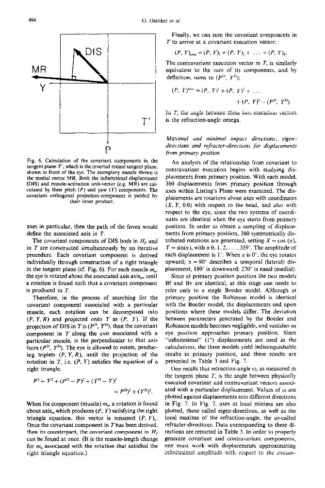
Let us consider a displacement (DIS, cf, Fig. 5), from initial position Ei to final position Ef (with Ei and Ef both being elements of Es), and let Ri and Rf be the matrices representing these eye positions. The matrix RI, = &*R;’ represents the displacement in the local eye-coordinate system associated with the initial eye position, Ei. After decomposing RD into (P, Y, R), the displacement may be projected into the tangent plane, T, as the vector (P, Y). = (P,, YD). An inverted image of the retina1 tangent plane, T’, as it would appear in front of the eye, is demonstrated in Fig. 6. Using the Bf or Rb model for each of the eye positions E, and Ef, elements of space M, i.e. sex- tuplets of lengths of the six extraocular muscles can be obtained. (Bf and Bv versions yield identical muscle lengths.) The difference between these sextu- plets (subtracting initial from final) yields a con- travariant vector of space HP. Associated with each muscle m,, (where n = 1,2, . . , 6), there is an axis axis,, about which that muscle would rotate the eye if it alone were to contract while the eye was in position Ei. Each of these six axes has its counterpart in T, axis,,,. If the eye is rotated about any one of these
494 G. Ostriker et nl
MR I
Y
I-’
Fig. 6. Calculation of the covariant components in the tangent plane T’, which is the inverted retinal tangent plane, shown in front of the eye. The exemplary muscle shown is the medial recuts MR. Both the infinitesimal displacement (DIS) and muscle-activation unit-vector (e.g. MR) are cal- culated by their pitch (P) and yaw (Y) components. The covariant orthogonal proj~tion-component is yielded by
their inner product.
axes in particular, then the path of the fovea would define the associated axis in T.
The covariant components of DIS both in lip and in T are constructed simultaneously by an iterative procedure. Each covariant component is derived individually through construction of a right triangle in the tangent plane (cf. Fig. 6). For each muscle m,, the eye is rotated about the associated axis axis,, until a rotation is found such that a covariant component is produced in T.
Therefore, in the process of searching for the covariant component associated with a particular muscle, each rotation can be decomposed into (P, Y, R) and projected onto T as (P, Y). If the projection of DIS in Tis (P”, YD), then the covariant component in 7’ along the axis associated with a particular muscle, is the perpendicular to that axis from (P”, YD). The eye is allowed to rotate, produc- ing triplets (P, Y, R), until the projection of the rotation in T, i.e. (P, Y) satisfies the equation of a right triangle:
P2+ Yz+(Pn-P)*+(Yu- Y)?
= Pn)* + (Yn)*.
When for component (muscle) m,, a rotation is found about axis,, which produces (P, Y) satisfying the right triangle equation, this vector is renamed (P, Y),. Once the covariant component in T has been derived, then its counterpart, the covariant component in HP can be found at once. (It is the muscle-length change for m, associated with the rotation that satisfied the right triangle equation.)
Finally, we can sum the covariant components in T to arrive at a covariant execution vector:
(P. I?,,, = (P, Y), + (P, Y)? + . . . + (P, n,.
The contravariant execution vector in T, is similarly equivalent to the sum of its components, and by definition, sums to (P”, YD):
(P, Y),,,, = (P, Y)’ + (P, Y)‘+ . . .
+(P, ry=p, YD).
in T, the angle between these two execution vectors is the refraction-angle omega.
Maximal and minimal impact directions; eigen- directions and refacter-directiorrs for displacements from primary position
An analysis of the relationship from covariant to contravariant execution begins with studying dis- placements from primary position. With each model, 360 displacements from primary position through axes within Listing’s Plane were examined. The dis- placements are rotations about axes with coordinates (X, Y, 0.0) with respect to the head, and also with respect to the eye, since the two systems of coordi- nates are identical when the eye starts from primary position. In order to obtain a sampling of displace- ments from primary position, 360 symmetrically dis- tributed rotations are generated, setting X = cos (cz), Y = sin(a), with tl0, 1, 2, . . . ,359”. The amplitude of each displacement is 1”. When LT is o”, the eye rotates upward; c( = 90” describes a temporal (lateral) dis- placement; 180” is downward; 270” is nasal (medial).
Since at primary position position the two models Bf and Bv are identical, at this stage one needs to refer only to a single Boeder model Although at primary position the Robinson model is identical with the Boeder model, the displacements end upon positions where these models differ. The deviation between parameters generated by the Boeder and Robinson models becomes negligible, and vanishes as eye position approaches primary position. Since “infinitesimal” (1”) displacements are used in the calculations, the three models yield indistinguishable results in primary position, and these results are presented in Table 3 and Fig. 7.
One recalls that refraction-angle c1), as measured in the tangent plane T, is the angle between physically executed covariant and contravariant vectors associ- ated with a particular displacement. Values of o are plotted against displacements into different directions in Fig. 7. In Fig. 7, axes at local minima are also plotted, those called eigen-directions, as well as the local maxima of the refraction-angle, the so-called refracter-directions. Data corresponding to these di- rections are reported in Table 3. In order to properly generate covariant and contravariant components, one must work with displacements approximating infinitesimal amplitude with respect to the circum-
Tensorial computer model of gaze 495
Fig. 7. Direction dependence of the refraction-angle of the displacement from primary position. The inset (upper left) shows the orientation of the refraction-diagram in the context of the eye in primary position. The diagram plots the magnitude of error-angle (refraction), if a physical displacement of the eye (DIS) is measured covariantly in the tangent plane and the covariant vector is executed as if it were contravariant. In the graphic presentation, a DIS vector into any direction cuts a segment from the “petal” curves: its length is proportional in the diagram to the calculated refraction-angle into the given DIS direction. For the shown displacement (DIS) the refraction-angle is lo”, while for displacement into direction E it is zero (E: eigen-direction). To another set of directions (R) the refraction-angle is maximal (R: refracter-direction). These E and R directions, which are least and most sensitive to covariant-contravariant difference, are not the same for models Bf, Bv and Rb, and vary with eye position.
Results are shown in a tabulated format in Table 3.
ference of the eye. It appears that the amplitude of displacements employed (3.6” or less) is sufficiently small, since the results yield graphic presentation of data that are indistinguishable when the 1” increment is further reduced, e.g. to 0.36” increment.
Eigen-directions and refracter -directions from second- ary and tertiary positions
For each of three models, a 60 x 60” “grid” per- taining to displacements from 7 x 7 = 49 different initial positions, is demonstrated in Fig. 8. Initial positions are chosen so that they follow Listing’s Law, and they span a region around primary position where P and Y vary from -30.0 to 30.0”. Just as in Fig. 7 refraction-angle w is plotted against dis- placement directions, but from non-primary initial positions, displacement directions must be referred to the system of coordinates attached to the eye. Initial positions were determined by triplets (P, Y, R), as follows: first P and Y were selected, with each variable taking on all possible multiples of 10 from -30.0 to 30.0”. Then, in order to ensure that initial positions adhered to Listing’s Law, R was determined such that the movement from primary position de-
scribed by (P, Y, R) would end upon a position which could be arrived at through a single rotation about an axis within Listing’s Plane.
From each initial position, 72 displacements (at 5”
Table 3. Displacements from primary position*
Direction Refraction-angle o
I. Eigen-directions (directions with minimal refraction-angle)
74 0.0343 163 0.1667 254 0.0000 345 0.1480
II. Refracter-directions (directions with maximal refraction-angle)
37 15.179 111 15.162 216 15.099 292 15.119
*All data are shown in degrees. Three hundred and sixty displacements were examined at 1” increments. Degrees mean clockwise rotations when observing subject (left eye): 0” = upward, 90” = temporal (lateral), 180” = downward, 270” = nasal (medial) displacement. In tangent plane T’, o is the refraction-angle between covariant and contravariant execution vectors.
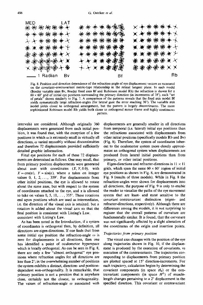
496 G. Ostriker et af.
Fig. 8. Position and direction dependence of the refraction-angle of eye displacement vectors as measured on the covariant-contravariant metric-type relationship in the retinal tangent plane. In each model (Boeder variable axes Bv, Boeder fixed axes Bf and Robinson model Rb) the refraction is shown for a 40 x 60” grid of initial eye positions surrounding the primary direction (in increments of lo”), each “set of petals” shown similarly to Fig. 7. A comparison of the patterns reveals that the fixed axis model Bf yields systematically large refraction-angles (for lateral gaze the error reaching 30”). The variable axis model yields closer to orthogonal arrangement, but the pattern is largely discontinuous. The more sophisticated Robinson model Rb yields both closer to orthogonal motor frame and highly continuous
pattern.
intervals) are considered. Although originally 360 displacements were generated from each initial pos- ition, it was found that, with the exception of a few positions in which w is extremely small in virtually all directions, w varied smoothly without discontinuities and therefore 72 displacements provided sufficiently detailed graphic info~ation.
Final eye positions for each of these 72 displace- ments are determined as follows. One may recall, that from primary position displacements were generated about axes with coordinates (X, Y,O.O), with X = cos(tr), Y = sin(E), where LX takes on integer values 0, 1, 2, . . . , 359”. For displacements from other initial positions, the eye is allowed to rotate about the same axes, but with respect to the system of coordinates attached to the eye, and CI is allowed to take on values 0, 5, 10,. . . , 355”. These rotations end upon positions which are used as intermediates, i.e. the direction of the visual axis is retained, but a rotation is added about the visual axis so that the final position is consistent with Listing’s Law. consistent with Listing’s Law.
As has been noted in the Introduction, if a system of coordinates is orthogonal then, by definition, all directions are eigen-directions. If one finds that from some initial eye position the ~fraction-angle w is zero for displacements in all directions, then one has identified a point of oculomotor hyperspace which is locally orthogonal. As can be seen in Fig. 8, there are only a few “almost orthogonal” eye pos- itions where refraction angles for all directions are less than 2”; in the ove~helming number of positions the system exhibits a distinct, direction- and position- dependent non-orthogonality. It is remarkable, that primary position is not a position that is anywhere close, certainly not the closest, to orthogonality. The values of refraction-angle w associated with
displacements are generally smaller in all directions from temporal (i.e. lateral) initial eye positions than the refractions associated with displacements from other initial positions (specifically models Rb and Bv) (Fig. 8). Therefore, the system of coordinates inher- ent to the oculomotor system more closely approxi- mates an orthogonaf system when ~spla~ments are produced from lateral initial positions than from primary, or other initial positions.
Eigen-directions and refracter-directions in 11 x 11 grids, which span the same 60 x 60” region of initial eye positions as shown in Fig. 8, are demonstrated in Fig. 9 (results of three models). While in Fig. 8 the refraction-angles were shown for ~spia~ments into all directions, the purpose of Fig. 9 is only to enable the reader to visualize the paths of the eye movement system that are least- and most sensitive to the covariant-contravariant distinction (eigen- and refracter-directions, respectively). Although there are differences among the models, it is not su~~sing to register that the overall patterns of curvature are fundamentally similar. It is found, that the curvature was not significantly affected by a slight alteration of the coordinates of the origin and insertion points.
~r~je~tori~s from primary position
The visual axis changes with the position of the eye along trajectories shown in Fig. 10, if the displace- ment is produced by the execution of covariants, vs execution of the contravariants. The trajectories cor- responding to displacements from primary position are plotted spaced at 15” dir~tion-increments. For each trajectory, calculation begins by determining the covariant components (in space HP) or the con- travariant components (in space HP) of muscle- length changes pertaining to a 2” displacement in the specified direction. This covariant or contravariant
Tensorial computer model of gaze 497
EIGEN-DIRECTIONS REFRACTER-DIRECTIONS
Bv
Rb
Fig. 9. Trajectories of eigen-directions (E) and refracterdirection (R) for the three different models, for an 11 x 11 (6” increment) displacement-grid. Inset (upper left) puts one pattern (E-B!) into the context of the eye. Directions, the least sensitive to covariant-contravariant distinction are shown in E, while trajectories in column R show the most sensitive directions. The discontinuous Bv model is biologically unlikely, while the fixed-axis Bf model is a physically crude approximation. The trajectories of Rb model seem to distinguish upper-lateral eye-displacement as general directions that are most a&&d by covariant-contravariant mixup, assuming that the CNS uses a metric-type transformation as calculated
in the tangent plane.
vector is used then to generate, in 50 steps, the respective trajectory. Each step involves: (1) updating the axes of rotation for all six extraocular muscles to the current eye position and then (2) six consecutive rotations about these axes. The amplitude of each of these six rotations is such that the change in length of the muscle corresponding to the particular axis of rotation, is precisely the corresponding component of the (co- or contravariant) generating vector. These trajectories shown in Fig. 10 were produced to demonstrate the feasibility of comparing theoretical models with experimental tests, not to conclude that
the gaze-stabilization system must be restricted to a covariant-contravariant calculation as calculated in the retinal tangent plane. It is already evident** that a thorough re-examination of the hierarchy of gaze system is required in order to arrive at not just potential-but to more conclusive results of tensorial modeling of gaze.
DISCUSSION
The chief goal of this paper is to illustrate a method of treating CNS functions in terms of biological
G. Ostriker er al. 498
CONTRAVARIANT COVARIANT
Fin. 10. Trajectories of contravariantly and covariantiy executed displacements from primary position (on the left and right side, respectively) for the Rb model. The con- travariant execution (in 15” direction-increments) is along straight lines. Dots along trajectories in the corresponding covariant execution indicate eye positions where at least one muscle looses its curved segment. 1 he covanant executfon (assuming a metric-type transformation in the tangent plane) yields characteristically curved trajectories that pro- vide an experimentally testable feature of the most primitive assumption of using a tangent-plane metric in the CNS.
(non-orthogonal) coordinate systems and its concep- tual implications. Traditionally, for reasons of sim- plicity, the Cartesian concept of reference frame was applied by using orthogonal coordinate systems where invariants could be quantitatively represented in a unique and simple manner. Indeed, for no other reason than convenience, an orthogonal frame of reference is chosen almost invariably whenever the coordinate system is allowed to be arbitrarily selected.
A central problem of geometrical transformations in biological systems therefore relates to the lack of o~hogon~ity in the actual reference frames in the body and, more importantiy, to the general character of the transformations which take place in the CNS. This lack of orthogonality results from both the gross anatomy and the microscopical morphology of the biological system as well from the development and diversity of cellular function. Indeed, differences in conduction velocity and length of pathways as well as synaptic transmission mechanisms, to name only a few, would create enormous difficulties if biological systems would be forced to utilize only those refer- ence systems that we as observers use in classical Newtonian mechanics.2s
An immediate consequence of the geometrical transformation within and between non-orthogonal reference frames is that in such general frames two distinct forms of vectorial representations are simul- taneously possible. These dual versions-the covar- iant and the contravariant forms-have quite different representational properties with respect to the invariant being expressed, also in the biological sense relating to sensory- and motor expressions respectively.24 The use by the CNS of various frames and vectorial expressions indicates that the brain is capable of such covariant-contravariant coordinate-
system-general, i.e. tensorial representation. These points have been treated in detail in previous publications2s25 suggesting that sensorimotor inte- gration requires a metric transformation. The mathe- matical problem of non-orthogonality is com- pounded by the biological fact that transformations occur between intrinsic reference frames that have inequal number of coordinate axes,
The present paper addresses two implications aris- ing from non-orthogonality. (1) Correct eye move- ment direction cannot be implemented by vectors which define the movement in a covariant manner (i.e. directly by intended vectors). Indeed, eye move- ments generated by covariant vectors are incorrectly directed. (2) The significant degree of non- orthogonality in the oculomotor transformation is determined by the difference between the movement executed by covariant and ~ontrav~ant execution (the “refraction-angle”; cf. Ref. 20). This deviation from the eigen-direction also relates to other param- eters, for instance timing. Further, the difference of covariants and contravariants and their identity in eigen-directions may serve an important role in the development of the neuronal networks underlying oculomotor coordination.‘8.22
Some predictions of this model
In addition to predicting the directions in which the largest eye movement distaxia should occur, as- suming a direct activation of the musc~ature by the motor intention vectors, the present model also gives a description of the eigenvectors of eye movement. The model predicts the direction of maxima1 and minimal movement-abnormalities following CNS le- sions which alter the metric of this transformation. Moreover, because it has been proposed that the cerebellum serves as a metric tensor of the motor space, 24 the effect of cerebellar lesion on eye move- ment coordination should be particularly clear when eye movements occur into directions with larger angles of refraction (i.e. further from the eigen- direction).
Two different kinds of experiments could be de- signed to test these assumptions. In the first type, damage of cerebellar structures relating to saecadic eye movement transformations should change the responses of eye movement in a manner predicted by Figs S-10. Furthermore, if the CNS utilizes the same retinal coordinate system for both optokinetic and saccadic eye movements, then optokinetic nystagmus, which relates direction of eye movement to the direction of image movement on the retina, should also demonstrate a predictable abnormality accord- ing to the model. An open question is if the directions of motor abno~ality are the same for vestibular and optokinetic nystagmus. This would be the case if the oculomotor metric of the transformation from retinal input to oculomotor response takes part also in the transformation of vestibular sensory input into eye movement.
Tensorial computer model of gaze 499
In such testing of the model one must also consider that the metric transformation by the cerebellum may have other aspects than coordination of the direc- tions of the muscular action. One such aspect may be the coordination of the muscular tone, which would also affect eye movements.
Practical implications of the computer model
The computer model developed here has another, more pragmatic use. It is based on general anatomical parameters which may be specified to describe a given oculomotor system, vertebrate or invertebrate. Among the uses of this modeling is that of predicting
the modifications of the functional geometry of eye movements which occur upon modifying the in- sertions of the extraocular muscles onto the eye globe or the modification of their origins in the orbit. Modifications of this type are often performed in corrective surgery in ophthalmology, especially in correction of strabismus or of extraocular muscle weakness. Because the results of the computation could be tabulated in the form of a nomogram which relates the anatomical parameters to characteristics of the sensorimotor transformation, the model is then quantitatively testable and thus may serve to predict the outcome of studies relating to the vestibulo- ocular transformation.
1.
2.
3.
4.
5. 6. I. 8.
9.
Anderson J. A., Silverstein J. W., Ritz S. A. and Randall S. J. (1977) Distinctive features, categorical perception, and probability learning: some applications of a neural model. Psycho/. Reu. 84, 413-451. Baker J., Goldberg J., Hermann G. and Peterson B. (1983) Convergence of canal inputs to secondary neurons in cat vestibular nuclei. Sot. neurosci. Abstr. 9, 315. Blanks R. H. I., Curthoys I. S. and Markham C.H. (1972) Planar relationships of semicircular canals in the cat. Am. J. Physiol. 223, 55-62. Blanks R. H. I., Curthoys I. S. and Markham C. H. (1975) Planar relationships of the semicircular canals in man. Acta oto-lur. 80, 185-196. Boeder P. (1961) The co-operation of extraocular muscles. Am. J. Ophthal. 51, 469-481. Boeder P. (1962) Co-operative action of extra-ocular muscles. Br. J. Ophthal. 46, 397-403. Carpenter R. H. S. (1977) Movement of the Eyes. Pion, London. Curthoys I. S., Curthoys E. J., Blanks R. M. I. and Markham C. H. (1975) The orientation of the semicircular canals in the guinea pig. Acta ore-tar. 80, 197-205. Ezure K. and Graf W. (1984) A quantitative analysis of the spatial organization of the vestibulo-ocular reflexes in lateral- and frontal-eyed animals-I. Orientation of semicircular canals and extraocular muscles. Neuroscience 12, 85-93.
10.
11. 12. 13.
14.
15.
16.
17.
18.
19.
Goldberg J., Baker J. Schor R. and Peterson B. (1982) Vertical canal contribution to horizontal eye movements. Sot. Neurosci. Absrr. 8, 419. Goldstein H. (1980) Classical Mechanics (2nd Edn). Addison-Wesley, Reading, Massachusetts. Helmholtz H. (1896) Handbuch der Physiologischen Oprik (Zweite A&age). Voss, Leipzig. Hering E. (1879) der Raumsinn und die Bewegungen des Auges. In Hundbuch der Physiologie (ed. Herman L.), Vol. 3/l, 343-601. F.C.W. Vogel, Leipzig. Krewson W. E. (1950) The action of the extraocular muscles. A method of vector-analysis computation. Trans. Am. oprhal. Sot. 48, 443486. Markham C. H. and Curthoys I. S. (1972) Convergence of labyrinthine influences on units in the vestibular nuclei of the cat. II. Electrical stimulation. Bruin Res. 43, 383-396. Nakayama K. (1974) Photographic determination of the rotational state of the eye using matrices. Am. J. Oprom. physiol. Opr. 51, 736742. Ostriker G., Pellionisz A. and Llinis R. (1982) Tensor network theory applied to the oculomotor system. CNS Activity expressed with natural non-orthogonal coordinates. Sot. Neurosci. Absrr. 8, 155. Ostriker G., Pellionisz A. and Llinas R. (1984) Tensorial computer movie display of the metaorganization of oculomotor metric network. Sot. Neurosci. Absrr. 10, 162. Pellionisz A. (1983) Sensorimotor transformation of natural coordinates via neuronal networks: conceptual and formal unification of cerebellar and tectal models. In COINS Technical Reporr, University of Massachusetts, Amherst, Masachusetts, on II. Workshop on Visuomotor Coordination in Frog and Toad-Models and Experiments (Mexico City, 17-19 Nov. 1982).
20. Pellionisz A. (1983) Brain theory: connecting neurobiology to robotics. Tensor analysis: utilizing natural coordinates to describe, understand and engineer functional geometries of intelligent organisms. J. rheor. Neurobiol. 2, 185-211.
21. Pellionisz A. (1984) Coordination: a vector-matrix description of transformations of overcomplete CNS coordinates and a tensorial solution using the Moore-Penrose generalized inverse. J. rheor. Biol. 101, in press.
22. Pellionisz A. (1984) Tensorial aspects of the multidimensional approach to the vestibule-oculomotor reflex and gaze. In Reoiews of Oculomoror Research. Vol. I. Adaotive Mechanisms in Gaze Control (eds Berthoz A. and Melvill-Jones G.). Elsevier; Holland.
.
23. Pellionisz A. and Llinls R. (1979) Brain modeling by tensor network theory and computer simulation. The cerebellum: distributed processor for predictive coordination. Neuroscience 4, 323-348.
24. Pellionisz A. and Llinas R. (1980) Tensorial approach to the geometry of brain function: cerebellar coordination via a metric tensor. Neuroscience 5, 1125-l 136.
REFERENCES
25. Pellionisz A. and Llinas R. (1982) Space-time representation in the brain. The cerebellum as a predictive space-time metric tensor. Neuroscience 7, 2949-2970.
26. Robinson D. A. (1968) The oculomotor control system: a review. Proc. IEEE 56, 1032-1049.
500 G. Ostriker et al.
27. Robinson D. A. (1975) A quantitative analysis of extraocular muscle cooperation and squint. Invest. Ophthal. 14, 801-825.
28. Robinson D. A. (1982) The use of matrices in analyzing the three-dimensional behavior of the vestibule-ocular reflex. Biol. Cybern. 46, 53-66.
29. Schultheis L. W. and Robinson D. A. (1981) Directional plasticity of the vestibulo-ocular reflex in the cat. In Vestibuhar and O~lomotor Physio~~y: ~nternat~onul Meeting of the &r&y Society (ed. Cohen B.), pp. 504-512. New York Academy of Sciences, New York.
30. Simpson J. I. (1983) Transformations of coordinates intrinsic to the vestibulo-ocular reflex. Sot. Neurosci. Abstr. 9, 315.
31. Simpson J. I., Graf W. and Leonard C. S. (1981) The coordinate system of visual climbing fibers to the flocculus. In Progress in Oculomotor Research (eds Fuchs A. F. and Becker W.), 475-484. Elsevier/North-Holland Biomedical Press, New York.
32. Simpson J. I. and Pellionisz A. (1984) The vestibule-ocular retlex in rabbit, as interpreted using the Moore-Penrose generalized inverse ~ansfo~ation of intrinsic coordinates. S’oc. Neuro~c~. Abstr. 10, 909.
33. Stark L. (1968) Neurological Control Systems. Plenum, New York. 34. Szentigothai J. (1952) Die Rolfe &r einzelnen Labyrinthrezeptoren bei der Orientation von Augen und Kopf im Raume.
Akademiai Kiad6, Budapest. 35. Volkmann A. W. (1869) Zur Mechanik der Augenmuskeln. Ber. siichs. Akad. Wiss. Math.-nat. Klasse 21, 28-69. 36. Westheimer G. (1957) Kinematics of the eye. J. opt. Sot. Am. 67, 967-974. 37. Young L. R. (1969) Biocybemetics of the vestibular system. In Riocybernetics of the Central Neroous System (Proc.
int. Symp., Washington, DC) (ed. Proctor L. D ), pp. 79-l 17. Little, Brown, Boston, Massachusetts.


![Pellionisz A. Prog Brain Res. Vol. 76 Elsevier Chapter 30 ... · nate systems that are intrinsic to the organism [58] and our task is simply 'letting the brain speak in its own terms'](https://static.documents.pub/doc/80x56/5f03e2c57e708231d40b401e/pellionisz-a-prog-brain-res-vol-76-elsevier-chapter-30-nate-systems-that.jpg)

