67
Other Nematodes – Other locations • Trichinella spiralis • Dracunculus medinensis • Capillaria spp. • Angiostrongylus spp. • Angiostrongylus spp. • Thelazia spp. • Gnathostoma spinigerum 1
Other Nematodes – Other locations
• Trichinella spiralis
• Dracunculus medinensis
• Capillaria spp.
• Angiostrongylus spp.• Angiostrongylus spp.
• Thelazia spp.
• Gnathostoma spinigerum
1

Trichinellosis Trichinella spiralis
• Man is not the normal host (in domestic and wild animals)
• Worldwide. Not particularly prevalent in tropical countries
• 11 million people infected• 11 million people infected
• Not soil-transmitted
• Two forms: adult and cystic
• White worm : ♀ 3-4 x 0. 06 mm
♂ 1.6 x 0.04 mm
2

Trichinella spiralis
(a) male (b) female(a) male (b) female
3
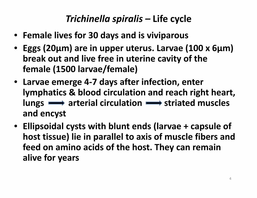
Trichinella spiralis – Life cycle
• Female lives for 30 days and is viviparous
• Eggs (20µm) are in upper uterus. Larvae (100 x 6µm) break out and live free in uterine cavity of the female (1500 larvae/female)
• Larvae emerge 4-7 days after infection, enter lymphatics & blood circulation and reach right heart, lungs arterial circulation striated muscles lungs arterial circulation striated muscles and encyst
• Ellipsoidal cysts with blunt ends (larvae + capsule of host tissue) lie in parallel to axis of muscle fibers and feed on amino acids of the host. They can remain alive for years
4

Larvae of Trichinella liberated from bear meat
5

Trichinella spp.
life cycle
6
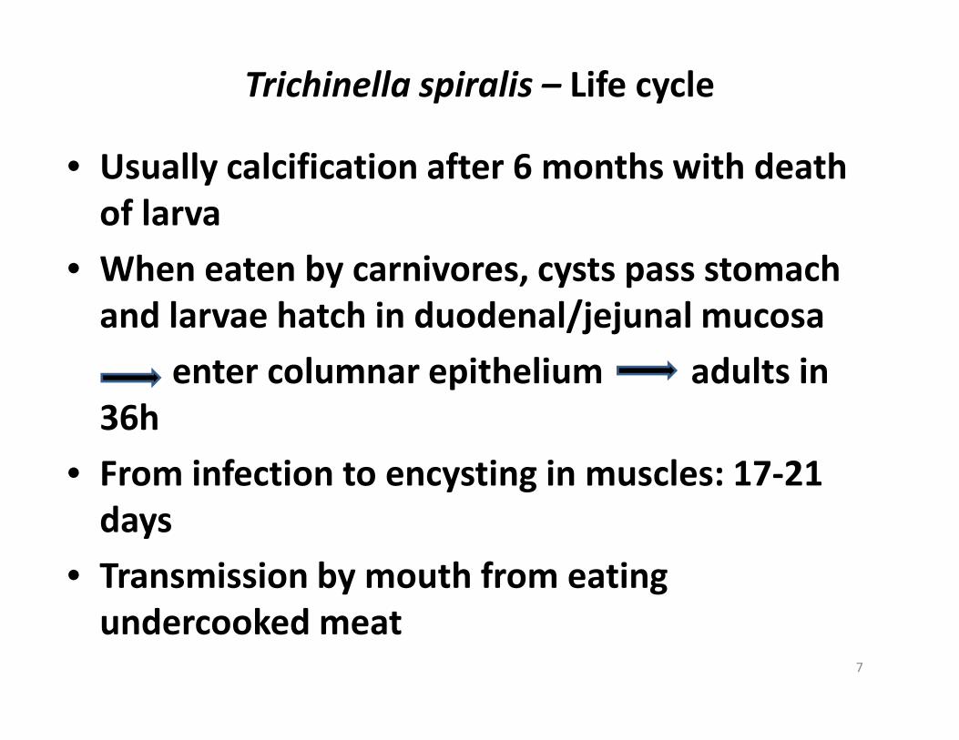
Trichinella spiralis – Life cycle
• Usually calcification after 6 months with death
of larva
• When eaten by carnivores, cysts pass stomach
and larvae hatch in duodenal/jejunal mucosa
enter columnar epithelium adults in enter columnar epithelium adults in
36h
• From infection to encysting in muscles: 17-21
days
• Transmission by mouth from eating
undercooked meat7

3 sub-species can infect man:
• T. spiralis spiralis : pigs , foxes
temperate regions
• T. spiralis nativa : polar bears• T. spiralis nativa : polar bears
Arctic regions
• T. spiralis nelsoni : wild pigs, lions, cheetah
Africa, South Europe
8

Trichinella spiralis – Pathology
Pathology is related to 3 phases:
• Enteric phase:
– Larva in duodenal/jejunal mucosa
– Pathology depends on number of larvae
• Migratory/invasive phase:• Migratory/invasive phase:
– After 5-7 days, females lay larvae in tissues
• Encystment phase:
– Larvae encyst in striated muscles but can travel to
brain and heart muscle where they cannot encyst
9

Encysted larvae of Trichinella sp. in muscle tissue, stained with
hematoxylin and eosin
300 x 400 x
10
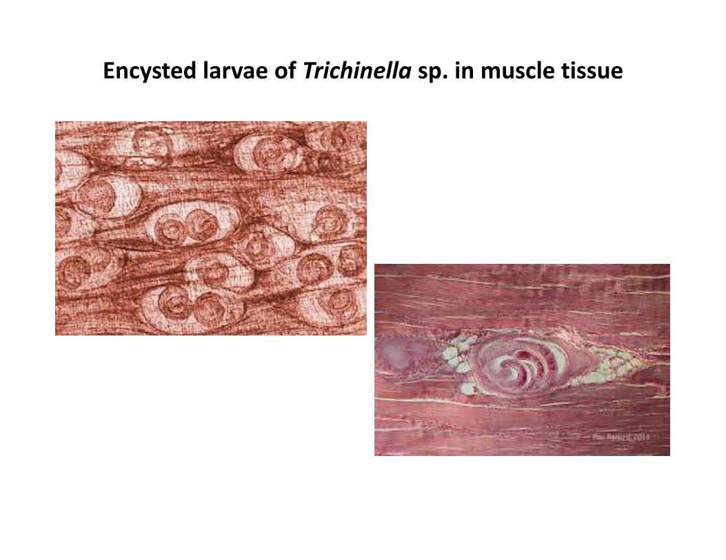
Encysted larvae of Trichinella sp. in muscle tissue

Trichinella
spiralis
Changes in
muscle muscle
capillaries
Trichinella spiralis – Clinical features
• If light : asymptomatic, self-limiting 2-3 w, low
mortality
– Light : ≤ 10 Larvae/g of muscle
– Moderate: 50-500 L/g
– Severe: ≥ 1000 L/g– Severe: ≥ 1000 L/g
• Incubation period (from ingestion to enteric
phase: 7 days; form ingestion to migratory
phase: 7-21 days)
• Symptoms depend on no. of larvae/g of muscle
13
Trichinella spiralis – Clinical features
• Enteric phase:
– Irritation & inflammation of duodenum/jejunum
• Nausea, vomiting, colic, sweating
• Skin rash
• In third of cases, pneumonitis for 5 days
• Encystment phase:• Encystment phase:– Cachexia, oedema, extreme dehydration
– After 2 months, tenderness of muscles decreases
– Congestive heart failure & damage to brain possible
– Jacksonian epilepsy has been described
– Gram- septicemia & permanent hemiplegia have occurred
14

Trichinella spiralis – Clinical features
• Migratory phase:
– Severe myalgia, periorbital oedema & eosinophilia
– Difficulty in mastication, breathing & swallowing
– High remittent fever with typhoidal symptoms
– Splinter hemorrhages under nails & in conjunctivae
Blood & albumin in urine– Blood & albumin in urine
– Hyper-eosinophilia from day 14, then decreases
– In severe cases, sub-pleural, gastric & intestinal
hemorrhages
– Rare, myocardial complications
– In 10-20% of cases, neurological complications15

Striated muscle Brain Heart
Basophilic
degeneration of fibres
Leptomeningitis Considerable damage
Formation of capsule
around L with
inflammatory
infiltration of
Granulomatous
nodules in basal
ganglia, medulla &
cerebellum
Cell infiltration
infiltration of
lymphocytes &
eosinophils
cerebellum
Deposit of fat at poles
of capsule
Cuffing in cortex Necrosis
After 6 months,
calcification and death
of larva
Fibrosis of myocardial
tissue
16

Trichinella spiralis – Immunity
• Good immunity to re-infection but only if the
cycle has gone to adult stage
• Mainly cell-mediated immunity but also
some humoral
17

Trichinella spiralis – Differential diagnosis
• Trichinellosis resembles many other conditions:
– Typhoid, encephalitis, myositis, tetanus, collagen
disorders (e.g. rheumatoid arthritis)
• Also resembles tissue stages of schistosomes,
hookworms, Strongyloides, etc.
18

Trichinella spiralis – Diagnosis
By demonstration of L by immunological or molecular methods
• Trichinoscopy: when encystment has started
– Samples of deltoid, biceps, pectoralis major are digested
with 15% pespsin + 1% HCL for hours at 37°c, then filtered,
centrifuged No L/g of muscle
– Muscle pressed between two slides
– Antigens detection by:
• direct immuno-fluorescence
• microfluorescence
• ELISA
• Western Blot test
• DELFIA (Dissociated Enhanced Lanthemide
FluoroImmunoAssay)
19

Trichinella spiralis – Management
• Mebendazole (10days)
• Thiabendazole , less well tolerated
• In severe infections: prednisolone to control
immunological response (Inflammation)immunological response (Inflammation)
20

Trichinella spiralis – Epidemiology/Prevention
• Man is not the normal host and is infected only
when eating raw/undercooked meat
PREVENTION:PREVENTION:
• Cook meat thoroughly
• Meat inspection
• Meat refrigeration to destroy cysts
• Cooking garbage fed to pigs
21

Keep your pigs healthy and happy!

Other Nematodes – Other locations
• Trichinella spiralis
• Dracunculus medinensis
• Capillaria spp
• Angiostrongylus spp• Angiostrongylus spp
• Thelazia spp
• Gnathostoma spinigerum
23

Dracunculiasis Dracunculus medinensis
• Also called Guinea worm. Related to filarial worms but not a “true” filaria as the vector is not a Dipteran
• The vector is a water flea (cyclopoid copepod) in freshwaterfreshwater
• Infection by drinking water with vectors containing the worm’s larvae
• Adult female can reach 60-80 cm long and about 2 mm Φ

Dracunculiasis Epidemiology
• Distribution limited to Sub-Sahel/Sahel Africa
• Use of small sources of water in semi-arid
countries
• Seasonal transmission related to rainfall• Seasonal transmission related to rainfall
• Incidence now low (mainly in 15-40 year old)
• No reports in Tanzania but in dogs

Dracunculus medinensis – Vector

Dracunculus medinensis - Life cycle
27

Dracunculus medinensis – Transmission

Dracunculus - Cycle
• Adult female (60-80 cm L x 2mm Φ) lives in
subcutaneous connective tissues of humans
• Can live everywhere in body but mostly in legs/ feet,
especially in late stages. Female worm is full of L1
• The female comes close to surface and a blister forms in
host’s skin. The blister bursts when in contact with host’s skin. The blister bursts when in contact with
water
• Female protrudes anterior end & discharges L1 (600x20
µm) into water (this goes on for 2-6 weeks) then the
female dies
• L1 is infective in water for 5-6 days. They must be
swallowed by the copepod

Dracunculus - Cycle
• In the copepod, the larvae penetrate the gut wall and
mould twice in the haemocoele (body cavity) to become
infective L3 (450x14 µm) in 2 weeks
• The infected vectors sink at the bottom of the pond and
can be ingested with water.
• In the stomach, the L3 is liberated, passes to the • In the stomach, the L3 is liberated, passes to the
intestine, connective tissues and becomes an adult.
• Male and female mate about 3 weeks after ingestion.
The male dies and the female moves in connective
tissues to lower extremities (8-10 months after
infection)
• The female begin producing eggs

Dracunculus – Clinical features
• First signs a few days before female pierces the skin
• Blister develops with burning & itching
• On exposure to water, blister ruptures and discharges
larvae
• If the worm lives close to joints arthritis• If the worm lives close to joints arthritis
• Calcification of worms joints of legs/feet become
stiff crippling
• In 50% cases, ulcer becomes infected with bacteria
spreading cellulitis
• Tetanus infection might be a complication
31

Dracunculus medinensis Clinical features
Dracunculus – Clinical features
• Inflammation makes the worm difficult to extract
• When worm is extricated the ulcer heals
• If the worm breaks severe inflammatory reaction
• Usually, one worm per year in one patient but can be up
to 20 wormsto 20 worms
• Some females fail to emerge and die in the body. They
are encysted and calcify. This may lead to sterile
subcutaneous abscess
• Migration to vital organs (rare) serious pathology
33
Dracunculus – Diagnosis
• Cannot be diagnosed for 8-10 months of infection
• When female appears see & palpate
• Blister is visible, itching & burning
• Larvae can be obtained by immersing female in water
container microscopecontainer microscope
• Serology is not useful
• Radiology of dead calcified worms is possible
• No evidence of acquired immunity
34
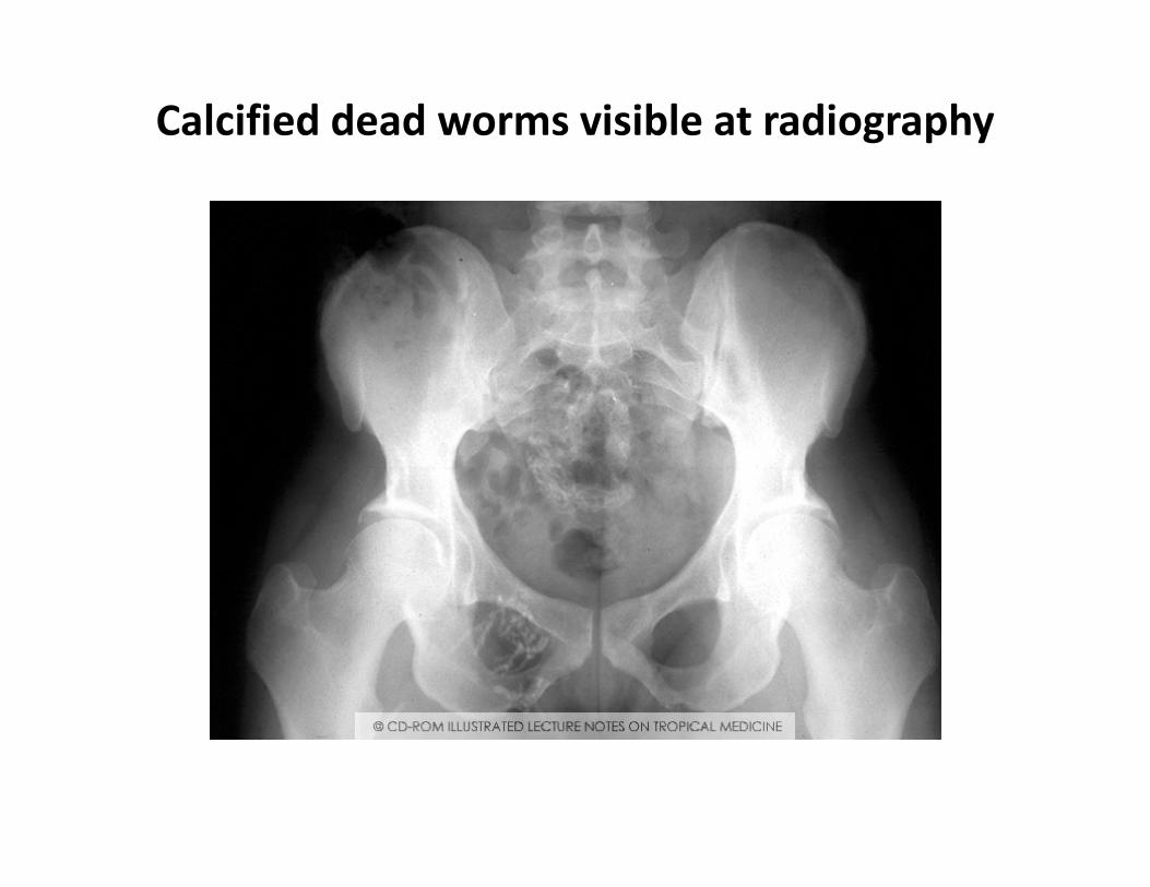
Calcified dead worms visible at radiography
Dracunculus – Management
• Slow extraction of female worm by rolling it on a
small stick (pull slowly each day)
• Be careful not to break the worm!
• Antibiotics & dressing ulcers to avoid bacterial
infection
• Tetanus vaccination is recommended
• Surgical removal possible
• No specific drug available
• Niridazole (12.5 mg/kg daily) reduces inflammation
and makes extraction easier
36
Dracunculus Control
• Can be eliminated (simple cycle, no animal
reservoir)
• Improve quality of drinking water (boreholes)
• Health education (filtering, boiling water)
• Chemical water control• Chemical water control
• Temephos (Abate) is used as insecticide (kills the
copepod)
• Surveillance of infected villages
37


The end....for now!

Other Nematodes – Other locations
• Trichinella spiralis
• Dracunculus medinensis
• Capillaria spp
• Angiostrongylus spp• Angiostrongylus spp
• Thelazia spp
• Gnathostoma spinigerum
40

Capillaria spp: 3 species with potential pathology for man
C. philippinensis C. hepatica C. aerophila
Philippines
Thailand
Few cases reported Few cases reported
Infection by ingestion of
undercooked, raw fish
Ingestion of embryonated eggs
in stool, contaminated food,
water, soil
Unsure. Similar to C. hepatica ?
Human intestinal capillariasis Human hepatic capillariasis
Animal parasite, rare in
humans
Human pulmonary capillariasis
Animal parasite, rare in humans
humans
Abdominal pain, diarrhea,
protein-losing enteropathy,
cachexia, death
Acute, sub-acute hepatitis,
eosinophilia, possible
dissemination to other organs.
May be fatal
Fever, cough, asthma,
pneumonia. May be fatal
Diagnosed by adults, eggs, L in
stool or intestinal biopsies
In severe infection, eggs, L,
adults in faeces
Diagnosed by adults/eggs in
liver tissue biopsy
Diagnosed by eggs in lung biopsy

Capillaria
philippinensis-
Life cycle
42
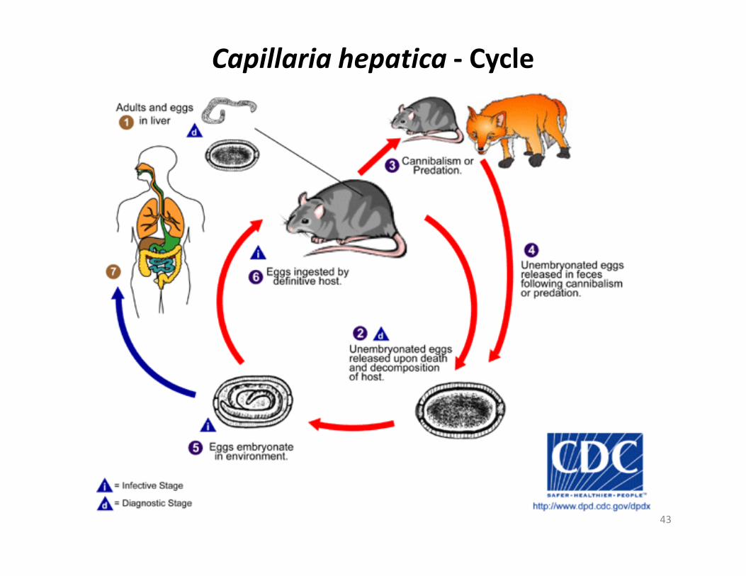
Capillaria hepatica - Cycle
43
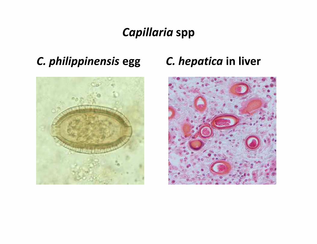
Capillaria spp
C. philippinensis egg C. hepatica in liver

Capillariasis - Management
• Mebendazole
• Albendazole
Other Nematodes – Other locations
• Trichinella spiralis
• Dracunculus medinensis
• Capillaria spp
• Angiostrongylus spp• Angiostrongylus spp
• Thelazia spp
• Gnathostoma spinigerum
46

Angiostrongyliasis Angiostrongylus spp
• The nematode (roundworm) Angiostrongylus
cantonensis, the rat lungworm, is the most common
cause of human eosinophilic meningitis.
• In addition, Angiostrongylus (Parastrongylus)
costaricensis is the causal agent of abdominal, or
intestinal, angiostrongyliasisintestinal, angiostrongyliasis
Distribution:
A. cantonensis: South-East Asia & Pacific Basin but
spreading elsewhere, including Africa
A. costaricensis: Costa Rica, Young children

Angiostrongylus cantonensis - Cycle
• Adults in pulmonary arteries of rats
• Eggs discharged into bloodstream and lodge as emboli in smaller vessels
• L1 break through respiratory tract, migrate up the trachea and passes out in faeces
• L1 enter molluscs (Acatina, Agriolimax, etc.) intermediate hosts
• Two moulds occur around 17th day
• When molluscs are eaten by rats, larvae are freed in stomach• When molluscs are eaten by rats, larvae are freed in stomach
• They pass to ileum, where they enter bloodstream and congregate in the CNS
• The anterior part of cerebrum is the favourite site where third mould takes place (6-7th day) and 4th mould on days 11-13
• Young adults emerge from day 12-14 and spread during the next 2 weeks on the arachnoid surface.
• From day 28-31,, they migrate to lungs via venous system and settle in pulmonary arteries
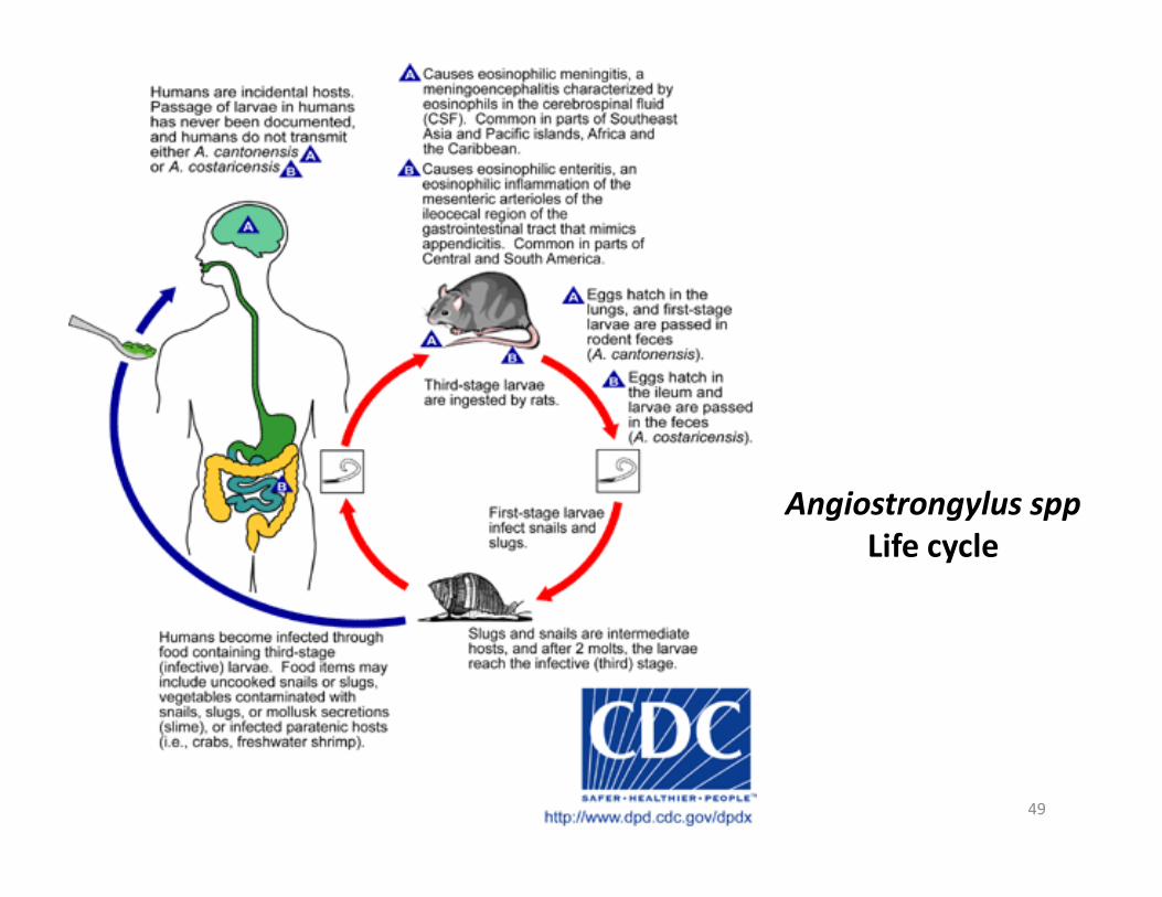
Angiostrongylus spp
Life cycle
49

Angiostrongylus cantonensis – Clinical features
• Humans can acquire the infection by eating raw or undercooked snails or slugs infected with the parasite
• They may also acquire the infection by eating raw produce that contains a small snail or slug, or part of one
• The disease can also be acquired by ingestion of contaminated or infected paratenic animals (crabs, freshwater shrimps)
• Clinical symptoms of eosinophilic meningitis are caused by the presence of larvae in the brain and by local host reactions.presence of larvae in the brain and by local host reactions.
• Symptoms : severe headaches, nausea, vomiting, neck stiffness, seizures, and neurologic abnormalities.
• Occasionally, ocular invasion occurs.
• Eosinophilia is present in most of cases.
• Most patients recover fully.
• Abdominal angiostrongyliasis mimics appendicitis, with eosinophilia.

Angiostrongylus cantonensis – Diagnosis
• In eosinophilic meningitis the cerebrospinal fluid
(CSF) is abnormal (elevated pressure, proteins,
and leukocytes; eosinophilia)
• On rare occasions, larvae have been found in the
CSF
• In abdominal angiostrongyliasis, eggs and larvae
can be identified in the tissues removed at
surgery (larvae similar to S. stercoralis but
smaller)

Angiostrongylus cantonensis – Management
• No drug with proven efficacy
• Relief of symptoms by analgesics,
corticosteroids, removal of CSF at frequent
intervals
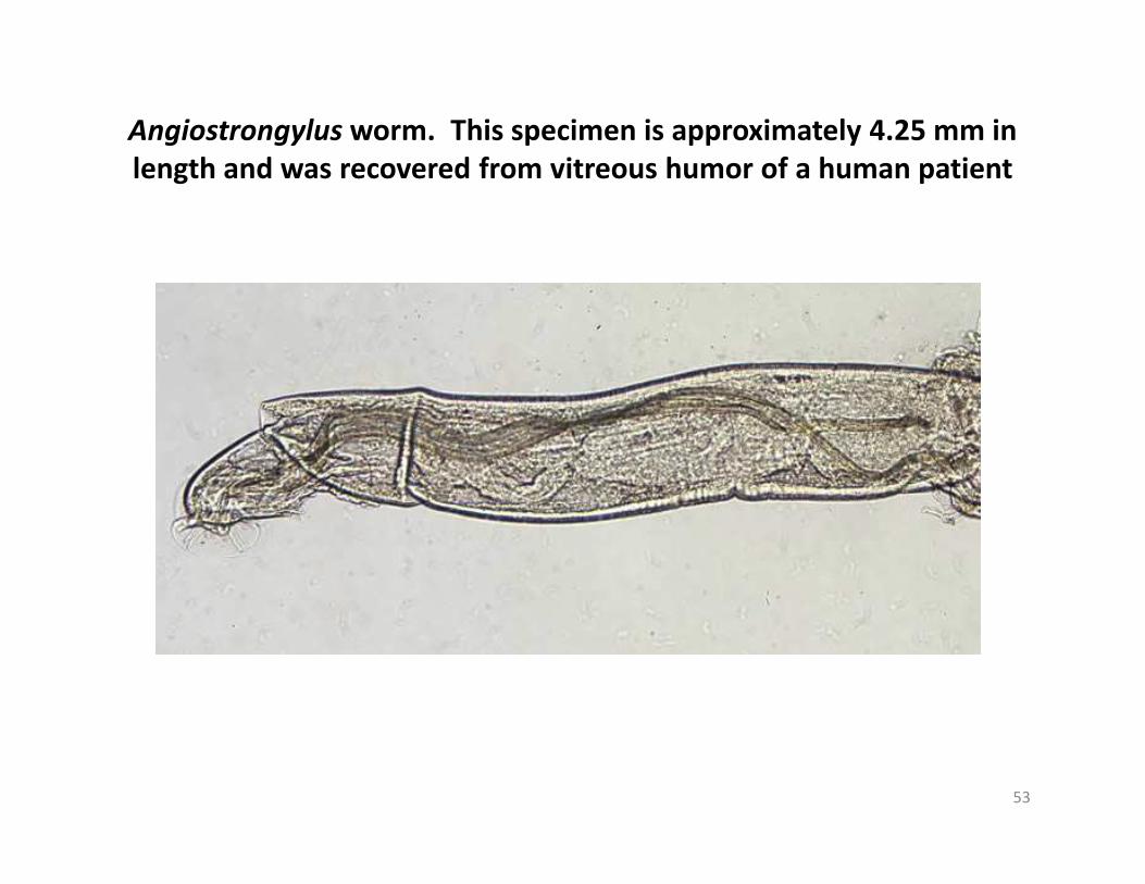
Angiostrongylus worm. This specimen is approximately 4.25 mm in
length and was recovered from vitreous humor of a human patient
53
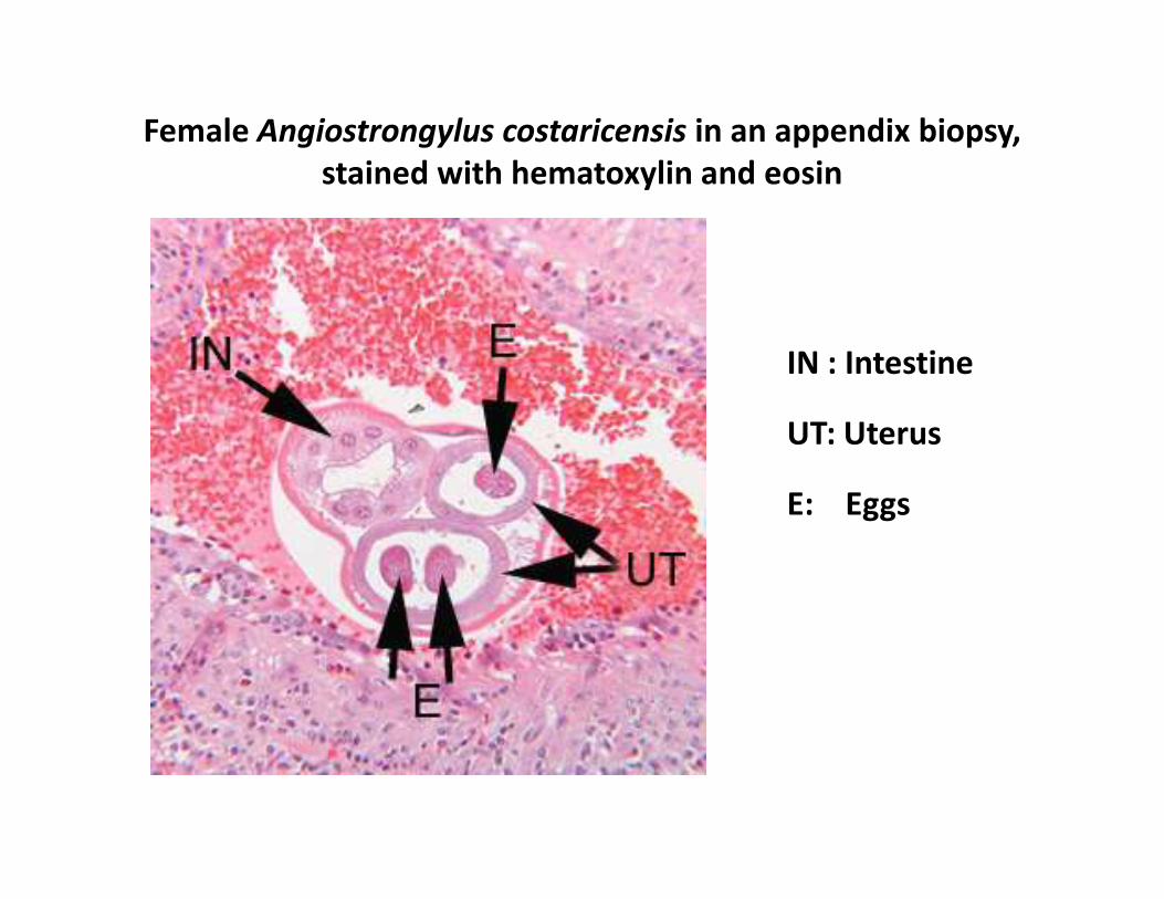
Female Angiostrongylus costaricensis in an appendix biopsy,
stained with hematoxylin and eosin
IN : Intestine
UT: UterusUT: Uterus
E: Eggs
Other Nematodes – Other locations
• Trichinella spiralis
• Dracunculus medinensis
• Capillaria spp
• Angiostrongylus spp• Angiostrongylus spp
• Gnathostoma spinigerum
• Thelazia spp
55
Gnathostoma spinigerum
• Adult worms are parasites of both wild & domestic felines and canines
• Widespread in Oriental, Palaeartic, Nearcticregions. Recently imported to Mexico
• Adults live in tumours in the stomach wall of cats & dogs
• Eggs are extruded form lesions and evacuated in • Eggs are extruded form lesions and evacuated in faeces to water, where they hatch
• G. spinigerum and G. hispidum are found in vertebrates
• Human gnathostomiasis is due to migrating immature worms

Gnathostoma – Cycle• In the natural definitive host (pigs, cats, dogs, wild animals), the
adult worms reside in a tumor which they induce in the gastric wall
• Un-embryonated eggs deposited when passed in the faeces
• Eggs become embryonated in water and release first-stage
larvae(L1)
• If ingested by a small crustacean (Cyclops, first intermediate host),
L1 develop into L2
• Following ingestion of the Cyclops by a fish, frog, or snake (second • Following ingestion of the Cyclops by a fish, frog, or snake (second
intermediate host), L2 migrate into the flesh and develop into L3
• When the second intermediate host is ingested by a definitive host,
L3 develop into adult parasites in the stomach wall
• Or, the second intermediate host may be ingested by paratenic host
(such as birds, snakes, and frogs) in which the L3 do not develop
further but remain infective to the next predator
• Humans are infected by eating undercooked fish/poultry containing
L3 , or by drinking water containing infective L2 in Cyclops

Gnathostoma spp
Cycle
58

Gnathostoma – Clinical features
• The clinical manifestations in human
gnathostomiasis are caused by migration of the
immature worms (L3s).
• Migration in the subcutaneous tissues causes
intermittent, migratory, painful, pruritic swellings
(cutaneous larva migrans).(cutaneous larva migrans).
• Migration to other tissues (visceral larva migrans)
can result in cough, hematuria, and ocular
involvement, with the most serious manifestations
eosinophilic meningitis with myeloencephalitis.
• High eosinophilia is present.
59
Gnathostoma – Diagnostic/Management
• Removal and identification of the worm is both diagnostic and therapeutic.
• Identification of gnathostomiasis is achieved by serology or microscopic observation of the larval serology or microscopic observation of the larval worms in tissue sections
• Surgical removal or treatment with Albendazoleor Ivermectin is recommended.
Other Nematodes – Other locations
• Trichinella spiralis
• Dracunculus medinensis
• Capillaria spp
• Angiostrongylus spp• Angiostrongylus spp
• Gnathostoma spinigerum
• Thelazia spp
61
Thelaziasis – Aetiology/Distribution
• Spirurid nematodes of the genus Thelazia
• Two species that have been implicated in human infection:
– T. callipaeda (the Oriental eye worm)
– T. californiensis (the California eye worm)
• Worldwide. Human infections have been recorded from the
United States, China, Russia, India, Japan, and Thailand
• Dogs and other canids, cattle, and horses are the usual definitive • Dogs and other canids, cattle, and horses are the usual definitive
hosts for Thelazia spp., although other mammals, including cats,
lagomorphs, cervids and humans, can also become infected
• Dipteran flies are utilized as intermediate hosts.
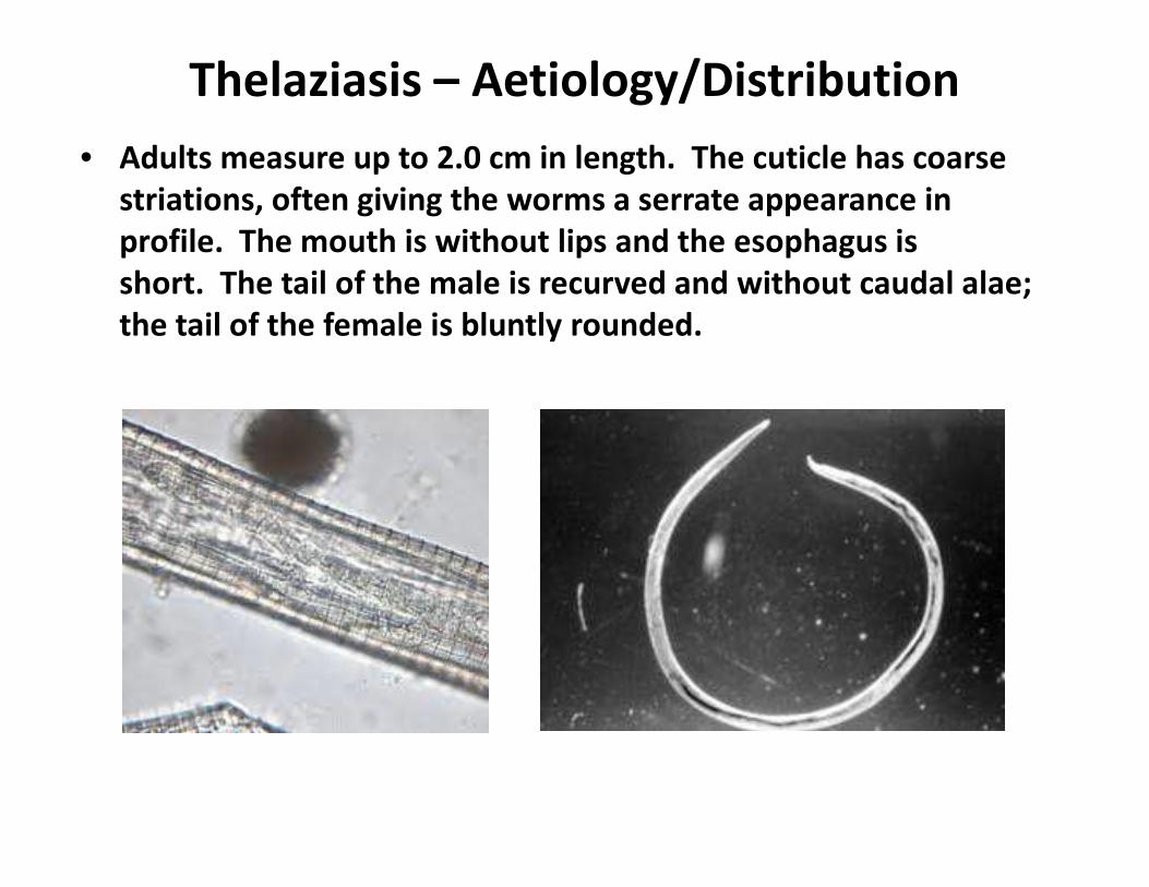
Thelaziasis – Aetiology/Distribution
• Adults measure up to 2.0 cm in length. The cuticle has coarse
striations, often giving the worms a serrate appearance in
profile. The mouth is without lips and the esophagus is
short. The tail of the male is recurved and without caudal alae;
the tail of the female is bluntly rounded.

Thelazia spp - Cycle
64
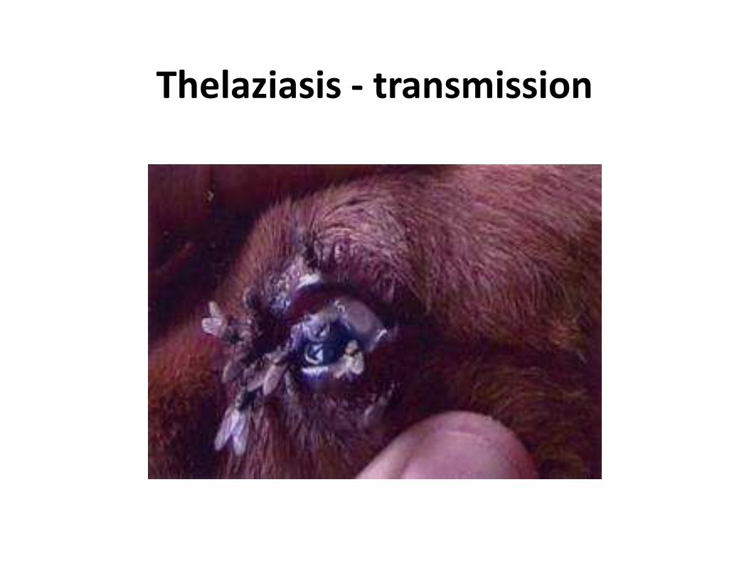
Thelaziasis - transmission

Thelaziasis
Thelaziasis – Clinical features /diagnosis/Treatment
• Clinical Features
– Adults in the eye cause varying degrees of inflammation and lacrimation.
– In heavier infections, photophobia, edema, conjunctivitis, and blindness may occur.
• Laboratory diagnosis• Laboratory diagnosis
– Identification is made by finding of adult worms in the conjunctival sac
• Treatment
– Treatment is usually limited to the complete removal of the adult worms from the eye
67


