Otodental syndrome: three familialcase reports Lourdes Santos-Pinto, DDS, PhD Maria del Pilar Oviedo, DDS, MS Ary Santos-Pinto, DDS, PhD Helda I. lost, DDS, PhD N.Sue Seale, DDS, MSD Anil K.Reddy, DDS, MPH O todental syndrome, also named oto- dentodysplasia, ~ otodental dysplasia, 2-4 and globodontia, 5’ 6 has been reported as a type of ectodermaldysplasia, with an autosomal domi- nant inheritance of variable expressivity) It wasfirst reported by Toledo et al. 7 as a multiple dental anomaly in three brothers in whom the authors observedabnor- mal crown morphologywith globe-shaped molars and short roots. Theclinical and radiographicaspects were not sufficient to allow differentiation between primary and permanent teeth. The histologic findings showed that the overall structure of enamel, dentin, cementum, and pulp appeared to be normal. In 1972, Levin and Jorgenson ~ reported a newsyn- drome that they named "familial otodentodysplasia", characterized by posterior teeth with abnormal mor- phology in association with high-frequency hearing loss. Absentor small premolars were also reported. Ra- diographic analysis of the abnormal teeth showed large pulp chambers, some of which were duplicated, and the root length was short compared with crown height. Witkop et al. 6 reported "globodontia" in otodental syndromeand observed that the patients had a long facies, anteverted nostrils, a long philtrum,and a full- cheek appearance. The primary canines and molars were larger than normal and presented with globe- shaped crowns having anomalous grooves extending from the labial and lingual onto the incisal or occlusal surface of the crowns. Localized yellow-white spots were present on the enamel, particularly on the labial surfaces of the primarycanines. Chen at al. 2 observed the presence of extra incisors in the maxillary anterior region and conical supernu- merary microdontic teeth on the palatal side of primary maxillary molars. Conical maxillary lateral incisors were reported by Stewart and Kinirons 5 in patients with otodental syn- drome. However that case report was considered as doubtful by Gorlin et al.8 Griffin 9 reporteda case diagnosed as a possible vari- ant of otodental syndrome. However, the clinical finding showed only fusion of a maxillary second per- manent molar with a supplemental premolar. The posterior teeth did not show globular deformities and the hearing impairment was not only for high-fre- quency sound. This case report describes the occurrenceof famil- ial otodental syndrome in three generations of a family, including the manifestations of the condition in both the primary and permanent dentitions of three affected patients. Case report The transmission of this disorder through genera- tions is depictedin Fig 1. Case 1 Patient II-13 (Fig 1) was examined for the first time, along with her brothers, when she was 6 years old by Toledo et al. 7 and all were reported as havingmultiple dental anomalies with size and shape alterations in posterior teeth. Recently, at age 30, she demonstrated a normal physical appearance but her lower anterior fao cial height wasexcessive with an abnormal lip posture. Oral examination of the maxillary arch revealed three erupted incisors, whichwere normal in size and shape. It wasimpossible to say if the canines werepri- mary or permanent. The two premolars on the right side andone on the left side were conical, andthe first and second molar crowns were remarkable for their rounded or globular shape. I m r~ ¯ Male affected [] Male unaffected ¯ Female ~ffected 0 Female unllffected * Hearing loss Fig 1.Distribution of the individuals in the family with otodental syndrome. 208 American Academy of Pediatric Dentistry PediatricDentistry - 20:3, 1998
Transcript
Otodental syndrome: three familial case reportsLourdes Santos-Pinto, DDS, PhD Maria del Pilar Oviedo, DDS, MS Ary Santos-Pinto, DDS, PhDHelda I. lost, DDS, PhD N. Sue Seale, DDS, MSD Anil K. Reddy, DDS, MPH
O todental syndrome, also named oto-dentodysplasia,~ otodental dysplasia,2-4
and globodontia,5’ 6 has been reported as atype of ectodermal dysplasia, with an autosomal domi-nant inheritance of variable expressivity) It was firstreported by Toledo et al.7 as a multiple dental anomalyin three brothers in whom the authors observed abnor-mal crown morphology with globe-shaped molars andshort roots. The clinical and radiographic aspects werenot sufficient to allow differentiation between primaryand permanent teeth. The histologic findings showedthat the overall structure of enamel, dentin, cementum,and pulp appeared to be normal.
In 1972, Levin and Jorgenson~ reported a new syn-drome that they named "familial otodentodysplasia",characterized by posterior teeth with abnormal mor-phology in association with high-frequency hearingloss. Absent or small premolars were also reported. Ra-diographic analysis of the abnormal teeth showed largepulp chambers, some of which were duplicated, andthe root length was short compared with crown height.
Witkop et al. 6 reported "globodontia" in otodentalsyndrome and observed that the patients had a longfacies, anteverted nostrils, a long philtrum, and a full-cheek appearance. The primary canines and molarswere larger than normal and presented with globe-shaped crowns having anomalous grooves extendingfrom the labial and lingual onto the incisal or occlusalsurface of the crowns. Localized yellow-white spotswere present on the enamel, particularly on the labialsurfaces of the primary canines.
Chen at al.2 observed the presence of extra incisorsin the maxillary anterior region and conical supernu-merary microdontic teeth on the palatal side of primarymaxillary molars.
Conical maxillary lateral incisors were reported byStewart and Kinirons5 in patients with otodental syn-drome. However that case report was considered asdoubtful by Gorlin et al.8
Griffin9 reported a case diagnosed as a possible vari-ant of otodental syndrome. However, the clinicalfinding showed only fusion of a maxillary second per-manent molar with a supplemental premolar. Theposterior teeth did not show globular deformities and
the hearing impairment was not only for high-fre-quency sound.
This case report describes the occurrence of famil-ial otodental syndrome in three generations of afamily, including the manifestations of the conditionin both the primary and permanent dentitions ofthree affected patients.
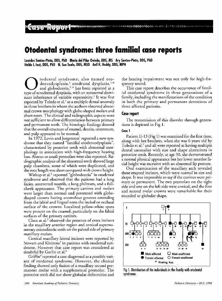
Case reportThe transmission of this disorder through genera-
tions is depicted in Fig 1.
Case 1Patient II-13 (Fig 1) was examined for the first time,
along with her brothers, when she was 6 years old byToledo et al.7 and all were reported as having multipledental anomalies with size and shape alterations inposterior teeth. Recently, at age 30, she demonstrateda normal physical appearance but her lower anterior faocial height was excessive with an abnormal lip posture.
Oral examination of the maxillary arch revealedthree erupted incisors, which were normal in size andshape. It was impossible to say if the canines were pri-mary or permanent. The two premolars on the rightside and one on the left side were conical, and the firstand second molar crowns were remarkable for theirrounded or globular shape.
I
m r~
¯ Male affected [] Male unaffected¯ Female ~ffected 0 Female unllffected
* Hearing loss
Fig 1. Distribution of the individuals in the family with otodentalsyndrome.
208 American Academy of Pediatric Dentistry Pediatric Dentistry - 20:3, 1998
In the mandibular arch, incisors and canines had thesame shape. Only one conical premolar was present oneach side, the left first permanent molar had been ex-tracted, and the remaining molars presented with manydevelopmental grooves radiating from the center pit onto all surfaces, dividing each crown into lobules of dif-ferent sizes.
The patient demonstrated a posterior bilateralcrossbite and a maxillary midline shift to the right dueto the missing lateral incisor. The mandibular arch was"U" shaped and the maxillary arch was "V" shaped andconstricted, so that the distance between the lingualsurfaces of the premolar measured 2.5 cm, resulting ina deep palate.
Dental radiographs showed an impacted maxillaryright lateral incisor and a periapical radiolucency overthe mandibular left first permanent molar. The rootsof affected teeth were short compared to crown size andtaurodontism was evident.
Case 2
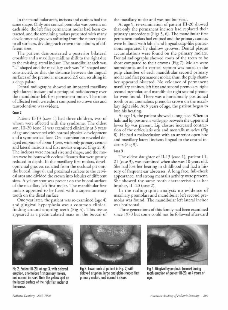
Patient 11-13 (case 1) had three children, two ofwhom were affected with the syndrome. The oldestson, 111-20 (case 2) was examined clinically at 3 yearsof age and presented with normal physical developmentand a symmetrical face. Oral examination revealed de-layed eruption of about 1 year, with only primary centraland lateral incisors and first molars erupted (Figs 2, 3).The incisors were normal size and shape, and the mo-lars were bulbous with occlusal fissures that were greatlyreduced in depth. In the maxillary first molars, devel-opmental grooves radiated from the occlusal pit ontothe buccal, lingual, and proximal surfaces to the cervi-cal area and divided the crown into lobules of differentsizes. A yellow spot was present on the buccal surfaceof the maxillary left first molar. The mandibular firstmolars appeared to be fused with a supernumerarytooth on the distal surface.
One year later, the patient was re-examined (age 4)and gingival hyperplasia was a common clinicalfinding around erupting teeth (Fig 4). This tissueappeared as a pedunculated mass on the buccal of
the maxillary molar and was not biopsied.At age 9, re-examination of patient 111-20 showed
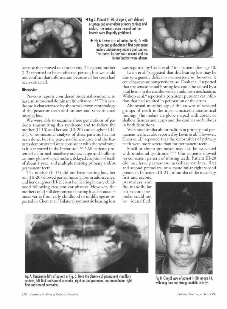
that only the permanent incisors had replaced theirprimary antecedents (Figs 5,6). The mandibular firstpermanent molars had erupted and the primary canineswere bulbous with labial and lingual cusp-like protru-sions separated by shallow grooves. Dental plaqueaccumulations were found on the primary molars.Dental radiographs showed roots of the teeth to beshort compared to their crowns (Fig 7). Molars weretaurodontic, and a vertical septum was noted in thepulp chamber of each mandibular second primarymolar and first permanent molar; thus, the pulp cham-ber appeared bisected. No evidence of permanentmaxillary canines, left first and second premolars, rightsecond premolar, and mandibular right second premo-lar were found. There was a horizontally positionedtooth or an anomalous premolar crown on the maxil-lary right side. At 9 years of age, the patient began tolose his hearing.
At age 14, the patient showed a long face. When inhabitual lip posture, a wide gap between the upper andlower lip was present. Lip closure increased contrac-tion of the orbicularis oris and mentalis muscles (Fig8). He had a malocclusion with an anterior open biteand maxillary lateral incisors lingual to the central in-cisors (Fig 9).
Case 3
The oldest daughter of 11-13 (case 1), patient HI-21 (case 3), was examined when she was 10 years old.She had lost her hearing in childhood and had a his-tory of frequent ear abscesses. A long face, full-cheekappearance, and strong mentalis activity were present.She showed the same tooth characteristics as herbrother, 111-20 (case 2).
In the radiographic analysis no evidence ofmaxillary premolars and mandibular left second pre-molar was found. The mandibular left lateral incisorwas horizontal.
Three generations of this family had been examinedsince 1970 but some could not be followed afterward
Fig 2. Patient 111-20, at age 3, with delayederuption, anomalous first primary molars,ana normal incisors. Note the yellow spot onthe buccal surface of the right first molar atthe arrow.
Fig 3. Lower arch of patient in Fig. 2, withdelayed eruption, large and globe-shaped firstprimary molars, and normal incisors.
Fig 4. Gingival hyperplasia (arrow) duringtooth eruption or patient 111-20, at 4 years ofage.
Pediatric Dentistry -20:3, 1998 American Academy of Pediatric Dentistry 209
Fig 5. Patient 111-20, at age 9, with delayederuption and anomalous primary canines andmolars. The incisors were normal but thelaterals were lingually positioned.
^ Fig 6. Lower arch of patient in Fig. 5, withlarge and globe-shaped first permanentmolars and primary molars and canines.The central incisors were normal and the
lateral incisors were absent.
because they moved to another city. The grandmother(1-2) reported to be an affected person, but we couldnot confirm that information because all her teeth hadbeen extracted.
DiscussionPrevious reports considered otodental syndrome to
have an autosomal dominant inheritance.1^4'6 This syn-drome is characterized by abnormal crown morphologyof the posterior teeth and canines and sensorineuralhearing loss.
We were able to examine three generations of pa-tients transmitting this syndrome and to follow themother (II-13) and her son (111-20) and daughter (HI-21). Chromosomal analysis of these patients has notbeen done, but the pattern of inheritance and the fea-tures demonstrated were consistent with the syndromeas it is reported in the literature.1'4'6'8 All patients pre-sented deformed maxillary arches, large and bulbouscanines, globe-shaped molars, delayed eruption of teethof about 1 year, and multiple missing primary and/orpermanent teeth.
The mother (11-13) did not have hearing loss, herson (111-20) showed partial hearing loss in adolescence,and her daughter (111-21) lost her hearing in early child-hood following frequent ear abscess. However, themother could still demonstrate hearing loss, because itsonset varies from early childhood to middle age as re-ported in Chen et al.2 Bilateral symmetric hearing loss
was reported by Cook et al.10 in a patient after age 40.Levin et al.3 suggested that this hearing loss may be
due to a genetic defect in neuroectoderm; however, itcould have some nongenetic cause. Cook et al.10 reportedthat the sensorineural hearing loss could be caused by alocal lesion in the cochlea with an unknown mechanism.Witkop et al.6 reported a persistent purulent ear infec-tion that had resulted in perforation of the drum.
Abnormal morphology of the crowns of selectedgroups of teeth is the most consistent anatomicalfinding. The molars are globe shaped with absent orshallow fissures and cusps and the canines are bulbousin both dentitions.
We found similar abnormalities in primary and per-manent teeth, as also reported by Levin et al.3 However,Chen et al.2 reported that the deformities of primaryteeth were more severe than for permanent teeth.
Small or absent premolars may also be associatedwith otodental syndrome.2~4'6 Our patients showedno consistent pattern of missing teeth. Patient 111-20did not have permanent maxillary canines, firstand second premolars, or a mandibular right secondpremolar. In patient III-21, primordia of the maxillaryfirst and secondpremolars andthe mandibularleft second pre-molar could notbe identified.
Fig 7. Panoramic film of patient in Fig. 5. Note the absence of permanent maxillarycanines, left first and second premolar, right second premolar, and mandibular rightfirst and second premolars.
Fig 8. Clinical view of patient 111-20, at age 14,with long face and strong mentalis activity.
210 American Academy ofPediatric Dentistry Pediatric Dentistry - 20:3, 1998
•P WH^̂ P
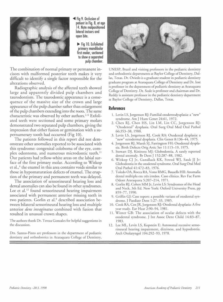
Fig 9. Occlusion ofpatient in Fig. 8, at age14. Note malpositionedlateral incisors andopen bite.
^ Fig 10. Exfoliatedprimary mandibular
first molar, sectionedto show a separated
pulp chamber.
The combination of normal primary or permanent in-cisors with malformed posterior teeth makes it verydifficult to identify a single factor responsible for thealterations observed.
Radiographic analysis of the affected teeth showedlarge and apparently divided pulp chambers andtaurodontism. The taurodontic appearance is a conse-quence of the massive size of the crown and largeappearance of the pulp chamber rather than enlargementof the pulp chambers extending into the roots. The samecharacteristic was observed by other authors.2'6 Exfoli-ated teeth were sectioned and some primary molarsdemonstrated two separated pulp chambers, giving theimpression that either fusion or gemination with a su-pernumerary tooth had occurred (Fig 10).
The patients followed in this report did not dem-onstrate other anomalies reported to be associated withthis syndrome: congenital coloboma of the eye, com-plex odontoma, and numerous microdontic teeth."Our patients had yellow-white areas on the labial sur-face of the first primary molar. According to Witkopet al.,6 the enamel in this area contains voids similar tothose in hypomaturation defects of enamel. The erup-tion of the primary and permanent teeth was delayed.
The association of sensorineural hearing loss anddental anomalies can also be found in other syndromes.Lee et al.12 found sensorineural hearing impairmentassociated with permanent anterior missing teeth intwo patients. Gorlin et al.8 described association be-tween bilateral sensorineural hearing loss and multipleanterior dens invaginatus combined with fusion thatresulted in unusual crown shapes.
The authors thank Dr. Tereza Gonzales for helpful suggestions inthe discussion.
Drs. Santos-Pinto are professors in the department of pediatricdentistry and orthodontics in Araraquara College of Dentistry,
UNESP, Brazil and visiting professors in the pediatric dentistryand orthodontic departments at Baylor College of Dentistry, Dal-las, Texas. Dr. Oviedo is a graduate student in pediatric dentistrygraduate program at Araraquara College of Dentistry and Dr. lostis professor in the department of pediatric dentistry at AraraquaraCollege of Dentistry. Dr. Scale is professor and chairman and Dr.Reddy is assistant professor in the pediatric dentistry departmentat Baylor College of Dentistry, Dallas, Texas.
References1. Levin LS, Jorgenson RJ: Familial otodentodysplasia: a "new"
syndrome. Am J Hum Genet 24:61, 1972.2. Chen RJ, Chen HS, Lin LM, Lin CG, Jorgenson RJ:
"Otodental" dysplasia. Oral Surg Oral Med Oral Pathol66:353-38, 1988.