+ All Categories
Home > Documents > Outcome analysis of cleft palate surgery–can static 2D MRI replace videofluroscopy?

Outcome analysis of cleft palate surgery–can static 2D MRI replace videofluroscopy?

Date post: 24-Nov-2016
Category:
Upload: ramesh-sharma
View: 214 times
Download: 1 times
Share this document with a friend
3
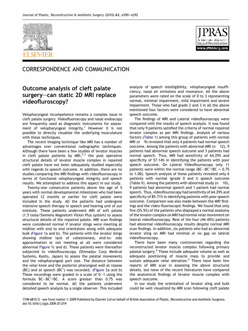
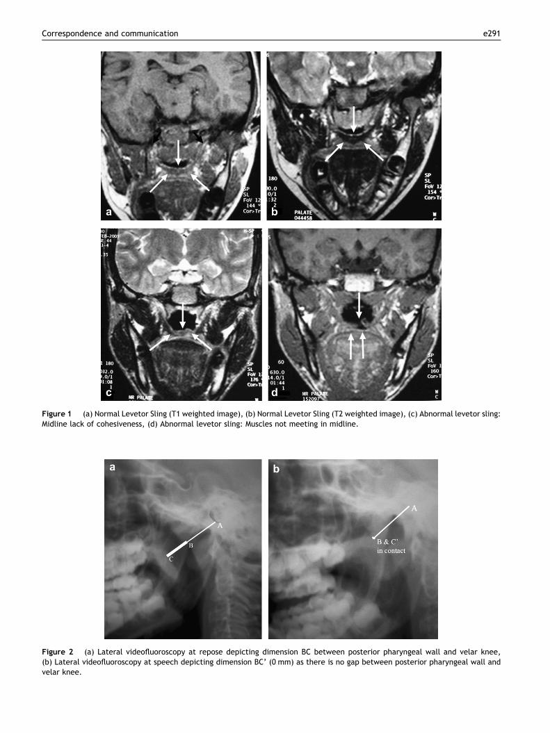
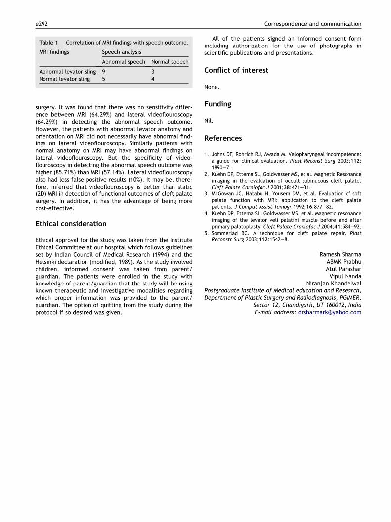
CORRESPONDENCE AND COMMUNICATION Outcome analysis of cleft palate surgeryecan static 2D MRI replace videofluroscopy? Velopharyngeal incompetence remains a complex issue in cleft palate surgery. Videofluoroscopy and nasal endoscopy are frequently used as diagnostic instruments for assess- ment of velopharyngeal integrity. 1 However it is not possible to directly visualize the underlying musculature with these techniques. The recent imaging technique like MRI has a number of advantages over conventional radiographic techniques. Although there have been a few studies of levator muscles in cleft palate patients by MRI, 2,3 the post operative structural details of levator muscle complex in repaired cleft palate have not been adequately studied especially with regards to speech outcome. In addition, there are no studies comparing the MRI findings with videofluoroscopy in terms of functional velopharyngeal integrity and speech results. We attempted to address this aspect in our study. Twenty-one consecutive patients above the age of 5 years with normal developmental milestones who had been operated 12 months or earlier for cleft palate were included in the study. All the patients had undergone intensive speech therapy in speech and hearing unit of our institute. These patients underwent MRI of soft palate (1.5 tesla/Siemens Magnetom Vision Plus system) to assess structural details of the repaired palate. MRI scan findings were considered normal if levator slings were meeting at midline with end to end orientation along with adequate bulk (Figure 1a and b). The patients with the levator slings showing midline lack of cohesiveness, end-to- side approximation or not meeting at all were considered abnormal Figure 1c and d). These patients were thereafter subjected to videoflouroscopy (Shimadzu Corp Medical Systems, Kyoto, Japan) to assess the palatal movements and the velopharyngeal port size. The distance between the velar knee and the posterior pharyngeal wall at repose (BC) and at speech (BC’) was recorded. (Figure 2a and b) These recordings were graded in a scale of 0e1 using the formula BCeBC’/BC. A score greater than 0.75 was considered to be normal. All the patients underwent detailed speech analysis by a single observer. This included analysis of speech intelligibility, velopharyngeal insuffi- ciency, nasal air emissions and resonance. All the above parameters were rated on the scale of 0 to 3 representing normal, minimal impairment, mild impairment and severe impairment. Those who had grade 2 and 3 in all the above mentioned four factors were considered to have abnormal speech outcome. The findings of MRI and Lateral videoflouroscopy were compared with the results of speech analysis. It was found that only 9 patients satisfied the criteria of normal repaired levator complex as per MRI findings. Analysis of various factors (Table 1) among this group of patients with normal MRI (n 9) revealed that only 4 patients had normal speech outcome. Among the patients with abnormal MRI (n 12), 9 patients had abnormal speech outcome and 3 patients had normal speech. Thus, MRI had sensitivity of 64.29% and specificity of 57.14% in identifying the patients with poor speech outcome. On Lateral Videoflouroscopy only 11 patients were within the normal range (BCeBC’/BC Z 0.75 to 1.00). Speech analysis of these patients revealed only 6 patients with normal (grade 0 and I) speech outcome (Table II). Among the patients with abnormal study (n 10), 9 patients had abnormal speech and 1 patient had normal speech. Thus, videoflouroscopy had sensitivity of 64.29% and specificity of 85.71% in identifying patients with poor speech outcome. Comparison was also made between the MRI find- ings and the video-fluoroscopic findings. We found that only five (55.5%) of the patients who displayed a normal anatomy of the levator complex on MRI had normal velar movement on lateral videoflouroscopy. Rest of the four (44.45%) patients had abnormal videoflouroscopy results despite normal MRI scan findings. In addition, six patients who had an abnormal levator sling on MRI had minimal or no gap on lateral videoflouroscopy. There have been many controversies regarding the reconstructed levator muscle complex following primary palatal surgery. 4 These include adequate volume as well as adequate positioning of muscle mass to provide and sustain adequate velar elevation. 5 There have been few reports of MRI scan in assessing the above structural details, but none of the recent literatures have compared the anatomical findings of levator muscle complex with speech outcome. In our study the orientation of levator sling and bulk could be well visualized by MRI scan following cleft palate 1748-6815/$ - see front matter ª 2009 Published by Elsevier Ltd on behalf of British Association of Plastic, Reconstructive and Aesthetic Surgeons. doi:10.1016/j.bjps.2009.07.019 Journal of Plastic, Reconstructive & Aesthetic Surgery (2010) 63, e290ee292
Transcript

Journal of Plastic, Reconstructive & Aesthetic Surgery (2010) 63, e290ee292

CORRESPONDENCE AND COMMUNICATION

Outcome analysis of cleft palatesurgeryecan static 2D MRI replacevideofluroscopy?

Velopharyngeal incompetence remains a complex issue incleft palate surgery. Videofluoroscopy and nasal endoscopyare frequently used as diagnostic instruments for assess-ment of velopharyngeal integrity.1 However it is notpossible to directly visualize the underlying musculaturewith these techniques.

The recent imaging technique like MRI has a number ofadvantages over conventional radiographic techniques.Although there have been a few studies of levator musclesin cleft palate patients by MRI,2,3 the post operativestructural details of levator muscle complex in repairedcleft palate have not been adequately studied especiallywith regards to speech outcome. In addition, there are nostudies comparing the MRI findings with videofluoroscopy interms of functional velopharyngeal integrity and speechresults. We attempted to address this aspect in our study.

Twenty-one consecutive patients above the age of 5years with normal developmental milestones who had beenoperated 12 months or earlier for cleft palate wereincluded in the study. All the patients had undergoneintensive speech therapy in speech and hearing unit of ourinstitute. These patients underwent MRI of soft palate(1.5 tesla/Siemens Magnetom Vision Plus system) to assessstructural details of the repaired palate. MRI scan findingswere considered normal if levator slings were meeting atmidline with end to end orientation along with adequatebulk (Figure 1a and b). The patients with the levator slingsshowing midline lack of cohesiveness, end-to- sideapproximation or not meeting at all were consideredabnormal Figure 1c and d). These patients were thereaftersubjected to videoflouroscopy (Shimadzu Corp MedicalSystems, Kyoto, Japan) to assess the palatal movementsand the velopharyngeal port size. The distance betweenthe velar knee and the posterior pharyngeal wall at repose(BC) and at speech (BC’) was recorded. (Figure 2a and b)These recordings were graded in a scale of 0e1 using theformula BCeBC’/BC. A score greater than 0.75 wasconsidered to be normal. All the patients underwentdetailed speech analysis by a single observer. This included

1748-6815/$ - see front matter ª 2009 Published by Elsevier Ltd on behalf ofdoi:10.1016/j.bjps.2009.07.019

analysis of speech intelligibility, velopharyngeal insuffi-ciency, nasal air emissions and resonance. All the aboveparameters were rated on the scale of 0 to 3 representingnormal, minimal impairment, mild impairment and severeimpairment. Those who had grade 2 and 3 in all the abovementioned four factors were considered to have abnormalspeech outcome.

The findings of MRI and Lateral videoflouroscopy werecompared with the results of speech analysis. It was foundthat only 9 patients satisfied the criteria of normal repairedlevator complex as per MRI findings. Analysis of variousfactors (Table 1) among this group of patients with normalMRI (n� 9) revealed that only 4 patients had normal speechoutcome. Among the patients with abnormal MRI (n� 12), 9patients had abnormal speech outcome and 3 patients hadnormal speech. Thus, MRI had sensitivity of 64.29% andspecificity of 57.14% in identifying the patients with poorspeech outcome. On Lateral Videoflouroscopy only 11patients were within the normal range (BCeBC’/BC Z 0.75to 1.00). Speech analysis of these patients revealed only 6patients with normal (grade 0 and I) speech outcome(Table II). Among the patients with abnormal study (n� 10),9 patients had abnormal speech and 1 patient had normalspeech. Thus, videoflouroscopy had sensitivity of 64.29% andspecificity of 85.71% in identifying patients with poor speechoutcome. Comparison was also made between the MRI find-ings and the video-fluoroscopic findings. We found that onlyfive (55.5%) of the patients who displayed a normal anatomyof the levator complex on MRI had normal velar movement onlateral videoflouroscopy. Rest of the four (44.45%) patientshad abnormal videoflouroscopy results despite normal MRIscan findings. In addition, six patients who had an abnormallevator sling on MRI had minimal or no gap on lateralvideoflouroscopy.

There have been many controversies regarding thereconstructed levator muscle complex following primarypalatal surgery.4 These include adequate volume as well asadequate positioning of muscle mass to provide andsustain adequate velar elevation.5 There have been fewreports of MRI scan in assessing the above structuraldetails, but none of the recent literatures have comparedthe anatomical findings of levator muscle complex withspeech outcome.

In our study the orientation of levator sling and bulkcould be well visualized by MRI scan following cleft palate

British Association of Plastic, Reconstructive and Aesthetic Surgeons.

Figure 1 (a) Normal Levetor Sling (T1 weighted image), (b) Normal Levetor Sling (T2 weighted image), (c) Abnormal levetor sling:Midline lack of cohesiveness, (d) Abnormal levetor sling: Muscles not meeting in midline.

Figure 2 (a) Lateral videofluoroscopy at repose depicting dimension BC between posterior pharyngeal wall and velar knee,(b) Lateral videofluoroscopy at speech depicting dimension BC’ (0 mm) as there is no gap between posterior pharyngeal wall andvelar knee.

Correspondence and communication e291

Table 1 Correlation of MRI findings with speech outcome.

MRI findings Speech analysis

Abnormal speech Normal speech

Abnormal levator sling 9 3Normal levator sling 5 4

e292 Correspondence and communication

surgery. It was found that there was no sensitivity differ-ence between MRI (64.29%) and lateral videoflouroscopy(64.29%) in detecting the abnormal speech outcome.However, the patients with abnormal levator anatomy andorientation on MRI did not necessarily have abnormal find-ings on lateral videoflouroscopy. Similarly patients withnormal anatomy on MRI may have abnormal findings onlateral videoflouroscopy. But the specificity of video-flouroscopy in detecting the abnormal speech outcome washigher (85.71%) than MRI (57.14%). Lateral videoflouroscopyalso had less false positive results (10%). It may be, there-fore, inferred that videoflouroscopy is better than static(2D) MRI in detection of functional outcomes of cleft palatesurgery. In addition, it has the advantage of being morecost-effective.

Ethical consideration

Ethical approval for the study was taken from the InstituteEthical Committee at our hospital which follows guidelinesset by Indian Council of Medical Research (1994) and theHelsinki declaration (modified, 1989). As the study involvedchildren, informed consent was taken from parent/guardian. The patients were enrolled in the study withknowledge of parent/guardian that the study will be usingknown therapeutic and investigative modalities regardingwhich proper information was provided to the parent/guardian. The option of quitting from the study during theprotocol if so desired was given.

All of the patients signed an informed consent formincluding authorization for the use of photographs inscientific publications and presentations.

Conflict of interest

None.

Funding

Nil.

References

1. Johns DF, Rohrich RJ, Awada M. Velopharyngeal incompetence:a guide for clinical evaluation. Plast Reconst Surg 2003;112:1890e7.

2. Kuehn DP, Ettema SL, Goldwasser MS, et al. Magnetic Resonanceimaging in the evaluation of occult submucous cleft palate.Cleft Palate Carniofac J 2001;38:421e31.

3. McGowan JC, Hatabu H, Yousem DM, et al. Evaluation of softpalate function with MRI: application to the cleft palatepatients. J Comput Assist Tomogr 1992;16:877e82.

4. Kuehn DP, Ettema SL, Goldwasser MS, et al. Magnetic resonanceimaging of the levator veli palatini muscle before and afterprimary palatoplasty. Cleft Palate Craniofac J 2004;41:584e92.

5. Sommerlad BC. A technique for cleft palate repair. PlastReconstr Surg 2003;112:1542e8.

Ramesh SharmaABMK PrabhuAtul Parashar

Vipul NandaNiranjan Khandelwal

Postgraduate Institute of Medical education and Research,Department of Plastic Surgery and Radiodiagnosis, PGIMER,

Sector 12, Chandigarh, UT 160012, IndiaE-mail address: [email protected]


Recommended