Overexpression of the stathmin gene in a subset ofhuman breast cancer
I Bieche1, S Lachkar2, V Becette3, C Cifuentes-Diaz4, A SobeJ2, R Lidereau1 and PA Curmi2
'Laboratoire d'Oncogenetique, Centre Ren6 Huguenin, 35 rue Dailly, 92211 St-Cloud, France; 2INSERM U440, 17 rue du Fer a Moulin, 75005 Paris, France;3Laboratoire d'Anatomo-cytopathologie, Centre Rene Huguenin, 35 rue Dailly, 92211 St-Cloud, France; 4INSERM, 17 rue du Fer a Moulin, 75005 Paris, France
Summary Stathmin is a highly conserved cytosolic phosphoprotein that destabilizes microtubules. Stathmin, which has been proposed as arelay protein integrating diverse cell signalling pathways, acts in vitro as a tubulin-sequestering protein, and its activity is dramatically reducedby phosphorylation. Interestingly, stathmin expression and phosphorylation are regulated during the control of cell growth and differentiation,and there is much evidence suggesting that in vivo stathmin plays a role in the control of microtubule dynamics during mitosis. Stathmin maythus be considered as one of the key regulators of cell division. We examined 50 human primary breast tumours for stathmin mRNA andprotein expression and screened for abnormalities in the chromosome region harbouring the stathmin gene. Overexpression of stathmin wasfound in 15 tumours (30%). At the present stage, no clear correlation emerged between stathmin expression and several prognosis markers.Interestingly, perfect matching was observed between stathmin mRNA overexpression, protein overexpression and strong staining forstathmin on paraffin-embedded tumour sections when specimens were available. Furthermore, a tentative link between loss of heterozygosity(LOH) in the 1 p32-1 pter region and stathmin overexpression was observed. Our results suggest that stathmin might play a role in breastcarcinogenesis and that stathmin-overexpressing tumours may represent a new subtype of breast cancer.
Keywords: human breast cancer; stathmin; protein phosphorylation; Western blotting; DNA; RNA
Stathmin (Sobel et al, 1989), also referred to as p 19 (Pasmantier etal, 1986), prosolin (Cooper et al, 1989), p18 (Hanash et al, 1988),pp20 (Peyron et al, 1989) and Opl8 (Hailat et al, 1990), is aubiquitous cytosolic phosphoprotein whose expression andphosphorylation is modulated during the activation of a widediversity of signal transduction pathways, such as cascades trig-gered by hormones (Sobel and Tashjian, 1983; Beretta et al, 1988,1989a), growth factors (Doye et al, 1990) and neurotransmitters(Chneiweiss et al, 1992). Stathmin has been proposed as a relayprotein integrating diverse cell signalling pathways (Sobel, 1991).Numerous data suggest that stathmin dysfunction might be asso-
ciated with tumorigenesis. Stathmin expression and phosphoryla-tion are probably linked to the control of cell differentiation (Doyeet al, 1992; Di Paolo et al, 1996) and proliferation (Braverman et al,1986; Cooper et al, 1990; Koppel et al, 1993; Balogh et al, 1996)(for a review see Sobel, 1991). The state of stathmin phosphoryla-tion changes markedly during the cell cycle (Strahler et al, 1992;Brattsand et al, 1994), and cell division also appears to requiremultisite phosphorylation of this protein (Larsson et al, 1995;Lawler et al, 1997). More obviously, it has recently been shownthat stathmin interferes with the dynamic instability of micro-tubules by destabilizing them in vitro (Belmont and Mitchison,1996) and in vivo (Marklund et al, 1996). We demonstrated thatthis phenomenon is related to a direct interaction of stathmin withtubulin dimers, leading to the sequestration of tubulin in a two-tubulin heterodimer-one-stathmin complex (T2S) (Curmi et al,
Received 30 April 1997Revised 10 March 1998Accepted 11 March 1998
Correspondence to: PA Curmi, INSERM U440, IFM, 17 rue du Fer a Moulin,75005 Paris, France
1997; Jourdain et al, 1997). Furthermore, it has been shown thatphosphorylation of stathmin dramatically reduces its affinity fortubulin and its microtubule-destabilizing activity (Marklund et al,1996; Curmi et al, 1997; Di Paolo et al, 1997; Horwitz et al, 1997;Larsson et al, 1997), giving an additional clue to the mechanisms ofthe in vivo control of microtubule reorganization during mitosis.Finally, the stathmin gene maps to lp35-36.1 (Ferrari et al, 1990),in a region (lp32-lpter) thought to harbour at least one tumour-suppressor gene (Bieche et al, 1994).The status of stathmin in tumours remains unclear, but a number
of reports support its participation in carcinogenesis. Over-expression of the protein has been regularly observed in acuteleukaemia (Hanash et al, 1988; Brattsand et al, 1993; Ghosh et al,1993; Luo et al, 1994), lymphomas (Brattsand et al, 1993; Ghoshet al, 1993; Nylander et al, 1995) and various carcinomas (Ghoshet al, 1993), while, in neuroblastomas, stathmin overexpressionhas been found to correlate negatively with N-myc amplification(Hailat et al, 1990). However, only a few of the above-mentionedreports examined stathmin phosphorylation in these tumours. Forexample, stathmin is not phosphorylated in acute leukaemia(Hanash et al, 1988), and, in neuroblastomas, a negative correla-tion between stathmin phosphorylation and N-mvc amplificationhas been reported (Hailat et al, 1990).The aim of this study was to investigate the status of stathmin in
a series of human malignant breast tumours. We studied, in parallel,stathmin genomic DNA, mRNA and protein (expression, phos-phorylation and immunohistolocalization) to determine whetheralterations of the gene or its product are involved in this verycommon human cancer. We show here that stathmin is overex-pressed in about one-third of breast carcinomas. Furthermore, wealso observed a trend towards a link between stathmin overexpres-sion and loss of heterozygosity (LOH) in the chromosomal
701
702 I Bieche et al
lp32-lpter region. Together, our results strengthen the idea thatstathmin dysfunction may be related to some mechanisms of thebreast tumorigenic process. Stathmin overexpression may thusdelineate a new subgroup of breast cancer.
PATIENTS AND METHODS
Tissue and blood samples
Fifty primary breast tumour samples classified grade I-III (I,n = 2;IL,n = 23; III,n = 25), were obtained at the Centre Rene Huguenin (StCloud, France). Adjacent normal breast tissue was also taken fromsix of the 50 patients. Normal breast tissue specimens were obtainedfrom eight women undergoing cosmetic breast surgery. Tissuesamples were immediately placed in liquid nitrogen until extractionof mRNA and protein. Breast tumour specimens were also fixed in10% neutral buffered formaldehyde or Bouin and embedded inparaffin for standard light microscopy. Morphological studies wereperformed on routinely processed tissue sections and the sameblocks were used for immunohistochemical detection. Blocks werecut into 3-gm sections, stained with haematoxylin-eosin and saffron(HES) and observed under the light microscope. This confirmedthe representative nature of the tumour specimens. Immuno-histochemistry was performed on 14 fixed, paraffin-embeddedtissue sections from the same tumour specimen.
Evaluation of 'classical' prognostic factors
The macroscopic size, histological type and steroid hormonereceptor status of each tumour, and the number of positive axillarynodes, were established at the time of surgery. The malignancy ofinfiltrating carcinomas was scored according to Bloom andRichardson's histoprognostic grading (Bloom and Richardson,1957). Oestrogen and progesterone receptors were assayed asdescribed by the European Organization for Research andTreatment for Cancer (EORTC Breast Cooperative GroupRevision, 1980), with a detection threshold of 10 fmol mg-'cytosolic protein.
DNA analysis
DNA was extracted from tumour tissue and blood leucocytes fromeach patient, according to standard methods (Sambrook et al, 1989).
Southern blot analysisTen micrograms of DNA from each sample was digested with theappropriate restriction endonuclease. The resulting fragmentswere separated by electrophoresis in agarose gel (leucocyte andtumour DNA samples from each patient were run in adjacentlanes), and blotted onto nylon membrane filters (Hybond N+,Amersham UK) according to standard techniques. The membranefilters were hybridized with nick-translated 32P-labelled probes,washed and autoradiographed at -80°C.
Polymorphic DNA probes used in this study to detect LOH onlp32-pter are D1S80, D1S76, D1S7, DIS57 and MYCLI. Adetailed description is given in Bieche et al (1994).
Determination of allele lossPaired normal and tumour DNA from each patient was analysedusing probe-enzyme combinations which identify restrictionfragment length polymorphisms (RFLPs) in a large proportion of
individuals. Normal DNA samples which were polymorphic at agiven locus were considered 'informative', whereas homozygoussamples were 'uninformative'. The signal intensity of fragmentswas determined by visual examination and confirmed by densito-metry. The amount of paired normal and tumour DNA loaded ontothe lanes (assessed with control probes on other chromosomes)was taken into account when judging the loss of allele in thetumour DNA. LOH was considered to occur when the intensity ofthe allele in the tumour DNA was less than 50% of that in corre-sponding normal tissue DNA. This partial loss is due either tocontaminating normal tissue or to tumour heterogeneity.
RNA analysis and quantification
RNA was extracted from normal and tumour tissue by using thelithium chloride/urea method (Auffray and Rougeon, 1980). Tenmicrograms of RNA was fractionated by electrophoresis on 1.2%agarose gels containing 6% formaldehyde and analysed by blothybridization after transfer onto nylon membrane filters (HybondN, Amersham). The filters were hybridized with a nick-translated32P-labelled human stathmin probe [1500-kb SmaI-Clal fragmentof plasmid p19.6 (Maucuer et al, 1990; Curmi et al, 1994)] in 50%formamide at 42'C. Membranes were washed in stringent condi-tions in 0.1 x SSPE (1 x SSPE: 150 mm sodium chloride, 9 mMsodium phosphate, 1 mM EDTA) and 0.1% SDS at 50°C andsubjected to autoradiography at -80°C. Membranes were rehy-bridized with a 36B4 cDNA control probe [0.7-kb PstI fragment asdescribed (Masiakowski et al, 1982)] corresponding to a ubiqui-tous RNA. This control probe served in each experiment as aninternal reference for the integrity of the RNA preparation and tonormalize the amount of RNA loaded on the gel. The relativeintensity of the mRNA bands was first assessed by visual exami-nation and then by densitometry. Stathmin transcript levels intumours were quantified relative to those in normal breast tissueby serial dilution of tumour RNA, until the Northern hybridizationsignals reached similar intensities. Stathmin transcript levels intumours were scored as B (basal), M (moderate) or H (high).
Immunohistochemistry (IHC)
Preliminary experiments on formalin- or Bouin-fixed tissuesections showed that this material was suitable for use with ouranti-stathmin antiserum. Fixed sections were deparaffinized twicein xylene, rehydrated through a graded series of ethanols from100% to 30% and then immersed in tap water. After three 10-minwashes in phosphate-buffered saline (PBS)-glycine 0.1 M, non-specific binding was blocked by three 10-min incubations in PBScontaining 3% bovine serum albumin (BSA). The primary anti-stathmin antiserum directed against peptide I of rat stathmin(Koppel et al, 1990) was applied at a 1:150 dilution to the slidesand incubated overnight at 4°C in a moist chamber. After six10-min washes with PBS-Tween 0.1%, fluorescein isothiocyanate(FITC)-conjugated goat anti-rabbit IgG antiserum (Tago, CA,USA) diluted 1:200 was applied to the slides, which were againincubated for 1 h at room temperature. After washing (nine 10-minwashes with PBS-Tween 0.1%), slides were mounted withMowiol (Mowiol 10%, glycerol 25%, Tris 100 mM). The prepara-tions were observed with a conventional fluorescence microscope.A negative control was used for each tumour, with a 100 molarexcess of antigen peptide during the staining procedure to
British Journal of Cancer (1998) 78(6), 701-709 0 Cancer Research Campaign 1998
Stathmin in breast cancer 703
neutralize the primary antibody specific binding. Immuno-histochemistry (IHC) results were analysed by two independentinvestigators, discordant results being reviewed together. ForKi-67 staining, sections were deparaffinized as described above,washed twice with PBS-glycine 100 mm, then boiled in citratebuffer pH 6 in a pressure cooker for 4 min. Sections were rinsed inPBS, and endogenous peroxidase activity was blocked by incuba-tion for 15 min at room temperature in 0.3% (v/v) hydrogenperoxide in PBS. Sections were then washed three times in PBSand incubated with rabbit anti-human Ki-67 antigen at 1:50 dilu-tion (Dako, Denmark). After three 5-min washes in PBS, thebinding of the primary antibody was visualized with the DakoLSAB-2 kit. Finally, sections were counterstained with haema-toxylin then mounted with Aquatex (Merk, France). The Ki-67labelling index represents the percentage of positively stainednuclei, reported to the total number of tumour cells (at least 1000cells by section) counted across photomicrographs of representa-tive fields of the section.
Protein analysis
Protein extraction and quantificationFrozen biopsy specimens were available for protein extraction in asubset of seven breast tumours. Samples (6-53 mg) were sonicatedtwice for 60 s on ice in 500 tl of extraction buffer (20 mM Tris-HCI pH 8, 10 ,tg ml-' leupeptin, 25 ,ug ml-' aprotinin, 10 jg ml-'pepstatin, 1 mm EGTA). The disrupted tissues were centrifuged at4°C and 100 000 r.p.m. for 6 min in a Beckman TL-100 centrifuge.Protein was assayed by the method of Bradford (1976) using BSAas standard.
Polyacrylamide gel electrophoresisOne-dimensional electrophoresis was performed on 13% poly-acrylamide gels (1 D PAGE) (Laemmli, 1970).
Two-dimensional PAGE (2D PAGE) was performed accordingto Garrels (1979) with modifications (Sobel and Tashjian, 1983).Isoelectric focusing gels contained 2% total ampholines(Pharmacia, Sweden), pH 5-8 and 3.5-10 in the proportion 4:1 forthe analysis of stathmin isoforms. The second dimension was runon 13% polyacrylamide gels. Proteins were either silver-stained onfixed gels as previously described, or immunoblotted (see below).
monoclonal antibody against actin (N350, Amersham) was used ata 1:1000 dilution. Bound antibodies were detected with ECL(Amersham) and the filters were exposed to XAR5 film (Kodak,NY, USA).The integral optical density of stathmin spots on trans-illumi-
nated autoradiograms was measured with a BioProfil (VilberLourmat, France) image analysis system, after background treat-ment. Absolute quantification of stathmin protein was performedby comparing the integrated optical density of the stathmin band intumour extracts with a standard scale constructed using recombi-nant protein (concentration assayed by amino acid analysis andmeasured simultaneously on the same film as experimentalsamples). Results with different exposure times differed by lessthan 10%. Results are expressed as absolute amounts of stathminper arbitrary unit of actin.
Quantification of the relative amounts of stathmin isoformsSeparation of stathmin isoforms was performed by 2D PAGE. Theamounts loaded on the gels were equilibrated for the total amountof stathmin in each sample (determined by ID Western blots).After separation, stathmin isoforms were revealed by Westernblotting as described above. Stathmin isoforms were identified bycomigrating paired tumour samples with a radiolabelled samplecontaining most of the known stathmin isoforms. The latterconsisted of extracts from [35S]methionine-labelled PC12 cellsstimulated by nerve growth factor (NGF)/forskolin (2.5 S NGF,200 ng ml- overnight; forskolin 100 gM for 1 h) (Doye et al,1990), after immunoprecipitation with an antistathmin antiserum.
Quantification was performed as described above. Results areexpressed as the relative amount of stathmin isoforms in eachtumour.
Statistical analysis
Differences were analysed for statistical significance by using thechi-square test with Yate's correction to adjust for the continuityof the chi-squared distribution, when appropriate. Differencesbetween the two populations were judged significant at a confi-dence level greater than 95% (P < 0.05).
Quantification of total stathmin in tumours by WesternblottingPreliminary 1D PAGE separation of equal amounts of tumourprotein revealed that samples contained, in addition to cellularprotein, variable amounts of plasma protein (the prominent visiblevariation concerend serum albumin). Use of the total protein contentto determine and compare the stathmin content of tumours was thusinappropriate. Instead, we expressed stathmin content relative to theamount of actin (considered as an intracellular reference).
Cell proteins were separated by 13% sodium dodecyl sulfatepolyacrylamide gel electrophoresis (SDS-PAGE) and transferredonto 0.2-jim nitrocellulose filters (Schleicher & Schuell,Germany) in a semi-dry electroblotting apparatus (transfer buffer:48 mM Tris, 39 mM glycine, containing 20% isopropanol). Themembrane was saturated with 5% non-fat dry milk in immunoblotsolution (12 mm Tris-HCl pH 7.4, 160 mm sodium chloride, 0.1I%Triton X- 100) and probed with the same antiserum used forstathmin detection in IHC, but at a 1:10000 dilution. A mouse
N
StU
P3 T3 P8 T8 T2 17 T5 T4 T13
athmir
36B4 i i
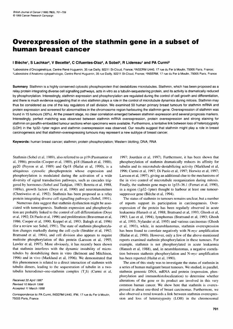
Figure 1 Total RNA extracted from breast specimen from womenundergoing cosmetic breast surgery (N), adjacent normal tissue from breastcancer patients (P) and breast tumour tissue (T) was electrophoresed onagarose gels. Northern blotting was performed with the 32P-labelledstathmin probe. Rehybridization of the blot with the 36B4 probedemonstrated that comparable amounts of RNA were loaded in each case.The normal breast tissues (N and P) show a low level of stathmin mRNA(scored B). Amounts of stathmin mRNA in breast tumours varied from basal(scored B: T2, T3, T4 and T13) to moderate (scored M: T5 and T8) andhigh (scored H: T7)
British Journal of Cancer (1998) 78(6), 701-7090 Cancer Research Campaign 1998
..
704 I Bieche et al
Table 1 Comparison between LOH on chromosome 1 p32-i pter andoverexpression of stathmin mRNA
1 p32-1 pter LOHStathminexpression Yes No p a
Basal 13 17 NSOverexpressing 8 2 P= 0.082
aX2 test; NS, not significant.
A
B M H
TI T2 T3 T5 T6 17
Actl
Stathmin
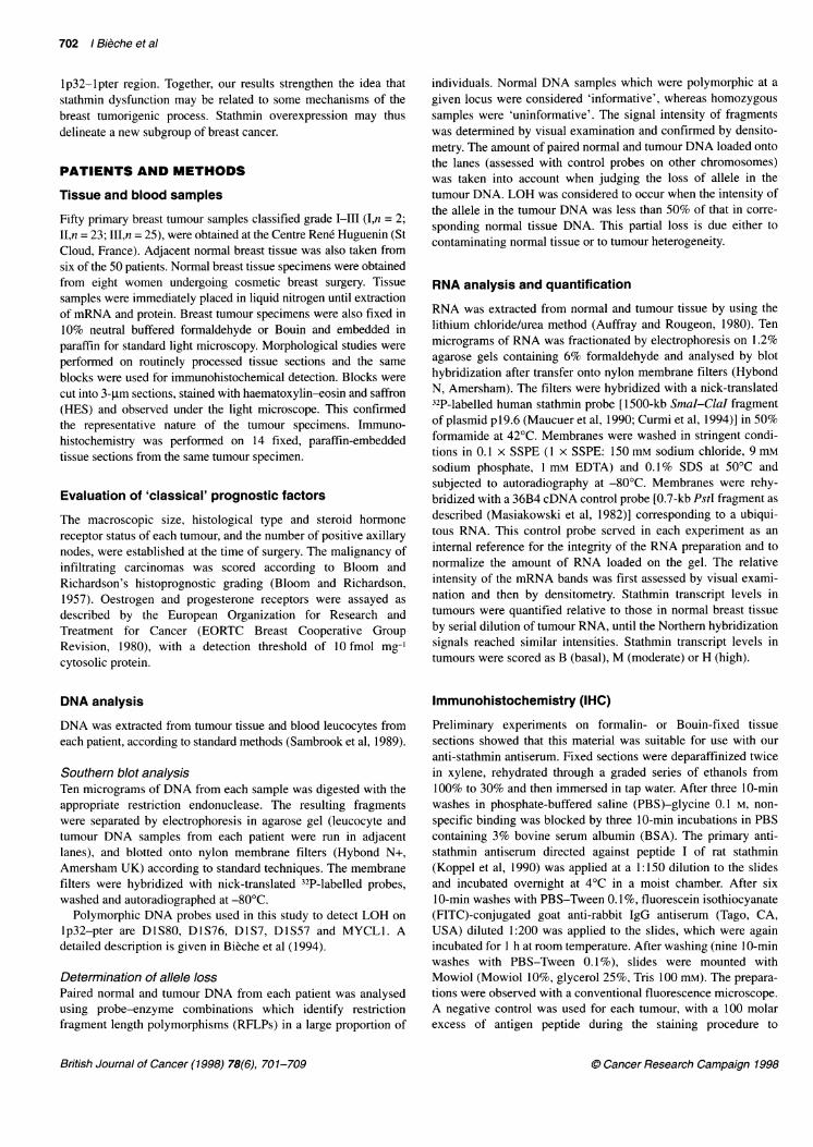
B
Figure 2 (A) Western blot analysis of stathmin in human breast tumours.Whole cellular proteins were prepared from frozen biopsies, separated onSDS-PAGE, electroblotted on nitrocellulose membrane and reacted with anti-stathmin antiserum and anti-actin monoclonal antibodies as described under'Patients and methods'. Tumours Ti, T2 and T3 (scored B by Northernblotting) expressed basal levels of stathmin. Tumours T4 and T5 (scored Mby Northern blotting) expressed moderate levels, whereas tumour T7 (scoredH by Northern blotting) expressed high levels of stathmin. (B) Normalizedexpression of stathmin. Normalized expression represents the ratio ofstathmin to actin in each tumour to the mean ratio of stathmin to actin innormal breast tissue. Three groups of tumours were identified matching thegroups based on stathmin mRNA scoring: (i) tumours Ti, T2 and T3expressed basal levels of stathmin (basal ratio), (ii) tumours T5 and T6showed moderate overexpression (ratio of stathmin to actin around 10 timesthe basal ratio) and (iii) tumour T7 expressed a high level of stathmin(stathmin/actin ratio about 75 times above the basal ratio)
RESULTS
Stathmin mRNA expression in normal breast tissue
RNA was extracted from normal breast specimens obtained fromeight women undergoing cosmetic breast surgery and six breast
cancer patients. Northern blot hybridization revealed a clearstathmin signal with a normal size of 1 kb, which was normalizedto the 36B4 control signal. Stathmin mRNA expression was weakin all the control specimens and was scored as basal (B) (Figure 1).
Stathmin mRNA overexpression in one-third of breasttumours
Northern blotting of the 50 human breast carcinomas showed thatall expressed a stathmin mRNA of the normal size. However,major differences in the amount of stathmin messenger were
observed: 35 tumours scored B (basal), ten gave a signal 3-5 timesthat of normal breast tissue (scored M) and five gave a greater thansixfold stronger signal (scored H) (Figure 1). The strongest expres-sion was 32-fold the basal level in tumour no. T7.
Correlation between stathmin mRNA overexpressionand clinical and pathological parameters
Overexpression of stathmin mRNA was not significantly associ-ated (X2 analysis) with standard prognostic feature includingmacroscopic tumour size, histopathological grade, lymph node or
steroid receptor status (data not shown) and with the Ki-67labelling index. The last feature showed great variations from4.7% to 21.2% in tumours scored B, and from 1.9% to 18.5% instathmin-overexpressing tumours.
Tentative link between stathmin mRNA levels and1 p32-1 pter LOH
Southern blots of restricted genomic DNA with the stathmincDNA probe (which is not polymorphic) showed no rearrange-
ment or amplification of the stathmin gene in the overexpressingtumours compared with the controls in any of the 50 human breasttumours tested (data not shown).
Forty of the 50 tumours have also been tested for LOH with fivepolymorphic DNA markers (DlS80, D1S76, D1S7, DlS57 andMYCLI) located in the lp32-pter region: 21 (52.5%) showedLOH on the telomeric region, while the remainder had a normalDNA profile. As shown in Table 1, we found a trend towards a linkbetween lp32-pter LOH and stathmin mRNA overexpression(P = 0.082). Indeed, 80% (8/10) of the tumours overexpressingthe stathmin gene showed deletions in the lp32-pter region,compared with 43% (13/30) of the tumours expressing basal levelsof stathmin mRNA.
High stathmin protein expression in tumours with highstathmin mRNA levels
Stathmin protein expression was investigated in seven tumours forwhich frozen samples were available. Four of these tumoursscored B for stathmin mRNA, two scored M and one scored H. Wefirst evaluated the stathmin content in each of these tumours byWestern blotting of 5 tg of high-speed centrifugation supernatantprotein. A parallel silver-stained gel, run with the same amount ofsample, revealed various degrees of contamination by serum
protein. Blots were thus probed with an anti-actin antibody tonormalize the amount of loaded tissue protein to this cellularprotein marker. As shown in Figure 2A, tumours scoring M and Hby Northern blotting contained far larger amounts of stathmin thantumours scoring B; tumours scoring M expressed about ten times
British Journal of Cancer (1998) 78(6), 701-709
Sampie Stathmin mRNA(nornmalizod unis) score
Normal breast 1.0tissue
Tumour Ti 0.7 ] B
T2 0.9 j
T3 1.3
T5 9.4
T6 13.4
T7 76.6 -H
0 Cancer Research Campaign 1998
Stathmin in breast cancer 705
Ti
T5
T2
T6
T3 T4
17 I IN P1 P2
Control
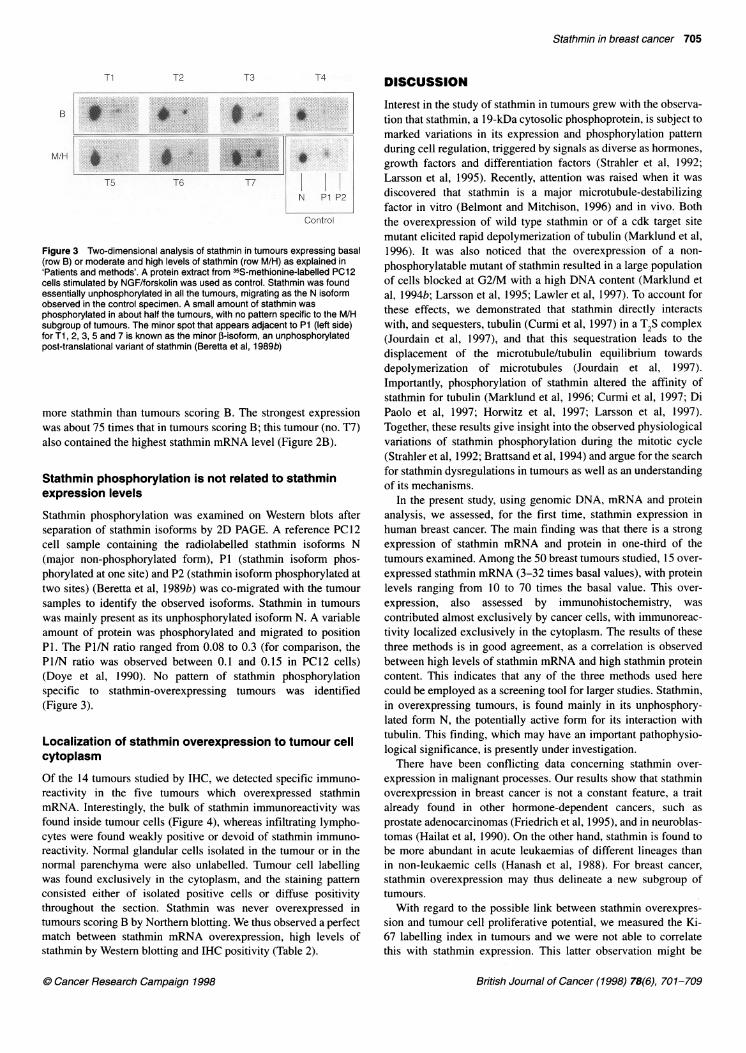
Figure 3 Two-dimensional analysis of stathmin in tumours expressing basal(row B) or moderate and high levels of stathmin (row M/H) as explained in'Patients and methods'. A protein extract from 35S-methionine-labelled PC12cells stimulated by NGF/forskolin was used as control. Stathmin was foundessentially unphosphorylated in all the tumours, migrating as the N isoformobserved in the control specimen. A small amount of stathmin was
phosphorylated in about half the tumours, with no pattern specific to the M/Hsubgroup of tumours. The minor spot that appears adjacent to P1 (left side)for Ti, 2, 3, 5 and 7 is known as the minor ,-isoform, an unphosphorylatedpost-translational variant of stathmin (Beretta et al, 1 989b)
more stathmin than tumours scoring B. The strongest expressionwas about 75 times that in tumours scoring B; this tumour (no. T7)also contained the highest stathmin mRNA level (Figure 2B).
Stathmin phosphorylation is not related to stathminexpression levels
Stathmin phosphorylation was examined on Western blots afterseparation of stathmin isoforms by 2D PAGE. A reference PC12cell sample containing the radiolabelled stathmin isoforms N(major non-phosphorylated form), P1 (stathmin isoform phos-phorylated at one site) and P2 (stathmin isoform phosphorylated at
two sites) (Beretta et al, 1989b) was co-migrated with the tumour
samples to identify the observed isoforms. Stathmin in tumourswas mainly present as its unphosphorylated isoform N. A variableamount of protein was phosphorylated and migrated to positionP1. The P1/N ratio ranged from 0.08 to 0.3 (for comparison, theP1/N ratio was observed between 0.1 and 0.15 in PC12 cells)(Doye et al, 1990). No pattern of stathmin phosphorylationspecific to stathmin-overexpressing tumours was identified(Figure 3).
Localization of stathmin overexpression to tumour cellcytoplasm
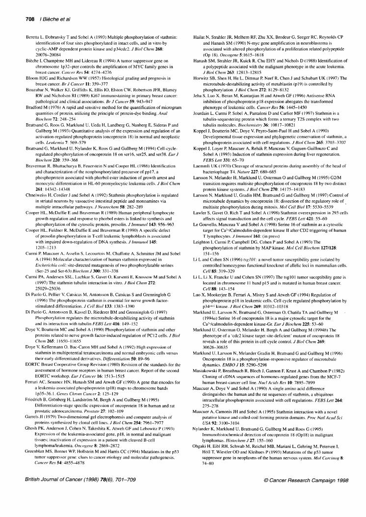
Of the 14 tumours studied by IHC, we detected specific immuno-reactivity in the five tumours which overexpressed stathminmRNA. Interestingly, the bulk of stathmin immunoreactivity was
found inside tumour cells (Figure 4), whereas infiltrating lympho-cytes were found weakly positive or devoid of stathmin immuno-reactivity. Normal glandular cells isolated in the tumour or in thenormal parenchyma were also unlabelled. Tumour cell labellingwas found exclusively in the cytoplasm, and the staining pattern
consisted either of isolated positive cells or diffuse positivitythroughout the section. Stathmin was never overexpressed intumours scoring B by Northern blotting. We thus observed a perfectmatch between stathmin mRNA overexpression, high levels ofstathmin by Western blotting and IHC positivity (Table 2).
DISCUSSION
Interest in the study of stathmin in tumours grew with the observa-tion that stathmin, a 19-kDa cytosolic phosphoprotein, is subject to
marked variations in its expression and phosphorylation patternduring cell regulation, triggered by signals as diverse as hormones,growth factors and differentiation factors (Strahler et al, 1992;Larsson et al, 1995). Recently, attention was raised when it wasdiscovered that stathmin is a major microtubule-destabilizingfactor in vitro (Belmont and Mitchison, 1996) and in vivo. Boththe overexpression of wild type stathmin or of a cdk target sitemutant elicited rapid depolymerization of tubulin (Marklund et al,1996). It was also noticed that the overexpression of a non-
phosphorylatable mutant of stathmin resulted in a large populationof cells blocked at G2/M with a high DNA content (Marklund etal, 1994b; Larsson et al, 1995; Lawler et al, 1997). To account forthese effects, we demonstrated that stathmin directly interactswith, and sequesters, tubulin (Curmi et al, 1997) in a T2S complex(Jourdain et al, 1997), and that this sequestration leads to thedisplacement of the microtubule/tubulin equilibrium towardsdepolymerization of microtubules (Jourdain et al, 1997).Importantly, phosphorylation of stathmin altered the affinity ofstathmin for tubulin (Marklund et al, 1996; Curmi et al, 1997; DiPaolo et al, 1997; Horwitz et al, 1997; Larsson et al, 1997).Together, these results give insight into the observed physiologicalvariations of stathmin phosphorylation during the mitotic cycle(Strahler et al, 1992; Brattsand et al, 1994) and argue for the searchfor stathmin dysregulations in tumours as well as an understandingof its mechanisms.
In the present study, using genomic DNA, mRNA and proteinanalysis, we assessed, for the first time, stathmin expression inhuman breast cancer. The main finding was that there is a strong
expression of stathmin mRNA and protein in one-third of thetumours examined. Among the 50 breast tumours studied, 15 over-
expressed stathmin mRNA (3-32 times basal values), with proteinlevels ranging from 10 to 70 times the basal value. This over-
expression, also assessed by immunohistochemistry, was
contributed almost exclusively by cancer cells, with immunoreac-tivity localized exclusively in the cytoplasm. The results of thesethree methods is in good agreement, as a correlation is observedbetween high levels of stathmin mRNA and high stathmin proteincontent. This indicates that any of the three methods used herecould be employed as a screening tool for larger studies. Stathmin,in overexpressing tumours, is found mainly in its unphosphory-lated form N, the potentially active form for its interaction withtubulin. This finding, which may have an important pathophysio-logical significance, is presently under investigation.
There have been conflicting data concerning stathmin over-
expression in malignant processes. Our results show that stathminoverexpression in breast cancer is not a constant feature, a traitalready found in other hormone-dependent cancers, such as
prostate adenocarcinomas (Friedrich et al, 1995), and in neuroblas-tomas (Hailat et al, 1990). On the other hand, stathmin is found to
be more abundant in acute leukaemias of different lineages thanin non-leukaemic cells (Hanash et al, 1988). For breast cancer,
stathmin overexpression may thus delineate a new subgroup oftumours.
With regard to the possible link between stathmin overexpres-
sion and tumour cell proliferative potential, we measured the Ki-67 labelling index in tumours and we were not able to correlatethis with stathmin expression. This latter observation might be
British Journal of Cancer (1998) 78(6), 701-709
B
M/H
. Cancer Research Campaign 1998
706 I Bi6che et al
D
F
Figure 4 Two breast invasive carcinomas scored H and considered as diffusely positive (A, C, E and B, D, F respectively) are presented afterhaematoxylin-eosin and saffron staining (A,B), and immunohistochemical detection of stathmin (C,D). The peptide absorbed anti-stathmin antiserum was usedas a control (E,F) (bar = 40 pm). The tumour on the left displays a massive and trabecular paftern of tumour proliferation (A). Immunohistochemical detection ofstathmin (C) shows positive cytoplasmic staining in the breast carcinoma cells, the tumour cell nuclei remaining unstained (arrow). The negative control (E)shows no reactivity in the previously immunostained tumour cells. The tumour on the right is from a carcinoma with isolated tumour cells or nests of tumour cellsin a stroma rich in lymphocytes (B). Immunohistochemical detection of stathmin (D) is found in the cytoplasm of tumour cells (arrow). The negative control (F)shows no reactivity in the previously immunostained tumour cells
because of the usually large variations found with this marker(Bouzubar et al, 1989). Studies on larger series will answer thisquestion. Data relating stathmin expression and the proliferationpotential of tumours reported in the literature are, apparently,somewhat confusing. In natural conditions, up-regulation ofstathmin has been found to be neither uncoupled from cell prolif-eration nor restricted to cell types with proliferative potential(Brattsand et al, 1993). In non-Hodgkin's lymphoma and inHodgkin's disease, Hodgkin and Reed-Sternberg cells frequently
express stathmin with strong staining intensity, but stathmin over-expression is only partly related to cell proliferation (Nylander etal, 1995). In contrast, stathmin transfection into lymphoblastoidcells results in a partial inhibition of cell proliferation (Brattsand etal, 1993), and antisense transfection into leukaemic cells reversesthe malignant phenotype (Jeha et al, 1996).To interpret these observations, one must consider that stathmin
is at the heart of a complex signalling network, being a directsubstrate for different kinases: the MAP kinase family (Leighton
British Journal of Cancer (1998) 78(6), 701-709
c_
0 Cancer Research Campaign 1998
Stathmin in breast cancer 707
Table 2 Summary of the results for stathmin expression andphosphorylation
Tumour no. Northern WB IHC 2DWB
Tl B 1 ud NT2 B 1 ud NPT3 B 1 ud NPT4 B 1 ud NT5 M 9 + NT6 M 13 + NT7 H 77 + NPT8 M ND + NDT9 M ND + NDT10 B ND UD NDT1l B ND UD NDT12 B ND UD NDT13 B ND UD NDT14 B ND UD ND
By Northern blotting, tumours were scored basal (B), moderate (M) or high(H). Western blotting (WB) showed basal levels of stathmin (1) or increasedlevels (ranging from 10 to 77 times the basal value; values are expressed asdefined in Figure 2). Immunohistochemical studies (IHC) showed tumourswith no stathmin reactivity (ud) or specific stathmin labelling in tumour cells(+). Note that a perfect match was found for stathmin expression at themRNA and protein levels (Northern, WB and IHC). Stathmin phosphorylationwas examined after separation of stathmin isoforms by 2D-PAGE asdescribed in Figure 3. No specific pattern of stathmin phosphorylation wasobserved for basal or overexpressing tumours. N, stathmin essentially in itsunphosphorylated state; NP, stathmin displaying a significant proportion of itsP1 phosphorylated form in addition to its unphosphorylated state. B, basal;UD, undetectable; N, 0.08 < P/N <0.15; M, moderate; ND, not determined;NP, 0.15 < P/N< 0.30. H, high
et al, 1993), cAMP-dependent protein kinase (Beretta et al, 1993),p34cdc2 kinase (Beretta et al, 1993; Brattsand et al, 1994; Larssonet al, 1995) and the Ca2+-calmodulin-dependent kinases II and IV(Marklund et al, 1994a; Le Gouvello et al, 1998). Furthermore,stathmin interacts with various protein partners, for which we haveidentified several candidates (Maucuer et al, 1995). One of these,CC2/tsg 1O1, interestingly being the product of a tumour sucepti-bility gene (Li and Cohen, 1996), was suggested to be implicatedin breast cancer (Li et al, 1997). The intricate regulation ofstathmin and of its partners being highly probable, we speculatethat stathmin overexpression might contribute to tumorigenesis indifferent ways.
1. it could represent a normal reaction to cell proliferation itself.In fact, a recent study in our laboratory showed that a high celldensity in culture induces stathmin expression, most likelytriggered by cell-cell contacts. Stathmin expression, in thatcase, is likely being up-regulated, in relation to the limitationof cell overgrowth at the stage preceding cell differentiation(Balogh et al, 1996). This cell culture result is in good agree-ment with the induction of stathmin expression during liverregeneration, stathmin displaying a delayed expression peakfollowing the mitotic peak and correlating with the slowdownin cell proliferation (Koppel et al, 1993). Stimulated expres-sion of stathmin may thus be part of a regulatory programmeaimed at limiting cell overproliferation, and also activated,although inefficiently, in transformed tumoral cells.
2. Alternatively, overexpression of stathmin might reflect analteration of stathmin itself, leading to the malignant pheno-type; mutations in the structural gene that are undetectable byblotting techniques would then remain to be identified.
C) Cancer Research Campaign 1998
3. Finally, cells might react to changes in stathmin proteinpartners (Maucuer et al, 1995) in a feedback pathway.
The other interesting finding in this study is the tentative linkbetween loss of heterozygosity in the lp32-lpter region andstathmin overexpression. Deletions of the short arm of chromo-some 1, especially the telomeric lp32-pter region, have beendetected by both molecular and cytogenetic approaches in breasttumours, suggesting that this region contains a breast tumour-suppressor gene (Bieche et al, 1994). Interestingly, this regionalso houses the stathmin gene (mapping to lp35-36.1). Stathminappears thus to be a good candidate for being one of the tumoursuppressor genes located in this chromosome region. The findingof a tentative link between LOH in the 1p32-lpter region andstathmin overexpression may appear surprising, but it is reminis-cent of the coexistence of p53 gene LOH and overexpression ofthe corresponding protein. In this case, mutations were foundeither in the regulatory or in the coding region of the p53 gene(Aka et al, 1993; Ohgaki et al, 1993; Greenblatt et al, 1994).Similar mutations may have occurred in the vicinity of or withinthe stathmin gene. Alternatively, DNA removal may have broughta powerful enhancer close to the stathmin gene to account for theincrease in mRNA levels.
In conclusion, our study has clearly established that a significantproportion of breast cancers overexpress stathmin and may definea new breast cancer subtype. Further studies with a larger popula-tion and longer follow-up will allow the evaluation of theprognostic significance of stathmin overexpression, as well as anexploration of the status of the stathmin protein partners in theoverexpressing tumours.
ACKNOWLEDGEMENTS
We are indebted to Dr A Maucuer for the human stathmin probeand to Dr P Chambon for probe 36B4. We also thank Dr DRickman for critical reading of this manuscript. This work wassupported by the Ligue Nationale Contre le Cancer (LNCC), theAssociation pour la Recherche contre le Cancer (ARC) and theInstitut National de la Sante et de la Recherche Medicale(INSERM).
REFERENCES
Aka K, Brunner JM, Bondy ML, Ligon K, Nishi T, del Giglio A, Moser RP, LevinVA and Saya H (1993) Detection of p53 alterations in human astrocytomasusing frozen tissue sections for the polymerase chain reaction. J Neurooncol16: 125-133
Auffray C and Rougeon F (1980) Purification of mouse immunoglobulin heavy-chain messenger RNAs from total myeloma tumor RNA. Eiur J Biochetn 107:303-314
Balogh A, Mege RM and Sobel A (1996) Cell density dependent expression ofstathmin in C2 myoblasts in culture. Exp Cell Res 224: 8-15
Belmont LD and Mitchison TJ ( 1996) Identification of a protein that interacts withtubulin dimers and increases the catastrophe rate of microtubules. Cell 84:623-631
Beretta L, Boutterin MC and Sobel A (1988) Phosphorylation of intracellularproteins related to the multihormonal regulation of prolactin: comparison ofnormal anterior pituitary cells in culture with the tumor-derived GH cell lines.Endocrinology 122: 40-51
Beretta L, Boutterin MC, Drouva S and Sobel A (1989a) Phosphorylation of a groupof proteins related to the physiological, multihormonal regulations of the variouscell types in the anterior pituitary gland. Endocrinology 125: 1358-1364
Beretta L, Houdouin F and Sobel A (I 989b) Identification of two distinct isoformsof stathmin and characterization of their respective phosphorylated forms.J Biol Chem 264: 9932-9938
British Journal of Cancer (1998) 78(6), 701-709
708 I Bieche et al
Beretta L, Dobransky T and Sobel A (1993) Multiple phosphorylation of stathmin:identification of four sites phosphorylated in intact cells, and in vitro bycyclic-AMP dependent protein kinase and p34cdc2. J Biol Cheimi 268:20076-20084
Bieche 1, Champeme MH and Lidereau R ( 1994) A tumor suppressor gene onchromosome I p32-pter controls the amplification of MYC family genes inbreast cancer. Cancer Res 54: 4274-4276
Bloom HJG and Richardson WW (1957) Histological grading and prognosis inbreast cancer. Br J Coniicer 11: 359-377
Bouzubar N, Walker KJ, Griffiths K, Ellis 10, Elston CW, Robertson JFR, BlameyRW and Nicholson RI (1989) Ki67 immunostaining in primary breast cancer:pathological and clinical associations. Br J Coincer 59: 943-947
Bradford M ( 1976) A rapid and sensitive method for the quantification of microgramquantities of protein, utilizing the principle of protein-dye binding. AnalBioclheni 72: 248-254
Brattsand G, Roos G, Marklund U, Ueda H, Landberg G, Nanberg E, Sideras P andGullberg M (1993) Quantitative analysis of the expression and regulation of anactivation-regulated phosphoprotein (oncoprotein 18) in normal and neoplasticcells. Leutke,niito 7: 569-579
Brattsand G, Marklund U. Nylander K, Roos G and Gullberg M (1994) Cell-cycle-regulated phosphorylation of oncoprotein 18 on serl6, ser25, and ser38. Eur JBioclhem 220: 359-368
Braverman R, Bhattacharya B, Feuerstein N and Cooper HL (1986) Identificationand characterization of the nonphosphorylated precursor of pp 1 7, aphosphoprotein associated with phorbol ester induction of growth arrest andmonocytic differentiation in HL-60 promyelocytic leukemia cells. J Biol Chein261: 14342-14348
Chneiweiss H. Cordier J and Sobel A (1992) Stathmin phosphorylation is regulatedin striatal neurons by vasoactive intestinal peptide and monoamines viamultiple intracellular pathways. J Neurochemn 58: 282-289
Cooper HL, McDuffie E and Braverman R (1989) Human peripheral lymphocytegrowth regulation and response to phorbol esters is linked to synthesis andphosphorylation of the cytosolic protein, prosolin. J Im)1otinol 143: 956-963
Cooper HL, Fuldner R, McDuffie E and Braverman R (1990) A specific defectof prosolin phosphorylation in T-cell leukemic lymphoblasts is associatedwith impaired down-regulation of DNA synthesis. J hniniunzol 145:1205-1213
Curmi P, Maucuer A, Asselin S, Lecourtois M, Chaffotte A, Schmitter JM and SobelA (1994) Molecular characterization of human stathmin expressed inEscherichio coli: site-directed mutagenesis of two phosphorylatable serines(Ser-25 and Ser-63) Biocheinz J300: 331-338
Curmi PA, Andersen SSL, Lachkar S, Gavet 0, Karsenti E, Knossow M and Sobel A( 1997) The stathmin tubulin interaction in vitro. J Biol Chein 272:25029-25036
Di Paolo G, Pellier V, Catsicas M, Antonsson B, Catsicas S and Grenningloh G(1996) The phosphoprotein stathmin is essential for nerve growth factor-stimulated differentiation. J Cell Biol 133: 1383-1390
Di Paolo G, Antonsson B. Kassel D, Riederer BM and Grenningloh G (1997)Phosphorylation regulates the microtubule-destabilizing activity of stathminand its interaction with tubulin FEBS Lett 416: 149-152
Doye V, Boutterin MC and Sobel A (1990) Phosphorylation of stathmin and otherproteins related to nerve growth factor-induced regulation of PC 12 cells. J BiolClhein 265: 1 1650-1 1655
Doye V, Kellermann 0, Buc-Caron MH and Sobel A (1992) High expression ofstathmin in multipotential teratocarcinoma and normal embryonic cells versustheir early differentiated derivatives. Diafjrentiation 50: 89-96
EORTC Breast Cooperative Group Revision (1980) Revision of the standards for theassessment of hormone receptors in human breast cancer. Report of the secondEORTC workshop. Ei]J Ctiancer 16: 1513-1515
Ferrari AC, Seuanez HN, Hanash SM and Atweh GF (1990) A gene that encodes fora leukemia-associated phosphoprotein (p1 8) maps to chromosome bandsI p35-36. 1. Genzes Chrom Cancer 2: 125-129
Friedrich B, Gronberg H, Landstrom M, Bergh A and Gullberg M (1995)Differentiation-stage specific expression of oncoprotein 18 in human and ratprostatic adenocarcinoma. Pi-ostate 27: 102-109
Garrels J1 (1979) Two-dimensional gel electrophoresis and computer analysis ofproteins synthesized by clonal cell lines. J Biol Chem 254: 7961-7977
Ghosh PK, Anderson J, Cohen N, Takeshita K, Atweh GF and Lebowitz P ( 1993)Expression of the leukemia-associated gene, p1 8, in normal and malignanttissues; inactivation of expression in a patient with cleaved B-cellIymphoma/leukemia. OnlcogenQe8: 2869-2872
Greenblatt MS, Bennet WP. Hollstein M and Harris CC (1994) Mutations in the p53tumor suppressor gene: clues to cancer etiology and molecular pathogenesis.Can1cer Res 54: 48:55-4878
British Journal of Cancer (1998) 78(6), 701-709
Hailat N, Strahler JR, Melhem RF, Zhu XX, Brodeur G, Seeger RC, Reynolds CPand Hanash SM (1990) N-myc gene amplification in neuroblastoma isassociated with altered phosphorylation of a proliferation related polypeptide(Op 18). O11cogente 5:1615-1618
Hanash SM, Strahler JR, Kuick R, Chu EHY and Nichols D (I1988) Identification ofa polypeptide associated with the malignant phenotype in the acute leukemia.JIBiol Chem 263: 12813-12815
Horwitz SB, Shen H, He L, Dittmar P, Neef R, Chen J and Schubart UK (1997) Themicrotubule-destabilizing activity of metablastin (p1 9) is controlled byphosphorylation. J Biol Chenm 272: 8129-8132
Jeha S, Luo X, Beran M, Kantarjian H and Atweh GF (1996) Antisense RNAinhibition of phosphoprotein p 18 expression abrogates the transformedphenotype of leukemic cells. Con1cer Res 56: 1445-1450
Jourdain L, Curmi P. Sobel A, Pantaloni D and Carlier MF (I1997) Stathmin is atubulin-sequestering protein which forms a ternary T2S complex with twotubulin molecules. Biochenmistnv 36: 10817-10821
Koppel J, Boutterin MC, Doye V, Peyro-Saint-Paul H and Sobel A (1990)Developmental tissue expression and phylogenetic conservation of stathmin, aphosphoprotein associated with cell regulations. J Biol Clietl 265: 3703-3707
Koppel J, Loyer P, Maucuer A, Rehak P, Manceau V, Guguen-Guillouzo C andSobel A ( 1993) Induction of stathmin expression during liver regeneration.FEBS Lett 331: 65-70
Laemmli UK (1970) Cleavage of structural proteins during assembly of the head ofbacteriophage T4. Nature 227: 680-685
Larsson N, Melander H, Marklund U, Osterman 0 and Gullberg M (1995) G2/Mtransition requires multisite phosphorylation of oncoprotein 18 by two distinctprotein kinase systems. J Biol Chem 270: 14175-14183
Larsson N, Marklund U, Gradin HM, Brattsand G and Gullberg M (1997) Control ofmicrotubule dynamics by oncoprotein 18: dissection of the regulatory role ofmultisite phosphorylation during mitosis. Mol Cell Biol 17: 553(-5539
Lawler S, Gavet 0, Rich T and Sobel A (1998) Stathmin overexpression in 293 cellsaffects signal transduction and the cell cycle. FEBS Lett 421: 55-60
Le Gouvello, Manceau V and Sobel A (1998) Serine 16 of stathmin as a cytosolictarget for Ca2+/Calmodulin-dependent kinase II after CD2 triggering of humanT lymphocytes. J lnrtnuol 161: (in press)
Leighton I, Curmi P, Campbell DG, Cohen P and Sobel A (1993) Thephosphorylation of stathmin by MAP kinase. Mol Cell Biochem 127/128:151-156
Li L and Cohen SN (1996) tsgIOl: a novel tumor susceptibility gene isolated bycontrolled homozygous functional knockout of allelic loci in mammalian cells.Cell 85: 319-329
Li L, Li X, Francke U and Cohen SN (1997) The tsgl01 tumor suceptibility gene islocated in chromosome 11 band p5 and is mutated in human breast cancer.Cell 88: 143-154
Luo X, Mookerjee B, Ferrari A, Mistry S and Atweh GF (1994) Regulation ofphosphoprotein p18 in leukemic cells. Cell cycle regulated phosphorylation byp34Xcl2 kinase. J Biol Chem 269: 10312-10318
Marklund U, Larsson N, Brattsand G, Osterman 0, Chatila TA and Gullberg M(I 994a) Serine 16 of oncoprotein 18 is a major cytosolic target for theCa2'/calmodulin-dependent kinase-Gr. Eur J Biochem 225: 53-60
Marklund U, Osterman 0, Melander H, Bergh A and Gullberg M (1994b) Thephenotype of a 'cdc2 kinase target site-deficient' mutant of oncoprotein 18reveals a role of this protein in cell cycle control. J Biol Chem 269:30626-30635
Marklund U, Larsson N, Melander Gradin H, Brattsand G and Gullberg M (1996)Oncoprotein 18 is a phosphorylation-responsive regulator of microtubuledynamics. EMBO J 15: 5290-5298
Masiakowski P, Breathnach R, Bloch J, Gannon F, Krust A and Chambon P (1982)Cloning of cDNA sequences of hormones-regulated genes from the MCF-7human breast-cancer cell line. Nucl Acids Res 10: 7895-7899
Maucuer A, Doye V and Sobel A (1990) A single amino acid differencedistinguishes the human and the rat sequences of stathmin, a ubiquitousintracellular phosphoprotein associated with cell regulations. FEBS Lett 264:275-278
Maucuer A, Camonis JH and Sobel A (1995) Stathmin interaction with a novelputative kinase and coiled-coil forming protein domains. Proc Notl Acad SciUSA 92: 3100-3104
Nylander K, Marklund U, Brattsand G, Gullberg M and Roos G (1995)Immunohistochemical detection of oncoprotein 18 (Opt8) in malignantlymphomas. Histochem J 27: 155-160
Ohgaki H, Eibl RH, Schwab M, Reichel MB, Mariani L, Gehring M, Petersen 1,Holl T, Wiestler OD and Kleihues P (1993) Mutations of the p53 tumorsuppressor gene in neoplasms of the human nervous system. Mol Carcinog 8:74-80
C) Cancer Research Campaign 1998
Stathmin in breast cancer 709
Pasmantier R, Danoff A, Fleischer N and Schubart UK (1986) P19, a hormonallyregulated phosphoprotein of peptide-hormone producing cells: secretagogue-induced phosphorylation in AtT-20 mouse pituitary tumor cells and in rat andhamster insulinoma cells. Endocrinology 19: 1229-1238
Peyron J, Aussel C, Ferrua B, Haring H and Fehlmann M (1989) Phosphorylation oftwo cytosolic proteins. An early event of T-cell activation. Biochem J 258:505-510
Sambrook J, Fritsch EF and Maniatis T (1989) Molecular Cloning: A LaboratoryManual, 2nd edn. Cold Spring Harbor Laboratory: Cold Spring Harbor, NY
Sobel A (1991) Stathmin: a relay phosphoprotein for multiple signal transduction?Trends Biochem Sci 16: 301-305
@) Cancer Research Campaign 1998
Sobel A and Tashjian Jr AH (1983) Distinct patterns of cytoplasmic proteinphosphorylation related to regulation of synthesis and release of prolactin byGH cells. JBiol Chem 258: 10312-10324
Sobel A, Boutterin MC, Beretta L, Chneiweiss H, Doye V and Peyro-Saint-Paul H( 1989) Intracellular substrates for extracellular signaling: characterization of aubiquitous, neuron-enriched phosphoprotein (stathmin). J Biol Chem 264:3765-3772
Strahler JR, Lamb BJ, Ungar DR, Fox DA and Hanash SM (1992) Cell cycleprogression is associated with distinct patterns of phosphorylation of Op 18.Biochem Biophvs Res Commun 185: 197-203