Molecular and cellular pharmacology Oxidative stress induced by tert-butylhydroperoxide interferes with the placental transport of glucose: in vitro studies with BeWo cells João R. Araújo 1 , Ana C. Pereira 1 , Ana Correia-Branco, Elisa Keating, Fátima Martel n Department of Biochemistry (U38-FCT), Faculty of Medicine, University of Porto, 4200-319 Porto, Portugal article info Article history: Received 22 February 2013 Received in revised form 10 October 2013 Accepted 16 October 2013 Available online 24 October 2013 Keywords: Glucose Oxidative stress Placenta Transport abstract Increased oxidative stress is implicated in the onset and progression of prevalent pregnancy disorders (e.g. gestational diabetes and fetal growth restriction), and in programming the fetus to develop metabolic diseases later in life. Since the molecular mechanisms underlying these effects of oxidative stress are largely unexplored, we aimed to investigate if the placental transport of glucose – the main energetic substrate for the fetus and placenta – is altered by oxidative stress. In a human syncytio- trophoblast (STB) cell model, the BeWo cell line, oxidative stress was induced by treatment with 100 mM tert-butylhydroperoxide (tert-BOOH) for 24 h. Tert-BOOH decreased the steady-state intracellular accumulation (A max ) of [ 3 H]2-deoxyglucose ([ 3 H]DG) mediated by both facilitative (GLUT) and non- facilitative (non-GLUT) glucose transporters. These effects were not associated with a change in the mRNA expression level of GLUT1, the major placental glucose transporter. Also, they seemed to be independent from phosphoinositide 3-kinase and protein kinase C signaling pathways and were unchanged either by inhibitors of free radical-generating enzymes or by free radical scavengers. In contrast, the dietary polyphenols quercetin, epigallocatechin-3-gallate and resveratrol completely reversed the inhibitory effect of tert-BOOH upon [ 3 H]DG accumulation through a specific effect on GLUT-mediated transport. Finally, tert-BOOH induced an increase in the transepithelial permeability to [ 3 H]DG in the apical-to-basal direction, apparently related to an increase in its paracellular transport. In conclusion, tert-BOOH-induced oxidative stress reduces STB accumulation of glucose associated with an increase in its transepithelial permeability. This effect may contribute to the deleterious consequences of pregnancy disorders associated with oxidative stress. & 2013 Elsevier B.V. All rights reserved. 1. Introduction One of the major functions of the placenta is to mediate the transport of nutrients from the mother to the fetus over the course of gestation. This function depends on the activity of specific transporters present at the apical (maternal-facing) and basal (fetal-facing) membranes of the syncytiotrophoblast (STB) epithe- lium (Jansson et al., 2009). Glucose is the primary substrate for energy metabolism in the feto-placental unit, and together with amino acids, it constitutes the primary stimuli for fetal secretion of the growth-promoting hormone insulin (Jansson et al., 2009). Since fetal glucose produc- tion is minimal (Magnusson et al., 2004), placental transport constitutes the primary source of glucose to the fetus (Baumann et al., 2002; Carter, 2012). Accordingly, alterations in glucose transport and metabolism at the STB level are strongly associated with aberrant fetal growth (Baumann et al., 2002; Desoye et al., 2011; Illsley, 2000; Magnusson et al., 2004), which increases the risk of perinatal complications and predispose the newborn to develop cardiovascular and metabolic diseases later in life (Jansson et al., 2009; Vo and Hardy, 2012). Placental transport of glucose occurs mainly through facilitative glucose transporters (GLUT). At least five different GLUT isoforms are expressed in the human STB: GLUT1, 3, 4, 9 and 12. However, the primary isoform responsible for glucose transport across the apical and basal membranes of the STB in term pregnancy is GLUT1 (Baumann et al., 2002; Carter, 2012; Jansson et al., 2009). GLUT1 distribution in the STB is asymmetric, with a greater expression and activity in the apical membrane, which assures that glucose is transported down its concentration gradient from maternal to fetal circulation (Baumann et al., 2002; Carter, 2012). An increasing amount of evidence implicates oxidative stress in the pathophysiology of prevalent pregnancy complications such as miscarriage (Myatt and Cui, 2004), preeclampsia (Siddiqui et al., 2010), fetal growth restriction (Son et al., 2004; Takagi et al., 2004) and gestational diabetes (Coughlan et al., 2004; Lappas et al., 2011; Peuchant et al., 2004). Moreover, increased oxidative stress at the Contents lists available at ScienceDirect journal homepage: www.elsevier.com/locate/ejphar European Journal of Pharmacology 0014-2999/$ - see front matter & 2013 Elsevier B.V. All rights reserved. http://dx.doi.org/10.1016/j.ejphar.2013.10.023 n Corresponding author. Tel.: þ351 22 0426654; fax: þ351 22 5513624. E-mail address: [email protected] (F. Martel). 1 João R. Araújo and Ana C. Pereira equally contributed to the work. European Journal of Pharmacology 720 (2013) 218–226
Transcript
Molecular and cellular pharmacology
Oxidative stress induced by tert-butylhydroperoxide interferes withthe placental transport of glucose: in vitro studies with BeWo cells
João R. Araújo 1, Ana C. Pereira 1, Ana Correia-Branco, Elisa Keating, Fátima Martel n
Department of Biochemistry (U38-FCT), Faculty of Medicine, University of Porto, 4200-319 Porto, Portugal
a r t i c l e i n f o
Article history:Received 22 February 2013Received in revised form10 October 2013Accepted 16 October 2013Available online 24 October 2013
Keywords:GlucoseOxidative stressPlacentaTransport
a b s t r a c t
Increased oxidative stress is implicated in the onset and progression of prevalent pregnancy disorders(e.g. gestational diabetes and fetal growth restriction), and in programming the fetus to developmetabolic diseases later in life. Since the molecular mechanisms underlying these effects of oxidativestress are largely unexplored, we aimed to investigate if the placental transport of glucose – the mainenergetic substrate for the fetus and placenta – is altered by oxidative stress. In a human syncytio-trophoblast (STB) cell model, the BeWo cell line, oxidative stress was induced by treatment with 100 mMtert-butylhydroperoxide (tert-BOOH) for 24 h. Tert-BOOH decreased the steady-state intracellularaccumulation (Amax) of [3H]2-deoxyglucose ([3H]DG) mediated by both facilitative (GLUT) and non-facilitative (non-GLUT) glucose transporters. These effects were not associated with a change in themRNA expression level of GLUT1, the major placental glucose transporter. Also, they seemed to beindependent from phosphoinositide 3-kinase and protein kinase C signaling pathways and wereunchanged either by inhibitors of free radical-generating enzymes or by free radical scavengers. Incontrast, the dietary polyphenols quercetin, epigallocatechin-3-gallate and resveratrol completelyreversed the inhibitory effect of tert-BOOH upon [3H]DG accumulation through a specific effect onGLUT-mediated transport. Finally, tert-BOOH induced an increase in the transepithelial permeability to[3H]DG in the apical-to-basal direction, apparently related to an increase in its paracellular transport. Inconclusion, tert-BOOH-induced oxidative stress reduces STB accumulation of glucose associated with anincrease in its transepithelial permeability. This effect may contribute to the deleterious consequences ofpregnancy disorders associated with oxidative stress.
& 2013 Elsevier B.V. All rights reserved.
1. Introduction
One of the major functions of the placenta is to mediate thetransport of nutrients from the mother to the fetus over the courseof gestation. This function depends on the activity of specifictransporters present at the apical (maternal-facing) and basal(fetal-facing) membranes of the syncytiotrophoblast (STB) epithe-lium (Jansson et al., 2009).
Glucose is the primary substrate for energy metabolism in thefeto-placental unit, and together with amino acids, it constitutesthe primary stimuli for fetal secretion of the growth-promotinghormone insulin (Jansson et al., 2009). Since fetal glucose produc-tion is minimal (Magnusson et al., 2004), placental transportconstitutes the primary source of glucose to the fetus (Baumannet al., 2002; Carter, 2012). Accordingly, alterations in glucosetransport and metabolism at the STB level are strongly associated
with aberrant fetal growth (Baumann et al., 2002; Desoye et al.,2011; Illsley, 2000; Magnusson et al., 2004), which increases therisk of perinatal complications and predispose the newborn todevelop cardiovascular and metabolic diseases later in life (Janssonet al., 2009; Vo and Hardy, 2012).
Placental transport of glucose occurs mainly through facilitativeglucose transporters (GLUT). At least five different GLUT isoformsare expressed in the human STB: GLUT1, 3, 4, 9 and 12. However,the primary isoform responsible for glucose transport across theapical and basal membranes of the STB in term pregnancy isGLUT1 (Baumann et al., 2002; Carter, 2012; Jansson et al., 2009).GLUT1 distribution in the STB is asymmetric, with a greaterexpression and activity in the apical membrane, which assuresthat glucose is transported down its concentration gradient frommaternal to fetal circulation (Baumann et al., 2002; Carter, 2012).
An increasing amount of evidence implicates oxidative stress inthe pathophysiology of prevalent pregnancy complications such asmiscarriage (Myatt and Cui, 2004), preeclampsia (Siddiqui et al.,2010), fetal growth restriction (Son et al., 2004; Takagi et al., 2004)and gestational diabetes (Coughlan et al., 2004; Lappas et al., 2011;Peuchant et al., 2004). Moreover, increased oxidative stress at the
Contents lists available at ScienceDirect
journal homepage: www.elsevier.com/locate/ejphar
European Journal of Pharmacology
0014-2999/$ - see front matter & 2013 Elsevier B.V. All rights reserved.http://dx.doi.org/10.1016/j.ejphar.2013.10.023
n Corresponding author. Tel.: þ351 22 0426654; fax: þ351 22 5513624.E-mail address: [email protected] (F. Martel).1 João R. Araújo and Ana C. Pereira equally contributed to the work.
European Journal of Pharmacology 720 (2013) 218–226
intrauterine environment has been demonstrated to program thefetus to develop metabolic and cardiovascular complications laterin life (Dong et al., 2013; Giussani et al., 2012). Given that there aremajor gaps both in the understanding of the cellular and mole-cular mechanisms that underlie the pathological and program-ming effects of oxidative stress during pregnancy, and in the studyof the impact of oxidative stress upon placental and fetal nutrition,we aimed to investigate the effect of oxidative stress upon theplacental uptake of glucose, by using a cell model of human STB,the BeWo cell line. This cell line is a well-characterized and widelyused cell model to investigate placental glucose transport, sinceGLUT expression (Baumann et al., 2007; Shah et al., 1999) andactivity (Illsley, 2000; Vardhana and Illsley, 2002) (particularly ofGLUT1) in BeWo cells are similar to that of primary culturedtrophoblast cells (Baumann et al., 2007) and human term placen-tae (Baumann et al., 2002).
2. Materials and methods
2.1. Materials
2-[1,2-3H(N)]deoxy-D-glucose – specific activity 60 mCi/mmol, andD-[14C(U)]sorbitol – specific activity 300mCi/mmol (American Radi-olabeled Chemicals, St. Louis, MO, USA); albumin from bovine serum,chelerythrine chloride, collagen type I, cytochalasin B (from Diechsleradematioidea), decane, 5,5′-dithiobis(nitrobenzoic) acid, 2,4-dinitrophe-nylhydrazine, (-)-cis-3,3′,4′,5,5′,7-hexahydroxy-fla-vane-3-gallate ((–)epigallocatechin-3-gallate), fetal calf serum, glutathione reductasefrom baker's yeast (Saccharomyces cerevisiae), Ham's F12K medium(Kaighn's modification), 2-(4-morpholinyl)-8-phenyl-1(4H)-benzo-pyran-4-one (LY-294002) hydrochloride, N-acetyl-L-cysteine, β-nicotinamide adenine dinucleotide 2′-phosphate reduced tetrasodiumsalt hydrate, β-nicotinamide adenine dinucleotide reduced disodiumsalt hydrate, 3,3′,4′,5,6-pentahydroxy-flavone (quercetin) dihydrate,β-(4-hydroxyphenyl)-2,4,6-trihydroxypropiophenone, 2′,4′,6′-trihy-droxy-3-(4-hydroxyphenyl)propiophenone, 3-(4-hydroxyphenyl)-1-(2,4,6-trihydroxyphenyl)-1-propanone (phloretin), 3,4′,5-trihydroxy-trans-stilbene (resveratrol), sodium pyruvate, phenol red sodium salt,sulforhodamine B, tert-butyl hydroperoxide (tert-BOOH), 2-thiobar-bituric acid, trichloroacetic acid sodium salt, and 2-vinylpyridine(Sigma, St. Louis, MO, USA); and dimethylsulfoxide, D(þ)-glucoseand Triton X-100 (Merck, Darmstadt, Germany).
Compounds to be tested were dissolved in decane, water, ordimethylsulfoxide. The final concentration of these solvents in thebuffer and culture medium was 1% (v/v).
2.2. BeWo cell culture
The BeWo cell line was obtained from the Deutsche Sammlungvon Mikroorganismen und Zellkulturen (DMSZ GmbH, ACC-458).Cells were used between passage numbers 16 and 49 and culturedas previously described (Araújo et al., 2008). For cell viability andcytotoxicity assays and for [3H]2-deoxyglucose ([3H]DG) apicaluptake studies, cells were seeded on collagen-coated 24-wellplastic cell culture clusters (2 cm2; ∅16 mm; TPP, Trasadingen,Switzerland), and for measurement of glutathione levels, lipidperoxidation products and protein carbonyl groups, cells wereseeded on collagen-coated 12-well plastic cells culture clusters(4 cm2; ∅21 mm; TPP). Experiments were performed 6–7 daysafter the initial seeding (90–100% confluence). For transepithelialtransport experiments, BeWo cells were seeded on collagen-coated Transwells inserts (0.4 μm pore size, 12 mm diameter;Corning Costar, NY, USA) and experiments were performed 9–10days after the initial seeding (100% confluence).
For cellular viability and cytotoxicity assays and quantificationof oxidative stress biomarkers, confluent BeWo cells were exposedfor 24 h to 1, 3, 10, 30, 100, 300 or 1000 μM of the oxidizing andfree radical generating agent tert-BOOH (Tormos et al., 2004), or itssolvent (decane), in fetal calf serum-free culture media.
2.3. Cellular viability and cytotoxicity assays
Cellular viability and cytotoxicity were quantified by measuringthe activity of extracellular lactate dehydrogenase (LDH) and bythe sulforhodamine B assay, respectively, as described elsewhere(Gonçalves et al., 2013).
2.4. Evaluation of tert-BOOH-induced oxidative stress in BeWo cells
Tert-BOOH has been previously shown by our group to induceoxidative stress in non-placental cell lines (Araújo et al., 2013;Couto et al., 2012; Gonçalves et al., 2013). To confirm that tert-BOOH induced oxidative stress in BeWo cells we measured totalglutathione (GSX), GSSG and GSH levels, and the generation oflipid peroxidation products and protein carbonyl groups, asdescribed previously (Gonçalves et al., 2013).
2.5. Transport studies
2.5.1. Apical uptakeThe transport experiments were performed in glucose-free
After treatment with tert-BOOH (100 μM; 24 h) or its solvent,BeWo cells were washed with buffer at 37 1C, and preincubated for20 min. Incubation was then initiated by the addition of buffercontaining 50 nM [3H]DG for various periods of time. [3H]DG is aD-glucose analog efficiently transported by GLUT transporters butpoorly transported by sodium-dependent glucose transporters (Shahet al., 1999). Incubationwas stopped by rinsing the cells with ice-coldbuffer, and then cells were solubilized with 0.1% (v/v) Triton X-100(5 mM Tris–HCl, pH 7.4). Radioactivity in the cells was measured byliquid scintillation counting and normalized for total cell protein,which was determined as described by Bradford (1976).
Tert-BOOH 100 μM and compounds to be tested (inhibitors ofintracellular signaling pathways and antioxidants) were presentduring both the preincubation and incubation periods. Controlswere run in the presence of the respective solvents, which did notsignificantly affect [3H]DG uptake (results not shown).
GLUT-mediated transport was measured as the Naþ-indepen-dent and cytochalasin B (50 mM)-sensitive component of [3H]DGaccumulation and non-GLUT-mediated transport was calculated asthe Naþ-independent and cytochalasin B (50 mM)-insensitivecomponent of [3H]DG accumulation. To measure Naþ-indepen-dent transport, cells were incubated in buffer in which 125 mMNaCl was isotonically replaced by LiCl. Cytochalasin B, a well-established inhibitor of GLUT isoforms (Araújo et al., 2008; Lappaset al., 2012; Shah et al., 1999), did not affect cellular viability at thetested concentration (50 mM) (results not shown).
2.5.2. Apical-to-basal transepithelial transportTransepithelial [3H]DG transport was determined by measuring
the apical-to-basal passage of [3H]DG across BeWo cell monolayersgrown in Transwellss. Both apical-to-basal transport and intracel-lular accumulation of [3H]DG were measured by adding buffer at37 1C containing 20 nM [3H]DG to the apical reservoir, after apreincubation period of 20 min. Transport was followed as a functionof time, as samples (50 ml) were taken from the basal side every30 min for 120 min and were replaced with equal volumes of buffer.
J.R. Araújo et al. / European Journal of Pharmacology 720 (2013) 218–226 219
Results were expressed as apparent permeability coefficient (Papp).Tert-BOOH was present in both apical and basal reservoirs through-out the experiment.
The effect of tert-BOOH upon paracellular transport acrossBeWo cells was assessed by (a) adding the paracellular markerphenol red (100 mM) to the apical reservoir at the beginning of theexperiment and measuring (spectrophotometrically at 560 nm)the apical-to-basal transport after 120 min, and by (b) measuringthe apical-to-basal transport of the paracellular marker [14C]sorbitol (500 nM), as described above.
BeWo cell monolayer integrity and confluence was evaluated atthe beginning and at the end of the experiment by measurementof transepithelial electrical resistance, using an epithelial voltohm-meter fitted with planar electrodes (EVOM; World PrecisionInstruments, Stevenage, UK). Measurements of transepithelialelectrical resistance showed that BeWo cells formed a confluentmonolayer from the beginning until the end of the permeabilitystudy both in the absence or presence of tert-BOOH (results notshown).
2.6. RNA extraction and real-time RT-PCR
RNA extraction, cDNA synthesis and real-time RT-PCR per-formed in BeWo cells are described in Supplementary data.
2.7. Quantification of lactate levels
Glucose metabolismwas assessed by quantification of lactate inthe extracellular medium after treatment of BeWo cells with100 mM tert-BOOH for 24 h. Lactate concentration was measuredby the lactate oxidase/peroxidase colorimetric assay, followingmanufacturer's instructions (Olympus Life and Material ScienceEuropa GmbH, Hamburg, Germany). Lactate production wasexpressed in nmol mg prot�1.
2.8. Calculations and statistics
The analysis of time-course of [3H]DG apical uptake wasperformed by using a non-linear regression analysis, as previouslydescribed by our group (Araújo et al., 2008).
Papp values were determined over a 120-min flux period, asdescribed by Santos et al. (2008).
Arithmetic means are given with S.E.M. Statistical significanceof the difference between various groups was evaluated by one-way analysis of variance followed by the Student–Newman–Keulspost-test. For comparison between two groups, Student's t-testwas used. Differences were considered to be significant whenPo0.05.
The value of n indicates the number of replicates of at least twodifferent experiments.
3. Results
3.1. Cytotoxicity of tert-BOOH and effect upon BeWo cell viability
In a first series of experiments, we evaluated the cytotoxic effectand the effect upon cell viability of exposure of BeWo cells toincreasing concentrations of tert-BOOH (1–1000 mM) for 24 h. Expo-sure of cells up to 100 mM tert-BOOH did not affect cell viability(assessed with the extracellular lactate dehydrogenase assay) (Fig. 1A)and caused no cytotoxicity (assessed with the sulforhodamine B assay)(Fig. 1B). In contrast, higher concentrations (300–1000 mM) of tert-BOOH significantly decreased cell viability and induced cytotoxicity(Fig. 1A and B).
3.2. Tert-BOOH efficiently induces oxidative stress in BeWo cells
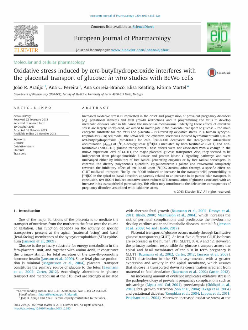
Treatment of BeWo cells with 100 μM tert-BOOH for 24 hincreased cellular GSX and GSSG levels. GSH levels did not change,probably due to its greater intracellular pool size, in comparison withGSSG, as high and similar GSH/GSSG ratios were found in control andtert-BOOH-treated cells (8.671.6 and 9.172.4, respectively; n¼9)(Fig. 2).
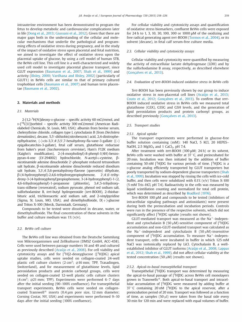
Under the same condition, tert-BOOH induced an increase in thelevels of the lipid peroxidation product malonaldehyde, an indicator ofoxidative damage to lipids (Fig. 3A), and of protein carbonyl groups, anindicator of oxidative damage to proteins (Fig. 3B).
Since exposure of BeWo cells to 100 μM tert-BOOH for 24 hinduced an increase in oxidative stress biomarkers while main-taining cellular viability and not causing cytotoxicity, this condi-tion was used in subsequent experiments aimed at determiningthe effect of oxidative stress upon glucose transport in BeWo cells.
3.3. Tert-BOOH decreases the steady-state accumulation of [3H]DGin BeWo cells
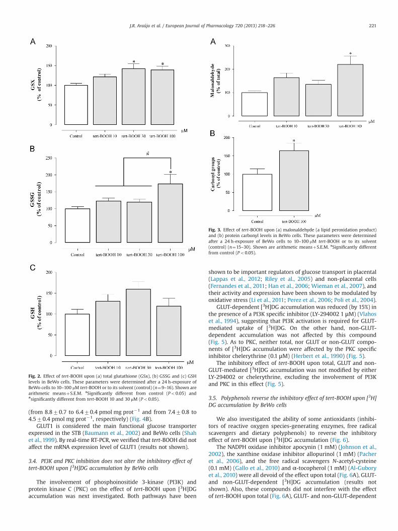
In a first series of experiments, we determined the effect oftert-BOOH upon [3H]DG apical uptake over time (Fig. 4). Tert-BOOH caused a significant decrease in [3H]DG intracellular accu-mulation at steady-state (Amax), as a result of a 2.6� increase inthe rate constant of [3H]DG outward transport (kout) and of a notso marked (1.7� ) increase in the rate constant of [3H]DG inwardtransport (kin) (Fig. 4A). Moreover, we verified that tert-BOOHdecreased the Amax of both GLUT- and non-GLUT mediated uptake
Fig. 1. Effect upon cell viability (a) and cytotoxicity (b) of tert-BOOH in BeWo cells.These parameters were determined after a 24 h-exposure of BeWo cells to 1–1000 mM tert-BOOH or to its solvent (control) (n¼9–12). Shown are arithmeticmeansþS.E.M. nSignificantly different from control (Po0.05) and ♯significantlydifferent from tert-BOOH 1–100 μM (Po0.05).
J.R. Araújo et al. / European Journal of Pharmacology 720 (2013) 218–226220
(from 8.870.7 to 6.470.4 pmol mg prot�1 and from 7.470.8 to4.570.4 pmol mg prot�1, respectively) (Fig. 4B).
GLUT1 is considered the main functional glucose transporterexpressed in the STB (Baumann et al., 2002) and BeWo cells (Shahet al., 1999). By real-time RT-PCR, we verified that tert-BOOH did notaffect the mRNA expression level of GLUT1 (results not shown).
3.4. PI3K and PKC inhibition does not alter the inhibitory effect oftert-BOOH upon [3H]DG accumulation by BeWo cells
The involvement of phosphoinositide 3-kinase (PI3K) andprotein kinase C (PKC) on the effect of tert-BOOH upon [3H]DGaccumulation was next investigated. Both pathways have been
shown to be important regulators of glucose transport in placental(Lappas et al., 2012; Riley et al., 2005) and non-placental cells(Fernandes et al., 2011; Han et al., 2006; Wieman et al., 2007), andtheir activity and expression have been shown to be modulated byoxidative stress (Li et al., 2011; Perez et al., 2006; Poli et al., 2004).
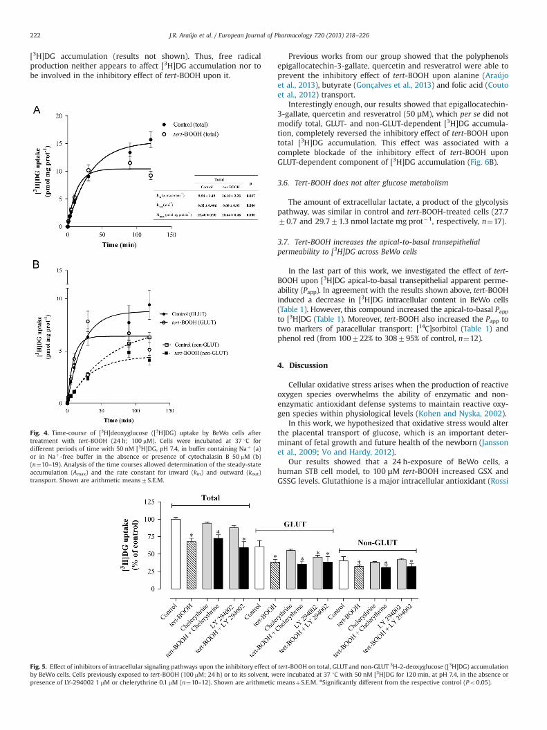
GLUT-dependent [3H]DG accumulation was reduced (by 15%) inthe presence of a PI3K specific inhibitor (LY-294002 1 μM) (Vlahoset al., 1994), suggesting that PI3K activation is required for GLUT-mediated uptake of [3H]DG. On the other hand, non-GLUT-dependent accumulation was not affected by this compound(Fig. 5). As to PKC, neither total, nor GLUT or non-GLUT compo-nents of [3H]DG accumulation were affected by the PKC specificinhibitor chelerythrine (0.1 μM) (Herbert et al., 1990) (Fig. 5).
The inhibitory effect of tert-BOOH upon total, GLUT and non-GLUT-mediated [3H]DG accumulation was not modified by eitherLY-294002 or chelerythrine, excluding the involvement of PI3Kand PKC in this effect (Fig. 5).
3.5. Polyphenols reverse the inhibitory effect of tert-BOOH upon [3H]DG accumulation by BeWo cells
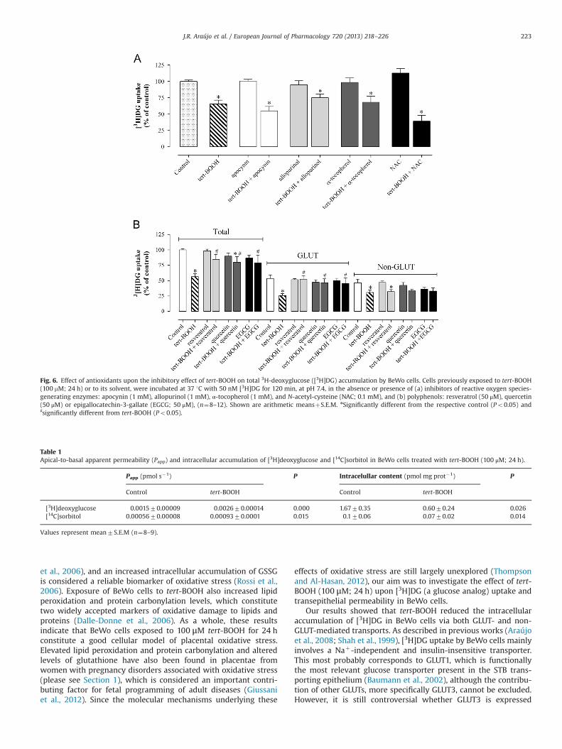
We also investigated the ability of some antioxidants (inhibi-tors of reactive oxygen species-generating enzymes, free radicalscavengers and dietary polyphenols) to reverse the inhibitoryeffect of tert-BOOH upon [3H]DG accumulation (Fig. 6).
The NADPH oxidase inhibitor apocynin (1 mM) (Johnson et al.,2002), the xanthine oxidase inhibitor allopurinol (1 mM) (Pacheret al., 2006), and the free radical scavengers N-acetyl-cysteine(0.1 mM) (Gallo et al., 2010) and α-tocopherol (1 mM) (Al-Guboryet al., 2010) were all devoid of the effect upon total (Fig. 6A), GLUT-and non-GLUT-dependent [3H]DG accumulation (results notshown). Also, these compounds did not interfere with the effectof tert-BOOH upon total (Fig. 6A), GLUT- and non-GLUT-dependent
Fig. 3. Effect of tert-BOOH upon (a) malonaldehyde (a lipid peroxidation product)and (b) protein carbonyl levels in BeWo cells. These parameters were determinedafter a 24 h-exposure of BeWo cells to 10–100 mM tert-BOOH or to its solvent(control) (n¼15–30). Shown are arithmetic meansþS.E.M. nSignificantly differentfrom control (Po0.05).
Fig. 2. Effect of tert-BOOH upon (a) total glutathione (GSx), (b) GSSG and (c) GSHlevels in BeWo cells. These parameters were determined after a 24 h-exposure ofBeWo cells to 10–100 mM tert-BOOH or to its solvent (control) (n¼9–16). Shown arearithmetic meansþS.E.M. nSignificantly different from control (Po0.05) and#significantly different from tert-BOOH 10 and 30 μM (Po0.05).
J.R. Araújo et al. / European Journal of Pharmacology 720 (2013) 218–226 221
[3H]DG accumulation (results not shown). Thus, free radicalproduction neither appears to affect [3H]DG accumulation nor tobe involved in the inhibitory effect of tert-BOOH upon it.
Previous works from our group showed that the polyphenolsepigallocatechin-3-gallate, quercetin and resveratrol were able toprevent the inhibitory effect of tert-BOOH upon alanine (Araújoet al., 2013), butyrate (Gonçalves et al., 2013) and folic acid (Coutoet al., 2012) transport.
Interestingly enough, our results showed that epigallocatechin-3-gallate, quercetin and resveratrol (50 μM), which per se did notmodify total, GLUT- and non-GLUT-dependent [3H]DG accumula-tion, completely reversed the inhibitory effect of tert-BOOH upontotal [3H]DG accumulation. This effect was associated with acomplete blockade of the inhibitory effect of tert-BOOH uponGLUT-dependent component of [3H]DG accumulation (Fig. 6B).
3.6. Tert-BOOH does not alter glucose metabolism
The amount of extracellular lactate, a product of the glycolysispathway, was similar in control and tert-BOOH-treated cells (27.770.7 and 29.771.3 nmol lactate mg prot�1, respectively, n¼17).
3.7. Tert-BOOH increases the apical-to-basal transepithelialpermeability to [3H]DG across BeWo cells
In the last part of this work, we investigated the effect of tert-BOOH upon [3H]DG apical-to-basal transepithelial apparent perme-ability (Papp). In agreement with the results shown above, tert-BOOHinduced a decrease in [3H]DG intracellular content in BeWo cells(Table 1). However, this compound increased the apical-to-basal Pappto [3H]DG (Table 1). Moreover, tert-BOOH also increased the Papp totwo markers of paracellular transport: [14C]sorbitol (Table 1) andphenol red (from 100722% to 308795% of control, n¼12).
4. Discussion
Cellular oxidative stress arises when the production of reactiveoxygen species overwhelms the ability of enzymatic and non-enzymatic antioxidant defense systems to maintain reactive oxy-gen species within physiological levels (Kohen and Nyska, 2002).
In this work, we hypothesized that oxidative stress would alterthe placental transport of glucose, which is an important deter-minant of fetal growth and future health of the newborn (Janssonet al., 2009; Vo and Hardy, 2012).
Our results showed that a 24 h-exposure of BeWo cells, ahuman STB cell model, to 100 μM tert-BOOH increased GSX andGSSG levels. Glutathione is a major intracellular antioxidant (Rossi
Fig. 4. Time-course of [3H]deoxyglucose ([3H]DG) uptake by BeWo cells aftertreatment with tert-BOOH (24 h; 100 mM). Cells were incubated at 37 1C fordifferent periods of time with 50 nM [3H]DG, pH 7.4, in buffer containing Naþ (a)or in Naþ-free buffer in the absence or presence of cytochalasin B 50 mM (b)(n¼10–19). Analysis of the time courses allowed determination of the steady-stateaccumulation (Amax) and the rate constant for inward (kin) and outward (kout)transport. Shown are arithmetic means7S.E.M.
Fig. 5. Effect of inhibitors of intracellular signaling pathways upon the inhibitory effect of tert-BOOH on total, GLUT and non-GLUT 3H-2-deoxyglucose ([3H]DG) accumulationby BeWo cells. Cells previously exposed to tert-BOOH (100 mM; 24 h) or to its solvent, were incubated at 37 1C with 50 nM [3H]DG for 120 min, at pH 7.4, in the absence orpresence of LY-294002 1 mM or chelerythrine 0.1 mM (n¼10–12). Shown are arithmetic meansþS.E.M. nSignificantly different from the respective control (Po0.05).
J.R. Araújo et al. / European Journal of Pharmacology 720 (2013) 218–226222
et al., 2006), and an increased intracellular accumulation of GSSGis considered a reliable biomarker of oxidative stress (Rossi et al.,2006). Exposure of BeWo cells to tert-BOOH also increased lipidperoxidation and protein carbonylation levels, which constitutetwo widely accepted markers of oxidative damage to lipids andproteins (Dalle-Donne et al., 2006). As a whole, these resultsindicate that BeWo cells exposed to 100 μM tert-BOOH for 24 hconstitute a good cellular model of placental oxidative stress.Elevated lipid peroxidation and protein carbonylation and alteredlevels of glutathione have also been found in placentae fromwomen with pregnancy disorders associated with oxidative stress(please see Section 1), which is considered an important contri-buting factor for fetal programming of adult diseases (Giussaniet al., 2012). Since the molecular mechanisms underlying these
effects of oxidative stress are still largely unexplored (Thompsonand Al-Hasan, 2012), our aim was to investigate the effect of tert-BOOH (100 μM; 24 h) upon [3H]DG (a glucose analog) uptake andtransepithelial permeability in BeWo cells.
Our results showed that tert-BOOH reduced the intracellularaccumulation of [3H]DG in BeWo cells via both GLUT- and non-GLUT-mediated transports. As described in previous works (Araújoet al., 2008; Shah et al., 1999), [3H]DG uptake by BeWo cells mainlyinvolves a Naþ-independent and insulin-insensitive transporter.This most probably corresponds to GLUT1, which is functionallythe most relevant glucose transporter present in the STB trans-porting epithelium (Baumann et al., 2002), although the contribu-tion of other GLUTs, more specifically GLUT3, cannot be excluded.However, it is still controversial whether GLUT3 is expressed
Fig. 6. Effect of antioxidants upon the inhibitory effect of tert-BOOH on total 3H-deoxyglucose ([3H]DG) accumulation by BeWo cells. Cells previously exposed to tert-BOOH(100 mM; 24 h) or to its solvent, were incubated at 37 1C with 50 nM [3H]DG for 120 min, at pH 7.4, in the absence or presence of (a) inhibitors of reactive oxygen species-generating enzymes: apocynin (1 mM), allopurinol (1 mM), α-tocopherol (1 mM), and N-acetyl-cysteine (NAC; 0.1 mM), and (b) polyphenols: resveratrol (50 μM), quercetin(50 μM) or epigallocatechin-3-gallate (EGCG; 50 μM), (n¼8–12). Shown are arithmetic meansþS.E.M. nSignificantly different from the respective control (Po0.05) and♯significantly different from tert-BOOH (Po0.05).
Table 1Apical-to-basal apparent permeability (Papp) and intracellular accumulation of [3H]deoxyglucose and [14C]sorbitol in BeWo cells treated with tert-BOOH (100 μM; 24 h).
Papp (pmol s�1) P Intracelullar content (pmol mg prot�1) P
J.R. Araújo et al. / European Journal of Pharmacology 720 (2013) 218–226 223
(Brown et al., 2011) or not (Shah et al., 1999) in BeWo cells.Concerning non-GLUT-mediated [3H]DG uptake, this could corre-spond to adsorption of [3H]DG to the cell membrane of BeWo cells.
Our results also demonstrated that the effect of tert-BOOH wasnot associated with a change in the mRNA level of GLUT1. Thissuggests that it rather involves changes in protein levels of GLUT1,post-translational GLUT1 modifications, such as oxidation (Burtonand Jauniaux, 2011; Fiorentini et al., 1999) phosphorylation (Hanet al., 2006), nitration (Webster et al., 2008) and ubiquitination(Fernandes et al., 2011), or changes in GLUT1 intrinsic activity.These parameters should be accessed in a future work in order toincrease knowledge about the impact of tert-BOOH-induced oxi-dative stress upon GLUT1-mediated transport.
We also verified that the inhibitory effect of tert-BOOH upon[3H]DG accumulation was not associated with an alteration inglycolytic metabolism, as assessed by measurement of lactateproduction. Since the first enzyme of glycolysis (hexokinase) is ofhigh affinity (Moraes and Reithmeier, 2012), the reduction in [3H]DG accumulation caused by tert-BOOH was probably not suffi-ciently marked to elicit a decrease in the glycolysis rate.
Very few studies relating oxidative stress with placental glucoseuptake have been performed. Interestingly and according to ourresults, Lappas et al. (2012) and Li et al. (2004) showed that oxidativestress-generating agents reduced placental glucose uptake, althoughthis effect was associated with a reduction in GLUT1 gene expression.These different findings can be explained by the use of distinctoxidative stress-generating agents in these works.
Alterations in placental transport or intracellular metabolism ofglucose have been described in some pregnancy disorders asso-ciated with increased reactive oxygen species generation, namelyfetal growth restriction (Magnusson et al., 2004), gestationaldiabetes and chronic hypoxia (which is present in preeclampsia)(Baumann et al., 2002; Illsley, 2000). However, placental glucosetransport and lactate production are differently altered in thesepathologies, suggesting that besides oxidative stress, other factorsplay an important role in determining the rate of placental glucosetransport and metabolism observed in these pathologies.
PI3K and PKC signaling pathways are important regulators ofglucose transport (Fernandes et al., 2011; Han et al., 2006; Lappaset al., 2012; Riley et al., 2005; Wieman et al., 2007). Moreover,activation of both of these pathways as a consequence of tert-BOOH-induced cellular oxidative stress has also been demonstrated(Li et al., 2011; Perez et al., 2006). Therefore, we decided to clarifythe role of these two signaling pathways in the inhibitory effect oftert-BOOH upon [3H]DG accumulation by BeWo cells. The resultsindicated that neither PI3K nor PKC activation seemed to be involvedin this effect. Interestingly, the results of PI3K agree with those of avery recent paper, also relating oxidative stress with placental DGuptake (Lappas et al., 2012).
Antioxidants are compounds that inhibit or delay the produc-tion of reactive oxygen species and the consequent oxidation ofbiomolecules (Al-Gubory et al., 2010). These compounds havebeen used as supplements to counteract oxidative stress andimprove fetal health outcomes in females with, or at higher riskof developing, preeclampsia (Chappell et al., 1999), fetal growthrestriction (Rueda-Clausen et al., 2012) and gestational diabetes(Cederberg et al., 2001). So, we decided to investigate the ability ofdifferent antioxidants to revert tert-BOOH-induced inhibition of[3H]DG accumulation. Interestingly, we verified that the polyphe-nols quercetin, epigallocatechin-3-gallate and resveratrol totallyabolished the reduction in [3H]DG accumulation induced by tert-BOOH, by specifically blocking the effect of tert-BOOH upon GLUT-dependent [3H]DG accumulation. Since these compounds alonedid not interfere with GLUT-dependent [3H]DG accumulation, webelieve that the effect of polyphenols does not involve a directinteraction with glucose transporters.
Dietary polyphenols possess antioxidant properties due to theirability to scavenge reactive oxygen species, modulate transcriptionfactors, and induce histone modifications (Mitjavila and Moreno,2012). A previous work from our group showed that the inhibitoryeffect of quercetin upon tert-BOOH-induced reduction of butyrateuptake in intestinal cells is associated with its capacity to abolish lipidperoxidation (Gonçalves et al., 2013). Moreover, resveratrol is able toreverse the decrease in placental GLUT1 activity evoked by hypox-anthine/xanthine oxidase-induced oxidative stress via sirtuin 1, anenzyme with histone deacetylase activity (Lappas et al., 2012). Finally,GLUTs can be upregulated trough a hypoxia-inducible transcriptionalfactor-1(HIF-1)-mediated pathway in BeWo cells (Baumann et al.,2007), and epigallocatechin-3-gallate (Mandel et al., 2008), quercetin(Bach et al., 2010) and resveratrol (Lin et al., 2012) increase HIF-1activity and/or expression. Altogether, these observations suggest thatpolyphenols may exert a protective role against oxidative stress-induced inhibition of placental glucose transport through distinctantioxidant mechanisms.
The reactive oxygen species scavengers α-tocopherol and N-acetyl-L-cysteine, the NADPH oxidase inhibitor apocynin and thexanthine oxidase inhibitor allopurinol were not able to reversethe inhibitory effect of tert-BOOH upon [3H]DG accumulation.These results suggest that the inhibitory effect of tert-BOOH up-on DG accumulation does not seem to greatly depend uponreactive oxygen species generation. Curiously, other works alsoreported that N-acetyl-L-cysteine and α-tocopherol were notable to alter oxidative stress-induced reduction of DG transport(Fernandes et al., 2011; Lappas et al., 2012). This can be due to thefact that, in the case of endogenous antioxidants such as vitaminE and glutathione (for which N-acetyl-L-cysteine is a precursor(Gallo et al., 2010)), the interaction of multiple systems is neces-sary to counteract oxidative processes. For instance, the antiox-idant activity of vitamin E against lipid peroxidation needs tobe supported by the activity of GSH and vitamin C (Gallo et al.,2010).
Fetal glucose availability does not solely rely on placentaltransport across the apical membrane of the STB. Indeed, changesin glucose transport across the basal membrane of the STB willalso have a significant impact upon transplacental glucose trans-port (Baumann et al., 2002). So, in the last part of this work, weinvestigated the effect of tert-BOOH upon [3H]DG transepithelialpermeability in the apical-to-basal direction in BeWo cells. Theresults obtained showed that, although tert-BOOH decreased theintracellular accumulation of [3H]DG in BeWo cells, it significantlyincreased the transepithelial permeability of [3H]DG in the apical-to-basal direction. Moreover, tert-BOOH increased the apical-to-basal transport of the paracellular markers [14C]sorbitol andphenol red. Noteworthy, in intestinal epithelial cells, tert-BOOHwas recently found to decrease the expression (Kim et al., 2012)and to alter the phosphorylation pattern (Sheth et al., 2009) of thetight junction proteins zonula occludens-1 and occludin, in asso-ciation with an increase in paracellular transport activity. So, wesuggest that tert-BOOH, by affecting BeWo cell tight junctions,induces an increase in [3H]DG paracellular transport, and that thiseffect contributes to the observed increase in transepithelialpermeability to [3H]DG.
In conclusion, our work demonstrates that tert-BOOH-inducedoxidative stress in BeWo cells decreased DG accumulation, and thatthis effect was completely reversed by some dietary polyphenols.Moreover, tert-BOOH-induced oxidative stress increased the transe-pithelial permeability to DG across BeWo cell monolayers, byincreasing its paracellular transport. As a whole, we suggest thatoxidative stress reduces placental accumulation of glucose associatedwith an increase in its transepithelial permeability. This effect maycontribute to the deleterious consequences of pregnancy disordersassociated with oxidative stress.
J.R. Araújo et al. / European Journal of Pharmacology 720 (2013) 218–226224
Acknowledgments
This work was financially supported by Fundação para a Ciênciae a Tecnologia (FCT) and COMPETE, QREN and FEDER (PTDC/SAU-OSM/102239/2008, SFRH/BD/63086/2009 and SFRH/BPD/40170/2007).
The authors wish to thank Prof. João T. Guimarães (Departmentof Clinical Pathology, Centro Hospitalar S. João, Porto, Portugal) forlactate quantification.
Appendix A. Supplementary material
Supplementary data associated with this article can be found inthe online version at http://dx.doi.org/10.1016/j.ejphar.2013.10.023.
References
Al-Gubory, K.H., Fowler, P.A., Garrel, C., 2010. The roles of cellular reactive oxygenspecies, oxidative stress and antioxidants in pregnancy outcomes. Int. J.Biochem. Cell Biol. 42, 1634–1650.
Araújo, J.R., Correia-Branco, A., Pereira, A.C., Pinho, M.J., Keating, E., Martel, F., 2013.Oxidative stress decreases uptake of neutral amino acids in a human placentalcell line (BeWo cells). Reprod. Toxicol. 40, 76–81.
Araújo, J.R., Gonçalves, P., Martel, F., 2008. Modulation of glucose uptake in a humanchoriocarcinoma cell line (BeWo) by dietary bioactive compounds and drugs ofabuse. J. Biochem. 144, 177–186.
Bach, A., Bender-Sigel, J., Schrenk, D., Flugel, D., Kietzmann, T., 2010. The antioxidantquercetin inhibits cellular proliferation via HIF-1-dependent induction ofp21WAF. Antioxid. Redox Signal. 13, 437–448.
Baumann, M.U., Deborde, S., Illsley, N.P., 2002. Placental glucose transfer and fetalgrowth. Endocrine 19, 13–22.
Baumann, M.U., Zamudio, S., Illsley, N.P., 2007. Hypoxic upregulation of glucosetransporters in BeWo choriocarcinoma cells is mediated by hypoxia-induciblefactor-1. Am. J. Physiol.: Cell Physiol. 293, C477–C485.
Bradford, M.M., 1976. A rapid and sensitive method for the quantitation ofmicrogram quantities of protein utilizing the principle of protein–dye binding.Anal. Biochem. 72, 248–254.
Brown, K., Heller, D.S., Zamudio, S., Illsley, N.P., 2011. Glucose transporter 3 (GLUT3)protein expression in human placenta across gestation. Placenta 32, 1041–1049.
Burton, G.J., Jauniaux, E., 2011. Oxidative stress. Best Pract. Res. Clin. Obstet.Gynaecol. 25, 287–299.
Carter, A.M., 2012. Evolution of placental function in mammals: the molecular basisof gas and nutrient transfer, hormone secretion, and immune responses.Physiol. Rev. 92, 1543–1576.
Cederberg, J., Siman, C.M., Eriksson, U.J., 2001. Combined treatment with vitamin Eand vitamin C decreases oxidative stress and improves fetal outcome inexperimental diabetic pregnancy. Pediatr. Res. 49, 755–762.
Chappell, L.C., Seed, P.T., Briley, A.L., Kelly, F.J., Lee, R., Hunt, B.J., Parmar, K., Bewley, S.J.,Shennan, A.H., Steer, P.J., Poston, L., 1999. Effect of antioxidants on the occurrenceof pre-eclampsia in women at increased risk: a randomised trial. Lancet 354,810–816.
Coughlan, M.T., Vervaart, P.P., Permezel, M., Georgiou, H.M., Rice, G.E., 2004. Alteredplacental oxidative stress status in gestational diabetes mellitus. Placenta 25,78–84.
Couto, M.R., Gonçalves, P., Catarino, T., Araújo, J.R., Correia-Branco, A., Martel, F.,2012. The effect of oxidative stress upon the intestinal uptake of folic acid:in vitro studies with Caco-2 cells. Cell Biol. Toxicol. 28, 369–381.
Dalle-Donne, I., Rossi, R., Colombo, R., Giustarini, D., Milzani, A., 2006. Biomarkersof oxidative damage in human disease. Clin. Chem. 52, 601–623.
Desoye, G., Gauster, M., Wadsack, C., 2011. Placental transport in pregnancypathologies. Am. J. Clin. Nutr. 94, 1896S–1902S.
Dong, M., Zheng, Q., Ford, S.P., Nathanielsz, P.W., Ren, J., 2013. Maternal obesity,lipotoxicity and cardiovascular diseases in offspring. J. Mol. Cell. Cardiol. 55,111–116.
Fernandes, R., Hosoya, K., Pereira, P., 2011. Reactive oxygen species downregulateglucose transport system in retinal endothelial cells. Am. J. Physiol.: Cell Physiol300, C927–C936.
Fiorentini, D., Hakim, G., Zambonin, L., Landi, L., 1999. The effect of oxygen radicalson rat thymocyte glucose transport is independent of the site of theirgeneration. Free Radic. Biol. Med. 26, 661–668.
Gallo, C., Renzi, P., Loizzo, S., Loizzo, A., Piacente, S., Festa, M., Caputo, M., Tecce, M.F.,Capasso, A., 2010. Potential therapeutic effects of vitamin E and C on placentaloxidative stress induced by nicotine: an in vitro evidence. Open Biochem. J. 4,77–82.
Gonçalves, P., Gregório, I., Catarino, T.A., Martel, F., 2013. The effect of oxidativestress upon the intestinal epithelial uptake of butyrate. Eur. J. Pharmacol. 699,88–100.
Han, H.J., Heo, J.S., Lee, Y.J., Min, J.J., Park, K.S., 2006. High glucose-inducedinhibition of 2-deoxyglucose uptake is mediated by cAMP, protein kinase C,oxidative stress and mitogen-activated protein kinases in mouse embryonicstem cells. Clin. Exp. Pharmacol. Physiol. 33, 211–220.
Herbert, J.M., Augereau, J.M., Gleye, J., Maffrand, J.P., 1990. Chelerythrine is a potentand specific inhibitor of protein kinase C. Biochem. Biophys. Res. Commun. 172,993–999.
Illsley, N.P., 2000. Glucose transporters in the human placenta. Placenta 21, 14–22.Jansson, T., Myatt, L., Powell, T.L., 2009. The role of trophoblast nutrient and ion
transporters in the development of pregnancy complications and adult disease.Curr. Vas. Pharmacol. 7, 521–533.
Kim, H.J., Lee, E.K., Park, M.H., Ha, Y.M., Jung, K.J., Kim, M.S., Kim, M.K., Yu, B.P.,Chung, H.Y., 2012. Ferulate protects the epithelial barrier by maintaining tightjunction protein expression and preventing apoptosis in tert-butyl hydroper-oxide-induced caco-2 cells. Phytother. Res. 27, 362–367.
Kohen, R., Nyska, A., 2002. Oxidation of biological systems: oxidative stressphenomena, antioxidants, redox reactions, and methods for their quantifica-tion. Toxicol. Pathol. 30, 620–650.
Lappas, M., Andrikopoulos, S., Permezel, M., 2012. Hypoxanthine–xanthine oxidasedown-regulates GLUT1 transcription via SIRT1 resulting in decreased glucoseuptake in human placenta. J. Endocrinol. 213, 49–57.
Lappas, M., Hiden, U., Desoye, G., Froehlich, J., Hauguel-de Mouzon, S., Jawerbaum, A.,2011. The role of oxidative stress in the pathophysiology of gestational diabetesmellitus. Antioxid. Redox Signal. 15, 3061–3100.
Li, H., Gu, Y., Zhang, Y., Lucas, M.J., Wang, Y., 2004. High glucose levels down-regulateglucose transporter expression that correlates with increased oxidative stress inplacental trophoblast cells in vitro. J. Soc. Gynecol. Invest. 11, 75–81.
Li, Y.B., Gao, J.L., Lee, S.M., Zhang, Q.W., Hoi, P.M., Wang, Y.T., 2011. Bisdemethox-ycurcumin protects endothelial cells against t-BHP-induced cell damage byregulating the phosphorylation level of ERK1/2 and Akt. Int. J. Mol. Med. 27,205–211.
Magnusson, A.L., Powell, T., Wennergren, M., Jansson, T., 2004. Glucose metabolismin the human preterm and term placenta of IUGR fetuses. Placenta 25, 337–346.
Mandel, S.A., Amit, T., Weinreb, O., Reznichenko, L., Youdim, M.B., 2008. Simulta-neous manipulation of multiple brain targets by green tea catechins: a potentialneuroprotective strategy for Alzheimer and Parkinson diseases. CNS Neurosci.Ther. 14, 352–365.
Mitjavila, M.T., Moreno, J.J., 2012. The effects of polyphenols on oxidative stress andthe arachidonic acid cascade. Implications for the prevention/treatment of highprevalence diseases. Biochem. Pharmacol. 84, 1113–1122.
Myatt, L., Cui, X., 2004. Oxidative stress in the placenta. Histochem. Cell Biol. 122,369–382.
Pacher, P., Nivorozhkin, A., Szabo, C., 2006. Therapeutic effects of xanthine oxidaseinhibitors: renaissance half a century after the discovery of allopurinol.Pharmacol. Rev. 58, 87–114.
Perez, L.M., Milkiewicz, P., Ahmed-Choudhury, J., Elias, E., Ochoa, J.E., Sanchez Pozzi, E.J.,Coleman, R., Roma, M.G., 2006. Oxidative stress induces actin-cytoskeletal andtight-junctional alterations in hepatocytes by a Ca2þ-dependent, PKC-mediatedmechanism: protective effect of PKA. Free Radic. Biol. Med. 40, 2005–2017.
Peuchant, E., Brun, J.L., Rigalleau, V., Dubourg, L., Thomas, M.J., Daniel, J.Y., Leng, J.J.,Gin, H., 2004. Oxidative and antioxidative status in pregnant women witheither gestational or type 1 diabetes. Clin. Biochem. 37, 293–298.
Poli, G., Leonarduzzi, G., Biasi, F., Chiarpotto, E., 2004. Oxidative stress and cellsignalling. Curr. Med. Chem. 11, 1163–1182.
Riley, J.K., Carayannopoulos, M.O., Wyman, A.H., Chi, M., Ratajczak, C.K., Moley, K.H.,2005. The PI3K/Akt pathway is present and functional in the preimplantationmouse embryo. Dev. Biol. 284, 377–386.
Rossi, R., Dalle-Donne, I., Milzani, A., Giustarini, D., 2006. Oxidized forms ofglutathione in peripheral blood as biomarkers of oxidative stress. Clin. Chem.52, 1406–1414.
Rueda-Clausen, C.F., Morton, J.S., Dolinsky, V.W., Dyck, J.R., Davidge, S.T., 2012.Synergistic effects of prenatal hypoxia and postnatal high-fat diet in thedevelopment of cardiovascular pathology in young rats. Am. J. Physiol.: Regul.Integr. Comp. Physiol. 303, R418–R426.
Santos, A., Gonçalves, P., Araújo, J.R., Martel, F., 2008. Intestinal permeability toglucose after experimental traumatic brain injury: effect of gadopentetatedimeglumine administration. Basic Clin. Pharmacol. Toxicol. 103, 247–254.
Shah, S.W., Zhao, H., Low, S.Y., McArdle, H.J., Hundal, H.S., 1999. Characterization ofglucose transport and glucose transporters in the human choriocarcinoma cellline, BeWo. Placenta 20, 651–659.
Sheth, P., Samak, G., Shull, J.A., Seth, A., Rao, R., 2009. Protein phosphatase 2A playsa role in hydrogen peroxide-induced disruption of tight junctions in Caco-2 cellmonolayers. Biochem. J. 421, 59–70.
J.R. Araújo et al. / European Journal of Pharmacology 720 (2013) 218–226 225
Siddiqui, I.A., Jaleel, A., Tamimi, W., Al Kadri, H.M., 2010. Role of oxidative stress inthe pathogenesis of preeclampsia. Arch. Gynecol. Obstet. 282, 469–474.
Takagi, Y., Nikaido, T., Toki, T., Kita, N., Kanai, M., Ashida, T., Ohira, S., Konishi, I.,2004. Levels of oxidative stress and redox-related molecules in the placenta inpreeclampsia and fetal growth restriction. Virchows Arch. 444, 49–55.
Thompson, L.P., Al-Hasan, Y., 2012. Impact of oxidative stress in fetal programming.J. Pregnancy 2012, 582748.
Tormos, C., Javier Chaves, F., Garcia, M.J., Garrido, F., Jover, R., O'Connor, J.E., Iradi, A.,Oltra, A., Oliva, M.R., Saez, G.T., 2004. Role of glutathione in the induction ofapoptosis and c-fos and c-jun mRNAs by oxidative stress in tumor cells. CancerLett. 208, 103–113.
Vardhana, P.A., Illsley, N.P., 2002. Transepithelial glucose transport and metabolismin BeWo choriocarcinoma cells. Placenta 23, 653–660.
Vlahos, C.J., Matter, W.F., Hui, K.Y., Brown, R.F., 1994. A specific inhibitor ofphosphatidylinositol 3-kinase, 2-(4-morpholinyl)-8-phenyl-4H-1-benzopyran-4-one (LY294002). J. Biol. Chem. 269, 5241–5248.
Vo, T., Hardy, D.B., 2012. Molecular mechanisms underlying the fetal programmingof adult disease. J. Cell Commun. Signal. 6, 139–153.
Webster, R.P., Roberts, V.H., Myatt, L., 2008. Protein nitration in placenta –