Page 1
~ 397 ~
Journal of Pharmacognosy and Phytochemistry 2018; 7(4): 397-410
E-ISSN: 2278-4136
P-ISSN: 2349-8234
JPP 2018; 7(4): 397-410
Received: 15-05-2018
Accepted: 20-06-2018
Anusree K Dharman
Cell Culture Lab, Department of
Botany Union Christian College,
Aluva, Ernakulam, Kerala, India
M Anilkumar
Cell Culture Lab, Department of
Botany Union Christian College,
Aluva, Ernakulam, Kerala, India
Correspondence
M Anilkumar
Cell Culture Lab, Department of
Botany Union Christian College,
Aluva, Ernakulam, Kerala, India
Pharmacognostic studies in Solanum capsicoides all
Anusree K Dharman and M Anilkumar
Abstract
Detailed analysis of morphological and anatomical features of Solanum capsicoides All. Was done that
would be helpful for pharmacognostic identification. Phytochemical screening and histochemical test
were performed for the confirmation and localization of the phytoconstituents present in the species.
Among the morphological features anthocyanin pigmentation on leaf petiole, five different types of
trichomes, The inflorescence with bisexual flowers, orange red fruit and winged seeds were found to be
distinctive. Presence of sandy crystals and bicollateral vascular bundles, the unifying features of the
genus, were observed in the plant. Absence of collenchymatous hypodermis in fruit exocarp was a
distinguishing feature from other Solanum members. The phytochemicals identified in the plant were
flavonoids, coumarins, alkaloids, tannins, steroids, saponins, phenol, resin, glycoside, protein and
carbohydrate. The present study thus emphasis the pharmaceutical potential of the plant and the necessity
for its conservation.
Keywords: anatomy, histochemical test, morphology, phytochemical screening
Introduction
Solanum is one of the species rich genera in angiosperm and is also the largest genera in
Solanaceae [1]. Species coming under the genus Solanum includes vegetables [2], weeds and
medicinal herbs [3]. Solanum capsicoides All. (Cockroach Berry) (Syn. S. aculeatissimumJacq.)
is a medicinal plant and is native to eastern Brazil. It is used as the source of Kantakari in
ayurveda, an important therapeutic agent for dislodging tenacious phlegm.It is extensively
used for the treatment of diverse ailments like cough, bronchitis, asthma, influenza and enteric
fever.‘Kanakasavam, kantakarighrtham, pulikaranjasavam and suranadileham’ are the
important ayurvedic formulations that use kantakari as a constituent [4]. Over exploitation and
urbanization has drastically decreased the availability of this plant as a raw drug in the Indian
Ayurvedic industry, especially in Kerala. Pharmacognostic standardisation of this valuable
medicinal plant was not been reported so far. Hence, adulteration at raw drug level has become
a problem in the industry.
Morphological and anatomical studies of medicinal plants are relevant for their identification.
Now a days refined chemical and molecular methods are available for the identification of
plant material. But morpho-anatomical documentation is the simplest qualitative method to
avoid falsification and adulteration of the drug [5]. The structural analysis pinpoints
idiosyncratic aspects that can be effective in determining the accuracy of medicinal plant
species [6-8].
The medicinal potential of taxonomically related species can be studied using histochemical
techniques [9-10]. This technique is quick and inexpensive and can be used in search of new
pharmaceuticals [11-12]. The histochemical studies are rare in Solanum [13-14] though the
members are used for medicinal purpose from ancient times [15].
Secondary metabolites present in a plant can be considered as its chemical individuality as
their composition differ from species to species [16]. Phytochemical screening is crucial in the
discovery of new sources of therapeutic agents that are economically important (Akrout et al.,
2010) [17]. It is also essential for more pharmacological approaches.
The present paper reports morphological and anatomical characterization, histochemical
localization and preliminary screening of phytochemical constituents of S. capsicoides as
pharmacognostic tool for the raw drug industry.
Materials and Methods
Collection of Plant Materials
Mature plants with fruits were collected from Ernakulam district of Kerala State, India,
identified and herbarium voucher specimens were deposited at the Herbarium of Kerala Forest
Research Institute (KFRI), Peechi (KFRI-13056).
Page 2
~ 398 ~
Journal of Pharmacognosy and Phytochemistry Structural Characterization
Morpho-anatomical analysis was executed using 30 cm ruler,
graph paper, electronic balance, micrometer, Olympus
trinocular microscope with Magnus microscope camera
attached, TESCAN VEGA 3 SBH electron microscope and
Olympus Stereo zoom microscope attached with Nikon
camera.
Macroscopic features considered include plant habit, plant
height, leaf colour, texture, phyllotaxy, leaf type, leaf blade
shape, leaf area, root type, colour, texture, taste,
inflorescence, flower colour, flower diameter, sepal length,
petal length, stamen length, pistil length, fruit diameter, fruit
weight, seed number, seed size and seed weight, where as the
microscopic aspects numbered aretype of stomata, stomatal
index, guard cell area, type of trichomes, trichome length,
seed surface architecture and pollen viability.
Macroscopic measurements were executed using 30 cm ruler
and electronic balance. Microscopic scaling was performed
using a calibrated eye piece graticule.
The seed surface architecture was analysed using scanning
electron microscope.
Leaf Structure Analysis
Mature healthy leaves were collected and used for studying
various characters. Leaf area was measured using millimeter
graph method [18]. For studying the venation pattern, leaves
were cleared following the method of Gardner (1975) with
minor modifications [19]. The epidermal features were studied
using epidermal peels obtained from fresh leaves using
pointed needle and forceps. The peels were stained using
safranin and viewed under microscope.
Anatomical Studies
For microscopic analysis cross sections were prepared
following free hand sectioning method and stained with
toluidine blue.
Histochemical Localization
Histochemical localization was done on fresh plant sections
using various reagents such as Lugol’s solution for starch
(Jensen, 1962), Aqueous NaOH for flavonoids (Johansen,
1940), Bromophenol Blue for protein (Mazia, 1953), Aqueous
Ferric chloride for phenol (Johansen, 1940), Wagners Reagent
for alkaloid (Furr and Mahlberg,1981) and Schiff’s reagent
for lignin (McLean and Cook, 1941) [20-24]. Plant sections not
treated with any chemicals were used as negative control.
Powder Analysis
Leaf, fruit, stem and root were air dried and powdered. Fine
powder was used for microscopic characterization and also
for macroscopic analysis. Drug powder was treated with
twelve different reagents and the colour change was noticed
under day light.
Physico-Chemical Parameters
Parameters studied include pH value (1% solution and 10%
solution), moisture content, extractive values (water soluble,
ethanol soluble, methanol soluble, chloroform soluble and
ethyl acetate soluble) and ash values (total ash, acid insoluble
ash) following WHO guidelines.
Preliminary Phytochemical Screening
Powdered leaf, fruit, stem and root were soaked directly in
extractive solvents such as ethyl acetate, chloroform and
methanol in the ratio 1:5 and were kept for 48 hrs. Using
Whatman Filter paper No.1, the extract was filtered and was
concentrated in water bath. Evaporated extracts were used to
investigate the presence of various phytochemical constituents
following standard procedures (WHO, 2002) [25].
Results
Structural characterization
Habit: Solanum capsicoides is a short lived perennial plant
and is suffrutescent. It is branching and acanceous with
determinate growth reaching a height of 50cm- 1m.The stem
is cylindrical, pubescentand green in colour (Fig 1). Plant
possesses simple leaves arranged alternately at the base but as
pairs in inflorescence portion.
Fig 1: Whole Plant
Inflorescence: The inflorescence of the plant is scorpioid
cyme with 4 flowers (Fig. 2A) in extra-axiallary position (Fig.
2B).Flowers are stellate, pentamerous, actinomorphic,
pedicellate with valvate aestivation and entomophilous. Both
peduncle and pedicel are armed with prickles.Calyx: 5,
gamosepalous, green in colour and is armed with prickles
(Fig.2C) Corolla: 5, gamopetalous, white coloured and light
greenish towards base (Fig.2D). Androecium: 5, equal,
epipetalous, basifixed, and connivent anthers alternately
arranged to petals. The filament tube is minute and pale green
in colour. The antheris ovate, dark yellow coloured towards
base which gradually fades to pale yellow at the tip (Fig.2E-
F). Gynoecium: Ovary superior, bicarpellary, with terminal
long style and a well developed green coloured bilobed
stigma (Fig.2G-H). The ovary is bilocular with axile
placentation (Fig.2I).
Page 3
~ 399 ~
Journal of Pharmacognosy and Phytochemistry
Fig 2: Inflorescence:(A) Inflorescence; (B) Extra-axillary position of inflorescence;(C) Calyx;(D) Corolla; (E) Epipetalous stamen; (F) Single
Stamen; (G) Section of flower showing superior ovary; (H) Gynoecium; (I)Ovary T.S.
Fruits: Globose berry, but different from botanically defined
berry because the fruit is only slightly juicy and the fruit
release the seeds at maturity by dehiscence of pericarp. Fruit
possess persistent calyx and the pedicel is curved. Both the
pedicle and calyx are equipped with prickles. Usually 1 fruit
is developed from each inflorescence. Young fruits are pale
green at the base which gradually turns white towards the
apex with green coloured longitudinal stripes (Fig. 3A).
Mature fruits are bright orange- red in colour (Fig.3B). Inside
portion of the fruit wall is white coloured and spongy. The
flesh is slightly juicy but brittle and can be easily broken.
Fruit is filled with numerous seeds. The septum present in the
ovary has degenerated and two placental bodies are clearly
visible (Fig.3C).
Seeds: The seeds are flattened, winged, considerably round
and straw coloured. A little mucilage coating is present on the
seed which make them sticky in texture (Fig. 3D-E). The seed
surface exhibit reticulate pattern. The lumen is shallow and
the convoluted cells displays sinuous pattern (Fig.3F-G).
Fig 3: Fruit:(A)Young Fruit; (B) Mature Fruit; (C) Matured fruit halved showing inside of fruit wall, seeds and placental bodies; (D) Mature
Seeds; (E)SEM image of seed; (F)Seed surface towards hilum showing reticulate architecture; (G) Lumen surrounded by convoluted cells with
sinuous pattern
Root: Root is long, cylindrical and branched having long thin
rootlets. External surface is rough in texture and brown in
colour (Fig.4). Fracture not easy and the plane of fracture
arefibrous. It tastes bitter and is odourless.
Page 4
~ 400 ~
Journal of Pharmacognosy and Phytochemistry
Fig 4: Root
Leaf structure analysis
Plants possess simple, dorsiventral, cordate membranous
leaves, dark green coloured in the adaxial surface and pale
green coloured in abaxial surface, arranged alternately at the
base but as pairs in inflorescence portion. The pubescent
lamina is symmetrical, with coarsely lobed margin, leptophyll
in size and has marginal petiolar attachment. Petiole portion
facing the adaxial surface of leaf is purple green in colour.
Prickles are present along the major veins on both adaxial and
abaxial surface (Fig. 5A-B).
Fig 5: Leaf: (A) Adaxial surface; (B) Abaxial surface
Epidermal features: The epidermal cells are irregular in shape
and anticlinal walls are undulating. The species is
hypoamphistomatic in which bothanomocytic and anisocytic
stomata are present (Fig.6A-B). Both glandular and non-
glandular trichomes are present on entire plant. Two types of
non-glandular trichomes were observed such as multicellular
hair with pointed tip having 4-6 cells (Fig. 6C) and bicellular
trichome with blunt end (Fig. 6D). Glandular trichomes are of
three types, unicellular claviform trichome with bicellular
base (Fig. 6E); multicellular capitate trichome with unicellular
base (Fig. 6F); multicellular glandular trichome (Fig. 6G).
Fig 6: Epidermal Features: (A)Epidermal peel from adaxial surface showing anisocytic stomata; (B) anisocytic and anomocytic stomata on
abaxial surface; (C) Multicellular eglandular trichome; (D)Bicellular eglandular trichome; (E)Claviform glandular trichome; (F)Capitate
glandular trichome; (G) multicellular glandular trichome
Venation pattern: The venation is pinnate, ornamented and the
primary vein is straight. The primary vein gives off secondary
veins which number 5-8 on either side. The spacing between
the secondary veins is not regular. Intersecondary veins are
present. The highest order of the vein was observed as 6
degree. Marginal ultimate venation is looped and complete.
The areoles are imperfect and random. They are square,
quadrangular or pentangular in shape. Vein endings may or
may not be present in the areole. Vein endings entering the
areoles were simple or branched. Simple veinlets were either
curved or linear. Branched veinlets observed were crescent
shaped and Y shaped and was both symmetric and
asymmetric. Veinlets are mostly uniseriate but at some
junctions of veinlets they are biseriate. The tracheids are
uniseriate, sometimes heavily thickened or less thickened.
Both conventional and dilated tracheids were observed.
Dilated tracheids were of two types; spindle shaped and
gnarled (Fig. 7A-O).
Page 5
~ 401 ~
Journal of Pharmacognosy and Phytochemistry
Fig 7: Leaf Venation Pattern:(A) areole lacking vein ending; (B) areole with vein ending; (C) curved conventional tracheid; (D) Principal
asymmetric vein ending with heavily thickenedtracheids in between and at the tip of veinlet; (E) Veinlet biseriate at one portion bearing less
thickened dilated tracheid; (F) Veinlet terminates in to two and one of them again divide dichotomously with one branch terminates in dilated
tracheid; (G) Biseriate veinlet divides into two; spindle shaped tracheid and uniseriate veinlet with dilated end ; (H) Curved veinlet with
conventional tracheid; (I) Gnarled tracheid; (J) Asymmetric Y shaped veinlets; (K) Conventional tracheid between veinlet; (L)Asymmetric Y
shaped veinlet biseriate at junction; (M)Symmetric Y shaped veinlets with spindle shaped tracheids; (N)Crescent shaped veinlets; (O) Ultimate
branching – complete and looped
Quantitative macroscopic and microscopic features are shown in Table I.
Table I: Quantitative microscopic and macroscopic features of S. capsicoides All.
Plant Parts Feature Quantified Measurement
LEAF
Leaf Area (cm2) 79.67±3.95
Trichome Length
Multicellular Eglandular (mm) 1.66-2.47
Bicellular Eglandular ( µm) 45-97.5
Claviform Glandular ( µm) 56.25-150
Capitate Glandular ( µm) 26.25-56.25
Guard cell area (µm2) Upper epidermis 395.41±18.27*
Lower epidermis 500.61±31*
Stomatal Index (%) Upper epidermis 6.25±0.61*
Lower epidermis 15.46±0.53*
Prickle Length (mm) Upper epidermis 6-10
Lower epidermis 4-9
STEM Trichome Length
Multicellular Eglandular (mm) 4.05-4.75
Bicellular Eglandular ( µm) 48.75 -75
Claviform Glandular ( µm) 67.5 -97.5
Capitate Glandular ( µm) 56.25 -86.25
Prickle Length (mm) 2-6
FRUIT
Fruit Diameter (cm) 7.2-7.9
Fruit Weight (g) 3.78-5.53
Seed Number 165-209
Seed Size(mm) 5
Seed Weight(g) 0.0017-0.0019
POLLEN Pollen Viability (%) 92.05±1.34*
FLOWER Flower Diameter (cm) 2-2.2
Sepal Length (mm) 2-5
Page 6
~ 402 ~
Journal of Pharmacognosy and Phytochemistry Petal length (mm) 12-14
Stamen Length (mm) 8-10
Pistil Length (Long) (mm) 10-11
Pistil Length (Short) (mm) 2-3
* Each value represents mean ± standard error
Abbreviations: mm- millimeter; cm- centimeter; µm- micrometer; g-gram
Anatomical Studies
Stem
Microscopic analysis revealed single layered barrel shaped
epidermal cells covered with cuticle. The epidermal layer
possesses appendages such as non-glandular and glandular
trichomes but lack stomata. Epidermis is followed by a single
layer of chlorenchyma cells. Cortex consists of 6 layers of
angular collenchyma cells followed by 4-6 layers of
parenchyma which are large sized and isodiammetric. The
vascular bundle is bicollateral, amphiphloic siphonostele. The
xylem is endarch, with large vessels. Both internal and
external phloem is 4-5 layered and is covered with
sclerenchymatous bundle sheath. Pith is parenchymatous.
Cortex and pith encompass black powdery mass, the sandy
crystals (Fig. 8A). As the secondary growth proceeds, the
epidermis followed by the chlorenchymatous layer turned
brown in colour. The parenchyma layer reduced in thickness
and became flattened. The vasculature became continuous
with the development of secondary xylem and secondary
phloem and the bundle sheath expanded in thickness. The
parenchyma in cortex and pith consists of sandy crystals and
starch grains (Fig. 8B).
Fig 8: Stem anatomy: (A) Transverse section of stem in primary growth; (B) Stem in secondary growth. Abbreviations:EP-epidermis, CO-
collenchyma, PA-parenchyma, SC-sandy crystals, EPH-external phloem, XY-xylem, IPH-internal phloem, SCL-sclerenchyma, PI-pith, SXY-
secondary xylem, SPH-secondary phloem
Leaf
In the cross section, the epidermis is single layered with
rounded cells and thin cuticle. Trichomes are present along
the epidermal layer. In midrib the epidermis is followed by 2
layers of angular collenchyma and by parenchyma, the ground
tissue. Single vascular bundle is present in the centre which is
bicollateral and arc shaped. Sandy crystals are present in
parenchyma and in the phloem (Fig. 9A). The microscopic
view of lamina affirms the dorsiventral organization of leaf.
The epidermis is followed by single layer of compactly
arranged palisade parenchyma followed by 3 layers of loosely
arranged spongy parenchyma. Stomata are visible on both
upper and lower epidermis (Fig.9B).
Fig 9: Leaf Anatomy: (A) Midrib cross section; (B) Transverse section of lamina. Abbreviations: EP-epidermis, CO-collenchyma, PA-
parenchyma, SC-sandy crystals, PAL-palisade parenchyma, SPO-spongy parenchyma, ST-stomata, UEP-upper epidermal cell, LEP-lower
epidermal cell
Page 7
~ 403 ~
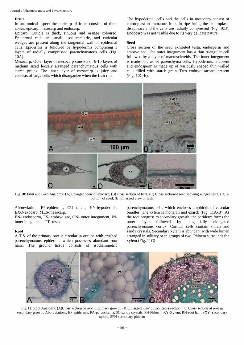
Journal of Pharmacognosy and Phytochemistry Fruit
In anatomical aspect the pericarp of fruits consists of three
zones: epicarp, mesocarp and endocarp.
Epicarp: Cuticle is thick, sinuous and orange coloured.
Epidermal cells are small, isodiammetric, and cuticular
wedges are present along the tangential wall of epidermal
cells. Epidermis is followed by hypodermis comprising 3
layers of radially compressed parenchymatous cells (Fig.
10A).
Mesocarp: Outer layer of mesocarp consists of 6-10 layers of
medium sized loosely arranged parenchymatous cells with
starch grains. The inner layer of mesocarp is juicy and
consists of large cells which disorganize when the fruit ripe.
The hypodermal cells and the cells in mesocarp consist of
chloroplast in immature fruit. In ripe fruits, the chloroplasts
disappears and the cells are radially compressed (Fig. 10B).
Endocarp was not visible due to its very delicate nature.
Seed
Cross section of the seed exhibited testa, endosperm and
embryo sac. The outer integument has a thin triangular cell
followed by a layer of macrosclereids. The inner integument
is made of crushed parenchyma cells. Hypodermis is absent
and endosperm is made up of variously shaped thin walled
cells filled with starch grains.Two embryo sacsare present
(Fig. 10C-E).
Fig 10: Fruit and Seed Anatomy: (A) Enlarged view of exocarp; (B) cross section of fruit; (C) Cross sectionof seed showing winged testa; (D) A
portion of seed; (E) Enlarged view of testa
Abbreviation: EP-epidermis, CU-cuticle, HY-hypodermis,
EXO-exocarp, MES-mesocarp,
EN- endosperm, ES- embryo sac, ON- outer integument, IN-
inner integument, TT- testa
Root
A T.S. of the primary root is circular in outline with crushed
parenchymatous epidermis which possesses abundant root
hairs. The ground tissue consists of isodiammetric
parenchymatous cells which encloses amphicribral vascular
bundles. The xylem is monarch and exarch (Fig. 11A-B). As
the root progress to secondary growth, the periderm forms the
outer layer followed by tangentially elongated
parenchymatous cortex. Cortical cells contain starch and
sandy crystals. Secondary xylem is abundant with wide lumen
arranged in solitary or in groups of two. Phloem surrounds the
xylem (Fig. 11C)
Fig 11: Root Anatomy: (A)Cross section of root in primary growth; (B) Enlarged view of root cross section; (C) Cross section of root in
secondary growth. Abbreviations: EP-epidermis, PA-parenchyma, SC-sandy crystals, PH-Phloem, XY-Xylem, RH-root hair, SXY- secondary
xylem, SPH-secondary phloem
Page 8
~ 404 ~
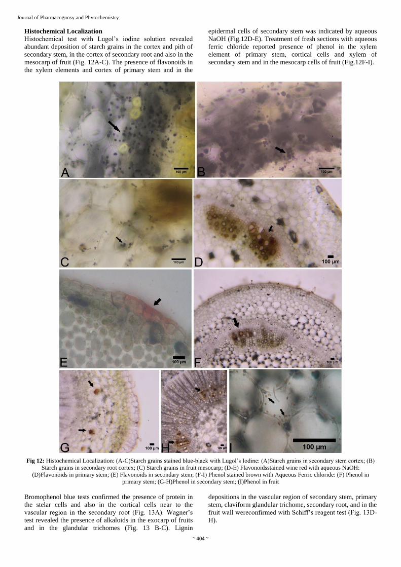
Journal of Pharmacognosy and Phytochemistry Histochemical Localization
Histochemical test with Lugol’s iodine solution revealed
abundant deposition of starch grains in the cortex and pith of
secondary stem, in the cortex of secondary root and also in the
mesocarp of fruit (Fig. 12A-C). The presence of flavonoids in
the xylem elements and cortex of primary stem and in the
epidermal cells of secondary stem was indicated by aqueous
NaOH (Fig.12D-E). Treatment of fresh sections with aqueous
ferric chloride reported presence of phenol in the xylem
element of primary stem, cortical cells and xylem of
secondary stem and in the mesocarp cells of fruit (Fig.12F-I).
Fig 12: Histochemical Localization: (A-C)Starch grains stained blue-black with Lugol’s Iodine: (A)Starch grains in secondary stem cortex; (B)
Starch grains in secondary root cortex; (C) Starch grains in fruit mesocarp; (D-E) Flavonoidsstained wine red with aqueous NaOH:
(D)Flavonoids in primary stem; (E) Flavonoids in secondary stem; (F-I) Phenol stained brown with Aqueous Ferric chloride: (F) Phenol in
primary stem; (G-H)Phenol in secondary stem; (I)Phenol in fruit
Bromophenol blue tests confirmed the presence of protein in
the stelar cells and also in the cortical cells near to the
vascular region in the secondary root (Fig. 13A). Wagner’s
test revealed the presence of alkaloids in the exocarp of fruits
and in the glandular trichomes (Fig. 13 B-C). Lignin
depositions in the vascular region of secondary stem, primary
stem, claviform glandular trichome, secondary root, and in the
fruit wall wereconfirmed with Schiff’s reagent test (Fig. 13D-
H).
Page 9
~ 405 ~
Journal of Pharmacognosy and Phytochemistry
Fig 13: Histochemical Localization:(A)Protein stained blue with Bromophenol blue in secondary root; (B-C) Alkaloid stained reddish brown
with Wagner’s Reagent: (B)Alkaloid in fruit; (C) Alkaloid in trichome; (D-H) Lignin stained majenta with Schiff’s reagent: (D)Lignin in
secondary stem; (E) Lignin in primary stem; (F)Lignin in trichome; (G)Lignin in secondary root; (H)Lignin in fruit
Powder analysis
The powder form of stem and root is cream coloured where as
leaf is in green colour and fruit in brown-orange colour (Fig.
14A-D).The stem powder under microscope revealed the
presence of cells with crystals (Fig.14 E), starch grains (Fig.
14 F), spiral xylem vessel (Fig. 14 G) and fiber (Fig. 14 H).
The leaf powder showed the presence of starch grains (Fig. 14
I), multicellular non-glandular trichome (Fig.14 J) and spiral
thickening of xylem vessel (Fig. 14 K). The fruit powder
confirmed cell with starch grains (Fig. 14 L), free starch
grains (Fig. 14 M), fibers (Fig.14 N), endosperm cells (Fig. 14
O) and fruit wall (Fig. 14 P). The root powder under
microscope presented fiber (Fig. 14 Q), starch grains (Fig. 14
R) and pitted vessels (Fig.14 S). The colour observed under
day light after the reaction of drug powders to twelve different
reagents is presented in Table II.
Page 10
~ 406 ~
Journal of Pharmacognosy and Phytochemistry
Fig 14: Powder Analysis: (A-D) Drug powder as such: (A) Stem; (B) Leaf; (C) Fruit; (D) Root; (E-H) Powder analysis of stem: (E) ruptured cell
with crystals; (F) starch grains; (G) xylem vessel with spiral thickening; (H) fiber; (I-K) Powder analysis of leaf: (I) starch grains; (J) trichome;
(K) spiral thickening of xylem vessel; (L-P) Powder analysis of fruit: (L) cell with starch grain; (M) starch grains; (N) fiber; (O)endosperm cell;
(P)fruit wall; (Q-S) Powder analysis of root: (Q) fiber; (R) starch grains; (S) pitted vessel
Table II: Reaction to chemicals
Reagents Stem Leaf Fruit Root
Powder as such Cream Dark green Brown Red Cream
Conc. H2SO4 Brown Pale green Deep red Brown
Conc. HCl Brownish Yellow Light green Yellow Flesh colour
Glacial acetic acid Cream Yellow No colour change Cream
Iodine solution Brown Reddish brown Reddish brown Reddish brown
Aq. Ferric chloride Orange Orange Deep yellow Brown
Aq. NaOH (5%) Cream Pale green Yellow Pale yellow
Aq. KOH (5%) Cream No colour change Yellow Pale yellow
Ammonia solution Cream Pale green Deep yellow Pale yellow
Distilled water Cream No colour change Light yellow No colour change
Acetone White Green No colour change White
Ethyl acetate No colour change Pale green No colour change White
Abbreviations: Conc. - Concentration; Aq. – Aqueous
Physicochemical Parameters
The pH value of drug solution ranges between 6-6.8. The
moisture content was high in leaf (86.75%) followed by other
parts. In leaf, stem and root the extractive value was highest
for water soluble matters where as in fruit, ethanol soluble
extractive was highest. In allthe four parts the total ash
content was highest than acid insoluble ash content (Table
III).
Page 11
~ 407 ~
Journal of Pharmacognosy and Phytochemistry Table III: Physicochemical parameters
Parameters Stem Leaf Fruit Root
pH value 1% solution 6.8 6.2 6.3 6.4
10% solution 6 6 6 6
Moisture content 83.66% 86.75% 79.1% 76.66%
Extractive values
Water soluble 15% 16% 7% 8%
Ethanol soluble 3% 5% 18% 3%
Methanol soluble 6.25% 11.25% 16.72% 3.37%
Chloroform soluble 0.5% 0.12% 0.08% 0.25%
Ethyl acetate soluble 4.37% 3.12% 0.17% 1%
Ash Value Total ash 68% 49% 87% 80%
Acid insoluble ash 49% 34% 84% 31%
Preliminary phytochemical screening
Phytochemical screening of different extracts revealed that
among the stem extracts ethyl acetate extract contains steroids
and protein where as methanolic extract has shown the
presence of coumarin, steroids, resin, protein and
carbohydrate. Chloroform extracts has not revealed the
presence of any phtyoconstituents. In leaf, methanolic extract
is rich in phytochemicals such as coumarin, alkaloids,
steroids, saponin, resin and carbohydrate. Only coumarin is
present chloroform extract while ethyl acetate extract contains
coumarins and resin. Among the fruit extracts ethyl acetate
extract contains alkaloid, steroid and resin. The chloroform
extract has shown the presence of alkaloids, steroids and
protein. Steroids, tannin, phenol, glycoside and carbohydrate
are present in methanolic extract. The phtyoconstituents
present in ethyl acetate root extract are steroids, resin, protein
and carbohydrate. Chloroform extract contain only protein
and methanolic extract have shown the presence of steroid,
resin and carbohydrate (Table IV).
Table IV: Preliminary phytochemical screening of stem, leaf, fruit
and root
Tests Stem Leaf Fruit Root
E C M E C M E C M E C M
Flavonoids - - - - - - - - - - - -
Coumarins - - + + + + - - - - - -
Alkaloids Mayer’s test - - - - - + + + - - - -
Wagner’s test - - - - - + + + - - - -
Tannin - - - - - - - - + - - -
Steroids / Terpenoids + - + - - + + + + + - +
Saponins - - - - - + - - - - - -
Quinines - - - - - - - - - - - -
Anthraquinones - - - - - - - - - - - -
Phenol - - - - - - - - + - - -
Resin - - + + - + + - - + - +
Glycoside - - - - - - - - + - - -
Protein Xanthoprotein test + - + - - - - + - + + -
Biuret test - - - - - - - - - - - -
Carbohydrate - - + - - + - - + + - +
Discussion
Pharmacognostic parameters are widely used for the
identification of medicinal plants and also to detect
adulteration in drugs [26]. Morphological features of
S.capsicoides are similar to other related species of Solanum
but it can be distinguished by its orange red fruit and straw
coloured winged seeds [27]. The fruit type in many other
Solanum species is berry [28] as that of S. capsicoides. In
Solanaceae the use of fruit type as taxonomic character have
proved to be systematically helpful [29]. The winged nature of
the seeds enable them to disperse by wind [30] or by floating
through water [31]. Seed morphology, especially the
sculpturing of outer seed coat, is a powerful tool for analyzing
the taxonomic relationship among plant families [32] as it has
been proved to be different among the species of same genus [33-34]. Seed surface of S.capsicoides resembles S. torvum as
both have convoluted cells with sinuous pattern and lack
fibrils [35]. The extra axillary position of inflorescence can be
found in many species of Solanum including S.capsicoides.
According to Anup and Singh (1984), the inflorescence in
Solanum shifts from axillary to extra axillary position by the
activation of intercalary meristem which maintains the main
axis always vegetative and the flowers are produced laterally [36].
The morpho-anatomical features of the leaf agree with the
report of Ferreira et al (2013) [37]. The presence of leaf
trichomes can be correlated with water control mechanism
and defense function [38]. Non glandular trichomes act as a
mechanical barrier that deters the insect movement and
feeding [39]. Glandular trichomes have heads that contain
various compounds which includes terpenes, flavonoids,
alkaloids, acyl-sugars and defense related proteins [40] that
provide protection against herbivores and pathogens [41]. The
presence of glandular trichomes is a prominent character of
genus Solanum [42]. Types of trichomes can be used as an
important taxonomic tool in the intrageneric classification [43-
44]. Stomatal characters are important distinguishing feature in
Solanaceae [45-46]. The distribution, stomatal size and stomatal
index can be used for species delimitation as it is found to be
constant for certain species [47-48]. Amphistomatic leaves and
the presence of anisocytic and anomocytic stomata are
common in Solanaceae [42]. Leaf architectural features are
powerful systematic indicators [49]. Leaf venation pattern is
species specific suggesting that it is under strict genetic
control [50]. The minor veins as well as the vein endings inside
the areole help in the transportation of water and
photosynthates [51-52]. According to Mohan and Inamdar
(1994), the dilated tracheids are involved in mechanical
support [53]. The presence of sandy crystals and the
bicollateral vascular bundles were reported in various other
Solanum species [54]. The functions of calcium oxalate crystals
include deterring herbivore and aluminium detoxification [55].
It has been used widely for solving taxonomic problems [56-57].
The cuticle deposition pattern in the fruit wall is a changeable
feature in Solanum [58]. The collenchymatous hypodermis
which is commonly found in fruit wall of various Solanum
species [58-59] is absent in S.capsicoides. The hypodermal cells
are usually concerned with mechanical support and
sometimes the dehiscence mechanism [60-61]. The secondary
metabolites present in S.capsicoides can be attributed to its
medicinal properties. Histochemical screening helps to easily
recognize the cell compartment in which the metabolites
accumulate [8, 11, 62]. The site of synthesis and accumulation
differ in secondary compounds. It has been reported that the
lipophilic compounds accumulate in membranes, vesicles,
Page 12
~ 408 ~
Journal of Pharmacognosy and Phytochemistry extracellular sites or dead cells where as hydrophilic
compounds accumulates in vacuoles [63-64]. Presence of
alkaloids in members of Solanaceae has been reported earlier [42]. Studies indicate that tropane alkaloids are present in
Solanaceae. It has anticholinergic activity and is used to treat
smooth muscle spasms, hypersecretion and pain.Other
properties of alkaloids are that they inhibit increase in blood
glucose level [65] and act as precursors in the synthesis of
corticosteroid drugs which have anti inflammatory property [66]. In the present study flavonoids were not detected in
phytochemical screening but were shown positive in
histochemical test. This may be due to the lower
concentration of flavonoids. In histochemical localization
transverse sections were treated with specific stain so that the
components reacts directly with the stain and impart
characteristic colour. Phenolic compounds have significant
medicinal properties including prevention and curing of
various skin disorders [67-68] and tannins are used in the
treatment of kidney inflammation, diarrhoea, skin bleeding
and transudates [69-70]. Coumarin compounds are very effective
in prevention and curing of diseases. Its activities includes
anti inflammatory, anticoagulant, antibacterial, antifungal,
antiviral, anticancer, anti-hypertensive, antitubercular,
anticonvulsant, anti- adipogenic, Cytochrome P450 inhibiting,
anti-hyperglycemic and neuroprotective [71]. Medicinally and
agrochemically beneficial properties have been attributed to
plant steroids which comprises anti tumor, immune
suppressive, hepatoprotective, antibacterial, plant growth
hormone regulator, sex hormone, antihelminthic, cytotoxic
and cardiotonic activity [72]. Saponin, an important glycosides
has features like red blood cell coagulation, precipitation,
haemolytic activity, cholesterol binding properties etc to its
credit [73-75]. Resins also have antimicrobial and wound
healing activity though their action confined in their chemical
composition. Plant proteins form an important source of food
for all living organism [69]. Singh et al. (2015) hypothesized
that the bioactive proteins from Solanaceae have quorum
quenching properties that can be utilized to establish a
therapeutic strategy against virulence [76].
Conclusion
Due to diminishing supply and overuse the availability of raw
drug has now become a serious problem in the Ayurvedic
scenario. The pharmacognostic standards derived from this
study can be used as powerful tool for the detection of
adulteration and authentication of the raw drug Solanum
capsicoides All. This will also shed light into the new areas
where researchers can intervene in developing new
therapeutic drugs for future use.
Conflict Of Interest
The authors have no conflict of interest.
Acknowledgements
The First author wish to thank UGC for the Junior Research
Fellowship; Dr.Nesy E.A. and Dr. Jaseela F., K.K.T.M. Govt.
College, Pullut for providing the trilacunar microscope
facility; Dr. M.G. Sanilkumar and Ms. Nithya, SN College,
Maliankara for helping in stereozoom microscopy and Mr.
Jishnu, Maharajas College, Ernakulam for electron
microscopic photography.
References
1. Weese TL, Bohs L. A Three-Gene Phylogeny of the
Genus Solanum (Solanaceae). Syst Bot. 2007; 32(2):445-
463.
2. Omidiji MO. Interrelationships of Solanum Species in
different series of the Subgenus Leptostemonum (Dun)
Bitt. Crop Res. 1982; 22:13-21.
3. Caicedo AL, Schaal BA. Heterogeneous evolutionary
processes affect R gene diversity in National populations
of Solanum pimpinellifolium. Proceedings of the National
Academy of Sciences, USA; 2004; 101(50):17444-
17449.
4. Sivarajan VV, Balachandran I. Ayurvedic drugs and their
plant sources. Oxford and IBH publishing co. Pvt Ltd,
New Delhi, 1994, 211
5. Sharma S, Hullatti KK, Prasanna SM, Sharma P.
Comparative morpho-anatomical and preliminary
phytochemical studies of Cuscuta reflexa and Cassytha
filiformis. Int. J Pharma Pharmaceut Sci. 2010; 2(1):59-
64.
6. Argyropoulou C, Akoumianaki-Ioannidou A,
Christodoulakis NS, Fasseas C. Leaf anatomy and
histochemistry of Lippiacitriodora (Verbenaceae). Aust.
J Bot. 2010; 58:398-409.
7. Ferreira PRF, Mendes CSO, Reis SB, Rodrigues CG,
Oliveira DA, Mercadante-Simões MO. Morphoanatomy,
histochemistry and phytochemistry of Psidium guineense
Sw. (Myrtaceae) leaves. J Pharm. Res. 2011; 4:942-944.
8. Coelho VPM, Leite JPV, Nunes LG, Ventrella MC.
Anatomy, histochemistry and phytochemical profile of
leaf and stem bark of Bathysa cuspidate (Rubiaceae).
Aust. J Bot. 2012; 60:49-60.
9. Araujo ND, Coelho VPM, Ventrella MC, Agra MF. Leaf
anatomy and histochemistry of three species of Ficus
sect. Americanae supported by light and electron
microscopy. Microsc. Microanal. 2014; 20:296-304.
10. Mercandante – Simões MO, Mazzotttini-Dos-Santos HC,
Nery LA, Ferreira PRB, Riberio LM, Royo VA et al.
Structure, histochemistry and phytochemical profile of
the sobol and aerial stem of Tontelea micrantha
(Celastraceae - Hippocrateoideae). An. Acad. Bras.
Cienc. 2014; 83:1167-1179.
11. Adams SJ, Kuruvilla GR, Krishnamurthy KV, Nagarajan
M, Venkatasubramanian P. Pharmacognostic and
phytochemical studies on Ayurvedic drugs Ativisha and
Musta. Rev. Bras. Farmacogn. 2013; 23(3):398-409.
12. Santos AV, Defavieri ACV, Bizzo HR, Gil RASS, Sato
A. In vitro propagation, histochemistry, and analysis of
essential oil from conventionally propagated and In vitro
propagated plants of Varronia curassavica Jacq. In Vitro
Cell Dev. Biol. Plant. 2013; 49:405- 413.
13. Araujo ND, Coelho VPM, Agra MF. The
pharmacobotanical comparative study of leaves of
Solanum crinitum Lam., Solanum gomphodes Dunal and
Solanum lycocarpum A. St.-Hil (Solanaceae). Rev. Bras.
Farmacogn. 2010; 20:666-674.
14. Picoli EAT, Isaias RMS, Ventrella MC, Miranda RM.
Anatomy, histochemistry and micromorphology of leaves
of Solanum granuloso-leprosum Dunal. Biosci. 2013;
29:655-666.
15. Gokhale GK, Purohit AP. Pharmacognosy. Edn 21,
NiraliPrakashan, Pune, 2000, 483-493.
Page 13
~ 409 ~
Journal of Pharmacognosy and Phytochemistry 16. Keller U, Tudzynski P. Ergot alkaloids. In: Osiewacz
HD, editors. The Mycota. Industrial application.
Springer, Berlin, 2002; 8:157-181.
17. Akrout A, Jani H, Zammouri T, Maghreb H, Efate M.
Phytochemical screening and mineral contents of annual
plants growing wild in the southern of Tunisia. Journal of
Phytology. 2010; 2(1):34-40.
18. Pandey K, Singh H. A Simple, cost-effective method for
leaf area estimation. Journal of Botany. Article ID
658240.2011; 2011: 1-6. doi:10.1155/2011/658240.
Available from:
https://www.hindawi.com/journals/jb/2011/658240/
19. Gardner R. An Overview of Botanical Clearing
Technique. Stain Technology. 1975; 50(2):99-105.
20. Jensen WA. Botanical Histochemistry: principles and
practice. W H Freeman & Company, San Francisco,
1962.
21. Johansen DA. Plant Microtechnique. Edn 1, McGraw
Hill Co. Inc, New York & London, 1940, 95-102.
22. Mazia D, Brewer PA, Alfert M. The cytochemistry
staining and measurement of protein with mercuric
bromophenol blue. Biol Bull. 1953; 104:57-67.
23. Furr Y, Mahlberg PG. Histochemical analysis of laticifers
and glandular trichomes in Cannabis sativa. J Nat. Prod.
1981; 44:153-159.
24. McLean RC, Cook WRI. Plant Science Formulae.
Macmillan & Co., Ltd, London, 1941.
25. WHO. Quality control methods for medicinal plant
materials. AITBS Publishers & Distributors, Delhi, 2002.
26. Trease, Evans. Pharmacognosy Textbook. Edn 15,
Elsevier Limited, United Kingdom, 2006.
27. Bryson CT, Reddy KN, Byrd JD. Jr. Growth.
development and morphological differences among
native and non native prickly nightshades (Solanum spp.)
of the southeastern United States. Invasive Plant Science
and Management. 2012; 5(3):241-352.
28. Feliciano EA, Salimena FRG. Solanaceae in the Serra
Negra. Rio Preto. Minas Gerais. Rodriguesia. 2001;
62(1):55-76.
29. Knapp S. Tobacco to tomatoes, a phylogenetic
perspective on fruit diversity in the Solanaceae. Journal
of Experimental Botany. 2002; 53:2001-2022.
30. Levin RA, Myers NR, Bohs L. Phylogenetic relationships
among the “spiny solanums” (Solanum subgenus
Leptostemonum, Solananceae). American Journal of
Botany. 2006; 93(1):157-169.
31. Pier. Pacific Islands Ecosystems at Risk. Honolulu, USA.
University of Hawai. 2014. Available from:
http://www.hear.org/pier/index.html
32. Takhtajan A. Evolutionary trends in flowering plants.
Columbia University Press, New York, 1991.
33. Bona M. Seed coat Microsculpturing of Turkish
Lepidium (Brassicaceae) and its Systematic application.
Turk J Bot. 2013; 37:662-668.
34. Mostafavi G, Assadi M, Nejadsattari T, Sharifnia F,
Mehregan I. Seed micromorphological Survey of the
Minuartiaspecies (Caryophyllaceae) in Iran. Turk J Bot.
2013; 37:446-454.
35. Hasan SMZ, Lester RN. Comparative Micromorphology
of the Seed Surface of Solanum melongena L. (eggplant)
and Allied Species. Pertanika. 1990; 13(1):1-8.
36. Anup G. (nee Ratnakar), Singh V. Nature of the
flowering Shoots in Solanum nigrum L. and
Lycopersicon lycopersicum (L.) Karst. (Solanaceae).
Proc. Indian natn. Sci. Acad. 1984; 50(3):321-325.
37. Ferreira RA, Silva CKL, Silva RML, Branco JO. Leaf
morphoanatomy of Solanum capsicoides All.
(Solanaceae) from Resting Area. Latin American Journal
of Pharmacy. 2013; 32(2):287-291.
38. Kim HJ, Han JH, Kim S, Lee HR, Shin JS, Kim JH et al.
Trichome density of main stem is tightly linked to
PepMoV resistance in chili pepper (Capsicum annuum
L.). Theor. Appl. Genet. 2011; 122:1051-1058.
39. Baur R, Binder S, Benz G. Non glandular leaf trichomes
as short-term inducible defense of the gray alder,
Alnusincana (L), against the Chrysomelid beetle,
Agelasticaalni L. Oecologia. 1991; 87:219-226.
40. Shepherd RW, Wagner GJ. Phylloplane proteins:
emerging defenses at the aerial frontline? Trends Plant
Sci. 2007; 12:51-56.
41. Elle E, Van Dam NM, Hare JD. Cost of glandular
trichomes, a resistance character in Daturawrightii Regel
(Solanaceae). Evolution. 1999; 53:22-35.
42. Maiti RK, Villarreal LR, Trevino V, Vallades Cerda MC.
Some aspects on pharmacognosy of ten species of the
family Solanaceae utilised in traditional medicine.
Caldasia. 2002; 24:317.
43. Carolin RC. The trichomes of the Goodeniaceae. Proc.
Linn. Soc. N.S.W. 1971; 96:8-22.
44. Ellis RP. A procedure for standardizing comparative leaf
anatomy in the Poaceae. The leaf blade as viewed in
transverse section. Bothalia. 1976; 12:65-109.
45. Bir SS, Satija GK, Jain A. Stomatal structure in certain
Athyrioid ferns. Abstr. Proc. Soc. Adv. Botany.
Ludhiana, 1979.
46. Van cotthem WRJ. A Classification of stomatal types.
Bot. J. Linn. Soc. 1970; 63:235-246.
47. Ahmad KJ. Stomatal features of Acanthaceae in structure
function and ecology of stomata. Bishen Singh Mahendra
Pal Singh, Dehradun, 1979, 43-60.
48. Rajagopal T. Distribution patterns and taxonomic
importance of foliar stomata. Indian J Bot. 1979; 2:63-69.
49. Hickey LJ, Wolfe JA. The basic of angiosperm
phylogeny: vegetative morphology. Ann. Miss. Bot. Gdn.
1975; 62:538-589.
50. Ash AW, Ellis B, Hickey LJ, Johnson KR, Wilf P.
Manual of Leaf architecture; morphological description
and categorization of dicotyledons and net-veined
monocotyledonous angiosperms. Smithsonian Institution,
Washington DC, 1999.
51. Patel JD, Shah JJ. Developmental Studies on leaf
ofbrinjal (Solanum melongena L.) and Chilli (Capsicum
annuum L.). Proceedings of the Indian Academy of
Sciences - Section B. 1973; 80(5):197-206.
52. Esau K. Minor veins in Beta leaves: structure related to
function. Proc. Amer. Phil. Soc. III. 1967; 219-233.
53. Mohan JSS, Inamdar JA. Leaf architecture in some
Asclepidiaceae. Phytomorphology. 1984; 34:36-45.
54. Metacafe CR, Chalk L. Anatomy of the Dicotyledons II.
Claredon Press, Oxford, 1957.
55. Nakata PA. Advances in our understanding of calcium
oxalate crystal formation and function in plants. Plant
Sci. 2003; 164:901-909.
56. Okoli BE. On the probable function and taxonomic value
of calcium oxalate crystals in cucurbitaceae. Feddes
Repert. 1988; 99:139-142.
57. Mbagwu FN. Taxonomic studies on some Vignasavi
species (Leguminosae- Papilionoideae). Ph.D.
Dissertation. Michael Okpara University of Agriculture.
Umudike. Abia State. Nigeria, 2005.
Page 14
~ 410 ~
Journal of Pharmacognosy and Phytochemistry 58. Dottori N, Cosa MT. Anatomia y ontogenia de fruto y
semilla en Solanum hieronymi (Solanaceae). Kurtziana.
1999; 27:293-302.
59. Roth I. Fruit of Angiosperms. In: Linsbauer K, editors.
Encyclopaedia of plant anatomy. Gebruderv Borntraeger,
Berlin, 1977; 10:1-675.
60. Klemt F. Über den bau und die Entwicklung einiger
Solanace enfruchte. Inaugural Diss. Berlin, 1-35.
61. Dyki B, Jankiewicz LS, Staniaszek M. Anatomy and
surface micromorphology of Tomatillo fruit (Physalis
ixocarpa Brot.). Acta Societatis Botanicorum Poloniae.
1997; 66:21-27.
62. Bedetti CS, Modolo LV, Isaias RMS. The role of
phenolics in the control of auxin in galls of Piptadenia
gonoacantha (Mart.) MacBr (Fabaceae: Mimosoideae).
Biochem. Syst. Ecol. 2014; 55:53-59.
63. Matile P. Biochemistry and function of vacuoles in
plants, Annu. Rev. Plant Physiol. 1978; 29:193-213.
64. Wink M. Physiology of the accumulation of secondary
metabolites with special reference to alkaloids, in Cell
Culture and Somatic Cell Genetics of Plants. Constabel
F, Vasil I K, editors. Academic Press, San Diego. CA.
1987; 4:17-42.
65. Yoshikawa M, Nakamura S, Ozaki K, Kumahara A,
Morikawa T, Matsuda H. Structures of steroidal alkaloid
alisoglycosides, robenesides A and B and
antidiabetogenic constituents from the Brazilian
medicinal plant Solanum lycocarpum. J. Nat. Prod. 2007;
70:210-214.
66. Goswami A, Kotoky R, Rastogi RC, Ghosh AC. A one-
pot efficient process for 16 dehydropregnenolone acetate.
Org. Proc. Res. Dev. 2003; 7:306-308.
67. Santos SC, Mello JCP. Taninos. In: Farmacognosia da
planta ao medicamento. Simões CMO, Shenkel EP,
Gosmann G, Mello JCP, Mentz LA, Petrovick PR,
editors. UFSC, Porto Alegre, 2003, 615-656.
68. Dzialo M, Mierziak J, Korzun U, Preisner M, Szopa J,
Kulma A. The potential of Plant Phenolics in Prevention
and Therapy of Skin Disorder. Int J Mol Sci. 2016;
17(2):160
69. Bernhoft A. A brief review on bioactive compounds in
plants. In: Bioactive compounds in plants- benefits and
risks for man and animals. The Norwegian Academy of
Science and Letters, Oslo, 2010, 11-17.
70. Pansera MR, Santos ACA, Paese K, Wasum R, Rossato
M, Rota LD et al. Analise de taninos totais em planatas
aromáticas e medicinais cultivadas no Nordeste do Rio
Grande do Sul. Rev Bras Farmacog. 2003; 13:17-22.
71. Venugopala KN, Rashmi V, Odhav B. Review on natural
coumarin lead compounds for their Pharmacological
Activity. BioMed Research International. 2013, 1-14.
72. Patel SS, Jignasha K, Savjani. Systematic review of plant
steroids as potential anti Inflammatory agents: Current
status and future perspectives. The Journal of
Phytopharmacology. 2015; 4(2):121-125.
73. Okwu DE. Phytochemicals and Vitamin content of
Indigenous Spices of South Eastern Nigeria. J Sustain
agric Environ. 2004; 6:3-34.
74. Fluck H. Medicinal plants and their uses. W. Feulshom
and Com. Ltd, New York, 1973, 7-15.
75. Sodipo OA, Akiniji JA, Ogunbamosu JU. Studies on
certain characteristics of extracts of bark of Paninystalia
macruras (K Schemp) pierre Exbeille. Global J Pure
Appl Sci. 2000; 6:83-87.
76. Singh G, Tamboli E, Acharya A, Kumarasamy C, Mala
K, Raman P. Bioactive proteins from Solanaceae as
quorum sensing inhibitors against virulence in
Pseudomonas aeruginosa. Med Hypotheses. 2015;
84(6):539-542.