p38MAPK Cooperates with c-Jun in trans-Activating Matrix Metalloproteinase 9 * □ S Received for publication, January 18, 2010, and in revised form, March 11, 2010 Published, JBC Papers in Press, March 15, 2010, DOI 10.1074/jbc.M110.105429 Mathew Loesch ‡ , Hui-Ying Zhi ‡1 , Song-Wang Hou ‡1 , Xiao-Mei Qi ‡ , Rong-Shan Li § , Zainab Basir § , Thomas Iftner ¶ , Ana Cuenda , and Guan Chen ‡ ** 2 From the Departments of ‡ Pharmacology and Toxicology and § Pathology and **Research Services, the Clement J. Zablocki Veterans Affairs Medical Center, Milwaukee Medical College of Wisconsin, Milwaukee, Wisconsin 53226, the ¶ Section of Experimental Virology, Institute of Medical Virology and Epidemiology of Viral Diseases, University Hospital of Tu ¨bingen, Tu ¨bingen D-72076, Germany, and the Departamento de Inmunología y Oncología, Centro Nacional de Biotecnología, Consejo Superior de Investigaciones Científicas, Madrid 28049, Spain Mitogen-activated protein kinases (MAPKs) regulate gene expression through transcription factors. However, the precise mechanisms in this critical signal event are largely unknown. Here, we show that the transcription factor c-Jun is activated by p38MAPK, and the activated c-Jun then recruits p38as a cofactor into the matrix metalloproteinase 9 (MMP9) promoter to induce its trans-activation and cell invasion. This signaling event was initiated by hyperexpressed p38that led to increased c-Jun synthesis, MMP9 transcription, and MMP9-dependent invasion through p38interacting with c-Jun. p38requires phosphorylation and its C terminus to bind c-Jun, whereas both c-Jun and p38are required for the trans-activation of MMP9. The active p38/c-Jun/MMP9 pathway also exists in human colon cancer, and there is a coupling of increased p38and MMP9 expression in the primary tissues. These results reveal a new paradigm in which a MAPK acts both as an activator and a cofactor of a transcription factor to regulate gene expression leading to an invasive response. MAPKs 3 (including extracellular signal-regulated kinases (ERKs), c-Jun N-terminal kinases (JNKs), and p38s) are critical signaling cascades that convert upstream signals into biological responses such as cell proliferation, invasion, and transforma- tion (1). MAPKs are believed to do so by phosphorylating and activating a group of transcription factors, which through bind- ing regulatory DNA elements lead to altered gene transcription. c-Jun is a major component of the AP-1 transcription factor downstream of MAPKs, whereas AP-1 is composed of homodimers of the Jun family or its heterodimers with another transcription factor such as c-Fos to bind the consensus DNA elements TGAg/cTCA (2). c-Jun is activated by JNK through phosphorylation at Ser-63, Ser-73, Thr-91, and Thr-93, and by ERK and p38 via increased gene expression. Activated c-Jun/ AP-1 leads to a cell type-specific biological response through integrated gene expression (1). However, the exact mechanism by which c-Jun converts a MAPK activity into a target gene expression remains mostly unknown. p38 MAPKs consist of four family members (, , , and ) in which p38is ubiquitously present, whereas p38is highly expressed in certain cancers (3). In addition to well established regulatory effects in cytokine signaling and stress response, substantial evidence suggests that the p38pathway functions as a tumor suppressor (4 – 8). p38, on the other hand, is a 43-kDa protein with an unique C-terminal motif, KETXL, that can dock with the PDZ (PSD-95/Dlg/ZO-1 homology) domain of other proteins (9, 10). In contrast to p38, our recent studies showed that p38is induced by Ras and required for Ras trans- formation and invasion (11, 12), indicating its oncogenic activ- ity. The underlying mechanisms for p38involvement in Ras tumorigenesis, however, have not been established. In this report, we show that p38acts both as an activator and a cofac- tor for c-Jun in trans-activating MMP9, a critical matrix metal- loproteinase involved in cancer invasion and metastasis (13, 14). These results reveal a novel paradigm by which p38increases c-Jun synthesis and activated c-Jun then recruits p38as a cofactor onto a target gene promoter through AP-1 recog- nition leading to an increased gene expression and invasion. EXPERIMENTAL PROCEDURES Reagents, Cell Culture, and cDNA Constructs—Cell culture materials were supplied by Invitrogen and chemicals by Sigma. p38 isoform-specific antibodies were purchased from RD Systems. Glyceraldehyde-3-phosphate dehydrogenase, c-Jun, MMP9, and MMP2 antibodies were from Santa Cruz Biotech- nology. Phosphorylated p38 (p-p38) and p-c-Jun (Ser-63/73) antibodies were from Cell Signaling. Mouse monoclonal anti- bodies against FLAG (M2) were from Sigma. IEC-6 cells as well as the procedure for establishing the Ras-transformed subline (IEC-6/K-Ras) were described previously (11). Human colon cancer cell lines were purchased from the American Type Cul- ture Collection. p38/and p38/mouse embryonic fibroblasts (MEFs) have been described previously (10), and early passages of these cells were immortalized by infection * This work was supported by National Institutes of Health Grant 2R01 CA91576. This work was also supported by the Department of Veterans Affairs (Merit Review) and the Breast Cancer Show House (Cancer Center, Medical College of Wisconsin). □ S The on-line version of this article (available at http://www.jbc.org) contains supplemental Figs. S1–S3. 1 Both authors contributed equally to this work. 2 To whom correspondence should be addressed. E-mail: [email protected]. 3 The abbreviations used are: MAPK, mitogen-activated protein kinase; ERK, extracellular signal-regulated kinase; JNK, c-Jun N-terminal kinase; PDZ, PSD-95/Dlg/ZO-1 homology; MMP9, matrix metalloproteinase 9; MEF, mouse embryonic fibroblast; Luc, luciferase; WT, wild type; Tet-on, tetracy- cline-inducible; ChIP, chromatin immunoprecipitation; qRT, quantitative reverse transcription; sh, small hairpin; WB, Western blot; SB, SB203580. THE JOURNAL OF BIOLOGICAL CHEMISTRY VOL. 285, NO. 20, pp. 15149 –15158, May 14, 2010 Printed in the U.S.A. MAY 14, 2010 • VOLUME 285 • NUMBER 20 JOURNAL OF BIOLOGICAL CHEMISTRY 15149 by guest on June 18, 2018 http://www.jbc.org/ Downloaded from
Transcript
p38� MAPK Cooperates with c-Jun in trans-Activating MatrixMetalloproteinase 9*□S
Received for publication, January 18, 2010, and in revised form, March 11, 2010 Published, JBC Papers in Press, March 15, 2010, DOI 10.1074/jbc.M110.105429
Mathew Loesch‡, Hui-Ying Zhi‡1, Song-Wang Hou‡1, Xiao-Mei Qi‡, Rong-Shan Li§, Zainab Basir§, Thomas Iftner¶,Ana Cuenda�, and Guan Chen‡**2
From the Departments of ‡Pharmacology and Toxicology and §Pathology and **Research Services, the Clement J. ZablockiVeterans Affairs Medical Center, Milwaukee Medical College of Wisconsin, Milwaukee, Wisconsin 53226, the ¶Section ofExperimental Virology, Institute of Medical Virology and Epidemiology of Viral Diseases, University Hospital of Tubingen,Tubingen D-72076, Germany, and the �Departamento de Inmunología y Oncología, Centro Nacional de Biotecnología, ConsejoSuperior de Investigaciones Científicas, Madrid 28049, Spain
Mitogen-activated protein kinases (MAPKs) regulate geneexpression through transcription factors. However, the precisemechanisms in this critical signal event are largely unknown.Here, we show that the transcription factor c-Jun is activated byp38� MAPK, and the activated c-Jun then recruits p38� as acofactor into thematrix metalloproteinase 9 (MMP9) promoterto induce its trans-activation and cell invasion. This signalingeventwas initiated by hyperexpressed p38� that led to increasedc-Jun synthesis, MMP9 transcription, and MMP9-dependentinvasion through p38� interacting with c-Jun. p38� requiresphosphorylation and its C terminus to bind c-Jun, whereas bothc-Jun and p38� are required for the trans-activation of MMP9.The active p38�/c-Jun/MMP9 pathway also exists in humancolon cancer, and there is a coupling of increased p38� andMMP9 expression in the primary tissues. These results reveal anew paradigm in which a MAPK acts both as an activator and acofactor of a transcription factor to regulate gene expressionleading to an invasive response.
MAPKs3 (including extracellular signal-regulated kinases(ERKs), c-Jun N-terminal kinases (JNKs), and p38s) are criticalsignaling cascades that convert upstream signals into biologicalresponses such as cell proliferation, invasion, and transforma-tion (1). MAPKs are believed to do so by phosphorylating andactivating a group of transcription factors, which through bind-ing regulatoryDNAelements lead to altered gene transcription.c-Jun is a major component of the AP-1 transcription factordownstream of MAPKs, whereas AP-1 is composed ofhomodimers of the Jun family or its heterodimers with anothertranscription factor such as c-Fos to bind the consensus DNA
elements TGAg/cTCA (2). c-Jun is activated by JNK throughphosphorylation at Ser-63, Ser-73, Thr-91, and Thr-93, and byERK and p38 via increased gene expression. Activated c-Jun/AP-1 leads to a cell type-specific biological response throughintegrated gene expression (1). However, the exact mechanismby which c-Jun converts a MAPK activity into a target geneexpression remains mostly unknown.p38MAPKs consist of four familymembers (�,�, �, and �) in
which p38� is ubiquitously present, whereas p38� is highlyexpressed in certain cancers (3). In addition to well establishedregulatory effects in cytokine signaling and stress response,substantial evidence suggests that the p38� pathway functionsas a tumor suppressor (4–8). p38�, on the other hand, is a43-kDa protein with an unique C-terminal motif, KETXL, thatcan dock with the PDZ (PSD-95/Dlg/ZO-1 homology) domainof other proteins (9, 10). In contrast to p38�, our recent studiesshowed that p38� is induced by Ras and required for Ras trans-formation and invasion (11, 12), indicating its oncogenic activ-ity. The underlying mechanisms for p38� involvement in Rastumorigenesis, however, have not been established. In thisreport, we show that p38� acts both as an activator and a cofac-tor for c-Jun in trans-activatingMMP9, a critical matrix metal-loproteinase involved in cancer invasion and metastasis (13,14). These results reveal a novel paradigm by which p38�increases c-Jun synthesis and activated c-Jun then recruits p38�as a cofactor onto a target gene promoter through AP-1 recog-nition leading to an increased gene expression and invasion.
EXPERIMENTAL PROCEDURES
Reagents, Cell Culture, and cDNA Constructs—Cell culturematerials were supplied by Invitrogen and chemicals by Sigma.p38 isoform-specific antibodies were purchased from RDSystems. Glyceraldehyde-3-phosphate dehydrogenase, c-Jun,MMP9, and MMP2 antibodies were from Santa Cruz Biotech-nology. Phosphorylated p38 (p-p38) and p-c-Jun (Ser-63/73)antibodies were from Cell Signaling. Mouse monoclonal anti-bodies against FLAG (M2) were from Sigma. IEC-6 cells as wellas the procedure for establishing the Ras-transformed subline(IEC-6/K-Ras) were described previously (11). Human coloncancer cell lines were purchased from the American Type Cul-ture Collection. p38��/� and p38��/� mouse embryonicfibroblasts (MEFs) have been described previously (10), andearly passages of these cells were immortalized by infection
* This work was supported by National Institutes of Health Grant 2R01CA91576. This work was also supported by the Department of VeteransAffairs (Merit Review) and the Breast Cancer Show House (Cancer Center,Medical College of Wisconsin).
□S The on-line version of this article (available at http://www.jbc.org) containssupplemental Figs. S1–S3.
1 Both authors contributed equally to this work.2 To whom correspondence should be addressed. E-mail: [email protected] The abbreviations used are: MAPK, mitogen-activated protein kinase; ERK,
with retroviruses expressing H-Ras and E1A. c-Jun�/� andc-Jun�/� cells were provided by R. Wisdom (15) and havebeen used previously in our laboratory (16). All cell cultureswere maintained in minimum Eagle’s medium or Dulbecco’smodified Eagle’smediumcontaining 10% serumand antibioticsat 37 °C, 5% CO2.An AP-1 luciferase reporter (AP-1 Luc, 3 AP-1 repeats fused
to a lucifease reporter gene containing aminimal Fos promoter)was described previously (4, 17), whereas the wild-type (WT)and mutant human MMP-9 promoter (MMP9-Luc) werereported before (18). The Tet-on inducible expression system(T-Rex) was purchased from Invitrogen. A full-length humanp38�, its AGF mutant (p38�/AGF), and the c-Jun luciferasepromoter (c-Jun Luc, containing �225 to �150 of the pro-moter) were provided by J. Han (19, 20) and used previously inour laboratory (21). To generate a Tet-on system, a full-lengthhuman p38� or p38�/AGF cDNA was cloned into pcDNA4vector, which was then cotransfected with pcDNA6/TR intoIEC-6 cells and selected/maintained as we described previously(12). Expression of p38� or p38�/AGF was induced by additionof 1 �g/ml of tetracycline that alone has been shown to have noeffects on cell invasion or endogenous p38� protein expression(data not shown). The C-terminal truncated p38� mutants(p38��4 and p38��13) were generated by PCR and cloned intoFLAG-tagged pcDNA3 vector as described (22).Invasion/Migration Assays—Invasion assays were carried
out using the BioCoat Matrigel Invasion Chamber (BD Bio-sciences, Bedford, MA) by using 20% fetal bovine serum as achemoattractant according to the manufacturer’s instruction,aswe described previously (12). Invaded cells on the low surfaceof membrane were then fixed, stained, and counted. On theother hand, wound assays were used to assess the ability of thecell to migrate into a scratched area in serum-free medium.Immunohistochemistry Studies of Primary Human Colon
Cancer Tissues—Immunohistochemistry analyses were con-ducted in accordance with Institutional Review Board approvalfrom the Medical College of Wisconsin and were performed asdescribed previously (22, 23). A rabbit anti-p38� (1:1200; R&Dcatalog no. AF1644) and a goat anti-MMP9 (1:150; Santa CruzBiotechnology) were used as primary antibodies. Stainingresults were scored by two observers (22), and a consensusscore was assigned to each case, which were then analyzed forthe relationship between p38� and MMP9 by a double-blindprocedure.Chromatin Immunoprecipitation (ChIP) Assays—The ChIP
assay was performed essentially as described previously (16).Briefly, cells were fixed in 1% formaldehyde solution to cross-link DNA with associated proteins, which were then sonicatedand incubated with a specific antibody or IgG. For ChIP-re-ChIP assays, the first precipitates were washed and incubatedwith second antibody as described (24). Precipitated DNA wasthen extracted and used as a template for PCR. DNA was phe-nol-chloroform-extracted, ethanol-precipitated, and used as atemplate for PCR with primers that cover the AP-1 site of therat MMP-9 promoter within nucleotides �547 to �327 (5�-ATCCTGCTTCAAAGAGCCTG-3� (sense) and 5�-GTCT-GAAGGCCCTGAGTGGT-3� (antisense). Total chromatin
also was prepared in parallel and subjected to PCR as an inputcontrol.Real-time Quantitative Reverse Transcription PCR (qRT-
PCR)—Total RNA was prepared using a TRIzol extractionkit, and the qRT-PCR was performed using the ExpressOne-Step Syber GreenER qPCR kit (Invitrogen). Samples wereanalyzed by ��Ct method for fold changes in expression andthe ratio of MMP9 or c-Jun over �-actin (rat and human cells)or 18 S (mouse cells) was used for comparison. All experimentswere repeated at least three times. Some of qRT-PCR productswere also visualized on agarose gels. All primers were obtainedfrom Integrated DNA Technologies, and their sequences areas follows: rat �-actin, 5�-ATCTGCCACCACACCTTCTAC-AATG-3� (sense) and 5�-CTTCATGAGGTAGTCAGTCAG-GTC-3� (antisense); ratMMP2, 5�-CATCGCTGCACCATCG-CCCATCATC-3� (sense) and 5�-CCCAGGGTCCACAGCTC-ATCATCATCAAAG-3� (antisense); ratMMP9, 5�-GAAGAC-TTGCCGCGAGACCTGATCGATG-3� (sense) and 5�-GCA-CCAGCGATAACCATCCGAGCGAC-3� (antisense); human�-actin, 5�-GATATCGCCGCGCTCGTCGTCGAC-3� (sense)and 5�-CAGGAAGGAAGGCTGGAAGAGTGC-3� (antisense);human MMP9, 5�-TGGGCTACGTGACCTATGACAT-3�(sense) and 5�-GCCCAGCCCACCTCCACTCCTC-3� (anti-sense); rat c-Jun, 5�-GACCTTCTACGACGATGC-3� (sense)and 5�-CAGCGCCAGCTACTGAGGC-3� (antisense); mouse18 S RNA, 5�-AGGAATTCCCAGTAAGTGCG-3� (sense)and 5�-GCCTCACTAAACCATCCAA-3� (antisense); mousec-Jun, 5�-AGAGCGGTGCCTACGGCTACAGTAA-3� (sense)and 5�-CGACGTGAGAAGGTCCGAGTTCTTG-3� (anti-sense); mouse MMP9, 5�-CCAAGGGTACAGCCTGTT-CCT-3� (sense) and 5�-GCACGCTGGAATGATCTAAGC-3�(antisense).Zymography—To determine the level of MMP9 activity,
Tet-on p38� IEC-6 cells were plated in 100-mm dishes at adensity to allow them to reach about 80% confluence within24 h with and without Tet addition. Thereafter, the mediumwas replaced with 4 ml of fresh serum-free medium. To deter-mine the effects on MMP9 activity by its inhibitors, Tet cellswere treated with 10 nM ilomastat (Chemicon International) or1 �M SB-3CT (Calbiochem) or a solvent control. After addi-tional 24 h, the media were collected and concentrated by cen-trifugation using the Amicon Ultra-4 Ultracel-50k filters (Mil-lipore). Concentrated samples were measured for the proteinconcentration by BCA protein assay reagents (Bio-Rad). Gela-tin zymography was used to assess MMP9 activity (25). Briefly,20 �g of protein from each sample was mixed with SDS samplebuffer in the absence of reducing reagents, which were sepa-rated on 10% SDS-polyacrylamide gels containing 0.1% gelatin.The gels were incubated with 2.5% Triton X-100 buffer for 1 hand then incubated in digestion buffer (50mMTris, pH 7.5, 200mM NaCl, 1 �M ZnCl2, and 5 mM CaCl2 overnight at 37 °C).Following staining with 5% Coomassie Brilliant Blue R-250, thegelatinolytic activities were detected as clear bands against ablue background.p38� Knockdown, Transfection, Infection, and Luciferase
Assays—Endogenous p38�was depleted by lentiviral-mediatedshRNA delivery (with two separate target sequences by includ-ing a sequence from luciferase gene as a control) as described
p38� Stimulates c-Jun and MMP9 Transcription
15150 JOURNAL OF BIOLOGICAL CHEMISTRY VOLUME 285 • NUMBER 20 • MAY 14, 2010
previously (22). The target sequences for individual genes are asfollows: luciferase (shLuc), GTGCGTTGCTAGTACCAAC;#1shp38�, CTCATGAAACATGAGAAGCTA; #2#1shp38�,GAAGGAGATCATGAAGGTGAC.To produce virus, lentiviral constructs were transfected into
packaging cells, and supernatants were collected and filtered48 h later. To deplete p38� protein expression, human coloncancer cells were double-infectedwith the viruses at a 2-h inter-val, which were processed for invasion assays and qRT-PCR/Western blot (WB) analyses 48 and 72 h later, respectively. Forreporter and promoter assays, AP-1 Luc, c-Jun-Luc, orMMP9-Luc was transiently coexpressed with various constructs, andlysates were prepared for the luciferase activity assays 48 h laterusing a dual luciferase kit from Promega (12). The proceduresfor immunoprecipitation and WB have been described previ-ously (11).Statistical Analyses—Results of multiple variables were ana-
lyzed by two-way analysis of variance followed by the Bonfer-roni post-test. Two variables were analyzed by Student’s t test.The immunohistochemistry results were analyzed by a �2 test,and the linear regression analysis was used for assessing therelationship between the normalized p38� content with thec-Jun or MMP9. A statistically significant difference is reachedwhen a p value is less than 0.05.
RESULTS
p38� Requires Phosphorylation and C Terminus to StimulateInvasion—To investigate signalingmechanisms for p38� onco-genic activity, we focused on how forced p38� expression leads
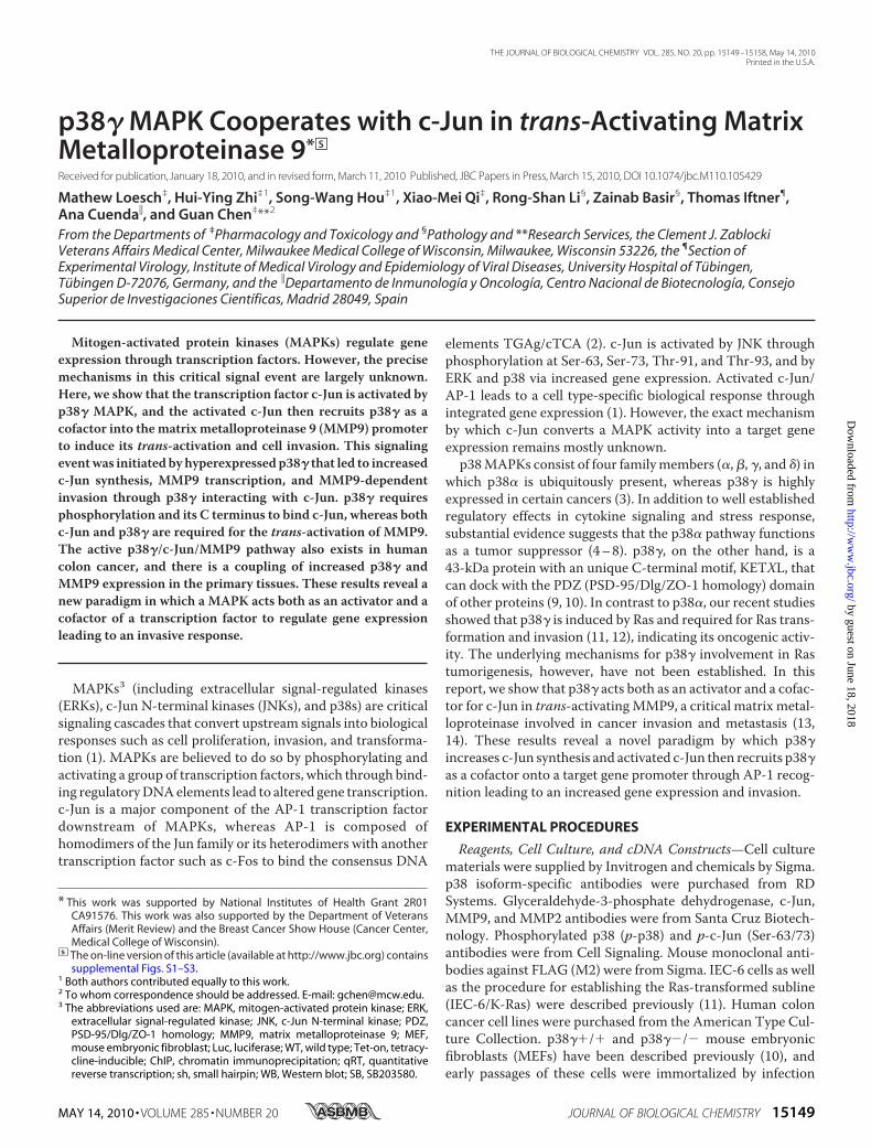
to an increased invasion. Becausenormal cells express little p38� (11),a tetracycline-inducible (Tet-on)system was used to express WTp38� and its nonphosphorablemutant p38�/AGF (by changing thedual phosphorylation motif TGY toAGF) in rat intestinal epithelialIEC-6 cells. Following an overnightincubation with and without Tet,cells were seeded in Matrigel-coated invasion chambers and ana-lyzed for invasion (12). Results inFig. 1, A and B, show that Tet-in-duced p38� stimulates invasionwhereas its AGF mutant is withouteffect, which couples with its activ-ity to increase c-Jun but not c-Fosprotein expression. The invasion-stimulatory activity of p38� is fur-ther confirmed by an increasedmigration inwound assays (Fig. 1C).These results indicate that p38�stimulates cell invasion and/ormigration by phosphorylation-de-pendent mechanisms.Recent studies indicate that the
C-terminal PDZ motif is requiredfor the invasive activity of c-Src (26)
and TAZ (tafazzin) proteins (27), and we then exploredwhether the C-terminal PDZ sequence of p38� plays a similarrole. To remove the PDZ motif (-ETPL), FLAG-tagged C-ter-minal truncated p38��4 and p38��13 that lack the last fourand 13 amino acids, respectively, were generated by PCR andstably expressed in IEC-6 cells by including FLAG-p38� andFLAG-p38�/AGF for comparison. To explore their potentialroles in Ras tumorigenesis, resistant clones were pooled andinfected with retrovirus expressing K-Ras and their invasiveactivity then compared. Consistent with the results in Tet-oncells, stable expression of p38� significantly increases invasionover the vector control, whereas its AGFmutant has much lesseffect (Fig. 1, D and E). Interestingly, the invasive activity alsowas decreased in cells expressing both p38��4 and p38��13(Fig. 1, D and E). These results together indicate that p38�requires both phosphorylation and its C terminus to stimulateinvasion.p38� Increases MMP9 Transcription by AP-1-dependent
Mechanisms—MMPs consist of at least 23 familymembers andhave long been associated with cancer invasion and metastasesbecause of their role in breaking down the extracellular matrix(28). Among these family proteins, MMP9 is one of the bestcharacterized AP-1 target genes involved in cancer invasion(13). Because p38 MAPKs were previously shown to regulatethe AP-1 target gene expression (29, 30), an AP-1 reporter (4)and a 670-bp human MMP9 promoter (18) were transientlyexpressed in IEC-6 cells to assess whether p38� increases theirluciferase activity compared with p38�. Results in Fig. 2, A andB, show that both p38� and p38� increase AP-1, but only p38�
FIGURE 1. p38� requires both phosphorylation and the C terminus to stimulate invasion. A–C, p38� butnot its AGF mutant, stimulates invasion and/or migration in IEC-6 cells. Cells were incubated with and withoutTet for 24 h and collected for protein expression by Western blotting (A) (p-p38 antibody reacts both withp-p38� and p-38�) and assessed for Matrigel invasion (B) and migration (C). Results of B and C are shown asrelative over those without Tet from at least three separate experiments (*, p � 0.05; error bars, S.D.). D andE, p38� increases invasion in IEC-6/K-Ras cells. Cells were transfected and selected, followed by retroviralinfection and a second antibiotic selection, and were then analyzed by Western blotting (D) and Matrigelinvasion (E) (*, p � 0.05 versus vector control; ** p � 0.05 versus p38�). GADPH, glyceraldehyde-3-phosphatedehydrogenase.
p38� Stimulates c-Jun and MMP9 Transcription
MAY 14, 2010 • VOLUME 285 • NUMBER 20 JOURNAL OF BIOLOGICAL CHEMISTRY 15151
stimulates the MMP9 promoter activity. These results are dif-ferent from those previously published (31), most likely as aresult of the different cell line used. To determinewhether p38�stimulates MMP9 via AP-1, Tet-on cells were expressed with aWT or AP-1 or NF-�B site-mutated MMP9 promoter con-struct (18), and luciferase activity was determined. Fig. 2Cresults show that Tet-p38� has a similar MMP9 stimulatoryactivity toward the WT and NF-�B mutated promoter, which,however, was abolished by the mutations on two AP-1 sites,indicating a required role of the AP-1 in p38� trans-activatingMMP9.To demonstrate theMMP9 stimulatory effects further, RNA
was prepared and analyzed by real-time qRT-PCR for MMP9
mRNA expression compared withMMP2. Results in Fig. 2D demon-strate that p38� significantly in-creases MMP9 but not MMP2expression; this is likely as a result ofthe lack of AP-1 site in the MMP2promoter. Importantly, analyses ofmedium collected from culturedTet-on p38� cells revealed thatthere is an increased secretedMMP9 protein expression by WBand an elevated MMP9 gelatinactivity by zymography (Fig. 2E),indicating the transcribed proteinbeing functionally active. Consis-tent with these results, analysis ofIEC-6/K-Ras cells stably transfectedwith p38�s also show that p38�increases MMP9 RNA expressionwhereas all of its mutants havemuch less effects (Fig. 2F), whichmore or less correlates with theirregulatory effects on AP-1 and/or MMP9 transcriptional activity(supplemental Fig. S1, A and B,respectively). These results togetherindicate that p38� requires bothphosphorylation and the C termi-nus to stimulate AP-1-dependentMMP9 transcription.p38� Binds the MMP9 Promoter
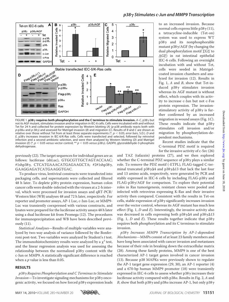
through a Complex Formation withc-Jun—Both human (18) and rat(32) MMP9 promoters contain twoAP-1 sites, and ChIP assays werenext performed to explore whetherp38� binds the endogenous MMP9promoter around this region usingprimers that span the functionaldistal AP-1 site (see Fig. 6C). Fol-lowing formaldehyde-induced DNAcross-linking with associated pro-teins, stably transfected p38� inIEC-6/K-Ras cells was isolated witha FLAG antibody, and the precipi-
tates were subjected to PCR analysis, as described previously(16). As a control, a set of plates were processed for FLAGimmunoprecipitation and WB analyses to explore whetherp38�may be recruited into theMMP9 promoter through inter-action with c-Jun and/or c-Fos proteins. Results in Fig. 3A (top)show that among these sublines, only precipitates from WTp38�-expressed cells contain theMMP9 promoter. Of interest,the immunoprecipitation/WB analyses (Fig. 3A, bottom)revealed that p38�, but not its mutants, increases c-Jun proteinexpression that couples with its c-Jun binding activity, whereasc-Fos remains undetectable. Because the MMP9 promoterbinding couples with the p38� activity to increase c-Jun expres-sion and bind c-Jun protein, these results suggest a scenario in
FIGURE 2. p38� stimulates MMP9 via AP-1. A–C, p38� increases MMP9 transcription. Cells were transientlyexpressed with the indicated plasmids, and luciferase activities were determined 48 h later (*, p � 0.05 versusvector or no Tet control where Tet was present for total 72 h in the Tet group, 24 h before the transfection, and48 h thereafter). D and E, p38� activates MMP9. Cells were incubated with and without Tet for 48 h, RNAs wereprepared for qRT-PCR, and levels of MMP9 and MMP2 RNAs were normalized to the �-actin and expressed asrelative over no Tet control (D, *, p � 0.05 versus no Tet). To assess the MMP9 activity, Tet-on cells were changedto a serum-free medium for the last 24 h, and concentrated medium was assayed for MMP9 activity by zymog-raphy and MMP9 protein expression by WB (E, top) in which cell lysates were also analyzed for protein expres-sion (E, bottom). F, stable transfected p38� increases MMP9 RNA expression. RNA was prepared and subjectedto qRT-PCR, and the ratio of MMP9/�-actin was expressed as a fold change over the vector alone (*, p � 0.05versus vector; **, p � 0.05 versus p38�).
p38� Stimulates c-Jun and MMP9 Transcription
15152 JOURNAL OF BIOLOGICAL CHEMISTRY VOLUME 285 • NUMBER 20 • MAY 14, 2010
which p38� first activates c-Jun by increasing its expression,and activated c-Jun then recruits p38� onto the MMP9 pro-moter via a complex formation.To demonstrate the c-Jun-mediated p38�-MMP9 promoter
binding further, Tet-on IEC-6 cells were next analyzed by ChIPand WB in which endogenous c-Jun was also purified andincluded as a positive control. Results in Fig. 3B show that inresponse to Tet addition, both p38� and c-Jun precipitates con-tain the MMP9 promoter by ChIP and its partner by WB, andthere is an increased c-Jun protein expression, further indicat-ing that p38� occupies the MMP9 promoter through interact-ing with c-Jun. Comparative analyses of c-Jun precipitates byChIP and WB reveal another interesting phenomenon.
Although c-Jun precipitates containmore c-Jun proteins in the absenceof Tet, c-Jun alone, in the absence ofTet-induced p38� expression, failsto bind the MMP9 promoter (Fig.3B, top, lanes 6 and 7 versus bottom,lanes 1 and 2). In response to Tet,however, c-Jun binds both the p38�protein and the MMP9 promoter,indicating a required role of p38� inc-Jun binding to the MMP9 pro-moter. Moreover, the interdepen-dent role of p38� and c-Jun in theMMP9 promoter binding was fur-ther demonstrated by the ChIP-re-ChIP assay, and Tet-p38� only acti-vates MMP9 without increasinganother AP-1 target gene vitamin Dreceptor expression (29) and with-out stimulating c-Jun and ATF-2phosphorylation (Figs. 2 and 3,B–D). Our previous studies havedemonstrated an increased p38�protein expression by a p38�/p38�inhibitor SB203580 (SB) in K-Ras-transformed IEC-6 cells as a resultof an antagonistic activity of p38�against p38� (33).We explored nextwhether endogenous p38� bindsendogenous c-Jun and the MMP9promoter in response to the SBtreatment. Results in Fig. 3E showthat there is a complex formationbetween p38� and c-Jun, which isincreased by SB treatment. Impor-tantly, both endogenous c-Jun andp38� bind to the MMP9 promoter,and the c-Jun binding activity isincreased by the SB-induced p38�/c-Jun up-regulation, further sup-porting the role of p38� in c-Junexpression and its MMP9 bindingactivity. The SB treatment, how-ever, failed to increase the p38�-MMP9 binding, which may result
from a general inhibitory activity of SB on MMP9 expression/activity (34, 35) and its effects on multiple AP-1 family mem-bers (36) and/or Raf pathways (37). These results together indi-cate a two-stagemechanism by whichMMP9 is trans-activatedby p38� MAPK; p38� first activates c-Jun by increasing itsexpression, and activated c-Jun then recruits p38� into theMMP9 promoter through a complex formation.p38� MAPK Both Stimulates de Novo c-Jun Synthesis and
Acts as a Cofactor for c-Jun-trans-Activating MMP9—Al-though p38 MAPKs are known to stimulate c-Jun promoteractivity (30), so far there have been no reports about the p38-induced increase in c-Jun protein expression. c-Jun promotercontains AP-1 and myocyte-enhancing factor 2 sites that are
FIGURE 3. p38� is recruited into the MMP9 promoter through interaction with c-Jun. A, p38� depends onits phosphorylation and C terminus to bind c-Jun protein and MMP9 promoter. Stably transfected p38�s fromIEC-6/K-Ras cells were isolated and precipitates subjected to PCR analyses for their activity in binding MMP9promoter (top) with a set of cells in parallel analyzed by immunoprecipitation (IP)/Western blotting for p38�interacting with c-Jun proteins (bottom). B and C, p38� and c-Jun bind the MMP9 promoter through a complexformation. Tet-on p38� IEC-6 cells were incubated with and without Tet and p38�/c-Jun proteins isolated bytheir specific antibodies, and precipitates subjected to PCR (B, top), Western blotting (B, bottom) or a secondIP/PCR for ChIP-re-ChIP (C). D, p38� does not induce c-Jun/ATF2 phosphorylation or vitamin D receptor (VDR)expression in IEC-6 cells. Cells were incubated with and without a p38 inhibitor SB203580 (SB) for 24 h and thenpulse-treated with a p38 activator arsenite (ARS) for 2 h, and analyzed by Western blotting (seesupplemental Fig. S2A for the relationship between c-Jun expression and c-Jun phosphorylation in response toTet addition). E, endogenous p38� forms a complex with c-Jun on the MMP9 promoter in K-Ras-transformedIEC-6 cells. Cells were treated with SB or solvent control (Co) as previously described in these cells (33), and p38�and c-Jun proteins were isolated and assessed for their MMP9 promoter binding activity by ChIP and theircomplex formation by Western blotting.
p38� Stimulates c-Jun and MMP9 Transcription
MAY 14, 2010 • VOLUME 285 • NUMBER 20 JOURNAL OF BIOLOGICAL CHEMISTRY 15153
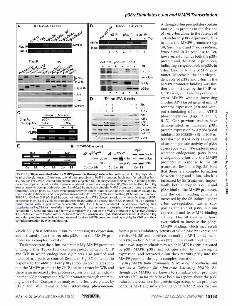
involved in its positive autoregulation by c-Jun/AP-1 (38)and/or its stimulation by p38 MAPKs (30). Experiments werethen performed to examine whether p38� up-regulates c-Juntranscription. In this regard, a c-Jun luciferase promoter (39)was transiently cotransfected with the indicated constructs inIEC-6 cells, and the luciferase activity was assessed. Results inFig. 4A show that p38�, but not its mutants or p38�, increasesc-Jun promoter activity. Moreover, analyses of stable trans-fected IEC-6/K-Ras cells further showed that only WT p38�increases c-Jun RNA expression (Fig. 4B). These results,together with the increased c-Jun protein expression (Fig. 3A),indicate that p38� stimulates de novo c-Jun synthesis by phos-
phorylation and C terminus-depen-dent mechanisms as observed forthe AP-1/MMP9 trans-activation(supplemental Fig. S1). To examinewhether p38� may additionally sta-bilize c-Jun proteins, Tet-p38� cellswere treated with a protein synthe-sis inhibitor cycloheximide and ana-lyzed for protein expression. Asillustrated in supplemental Fig. S2A,levels of expressed c-Jun proteinsare decreased similarly after cyclo-heximide with and without Tet andbecome undetectable at 9 h despitethe presence of p38� protein. Theseresults indicate that p38� activatesc-Jun primarily by stimulating itsexpression and not by increasing itsprotein stability and/or phosphory-lation (a slight increase in p-c-Jun at3 h is likely due to the stress insultfrom cycloheximide). The criticalrole of p38� in c-Jun and MMP9expression was further demon-strated in p38��/� and p38��/�MEFs (10) in which p38� knockoutdecreases c-Jun and MMP9 expres-sion that was restored by reexpress-ing WT p38� but not its mutants(Fig. 4, C–E). These results indicatea required role of p38� in c-Jun syn-thesis that may be critical for itsMMP9 trans-activating activity.c-Jun has a well established AP-1
binding activity through dimeriza-tion with itself or another transcrip-tion factor, but no studies havereported thus far that a MAPK canbind the AP-1 element to regulategene expression. Because c-Jundepends on p38� to bind theMMP9promoter (Fig. 3B), p38� may act asa cofactor for c-Jun in binding theAP-1 element in the regulation ofgene transcription. If this is the case,c-Jun activation alone, in the
absence of p38�, should not be active in transcription. To testthis possibility, c-Junwas transiently transfected into p38��/�and p38��/� MEFs together with the AP-1 reporter or theMMP9 promoter, and the luciferase activity was analyzed.Results in supplemental Fig. S2, B and C, show that c-Jun onlystimulates AP-1/MMP9 transcription in p38��/�, but not inp38��/� cells. Conversely, p38� transfection only increasesthe AP-1/MMP9 in c-Jun�/� but not in its knock-out coun-terparts (15) despite similar levels of endogenous p38� proteinexpression in both lines (33) (supplemental Fig. S2, D and E).These results together indicate that p38� and c-Jun are bothrequired for AP-1/MMP9 transcription, which thereby further
FIGURE 4. p38� stimulates c-Jun expression through a complex formation with c-Jun and plays an impor-tant role in c-Jun/MMP9 expression. A and B, p38� increases c-Jun promoter activity and RNA expression byphosphorylation and C terminus-dependent mechanisms. IEC-6 cells were transiently transfected with indi-cated constructs, and luciferase activity was analyzed 48 h later (A). To measure c-Jun RNA expression, p38�stably expressed IEC-6/K-Ras cells were subjected to qRT-PCR analysis, and the obtained signals were normal-ized to �-actin (B) (*, p � 0.05 for p38� versus vector for both A and B; error bars, S.D.). C–E, p38� plays a role inendogenous c-Jun and MMP9 expression. Immortalized p38��/� and p38��/� cells were analyzed for pro-tein expression by Western blotting and for c-Jun/MMP9 RNA expression by qRT-PCR (C). To reexpress p38�and its mutants, p38��/� cells were infected with retroviruses and selected with antibiotics, and resistantcells were then subjected to Western blotting (D) and qRT-PCR analyses (E) (*, p � 0.05 versus respective controlfor C and E).
p38� Stimulates c-Jun and MMP9 Transcription
15154 JOURNAL OF BIOLOGICAL CHEMISTRY VOLUME 285 • NUMBER 20 • MAY 14, 2010
reinforces our conclusion that p38� is an essential cofactor ofc-Jun in stimulating MMP9 expression.p38� Controls Endogenous c-Jun and MMP9 Expression in
Human Colon Cancer and Stimulates MMP9-dependentInvasion—To assess whether MMP9 activity is required forp38�-induced invasion, Tet-on cells were incubated with andwithout MMP9 inhibitor ilomastat and SB-3CT, and theireffects on invasion as well as MMP9 activity were assessed.Ilomastat suppresses both MMP2 and MMP9 activity (40),whereas SB-2CT is a specific MMP9 inhibitor (41). Results inFig. 5, A and B, show that a 24-h incubation of p38�-expressedIEC-6 cells with both inhibitors significantly reduces theMMP9 activity and almost completely abolishes p38�-inducedinvasion. SB-3CT (41) and ilomastat (25) were previouslyshown to decrease theMMP9 activity, which was speculated tooccur via a positive feedback regulation that linksMMP9 activ-ity to its transcription (41). Although these compounds mayaffect additional targets, a coupling of decreasedMMP9 activitywith reduced invasion by both inhibitors strongly suggests arole of MMP9 in p38� invasive activity.
To demonstrate whether endogenous p38�/c-Jun/MMP9pathways intrinsically exist in human colon cancer, a group ofcolon cancer cell lineswith knownRas statuswere examined fortheir protein expression. Results in Fig. 5C show that p38� pro-tein levels were increased in all three cell lines harboring Rasactivations/mutations (HCT116, SW480, and LH147T) com-pared with those without (Caco-2, HT-29, and T48), indicatingthat activated Ras positively controls endogenous p38� proteinexpression. Of great interest, p38� protein expression levelswere also significantly correlated with both c-Jun protein andMMP9 RNA expression in these cells (Fig. 5, C and D, andsupplemental Fig. S3), suggesting an intrinsic p38�/c-Jun/MMP9 pathway in human colon cancer. To demonstratewhether this pathway is functionally active, p38� proteins weredepleted by shRNA in HCT116 and SW480 cells, and its effectson c-Jun/MMP-9 expression as well as invasion were analyzed.Results in Fig. 5, E–H, show that silencing p38� by two separateshRNAs reduces c-Jun andMMP9expression/activity that cou-ples with a decreased invasion in both lines in which an inva-sion-suppressive effect was also achieved by the pharmacolog-
FIGURE 5. The p38�/c-Jun/MMP9 pathway is functionally active in stimulating invasion. A and B, inhibition of MMP9 activity blocks p38�-inducedinvasion. Tet-p38� expressed cells were incubated with MMP9 inhibitors for 24 h and subjected to Matrigel invasion (A) and zymography (B) (A, *, p � 0.05 versusp38�; **, p � 0.05 versus no Tet). Error bars, S.D. C and D, increased p38� protein expression couples with elevated c-Jun/MMP9 expression in human coloncancer cells. Human colon cancer cells were analyzed by Western blotting for p38�/c-Jun protein expression and by qRT-PCR for MMP9 RNA expression.Normalized MMP9 RNA levels are significantly higher in Ras-activated cells (**) than those without (*). E–H, p38� controls endogenous c-Jun/MMP9 expression,and both p38� and MMP9 are required for human colon cancer invasion. Human colon cancer cells were infected with lentiviral shLuc or shp38� and subjectedto Western blotting/zymography (E) and qRT-PCR (F) at 72 h and Matrigel invasion at 48 h (G). Effects of MMP9 inhibitors on invasion (H) were analyzed as in A(*, p � 0.05 versus shLuc or solvent control, F–H).
p38� Stimulates c-Jun and MMP9 Transcription
MAY 14, 2010 • VOLUME 285 • NUMBER 20 JOURNAL OF BIOLOGICAL CHEMISTRY 15155
icalMMP9 inhibition. These results together reveal an intrinsicp38�/c-Jun/MMP9 pathway that is functionally active in stim-ulating colon cancer invasion.
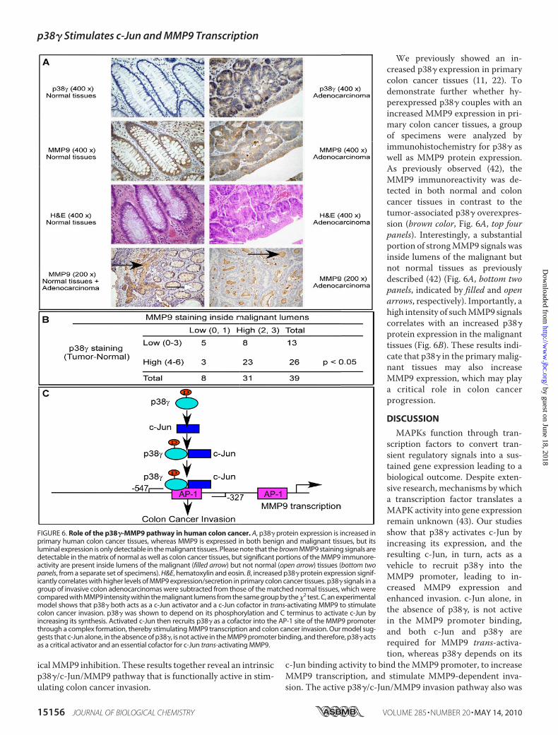
We previously showed an in-creased p38� expression in primarycolon cancer tissues (11, 22). Todemonstrate further whether hy-perexpressed p38� couples with anincreased MMP9 expression in pri-mary colon cancer tissues, a groupof specimens were analyzed byimmunohistochemistry for p38� aswell as MMP9 protein expression.As previously observed (42), theMMP9 immunoreactivity was de-tected in both normal and coloncancer tissues in contrast to thetumor-associated p38� overexpres-sion (brown color, Fig. 6A, top fourpanels). Interestingly, a substantialportion of strongMMP9 signals wasinside lumens of the malignant butnot normal tissues as previouslydescribed (42) (Fig. 6A, bottom twopanels, indicated by filled and openarrows, respectively). Importantly, ahigh intensity of suchMMP9 signalscorrelates with an increased p38�protein expression in the malignanttissues (Fig. 6B). These results indi-cate that p38� in the primarymalig-nant tissues may also increaseMMP9 expression, which may playa critical role in colon cancerprogression.
DISCUSSION
MAPKs function through tran-scription factors to convert tran-sient regulatory signals into a sus-tained gene expression leading to abiological outcome. Despite exten-sive research, mechanisms by whicha transcription factor translates aMAPK activity into gene expressionremain unknown (43). Our studiesshow that p38� activates c-Jun byincreasing its expression, and theresulting c-Jun, in turn, acts as avehicle to recruit p38� into theMMP9 promoter, leading to in-creased MMP9 expression andenhanced invasion. c-Jun alone, inthe absence of p38�, is not activein the MMP9 promoter binding,and both c-Jun and p38� arerequired for MMP9 trans-activa-tion, whereas p38� depends on its
c-Jun binding activity to bind theMMP9 promoter, to increaseMMP9 transcription, and stimulate MMP9-dependent inva-sion. The active p38�/c-Jun/MMP9 invasion pathway also was
FIGURE 6. Role of the p38�-MMP9 pathway in human colon cancer. A, p38� protein expression is increased inprimary human colon cancer tissues, whereas MMP9 is expressed in both benign and malignant tissues, but itsluminal expression is only detectable in the malignant tissues. Please note that the brown MMP9 staining signals aredetectable in the matrix of normal as well as colon cancer tissues, but significant portions of the MMP9 immunore-activity are present inside lumens of the malignant (filled arrow) but not normal (open arrow) tissues (bottom twopanels, from a separate set of specimens). H&E, hematoxylin and eosin. B, increased p38� protein expression signif-icantly correlates with higher levels of MMP9 expression/secretion in primary colon cancer tissues. p38� signals in agroup of invasive colon adenocarcinomas were subtracted from those of the matched normal tissues, which werecompared with MMP9 intensity within the malignant lumens from the same group by the�2 test. C, an experimentalmodel shows that p38� both acts as a c-Jun activator and a c-Jun cofactor in trans-activating MMP9 to stimulatecolon cancer invasion. p38� was shown to depend on its phosphorylation and C terminus to activate c-Jun byincreasing its synthesis. Activated c-Jun then recruits p38� as a cofactor into the AP-1 site of the MMP9 promoterthrough a complex formation, thereby stimulating MMP9 transcription and colon cancer invasion. Our model sug-gests that c-Jun alone, in the absence of p38�, is not active in the MMP9 promoter binding, and therefore, p38� actsas a critical activator and an essential cofactor for c-Jun trans-activating MMP9.
p38� Stimulates c-Jun and MMP9 Transcription
15156 JOURNAL OF BIOLOGICAL CHEMISTRY VOLUME 285 • NUMBER 20 • MAY 14, 2010
demonstrated in Ras-activated human colon cancer, whereashyperexpressed p38� was shown to couple with an increasedluminal MMP9 expression in primary cancer tissues. Theseresults together reveal a novel mechanism by which p38�MAPK acts both as a c-Jun activator and a c-Jun cofactor in thestimulation of MMP9 transcription leading to increased inva-sion (Fig. 6C). Because Ras-activated colon cancers are moremetastatic (44) and MMP9 is a target antimetastatic therapy(14), p38� may promote colon cancer progression throughtransduction of Ras signaling to MMP9 via the c-Jun-mediatedpromoter binding.Previous studies showed that Hog1 in yeast (corresponding
to mammalian p38�) is recruited to both promoter and codingregions of osmotic stress genes in stress response (45, 46). Inmammalian cells, activated p38� similarly was shown to inter-act with chromatin during cell differentiation (47). In all ofthese studies, however, mechanisms for p38�-DNAbindings aswell as biological consequences remain unknown. Here, weshow that the p38�-MMP9 promoter binding first requiresp38�-induced c-Jun expression and then its interaction withc-Jun protein. The signaling specificity of this regulation is sug-gested by the fact that MMP9 is stimulated by p38� but not byp38�, which is mediated by c-Jun (but not c-Fos or ATF2) viathe AP-1 (but not NF-�B) site without significant effects onMMP2 or another AP-1 target gene vitamin D receptor expres-sion. Although MMP9 may be one of the most documentedAP-1 targets in cancer invasion andmetastasis, it remains to bedetermined whether additional AP-1 target genes are involvedin the p38� invasive phenotype through the promoter binding.
It is important that p38� was shown to activate c-Jun byincreasing its expression, as up-regulated c-Jun protein expres-sion has been observed in primary human colon cancer tissues(48) where it may be involved in colon cancer invasion (49).Because c-Jun is positively autoregulated by c-Jun/AP-1 (38)and p38� increases c-Jun/AP-1/MMP9 transcription throughits c-Jun binding activity, the same c-Jun-mediated promoterbinding may likely operate in p38� stimulating c-Jun as well asMMP9 transcription. With regard to the c-Jun-p38� binding,there may be two mechanisms involved. c-Jun lacks a PDZdomain that is required for a direct interaction with a PDZmotif-containing protein such as p38�. Therefore, the require-ment of p38� C terminus for its c-Jun binding suggests thatboth proteins may interact indirectly inside cells through addi-tional PDZ proteins. On the other hand, phosphorylated p38�is known to be localized predominantly in the nucleus (33),which may be critical for its interaction with nuclear c-Jun as acofactor. Although these different scenarios require furtherstudy, the required role of the c-Jun binding activity in p38�stimulating c-Jun synthesis, MMP9 transcription, and invasionhighlights an essential role of this protein-complex in triggeringthe invasion cascade. c-Jun was previously shown to bind theMMP9 promoter in cardiac (50) but not in neuronal cells (51),and similarly we showed that c-Jun only binds theMMP9 in thepresence of p38�, indicating a determinant role of c-Jun cofac-tor abundances in its trans-activation of MMP9. Our resultssuggest that p38� may be one of these critical cofactors todetermine c-Jun transcriptional activity by increasing itsexpression and facilitating its target gene promoter binding.
Acknowledgments—We thank Drs. Jiahuai Han, Michael Dwinell,Ron Wisdom, and Craig Hauser for providing critical reagents, andGuan Chen’s laboratory members for useful discussion.
REFERENCES1. Chang, L., and Karin, M. (2001) Nature 410, 37–402. Shaulian, E., and Karin, M. (2002) Nat. Cell Biol. 5, E131–E1363. Loesch, M., and Chen, G. (2008) Front. Biosci. 13, 3581–35934. Chen, G., Hitomi, M., Han, J., and Stacey, D.W. (2000) J. Biol. Chem. 275,
38973–389805. Brancho, D., Tanaka, N., Jaeschke, A., Ventura, J. J., Kelkar, N., Tanaka, Y.,
Kyuuma, M., Takeshita, T., Flavell, R., and Davis, R. J. (2003) Genes Dev.17, 1969–1978
6. Qi, X., Tang, J., Pramanik, R., Schultz, R. M., Shirasawa, S., Sasazuki, T.,Han, J., and Chen, G. (2004) J. Biol. Chem. 279, 22138–22144
7. Dolado, I., Swat, A., Ajenjo, N., De Vita, G., Cuadrado, A., and Nebreda,A. R. (2007) Cancer Cell 11, 191–205
8. Sun, P., Yoshizuka, N., New, L., Moser, B. A., Li, Y., Liao, R., Xie, C., Chen,J., Deng, Q., Yamout, M., Dong, M.-Q., Frangou, C. G., Yates, J. R., III,Wright, P. E., and Han, J. (2007) Cell 128, 295–308
9. Hasegawa,M., Cuenda, A., Spillantini,M.G., Thomas, G.M., Buee-Scher-rer, V., Cohen, P., and Goedert, M. (1999) J. Biol. Chem. 274,12626–12631
10. Sabio, G., Arthur, J. S., Kuma, Y., Peggie, M., Carr, J., Murray-Tait, V.,Centeno, F., Goedert,M.,Morrice, N., and Cuenda, A. (2005) EMBO J. 24,1134–1145
11. Tang, J., Qi, X., Mercola, D., Han, J., and Chen, G. (2005) J. Biol. Chem.280, 23910–23917
13. Ozanne, B. W., Spence, H. J., McGarry, L. C., and Hennigan, R. F. (2007)Oncogene 26, 1–10
14. Lubbe,W., Zhou, Z. Y., Fu,W., Zuzga,D., Schulz, S., Fridman, R.,Muschel,R., Waldman, S. A., and Pitari, G. M. (2006) Clin. Cancer Res. 12,1876–1882
15. Johnson, R., Spiegelman, B., Hanahan, D., and Wisdom, R. (1996) Mol.Cell. Biol. 16, 4504–4511
17. Galang, C. K., Der, C. J., and Hauser, C. A. (1994)Oncogene 9, 2913–292118. Behren, A., Simon, C., Schwab, R.M., Loetzsch, E., Brodbeck, S., Huber, E.,
Stubenrauch, F., Zenner, H. P., and Iftner, T. (2005) Cancer Res. 65,11613–11621
19. Han, J., Jiang, Y., Li, Z., Kravchenko, V. V., andUlevitch, R. J. (1997)Nature386, 296–299
20. Li, Z., Jiang, Y., Ulevitch, R. J., and Han, J. (1996) Biochem. Biophys. Res.Commun. 228, 334–340
21. Pramanik, R., Qi, X., Borowicz, S., Choubey, D., Schultz, R.M., Han, J., andChen, G. (2003) J. Biol. Chem. 278, 4831–4839
22. Hou, S.W., Zhi, H., Pohl, N., Loesch,M., Qi, X., Li, R., Basir, Z., and Chen,G. (2010) Cancer Res. 70, 2901–2910
23. Zhi, H., Yang, X. J., Kuhnmuench, J., Berg, T., Thill, R., Yang, H., See,W. A., Becker, C. G., Williams, C. L., and Li, R. (2009) J. Pathol. 217,389–397
Mol. Cell. Biol. 19, 4289–430131. Simon, C., Simon, M., Vucelic, G., Hicks, M. J., Plinkert, P. K., Koitschev,
A., and Zenner, H. P. (2001) Exp. Cell Res. 271, 344–35532. Eberhardt, W., Schulze, M., Engels, C., Klasmeier, E., and Pfeilschifter, J.
(2002)Mol. Endocrinol. 16, 1752–176633. Qi, X., Pohl, N. M., Loesch, M., Hou, S., Li, R., Qin, J. Z., Cuenda, A., and
Chen, G. (2007) J. Biol. Chem. 282, 31398–3140834. Simon, C., Goepfert, H., and Boyd, D. (1998) Cancer Res. 58, 1135–113935. Underwood, D. C., Osborn, R. R., Bochnowicz, S., Webb, E. F., Rieman,
D. J., Lee, J. C., Romanic, A. M., Adams, J. L., Hay, D. W., and Griswold,D. E. (2000) Am. J. Physiol. Lung Cell Mol. Physiol. 279, L895–L902
36. Adiseshaiah, P., Li, J., Vaz, M., Kalvakolanu, D. V., and Reddy, S. P. (2008)Biochem. Biophy. Res. Commun. 371, 304–308
37. Hall-Jackson, C. A., Goedert, M., Hedge, P., and Cohen, P. (1999) Onco-gene 18, 2047–2054
38. Angel, P., Hattori, K., Smeal, T., and Karin, M. (1988) Cell 55, 875–88539. Han, T. H., and Prywes, R. (1995)Mol. Cell. Biol. 15, 2907–291540. Canning, M. T., Postovit, L.-M., Clarke, S. H., and Graham, C. H. (2001)
Exp. Cell Res. 267, 88–9441. Gu, Z., Cui, J., Brown, S., Fridman, R., Mobashery, S., Strongin, A. Y., and
Lipton, S. A. (2005) J. Neurosci. 25, 6401–6408
42. Illemann, M., Bird, N., Majeed, A., Sehested, M., Laerum, O. D., Lund,L. R., Danø, K., and Nielsen, B. S. (2006)Mol. Cancer Res. 4, 293–302
43. Whitmarsh, A. J. (2007) Biochim. Biophys. Acta 1773, 1285–129844. Oliveira, C., Velho, S., Moutinho, C., Ferreira, A., Preto, A., Domingo, E.,
Capelinha, A. F., Duval, A., Hamelin, R., Machado, J. C., Schwartz, S., Jr.,Carneiro, F., and Seruca, R. (2007) Oncogene 26, 158–163
45. de Nadal, E., Zapater, M., Alepuz, P. M., Sumoy, L., Mas, G., and Posas, F.(2004) Nature 427, 370–374
46. Pokholok, D. K., Zeitlinger, J., Hannett, N.M., Reynolds, D. B., and Young,R. A. (2006) Science 313, 533–536
47. Simone, C., Forcales, S. V., Hill, D. A., Imbalzano, A. N., Latella, L., andPuri, P. L. (2004) Nat. Genet. 36, 738–743
48. Magrisso, I. J., Richmond, R. E., Carter, J. H., Pross, C. B., Gilfillen, R. A.,and Carter, H. W. (1993) Lab. Invest. 69, 674–681
49. Allgayer, H., Wang, H., Shirasawa, S., Sasazuki, T., and Boyd, D. (1999)Br J. Cancer 80, 1884–1891
50. Goruppi, S., Patten, R. D., Force, T., and Kyriakis, J. M. (2007) Mol. Cell.Biol. 27, 993–1006
51. Rylski, M., Amborska, R., Zybura, K., Michaluk, P., Bielinska, B.,Konopacki, F. A., Wilczynski, G. M., and Kaczmarek, L. (2009)Mol. Cell.Neurosci. 40, 98–110
p38� Stimulates c-Jun and MMP9 Transcription
15158 JOURNAL OF BIOLOGICAL CHEMISTRY VOLUME 285 • NUMBER 20 • MAY 14, 2010