Page 1
Vol. 1, /463-1469, December /995 Clinical Cancer Research 1463
S The abbreviations used are: p53-Ab. autoantibodies to p53: PBS-T.
PBS with 0.05% Tween 20: SD. standard deviation.
Advances in Brief
p53 Antibodies in Patients with Various Types of Cancer: Assay,
Identification, and
Richard Lubin, Beata Schlichtholz,2
Jean Luc Teillaud, Edith Garay,3 Annette Bussel,
Christopher P. Wild, and Thierry Soussi4
Unite 301 Institut National de Ia Sante et de Ia Recherche M#{233}dicale.
Institut de G#{233}n#{233}tiqueMol#{233}culaire, 27 rue J. Dodu. 75010 Paris
IR. L.. B. S.. E. G., T. 5.1; Unite 255, Institut National de Ia Sante etde Ia Recherche M#{233}dicale, Institut Curie, 26 rue d’Ulm, 7523 1 Paris,
Cedex 05 IJ. L. TI: Polyclinique d’H#{233}matologie et d’Immunologie.H#{244}pitalSt. Louis. I avenue Claude Vellefaux. 75475 Paris IA. B.):
and IARC, 150 dOUf5 Albert Thomas, 69372 Lyon IC. WI, France
Abstract
Alteration of the p53 gene is the most frequent genetic
alteration in human cancer and leads to the accumulation of
mutant p53 in the nucleus of tumor cells. In addition, it hasbeen shown that patients with various types of neoplasia
have p53 antibodies in their sera which could be used as an
indirect diagnostic procedure for p53 alteration.
Using a new ELISA, we have analyzed the sera from
more than 1000 patients with various types of cancer and
from healthy blood donors. We demonstrate that p53 anti-bodies are detected mainly in cancer patients and are strictly
proportional to the occurrence ofp53 mutations. Using var-
ious immunological approaches, these antibodies were un-
ambiguously demonstrated to be directed toward the human
p53 protein. Isotyping analysis of these antibodies strongly
suggested that they correspond to a humoral response to the
p53 protein which accumulates in the tumor cell. This find-
ing suggests that serological analysis, combined with histo-
chemistry, is suitable for assessing the integrity of the p53
gene in cancer patients.
Introduction
Inactivation of the p53 tumor suppressor gene is the most
common genetic alteration in human cancers. and may occur
through point mutations or complex formation with cellular
proteins (I, 2). The recognition of the p53 alteration is an
important feature in clinical diagnosis. since it is an indepen-
Received 4/10/95: revised 7/19/95: accepted 7/28/95.
I This work was supported by grants from the Association de Recherche
sur Ic Cancer. Ligue Nationale contre Ic Cancer (comit#{233}de Paris), and
Ligue Nationale contre Ic Cancer. B. S. was supported by fellowships
froti� the French Minist#{232}rede Ia Recherche et de Ia Technologie and the
Soci#{233}t#{233}Francaise du Cancer. E. G. was supported by fellowships from
the CONACYT (Mexico) and Institut National de Ia Sante et de Ia
Recherche M#{233}dicale(France). R. L. was supported by a CIFRE contract.
2 Present address: University of Gdansk. Department of Biochemistry.Kladki 24. 80-822 Gdansk, Poland.
� Present address: UNAM, Instituto de Investigaciones Biomedicas, Dc-
partemento Biologica Molecular, Apto Postal 70228. (MS 10, Mexico, D.F.
4 To whom requests for reprints should he addressed. Phone: (33)1-42-
49-92-69: Fax: (33)1-42-06-95-31.
dent, unfavorable prognostic factor in breast. colon, and gastric
carcinomas (3-5). Furthermore, it has been shown that the
vulnerability of tumor cells to radiation or chemotherapy is
greatly reduced by mutations that abolish p53-dependent apop-
tosis (6). Analysis of p53 status by DNA sequencing is the most
accurate method described thus far. but it is not easy to use on
a routine clinical basis for a large number of samples. Fortu-
nately, it is known that most mutations induce an increase in the
p53 half-life. leading to an accumulation of p53 protein in the
nucleus of tumor cells. Immunohistochemical analyses have
been extensively used for the screening of the p53 alteration in
a wide variety of human cancers (7) for review. More recently.
a serological method has been proposed for diagnosis of the p53
alteration. p53-Abs5 have been detected in sera of patients with
various carcinomas (8-16). The presence of these antibodies
was shown to be dependent on the accumulation of p53 protein
in tumor cells (10, 12, 17), but some exceptions have been
reported ( I 5. 16). In breast carcinoma. the presence of p53-Abs
is correlated with poor prognostic factors such as high histolog-
ical grade and the absence of hormone receptors ( I 1 . I 8. 19). A
more recent study on 400 patients with breast carcinoma dem-
onstrated that the overall survival was worse in patients with
p53-Abs ( I 9). In lung cancer. p53-Abs can be detected several
years before clinical diagnosis of the tumor (20). Analyses of
this humoral response have demonstrated that p53-Ab recog-
nizes immunodominant regions localized in the amino and car-
boxy termini of the p53 protein. outside the mutational hot spot
(11, 21, 22).
In addition. serological analysis of p53 can be used as a
complementary procedure with molecular and immunohisto-
chemical methods, since it does not require tumor tissues and can
be easily used for h�llow-up of patients with p53 alterations (7).
In light of various reports. the frequency of these p53-Abs
for a given cancer remains a matter of debate. In breast carci-
nomas. which have been extensively studied, it niay range from
I (23) to 5% (13). 9% (8), 14% (1 1. 19), and 25.6�% (18). This
discrepancy can be partially explained by the various techniques
(ELISA, immunoprecipitation. or Western blot) used in these
studies. but it might also reflect some unsuspected bias in the
choice of patients (difference in clinical status, environmental.
or geographical factors). More surprising was the report by
Vojtesek et a!. (23) describing the near total absence of p53-Abs
( I : 1(X)) in sera of patients with breast carcinoma. These authors
raised the possibility that some of the previously described
p53-Abs could be directed toward an unknown Mr 53,000 pro-
tein unrelated to p53.
In the present report. we describe a novel ELISA for the
detection of p53-Abs in human sera. Using conventional immu-
Research. on August 7, 2020. © 1995 American Association for Cancerclincancerres.aacrjournals.org Downloaded from
Page 2
1464 p53 Antibodies in Patients with Various Types of Cancer
nological methods, we demonstrate that these antibodies are
directed against the p53 protein. We show that the prevalence of
p53-Ab is correlated with the prevalence ofp53 mutations in the
different cancer types. Isotyping of these antibodies has dem-
onstrated that most of them belong to the IgG subclass, rein-
forcing the notion that this humoral response is the result of an
active self-immunization process.
Materials and Methods
Sera and Antibodies. All sera were collected from var-
ious clinical laboratories in France between 1992 and 1994.
They were obtained after diagnosis, but prior to any treatment.
Sera were stored at -70#{176}C until use. Sheep antihuman IgG
peroxidase-conjugated antibodies (‘y chain specific and affinity
isolated; Silenius GAH) were used for the detection of human
p53 antibodies in the ELISA. To evaluate the subclasses of
immunoglobulin specific for the p53 protein, the following
mouse monoclonal antibodies were used: HP6001 (IgG1 sub-
class), HP6002 (IgG2 subclass), HP6050 (IgG3 subclass),
HP6024 (IgG4 subclass), DA4.4 (1gM subclass), and 2D7
(IgA1 ±2 subclass; Ref. 24). The specificity of each monoclonal
antibody was assessed using the pure immunoglobulin subclass.
Mouse monoclonal antibodies were detected using a goat anti-
mouse IgG (‘y chain specific and human adsorbed; Caltag Lab-
oratories, M30l07). HR231 , a mouse monoclonal antibody
which recognizes human p53, has been previously described
(25).
Immunoblotting and Immunoprecipitation For im-
munoblotting, we used wild-type human p53 protein expressed
in insect cells; 48 h after infection by the recombinant baculo-
virus, cells were lysed using RIPA [150 msi NaC1, 10 msi
Tris-HC1 (pH 8.00), 1% sodium deoxycholate, 0.1% sodium
lauryl sulfate, 1 mM EDTA] + SDS buffer for 30 mm at 4#{176}C.
The extract was centrifuged for 30 mm at 100,000 X g, and the
supernatant containing soluble p53 was stored at -80#{176}Cuntil
use. p53 protein corresponded to 20-40% of the total protein
content in our conditions. For the ELISA, a control extract
prepared from cells infected either with wild-type virus or with
a recombinant virus encoding a protein unrelated to p53 was
used. For competition experiments, human p53 protein was
purified by immunoaffinity using monoclonal antibody HR231.
As judged by staining and immunoblotting, the p53 protein was
more than 95% pure. Immunoblotting of human sera has been
previously described (I 1). For immunoprecipitation, full-length
wild-type p53 was obtained by in vitro transcription/translation.
For each immunoprecipitation, 10,000 cpm of labeled protein
were used as described by Soussi et a!. (26).
ELISA. Polystyrene flat-bottomed microtiter plates (Im-mulon B; Dynatech Laboratories) were coated with 50 p.1 p53
extract (corresponding to 40-80 ng p53 protein) in PBS buffer.
The same cell extract was used for all of the experiments
described in this report. Plates were dried for 48 h at 37#{176}Cand
then sealed in a polypropylene bag and stored at 4#{176}Cuntil use.
Such plates give reproducible results over a period of 6 months.
Before use, plates were washed five times with PBS-T; 100 p.1
blocking buffer (PBS-0.2% Tween 20, and 5.0% dried nonfat
milk) were added per well. After 1 h at 37#{176}C,the wells were
washed as described above. Then 50 p.) sera (diluted 1 :50 and
1:100 in PBS. 5.0% dried non-fat milk) were tested in duplicate.
The plates were incubated for I h at room temperature on an
ELISA plate shaker. After five washings in PBS-T, 100 p.l of an
antihuman IgG peroxidase conjugate diluted 1 :2500 in PBS-5%
nonfat milk were added and incubated for I h at 37#{176}C.Prelim-
mary experiments were performed with an antihuman immuno-
globulin antibody. but since we had demonstrated that all of
these p53-Abs include IgG. we used an antihuman IgG anti-
body. Plates were then washed five times and developed using
2,2’-azino-di(3-ethylbenzthiazoline) sulfonate substrate in ci-
trate buffer (Boehringer, catalogue no. 1204521 ). Absorbance
was read at 405 nm after 30 mm using an MR 5000 ELISA
reader (Dynatech Laboratories). All of these manipulations were
performed simultaneously on a plate coated with either p53
protein as described above or with the control extract. Each
serum was tested by duplicate replication at two different dilu-
tions (1 :50 and 1: 100). SDs were calculated using the four wells.
For the competition experiment, the ELISA procedure was
similar with the following exception: prior to ELISA, the sera
(diluted 1:100 in PBS-5% nonfat milk) were incubated with
various amounts of either purified p53 or ovalbumin (as a
negative control). Incubation was performed in 96-well plates
that had been washed with PBS-T prior to incubation. Plates
were incubated for 60 mm at room temperature on an ELISA
plate shaker. Sera were then tested as described above. Compe-
tition experiments were also performed simultaneously on a
plate coated either with p53 protein or with the control extract.
For isotyping the p53 antibodies, we used the protocol
described above with the following modifications: only one
dilution of sera was used ( I :50 in PBS-5% nonfat milk). After
incubation with the sera and washing. antihuman isotype-spe-
cific monoclonal antibodies were used to reveal the antibody-
antigen complex; 50 p.1 of antibodies were added to the well and
incubated for 1 h at 37#{176}C.The wells were then washed five
times with PBS-T, and 100 p.1 goat antimouse immunoglobulin
peroxidase-conjugated antibodies (Caltag. catalogue no.
M30107, human absorbed, diluted I :3000) were added to each
well. After I h at 37#{176}C,plates were washed five times and
bound antibodies were revealed by adding 100 p.1 3,3’5,5’-
tetramethylbenzidine substrate. The reaction was stopped after
10 mm by adding 100 p.1 1 M phosphoric acid. Plates were then
read at 450 nm.
Results
A New ELISA for the Detection of Anti-p53 Antibodies.We have recently reported the presence of p53-Abs in patients
with breast and lung cancers ( 1 1 , 2 1 ). Sera were tested using
Western blot or immunoprecipitation. To test several thousand
sera and to set up an assay for routine diagnosis, we developed
an ELISA for the detection of p53-Abs in sera. Preliminary
experiments showed that human sera can lead to important,
variable background signals (data not shown). This background
was shown to be independent of the protein extract coated in the
plate, and seemed to reflect a nonspecific interaction of serum
components with either the plastic or components used in the
ELISA. Thus, an ELISA was developed that used an internal
control to measure the nonspecific background of each serum.
As described in ‘ ‘Materials and Methods,’ ‘ each serum was
Research. on August 7, 2020. © 1995 American Association for Cancerclincancerres.aacrjournals.org Downloaded from
Page 3
POSITIVE SERA16 �
A
0I-
� 14
.-J 120cr 10I-z80C)
InQ.
0
B
NEGATIVE SERA
#{149} U - #{149} . #{149} ( f
SERA
C
M T Ab 6 32 84 132147 148
�. �--
I1.2 1.3 32.0250 4.0 2.5
‘4’
‘a bc d 5’
Clinical Cancer Research 1465
tested simultaneously with a cell extract containing p53 and
with a control extract which was devoid of p53. Theoretically. a
normal serum should give a similar signal with the two extracts,
leading to a ratio of p53:control very close to I .0. This ratio is
independent of the background of the serum. This assay was
developed using sera from breast cancer patients which were
previously shown to contain p53-Abs detected either by immu-
noprecipitation or Western blot. As shown in Fig. 1A, sera
containing p53-Abs led to a ratio value higher than 2.0, whereas
negative sera gave rise to a value of around 1.0. Fig. lB shows
a typical Western blot experiment using a cell extract from
insect cells infected with a recombinant baculovirus expressing
human wild-type p53. These sera did not detect any specific
protein when incubated with control extract obtained from cells
infected with a wild-type virus (data not shown). In Western blot
experiments. the p53 was fully denatured. whereas in ELISA.
the wild-type p53 was partially denatured during the coating
procedure. Using immunoprecipitation of in vitro translated
human wild-type native p53. we showed that the serum p53-Abs
were also able to recognize native p53 (Fig. 1 C).
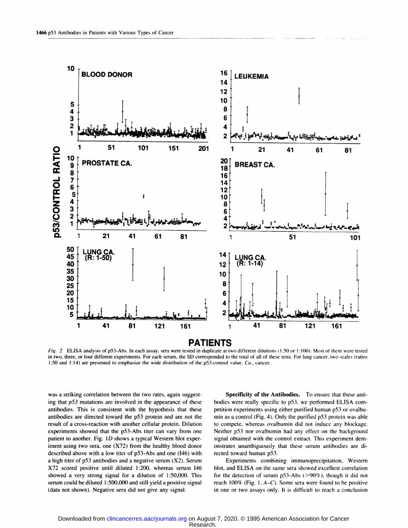
Absence of p53-Abs in Sera from Healthy Donors. We
tested 200 sera from healthy blood donors theoretically negative
for p53 antibodies (Fig. 2). Each serum was tested in duplicate
using two different dilutions ( 1 :50 and 1 : 100). In agreement
with our hypothesis. the mean ratio obtained with all of these
sera was I . I , with a SD of 0.4. Using a cutoff value correspond-
ing to the mean plus 2 SDs (97.7C/c of confidence), all but one
sera were negative in ELISA. This serum (X72) had a value (2.6
:: 1.5) which was reproducibly higher than this ratio. It was also
tested by Western blot, and p53 antibodies were readily detect-
able using this method (Fig. ID). Several samples ofserum from
the same woman over a period of 15 months were obtained from
January 1992 to July 1993. All were positive. with a slight
constant increase over this period. Examination of the clinical
records did not show the presence of any neoplastic disorder.
The woman was lost for continuous follow-up. and no further
studies were performed on this case. Taken together. these data
led us to conclude that the prevalence of p53 antibodies in the
normal population is very low, and that our ELISA could be
effectively used on a population with various types of cancer.
Prevalence of p53 Antibodies in Patients with Various
Types of Cancer. The ELISA was then used to detect p53-
Abs in sera of more than 1000 patients with various types of
neoplasias (Fig. 2 and Table 1 ). Each experiment was performed
using similar positive and negative controls. Positive controls
included two sera. One of them (LC84) had an ELISA ratio of
around 30. with p53-Abs recognizing p53 by both immunopre-
cipitation and Western blot (Fig. 1C). The second serum (BC2O)
also contained p53-Abs. as demonstrated by immunoprecipita-
tion and Western blot (Fig. 1B). It had an ELISA ratio slightly
higher than the cutoff value discussed above. This serum was
used as a threshold for delimiting positivity. p53-Abs were
found in the sera of patients with every type of neoplasia. The
frequency ranged from 24% for lung cancer to I % for prostate
carcinoma (Table 1 ). Statistical analysis show that the presence
of p53 antibodies is highly specific for cancer patients (P <
0.001).
Although the mechanisms involved in the appearance of
these antibodies are not fully understood, it has been strongly
33
57 1 #{149} � 109 1 1#{149}63 �1O3 i34�
* * * * * *N Ab 912202627334954555760636680909294969899 109
a #{149}� #{149}� -I� �
X72 X2 _________D #{149}bcd’���’
Fig. 1 Analysis of serum p53-Abs by various immunological meth-
ods. A, validation of ELISA using sera from patients with breast carci-
nomas already evaluated by immunoblotting ( 1 1 ). Number of positive
sera corresponds to serum name. B, Western blot analysis of p53-Abs
from patients with breast carcinoma using extract of insect cells infectedwith a recombinant p53 baculovirus. Sera were diluted I : 100. N. serum
from a healthy donor. Ab, positive control with a monoclonal antibody
specific for human p53. *. sera which were used above in the ELISA.
C. immunoprecipitation of in vitro translated p53 with sera from patients
with lung cancer. M, marker: T. sera from a blood donor: Ab. positive
control with a monoclonal antibody specific for human p53. Number
corresponds to serum name. The values below the photograph corre-
spend to the ELISA ratios obtained with these sera (see text). The lower
band corresponds to truncated p53 produced in vitro by internal initia-
tion. D. Western blot analysis: X72 and X2 sera were diluted I : 10: 1:25:1:100, and 1:2(X) (a-d). I46 serum was diluted 1:100; 1:2(X): l:l,0(�);
1:10.000. and l:50,()00 (a-c). Exposition time was 5 s. X72 corre-
sponded to the blood donor shown to be positive by ELISA (Fig. 3). X2
was a negative control (healthy donor). 146 was a patient with a
chondrosarcoma. p53 status in this patient was unknown. The various
bands observed in the Western blot experiment correspond to various
forms of p53 expressed in the insect cells.
suggested that they are associated with an alteration in the p.53
gene which might lead to p53 accumulation. The rate of p53
antibodies found in our ELISA study were compared with the
rate ofp53 mutations described in the literature (Fig. 3). There
Research. on August 7, 2020. © 1995 American Association for Cancerclincancerres.aacrjournals.org Downloaded from
Page 4
16
14
12
10
8
JTJ�I�_I�� ‘�y I J� �idJ
10BLOOD DONOR
543
�
0 � 51 101 151
� 1� PROSTATE CA.
_I 706�I-z40�
� �
#{149}4����l#{149}{�Y.�I4{sAiiiirs1’i�1�ji fJ!l�l�jSI1q#{149}.#{149}I�4uj5 jabf5l’..&’
201 1 21 41 61 81
201816
1�1
50 LUNG CA.45 (R:1-50)40353025201510
5
51
ILUNG CA.(R: 1-14)
101
10
8
6
if
1 41 81 121 161 1 41 81 121 161
1466 p53 Antibodies in Patients with Various Types of Cancer
141210
8642
LEUKEMIA
BREAST CA.
PATiENTSFig. 2 ELISA analysis of p53-Abs. In each assay, sera were tested in duplicate at two different dilutions ( I :50 or 1 : 100). Most of them were tested
in two, three, or four different experiments. For each serum, the SD corresponded to the total of all of these tests. For lung cancer, two scales (ratios
1 :50 and I : 14) are presented to emphasize the wide distribution of the p53:control value. (‘a., cancer.
was a striking correlation between the two rates, again suggest-
ing that p53 mutations are involved in the appearance of these
antibodies. This is consistent with the hypothesis that these
antibodies are directed toward the p53 protein and are not the
result of a cross-reaction with another cellular protein. Dilution
experiments showed that the p53-Abs titer can vary from one
patient to another. Fig. ID shows a typical Western blot exper-
iment using two sera, one (X72) from the healthy blood donor
described above with a low titer of p53-Abs and one (146) with
a high titer of p53 antibodies and a negative serum (X2). Serum
X72 scored positive until diluted I :200, whereas serum I46
showed a very strong signal for a dilution of 1 :50,000. This
serum could be diluted I :500,000 and still yield a positive signal
(data not shown). Negative sera did not give any signal.
Specificity of the Antibodies. To ensure that these anti-
bodies were really specific to p53. we performed ELISA com-
petition experiments using either purified human p53 or ovalbu-
mm as a control (Fig. 4). Only the purified p53 protein was able
to compete, whereas ovalbumin did not induce any blockage.
Neither p53 nor ovalbumin had any effect on the background
signal obtained with the control extract. This experiment dem-
onstrates unambiguously that these serum antibodies are di-
rected toward human p53.
Experiments combining immunoprecipitation, Western
blot, and ELISA on the same sera showed excellent correlation
for the detection of serum p53-Abs (>90%), though it did not
reach 100% (Fig. 1, A-C). Some sera were found to be positive
in one or two assays only. It is difficult to reach a conclusion
Research. on August 7, 2020. © 1995 American Association for Cancerclincancerres.aacrjournals.org Downloaded from
Page 5
Table / J)53-Abs in patieits tilt/i various types of neoplasias
>-C-)zuJ0Ui
U�2o
(1 The mutation data were compiled from Soussi et al. (7).
“ Three series of breast carcinomas from three different hospitals
were tested.
LUNG PANCREAS BLADDER BREAST ThYROID LEUKEMIA PROSTATE
DONOR
CANCER
Fig. 3 Comparison of the frequency of p53 mutations and p53 anti-
bodies. The mutation data were compiled from Soussi et al. (7). Due to
the heterogeneity noted in this compilation, a 10C/ SD was assigned to
these values.
Clinical Cancer Research 1467
Frequency
Neoplasia
p53
positive/total
C/( of
patients with
p53 antibodies
of p53
mutations”
(%)
Lung 10/42 24 60
Pancreas 14/73 19 44
Bladder 9/52 17 34
Breast” 14/106 13 22
Breast” 42/353 12 22
Breast” 14/l0() 14 22
Thyroid 4/108 4 13
Leukemia 3/92 3 12
Prostate 1/83 2 10
Healthy donor 1/208 0.5 0
concerning these sera. In ELISA, they gave borderline results,
with a ratio value comprised between 1 .5 and 2. Although we
have demonstrated that most of the p53 antibodies recognize
linear epitopes localized in the amino- and carboxyl terminus of
PS3, we cannot exclude an immune response leading to the
production of antibodies which recognized only native p53
(positive by immunoprecipitation and negative by Western
blot).
Isotype Analysis of p53 Antibodies. No isotypic analy-
sis of p53-Abs has been reported. Thus. to better define the
humoral response of cancer patients to p53, we analyzed the
isotype of these p53-Abs (Fig. 5). A total of 28 patients were
tested; 20 of them had been previously positive for p53 anti-
bodies, while 8 were negative. The latter sera remained negative
in the isotype-specific assay. Among the 20 positive sera, two
gave a very weak signal, and the results could not be interpreted.
Analysis of the other 18 patients showed that 15 sera contained
mostly IgG1 and IgG2. while the 3 others exhibited a predom-
inant IgA response. Several patients (6) also contained 1gM,
although none had p53-1gM as the only isotype. No IgG3 or
IgG4 was detected. This result strengthens the hypothesis of an
active humoral response against p53 and indicates that the
presence of p53-Abs is not due to cross-reacting low-affinity
1gM.
Discussion
p53-Abs were first described in sera of animals bearing
tumors (27-29). Later. Crawford ci a!. (8). using immunopre-
cipitation. described the presence of p53-Ab in sera of breast
cancer patients. Following this, Caron de Fromentel et a!. (9)
found similar antibodies in the sera of children with B-cell
lymphomas. These observations were ignored for several years
due to our lack of knowledge concerning p53 alterations. The
discovery that the p53 alteration can lead to p53 protein accu-
mulation shed new light on these findings, suggesting that these
p53 antibodies could be correlated with p53 alterations. Recent
studies have confirmed the presence of such antibodies in pa-
tients with either breast or lung cancer. Nevertheless, there are
some discrepancies concerning the frequency of these antibod-
ies in breast cancer. It may range from I % (23) to 5% ( 1 3), 9%
(8), 14% (1 1, 19). and 25.6% (18). Furthermore, the nature of
such antibodies has recently been questioned (23). and it has
been proposed that another Mr 53,000 protein could be the target
for such antibodies. Most of these previous studies were per-
formed on small series of samples using either immunoprecipi-
tation or Western blot. In light of the possibility that such a test
could be of clinical value, we have devised a simple ELISA
assay enabling the easy, rapid screening of large numbers of
samples.
We tested the sera of more than I 000 patients with various
types of neoplasias and 200 healthy volunteers. We demonstrate
here that p53-Ab are present primarily in patients with neopla-
sias. We show unambiguously that these antibodies are directed
toward human p53: (a) the antibodies recognized the p53 pro-
tein using three methods (immunoprecipitation, Western blot,
and ELISA). corresponding to the native or denatured state of
p53; (b) the antibodies were specifically blocked by human p53
protein, whereas control protein did not abolish the reaction; and
(c) the frequency of these serum p53 antibodies was correlated
with p53 alterations in human cancers. In view of these results,
along with our previous work showing that these p53-Abs
recognized immunodominant epitopes localized in the amino
and carboxyl termini of the p53 protein. and given the similarity
ofthe immune response ofpatients and animals immunized with
human wild-type p53. we conclude that these antibodies are
directed toward human p53. All of these observations are
strengthened by the observation that the presence of p53 anti-
bodies is directly linked to several clinical prognosis markers
(I 1. 19).
The most important question raised by the serological
analysis concerns its correlation with the p53 mutation and/or
p53 accumulation. Several studies have addressed this question
(10, 12, 15-17, 22, 30). All of these data suggest that most
patients with p53 antibodies have a p53 mutation which leads to
p53 accumulation. Nevertheless, exceptions exist ( 1 5. 16): cer-
tam patients have p53-Abs. yet no p.53 mutation is found in the
tumor. We should emphasize that assay of p53 antibodies cor-
responds to a global approach to assessing p53 alterations and
does not depend on sampling of the tumor, the composition of
which may be very heterogenous. Molecular analysis of tumor
Research. on August 7, 2020. © 1995 American Association for Cancerclincancerres.aacrjournals.org Downloaded from
Page 6
0.8
2 LC132
:�0 50 100 150
0.6
200
UC74
E�0 50 100 150 5
0.6
0.4
EC
aU,
0UiC�)z
0
Cl)
4
50 1X 150 200
IIgGi IgG2 IgG3 IgG4 gM IgA
2.5 BC542
2
1.5
0.5
0IgGi lgG2 lgG3 lgG4 gM
BC33
050�00150
�o-e. .50 100 150
PC51
-C--
I I
- IgGi lgG2 lgG3 lgG4 gM gA
1 LC132
0.5 I1 BC77
0:
- gd lgG2 IgG3 lgG4 gM gA IgGI IgG2 lgG3 lgG4 gM gA
Fig. 5 Isotyping of p53 antibodies. UC 132 and UC229, patients withbladder cancer; PC5 1 , patient with prostate carcinoma; BC542, BC33,
and BC77, patients with breast carcinoma; LC374 and LC 132. patients
with lung cancer.
ward an antigen. Furthermore, the finding of IgG 1 and IgG2 in
all sera supports the notion that these p53-Abs correspond to a
secondary response. Since all of these sera were taken at the
time of diagnosis, this suggests that p53-Abs were present prior
to the clinical manifestation of the cancer. In fact, we recently
observed p53 antibodies in the sera of two heavy smokers
several months before clinical detection of lung cancer (2 1 ),8
In a recently published study performed by Angelopoulou
et a!. (13), the authors tested over 1000 sera from patients with
various types of cancer using a direct ELISA. They found
p53-Abs only in patients with cancers, but the frequencies were
lower than those described in the present work. This could
reflect a bias in selection of the patient, but could also be due to
the difference in the assay. At present, the status of the assay of
serum p53-Ab is similar to that of the evaluation of p53 accu-
mulation in tumor tissues several years ago. Numerous discrep-
ancies were (and still are) observed due to the lack of standard-
ization of the technique itself, but also to methods for recording
the results. Assay of p53-Ab is still in its infancy because more
data are needed to standardize the approach. Serological analy-
sis of p53 alterations has several advantages, including fol-
low-up of patients during treatment and early detection of p53
alterations. In view of the use of p53 as a new tumoral marker,
the combined use of immunohistochemical and serological anal-
yses should be a valuable asset in clinical investigations.
1468 p53 Antibodies in Patients with Various Types of Cancer
6 R. Lubin, B. Bressac, N. Janin, and T. Soussi, unpublished results.
7 R. Lubin, I. Bouchet, and T. Soussi, manuscript in preparation. 8 Unpublished data.
0 50 100 150 200 0
p53 proteIn (ng/welI)
Fig. 4 ELISA competition experiment. Prior to the ELISA, sera (I:100 diluted) were incubated with various amounts of p53 protein or
ovalbumin (8, 40, and 160 ng/well). ELISA was then performed as
described in “Materials and Methods” using either p53 (#{149},0) orcontrol antigen (U, #{163}2)on the plates. ELISA was performed either with
ovalbumin (0, 0) or p53 protein (S, U). LC 132, patient with lung
cancer; TY68, patient with thyroid cancer; PC51, patient with prostate
carcinoma; UC74, patient with bladder carcinoma; BC33, patient with
breast carcinoma; X5, healthy donor.
tissues or biopsies corresponds to local analysis of p53 status,
and might be erroneous if the tumor is too heterogenous or too
highly contaminated by normal tissue. Furthermore, mutation is
not necessary for p53 accumulation (3 1 , 32), and we have been
able to detect p53 antibodies in such patients.6 However, it is
also clear that not all patients with a p53 alteration develop p53
antibodies. Davidoff et a!. (10) suggested that the type of
mutation could influence the production of p53 antibodies, but
given more recent results (16) this hypothesis requires further
investigation. It is also possible that, for an identical mutation,
the humoral response is dependent on the MHC class I or II
molecule specific to each individual. If we compare the fre-
quency of p53 alteration in the literature, the present work
indicates that 30-40% of patients with an alteration in the p53
gene develop p53 antibodies. In a more recent work, using a
new ELISA based on specific p53 peptides, we were able to
detect p53-Abs in sera of nearly 40% of patients with p53
mutations.7
It has been proposed that the p53-Ab is the result of a
self-immunization process toward a protein which is normally
expressed in minute quantities in the organism (22). This is
supported by the finding that the immunoglobulin subclasses of
these antibodies are characteristic of an immune response to-
0.3 � UC132 0.7 � UC229
� J :::��JJ0.2
IgGi lgG2 IgG3 igG4 gM gA IgGi lgG2 lgG3 lgG4 gM IgA
! 11PC51I.C) 0.5
F-z 0.25j
< 0.2 BC33
� 0.15
00.1CI) 0.05
IgA
2.5 � LC374.;j�0.:
IgGi lgG2 lgG3 lgG4 lgM IgA
Research. on August 7, 2020. © 1995 American Association for Cancerclincancerres.aacrjournals.org Downloaded from
Page 7
Clinical Cancer Research 1469
Acknowledgments
We are grateful to K. Ory and Y. Legros for discussion. Y. Legros
for his generous gift of purified p53 protein, B. Vojtesek and D. Lane for
communicating their results prior to publication, and J. Bram for reading
the manuscript.
References
1 . Caron de Fromentel, C., and Soussi, T. TP53 tumor suppressor gene:
a model for investigating human mutagenesis. Genes Chromosomes &
Cancer, 4: 1-15, 1992.
2. Hollstein. M., Sidransky, D., Vogelstein, B.. and Harris, C. C. p53
mutations in human cancers. Science (Washington DC) 253: 49-53. 1991.
3. Remvikos, Y., Tominaga. 0.. Hammel. P.. Laurent-Puig. P., Salmon,
R. J.. Dutrillaux, B., and Thomas, G. Increased p53 protein content of
colorectal tumours correlates with poor survival. Br. J. Cancer, 66:
758-764, 1992.
4. Thor, A. D., Moore, D. H., Edgerton, S. M.. Kawasaki. E. S..
Reihsaus, E., Lynch, H. T., Marcus, J. N., Schwartz, L., Chen, L. C.,
Mayall. B. H., and Smith, H. S. Accumulation ofp53 tumor suppressor
gene protein-an independent marker of prognosis in breast cancers. J.
Natl. Cancer Inst., 84: 845-855, 1992.
5. Starzynska. T., Bromley. M., Ghosh, A., and Stern, P. L. Prognostic
significance of p513 overexpression in gastric and colorectal carcinoma.
Br. J. Cancer, 66: 558-562, 1992.
6. Lowe, S. W., Ruley. H. E.. Jacks. T.. and Housman, D. E. p53-dependent apoptosis modulates the cytotoxicity of anticancer agents.
Cell, 74: 957-967. 1993.
7. Soussi, T., Legros. Y., Lubin, R., Ory. K.. and Schlichtholz. B.
Multifactorial analysis of p53 alteration in human cancer-a review. Int.
J. Cancer, 57: 1-9, 1994.
8. Crawford. L. V., Pim, D. C., and Bulbrook, R. D. Detection of
antibodies against the cellular protein p53 in sera from patients with
breast cancer. Int. J. Cancer, 30: 403-408, 1982.
9. Caron de Fromentel, C., May-Levin, F., Mouriesse. H., Lemerle, J.,
Chandrasekaran, K., and May, P. Presence of circulating antibodies
against cellular protein p53 in a notable proportion of children with
B-cell lymphoma. Int. J. Cancer. 39: 185-189, 1987.
10. Davidoff, A. M.. Iglehart, J. D., and Marks, J. R. Immune response
to p53 is dependent upon p53/HSP7O complexes in breast cancers. Proc.
Nail. Acad. Sci. USA. 89: 3439-3442. 1992.
11. Schlichtholz, B., Legros. Y., Gillet. D., Gaillard, C.. Marty, M..
Lane, D.. Calvo, F., and Soussi, T. The immune response to p53 inbreast cancer patients is directed against immunodominant epitopes
unrelated to the mutational hot spot. Cancer Res.. 52: 6380-6384. 1992.
12. Winter, S. F., Minna, J. D.. Johnson, B. E.. Takahashi, T., Gazdar,
A. F., and Carbone. D. P. Development of antibodies against p53 in lung
cancer patients appears to be dependent on the type of PS3 mutation.
Cancer Res, 52: 4168-4174. 1992.
13. Angelopoulou, K., Diamandis, E. P., Sutherland, D. J. A., Kellen,J. A.. and Bunting, P. 5. Prevalence of serum antibodies against the p.53
tumor suppressor gene protein in various cancers. Int. J. Cancer, 58:
480-487, 1994.
14. Marxsen, J., Schmiegel, W., Roder, C., Harder, R., JuhI, H., Hen-
nebruns, D., Kremer, B., and Kalthoff, H. Detection of the anti-p53
antibody response in malignant and benign pancreatic disease. Br. J.
Cancer, 70: 1031-1034, 1994.
15. Preudhomme, C., Lubin, R., Lepelley. P.. Vanrumbeke, M.. and
Fenaux, P. Detection of serum anti p53 antibodies and their correlationwith p53 mutations in myelodysplastic syndromes and acute myeloid
leukemia. Leukemia (Baltimore). 8: 1589-1591. 1994.
16. Wild, C. P., Ridanpaa, M.. Anttila, S., Lubin, R., Soussi, T.,
Husgafvel-Pursiainen. K., and Vainino, H. p53 antibodies in the sera of
lung cancer patients: comparison with p53 mutation in the tumour
tissue. Int. J. Cancer, 64: 176-181, 1995.
17. Guinee, D. G., Travis, W. D., Trivers, G. E., Debenedetti, V. M. G.,
Cawley, H., Welsh, J. A., Bennett, W. P., Jett, J., Colby. T. V., Tazelaar.
H., Abbondanzo, S. L., Pairolero, P., Trastek, V., Caporaso, N. E.,Liotta, L. A., and Harris, C. C. Gender comparisons in human lung
cancer: analysis of p53 mutations, anti-p53 serum antibodies and C-
erbB-2 expression. Carcinogenesis. 16: 993-1002. 1995.
18. Mudenda, B., Green, J. A., Green, B., Jenkins, J. R.. Robertson, L.,
Tarunina, M., and Leinster, S. J. The relationship between serum p53
autoantibodies and characteristics of human breast cancer. Br. J. Cancer,
69: 1115-1119. 1994.
19. Peyrat. J. P.. Bonneterre. J.. Lubin, R., Vanlemmens, L., Fournier,
J., and Soussi, T. Prognostic significance of circulating p53 antibodies
in patients undergoing surgery for locoregional breast cancer. Lancet,
345: 621-622. 1995.
20. Lubin, R., Zalcman, G., Bouchet, L., Tr#{233}daniel, J., Legros, Y.,
Cazals, D., Hirsh, A., and Soussi, T. Serum p53 antibodies as early
markers of lung cancer. Nat. Med., /: 701-702, 1995.
21. Schlichtholz, B., Tredaniel, J., Lubin, R., Zalcman, G., Hirsch, A.,
and Soussi, T. Analyses of p53 antibodies in sera of patients with lung
carcinoma define immunodominant regions in the p53 protein. Br. J.
Cancer, 69: 809-816, 1994.
22. Lubin. R., Schlichtholz, B., Bengoufa, D.. Zalcman, G.. Tredaniel.
J., Hirsch, A., Caron de Fromentel, C., Preudhomme, C., Fenaux, P.,Fournier, G., Mangin, P., Laurent-Puig, P., Pelletier, G., Schlumberger,
M.. Desgrandchamps. F.. Leduc. A., Peyrat. J. P.. Janin, N.. Bressac, B..and Soussi, T. Analysis of p53 antibodies in patients with various cancers
define B-cell epitopes of human p53: distribution on primary structure and
exposure on protein surface. Cancer Res.. 53: 5872-5876, 1993.
23. Vojtesek. B.. Kovarik, J., Dolezalova, H., Nenutil, R., Havlis, P.,
Brentani, R. R., and Lane, D. P. Absence of p53 autoantibodies in a
significant proportion of breast cancer patients. Br. J. Cancer, 7/:
1253-1256, 1995.
24. Jeffries, R., Reimer, C. B., Skvrail, F., de Lange. G., Ling. N. R.,
Lowe, J., Walker, M. R., Phillips. D. J., Aloisio. C. H., Wells, T. W.,
Vaerman, J. P., Magnuson, C. G., Kubagawa, H., Cooper, M., Vardtal.
F.. Vandvik, B., Haaijman, J. J., Makeda, 0., Sarnesto, A., Lando, Z.,Gergely. J., RadI, J., and Molinaro, G. A. Evaluation of monoclonal
antibodies having specificity for human IgG subclasses: results of an
IUIS/WHO collaborative study. Immunol. Lett.. /0: 223-252, 1985.
25. Legros. Y.. Lafon, C.. and Soussi, T. Linear antigenic sites defined
by the B-cell response to human p53 are localized predominantly in the
amino and carboxy-termini of the protein. Oncogene. 9: 2071-2076. 1994.
26. Soussi, T.. Caron de Fromentel, C., St#{252}rzbecher. H. W.. Ullrich, S..
Jenkins, J.. and May. P. Evolutionary conservation of the biochemical
properties of p53-specific interaction of Xenopus-laevis p53 with simian
virus 40 large T-antigen and mammalian heat shock proteins-70. J.
Virol., 63: 3894-3901. 1989.
27. Kress, M., May, E., Cassingena, R., and May, P. Simian virus
40-transformed cells express new species of proteins precipitable by
anti-simian virus 40 serum. J. Virol., 3/: 472-483, 1979.
28. Dc Leo, A. B., Jay, G., Appella, E., Dubois, G. C., Law, L. W., and
Old. L. J. Detection of a transformation-related antigen in chemicallyinduced sarcomas and other transformed cells of the mouse. Proc. NatI.
Acad. Sci. USA. 76: 2420-2424, 1979.
29. Rotter. V., Witte, 0. N., Coffman, R., and Baltimore, D. Abelson
murine leukemia virus-induced tumors elicit antibodies against a host
cell protein, p50. J. Virol., 36: 547-555, 1980.
30. Volkmann, M., Muller, M., Hofmann, W. J., Meyer, M., Hagelstein,
J.. Rath, U., Kommerell, B., Zentgraf, H., and Galle, P. R. The humoralimmune response to p53 in patients with hepatocellular carcinoma is
specific for malignancy and independent of the alpha-fetoprotein status.
Hepatology. /8: 559-565, 1993.
3 1 . Andersen. T. I.. HoIm, R., Nesland, J. M., Heimdal, K. R.. Ottestad,
L., and Borresen, A. L. Prognostic significance of TP53 alterations in
breast carcinoma. Br. J. Cancer, 68: 540-548, 1993.
32. MolI. U. M., Riou, G., and Levine, A. J. Two distinct mechanismsalter p53 in breast cancer-mutation and nuclear exclusion. Proc. NatI.
Acad. Sci. USA, 89: 7262-7266, 1992.
Research. on August 7, 2020. © 1995 American Association for Cancerclincancerres.aacrjournals.org Downloaded from
Page 8
1995;1:1463-1469. Clin Cancer Res R Lubin, B Schlichtholz, J L Teillaud, et al. identification, and characterization.p53 antibodies in patients with various types of cancer: assay,
Updated version
http://clincancerres.aacrjournals.org/content/1/12/1463
Access the most recent version of this article at:
E-mail alerts related to this article or journal.Sign up to receive free email-alerts
Subscriptions
Reprints and
[email protected] at
To order reprints of this article or to subscribe to the journal, contact the AACR Publications
Permissions
Rightslink site. Click on "Request Permissions" which will take you to the Copyright Clearance Center's (CCC)
.http://clincancerres.aacrjournals.org/content/1/12/1463To request permission to re-use all or part of this article, use this link
Research. on August 7, 2020. © 1995 American Association for Cancerclincancerres.aacrjournals.org Downloaded from