Page 1
p90 and UHRF1, Two Novel Regulators of the p53
Signaling Pathway
Chao Dai
Submitted in partial fulfillment of the
Requirements for the degree of
Doctor of Philosophy
in the Graduate School of Arts and Sciences
COLUMBIA UNIVERSITY
2014
Page 2
©2013
Chao Dai
All Rights Reserved
Page 3
ABSTRACT
p90 and UHRF1, Two Novel Regulators of the p53 Signaling Pathway
Chao Dai
To ensure proper and differentiated regulation of stress response pathways,
the p53 tumor suppressor calls for an intricate network of control of activation
and fine tuning of transcription activity, which is offered largely through post-
translational modifications. Accumulating evidence supports the indispensability
of acetylation in the activation of p53 function and indicates modulation of cell
fate decision; however the underlying molecular mechanisms are not well
understood and identification of the regulatory mechanisms controlling p53
acetylation remains an important step in furthering the understanding of p53
regulation in vivo. In this study we identify p90 and UHRF1 as two novel
members of the p53 regulatory network upstream of TIP60-mediated p53
acetylation.
Through biochemical purification, p90 was identified as a unique regulator for
p53. p90 (also called CCDC8, coiled-coil domain containing 8) interacts with p53
both in vitro and in vivo. Depletion of p90 by RNAi has no obvious effect on p53
stability or p53-mediated activation of p21, but specifically abrogates PUMA
activation. Moreover, p90 also interacts with the TIP60 acetyltransferase and
stimulates TIP60-dependent Lys120 acetylation of p53, therefore enhancing the
Page 4
apoptotic response of p53. These data reveal p90 as an upstream regulator of the
Tip60-p53 interaction and demonstrate that p90 is specifically required for p53-
mediated apoptosis upon DNA damage.
We also report that the epigenetic regulator UHRF1 (ubiquitin-like with PHD
and RING finger domains 1) interacts with TIP60 and induces degradation-
independent ubiquitination of TIP60. Moreover, UHRF1 markedly suppresses the
ability of TIP60 to acetylate p53. In contrast, RNAi-mediated inactivation of
UHRF1 increases endogenous p53 acetylation and significantly augments p53-
mediated apoptosis. To elucidate the mechanisms of this regulation, we found
that the interaction between TIP60 and p53 is severely inhibited in the presence of
UHRF1, suggesting that UHRF1 modulates TIP60-mediated functions in both
K120 acetylation-dependent and -independent manners. Consistent with this
notion, UHRF1 knockdown promotes activation of p21 and PUMA but not
HDM2. These findings demonstrate that UHRF1 is a critical negative regulator of
TIP60 and suggest that UHRF1-mediated effects on p53 may contribute, at least
in part, to its role in tumorigenesis.
This study provides insight for understanding the regulation of p53 acetylation
and cell fate decision. Both p90 and UHRF1 are previously unidentified members
of the p53 regulatory network. Although both function upstream of the TIP60-p53
Page 5
interplay, they act through distinct and opposing mechanisms to dynamically
regulate TIP60-mediated effects on p53 in vivo.
Page 6
i
TABLE OF CONTENTS
Table of Contents …………………..…………….………...….…i
List of Figures ……………………………………...….……...viii
Acknowledgements ….…………………………………..….…...xi
Dedication ………………………………..……..……….…...xiii
Copyright Notice …..……..………....………..………..………xiv
Chapter 1. Introduction………………………………..………… 1
1.1 p53 is a tumor suppressor………………………..………….…. 2
1.2 p53 functions as a sequence-specific transcription factor……..………..3
1.3 p53 centrally coordinates cellular responses to a wide range of stresses….. 5
1.4 p53 and the “big three”: growth arrest, apoptosis and senescence…..……6
1.4.1 Growth arrest ………………………………..…..…….. 7
1.4.2 Apoptosis ………………………………..…….…....…8
1.4.3 Senescence …………....…………………….……...…10
1.4.4 Tumor suppression: the “big three” and beyond ………..…...…12
1.4.5 Summary …..……..……...….…..………..............….14
Page 7
ii
1.5 Regulation of p53 function ………..…………………..…..……16
1.5.1 Regulation of p53 stability ………..…………….………….19
1.5.1.1 Ubiquitination overview ………..…………….………19
1.5.1.2 p53 ubiquitination by HDM2………..…………….….. 20
1.5.1.3 p53 ubiquitination by HDM2-independent E3 ubiquitin ligases. 22
1.5.1.4 p53 deubiquitination by USP7 ……..…………………..23
1.5.2 Regulation of p53 localization ……..…………………....…24
1.5.2.1 cytoplasmic targeting of p53 by ubiquitination …..…..……24
1.5.2.2 Nuclear import of p53 through deubiquitination by USP10…...25
1.5.3 p53 repression on promoters by HDM2 and HDMX……..……... 28
1.5.3.1 p53 is bound to DNA at homeostasis……..…………….. 28
1.5.3.2 Repression of p53 at promoters by HDM2/HDMX…………29
1.5.3.3 De-repression of p53 is required for transcription activation….30
1.5.4 Regulation of p53 transcription activity by post-translational
modifications……..…………………..…………………...…31
1.5.4.1 Phosphorylation ……...…………………..…………31
Page 8
iii
1.5.4.1a phorphorylation at Ser15/Ser20 ……..……...………32
1.5.4.1b phosphorylation at Ser46……..…………...………32
1.5.4.1c phosphorylation at Ser392 ……..……………….…33
1.5.4.2 Ubiquitin-like modifications……..……………….……34
1.5.4.3 Methylation ……..…………………..…………..…35
1.5.4.4 Acetylation……..…………………..…………...…37
1.5.4.4a Acetylation at the C-terminus ……..……………….38
1.5.4.4b Acetylation at Lys320 ……..……………………..39
1.5.4.4c Acetylation in the DNA binding domain….…..………39
1.5.4.4d Deacetylation by HDACs and SIRT1 ……..…………41
1.5.4.5 Concluding remarks ……..…………………..………42
1.6 Summary ..……………………………..…………………..43
Chapter 2. Differential Effects on p53-mediated Cell Cycle Arrest vs.
Apoptosis by p90 ..……..…………...…..……….……45
2.1 Introduction ..…………………..……….…………..………46
Page 9
iv
2.2 Results ..…………………..……………...……..…………48
2.2.1 Identification of p90 as a unique component of p53-associated
complexes..…………………..………………….…..…48
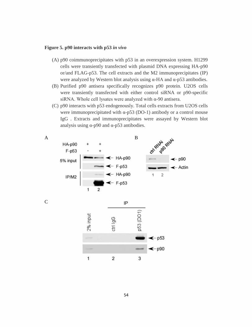
2.2.2 p90 is a bona fide p53 interacting protein..…………………....53
2.2.3 Inactivation of p90 attenuates p53-mediated activation of PUMA but
not p21..…………………..……………...……………61
2.2.4 p90 is Required for p53-Mediated Apoptosis upon DNA Damage …66
2.2.5 Mechanistic insights into p90-mediated effect on p53-dependent
apoptotic responses ..…………………..………..………..70
2.3 Discussion ..…………………..…………………………….75
2.4 Materials and methods ..…………………..…………………...78
Chapter 3. Negative Regulation of the TIP60-p53 Interplay by UHRF1….84
3.1 Introduction ..…………………..………….………..………85
3.2 Results …..………...………..………..………..…………..91
3.2.1 UHRF1 interacts with TIP60 both in vitro and in vivo.…..………91
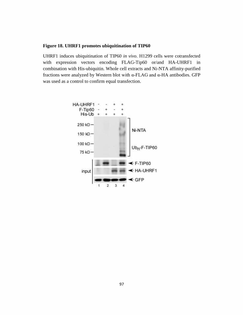
3.2.2 UHRF1 induces degradation-independent ubiquitination of TIP60....96
Page 10
v
3.2.3 UHRF1 depletion increases TIP60-mediated p53 acetylation at K120
and enhances apoptosis ..……...…..………..………..….104
3.2.4 UHRF1 inhibits TIP60-p53 interaction …..………....……….108
3.2.5 SRA and RING domains of UHRF1 are indispensable for UHRF1
suppression of TIP60-p53 interaction…..………..…...……..110
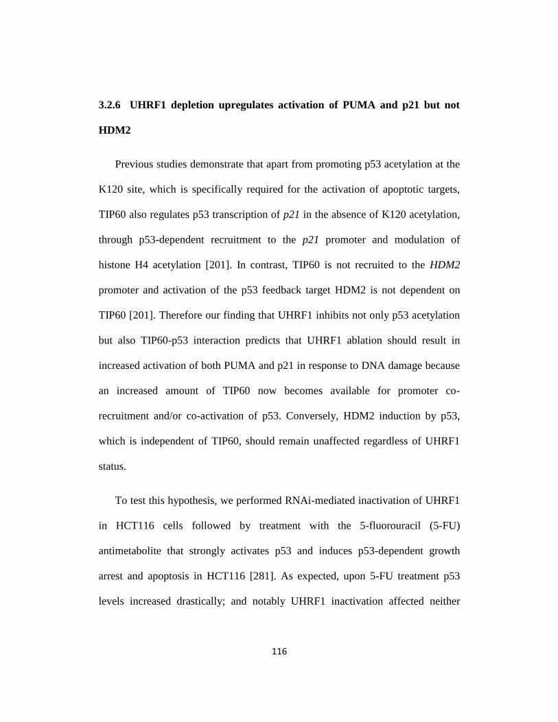
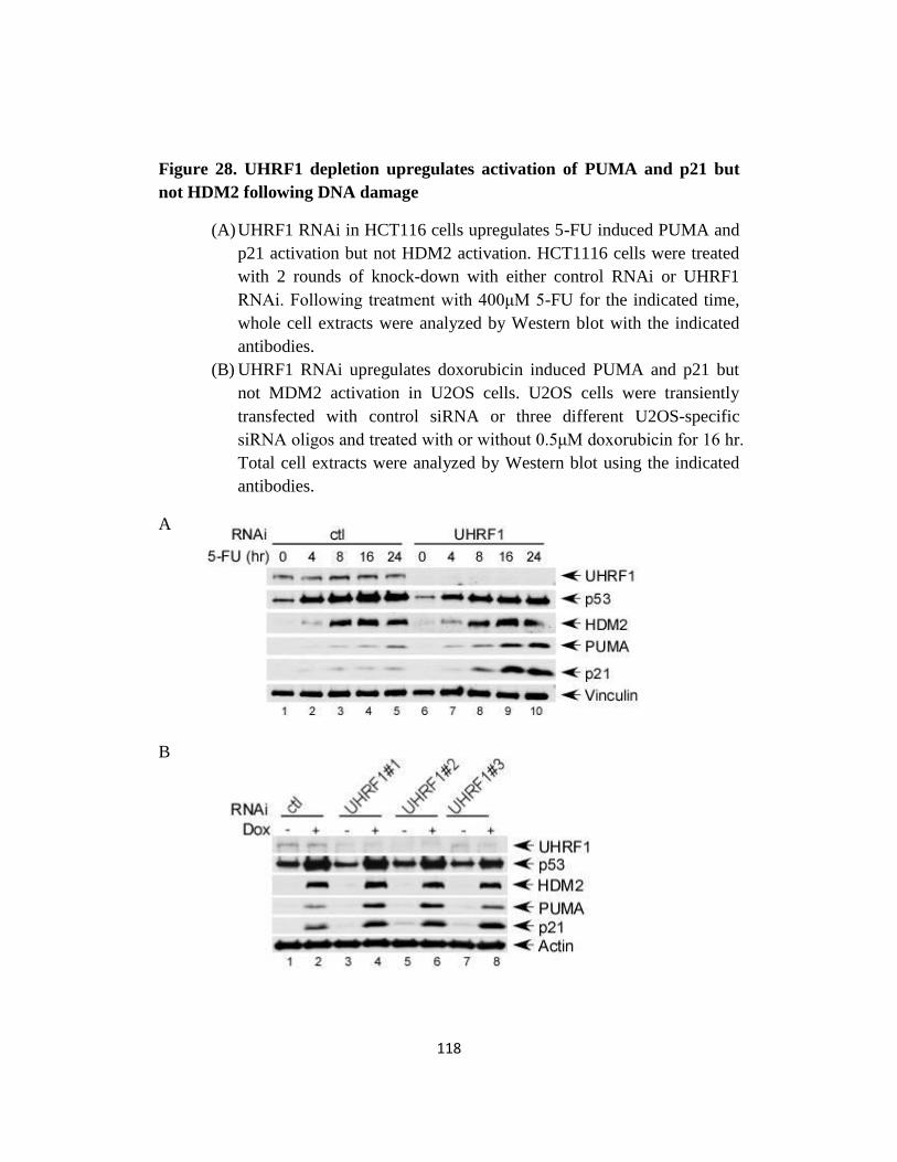
3.2.6 UHRF1 depletion upregulates activation of PUMA and p21 but not
HDM2…..………..………..………..………..…...…116
3.3 Discussion …..………..………..………..………..…….…127
3.4 Materials and methods …..………..………..………..…….…134
Chapter 4. Concluding Remarks and Future Directions…..……….…141
4.1 p90 and renal cell carcinoma …..………..………..………....…143
4.2 p90 and post-translational modifications …..………..………..…..144
4.3 p90 as a potential promoter specific cofactor for p53…..…………....145
4.4 The regulation of TIP60…..………..……..………...……..….146
4.5 UHRF1 and cancer therapy…..………..………..……………..148
4.6 Acetylation is required for all major steps of p53 activation…..………151
Page 11
vi
4.7 Other modifications/cofactors for p53 regulation of metabolism, antioxidant
defense and autophagy ………...……………………………..155
References…..……………………………………….....….…159
Page 12
vii
LIST OF FIGURES
Chapter 1
Fig 1. Overview of p53 domain structure and post-translational
modifications…..………..………..………..……..……18
Fig 2. Regulation of p53 stability and localization by ubiquitination…… 27
Chapter 2
Fig 3. Construction of p538KR stable line in H1299 ..……….………50
Fig 4. Identification of p90 as a component of a p53-containing protein
complex..…………………..……..……………..……52
Fig 5. p90 interacts with p53 in vivo..………..…………..………54
Fig 6. p90 interacts with the C-terminal domain of p53 in vitro ..………56
Fig 7. p90 interacts with p53 through its N-terminal fragment ..….……58
Fig 8. p90 interacts with p53 in the nucleus..…………………....…60
Fig 9. p90 inactivation reduces basal PUMA level and differentially affects
PUMA and p21 induction upon DNA damage ..……………....63
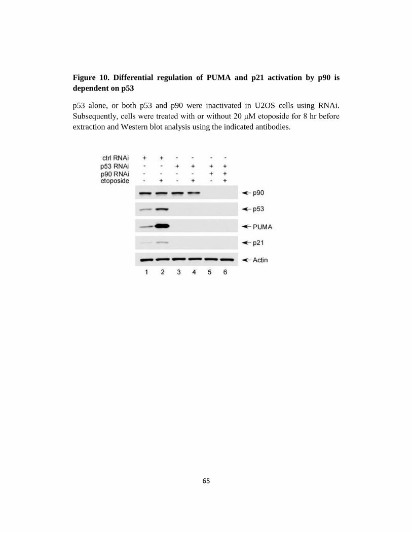
Fig 10. Differential regulation of PUMA and p21 activation by p90 is
dependent on p53..…………………..…………….……65
Page 13
viii
Fig 11. Inactivation of p90 attenuates p53-dependent PUMA activation in
time point experiments ..…………………..………….….67
Fig 12. Inactivation of p90 impairs p53-mediated apoptosis upon damage...69
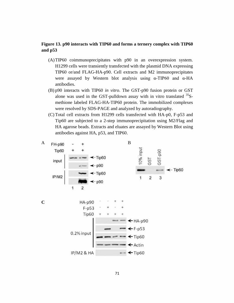
Fig 13. p90 interacts with TIP60 and forms a ternery complex with TIP60 and
p53…..………..………..………..…………………..71
Fig 14. p90 promotes TIP60-mediated p53 acetylaiton at K120.…..…….74
Chapter 3
Fig 15. Schematic representation of UHRF1 domain structure…..……...90
Fig 16. UHRF1 coimmunoprecipitates with TIP60 exogenously and
endogenously …..………..………..………..…….……93
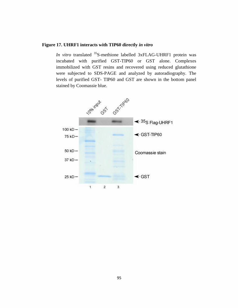
Fig 17. UHRF1 interacts with TIP60 directly in vitro…..…….……….95
Fig 18. UHRF1 promotes ubiquitination of TIP60 …..……………….97
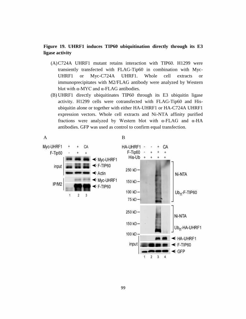
Fig 19. UHRF1 induces TIP60 ubiquitination directly through its E3 ligase
activity…..………..………..………..…….…....……99
Fig 20. UHRF1 does not promote TIP60 degradation or affect Tip60 at the
transcription level…..……….………..…….….....……101
Page 14
ix
Fig 21. UHRF1 ubiquitinates TIP60 through atypical ubiquitin lysine
linkages…..………..………..………..……..…….…103
Fig 22. UHRF1 suppresses TIP60-mediated p53 acetylation at K120…...105
Fig 23. UHRF1 depletion augments damage-induced apoptosis…..…... 107
Fig 24. UHRF1 suppresses TIP60-p53 interaction partially through promoting
TIP60 ubiquitination…..………..………..……….……109
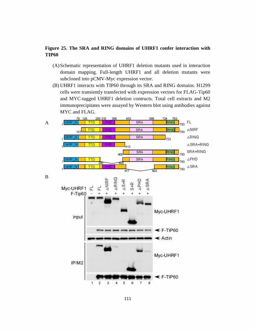
Fig 25. The SRA and RING domains of UHRF1 confer interaction with
TIP60…..………..………..………..………..…...…111
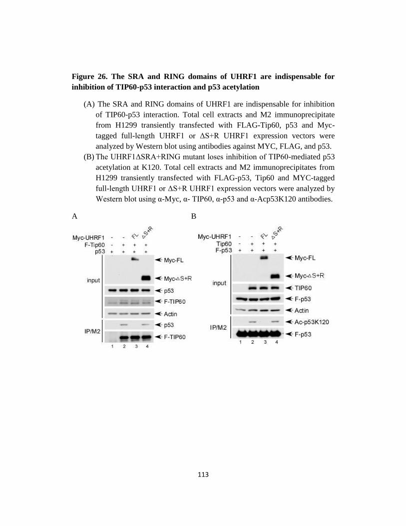
Fig 26. The SRA and RING domains of UHRF1 are indispensable for
inhibition of TIP60-p53 interaction and p53 acetylation…...…...113
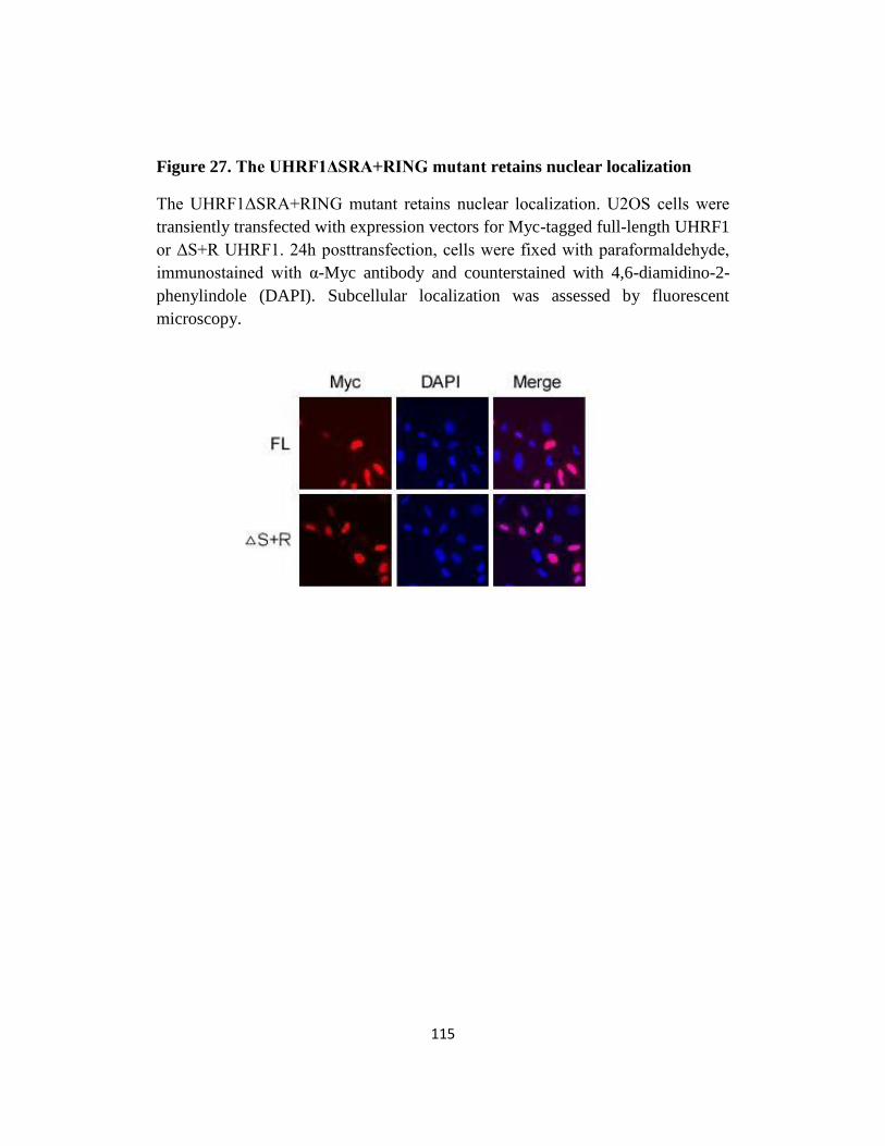
Fig 27. The UHRF1ΔSRA+RING mutant retains nuclear localization.…..115
Fig 28. UHRF1 depletion upregulates activation of PUMA and p21 but not
HDM2 following DNA damage…..………..…….…….…118
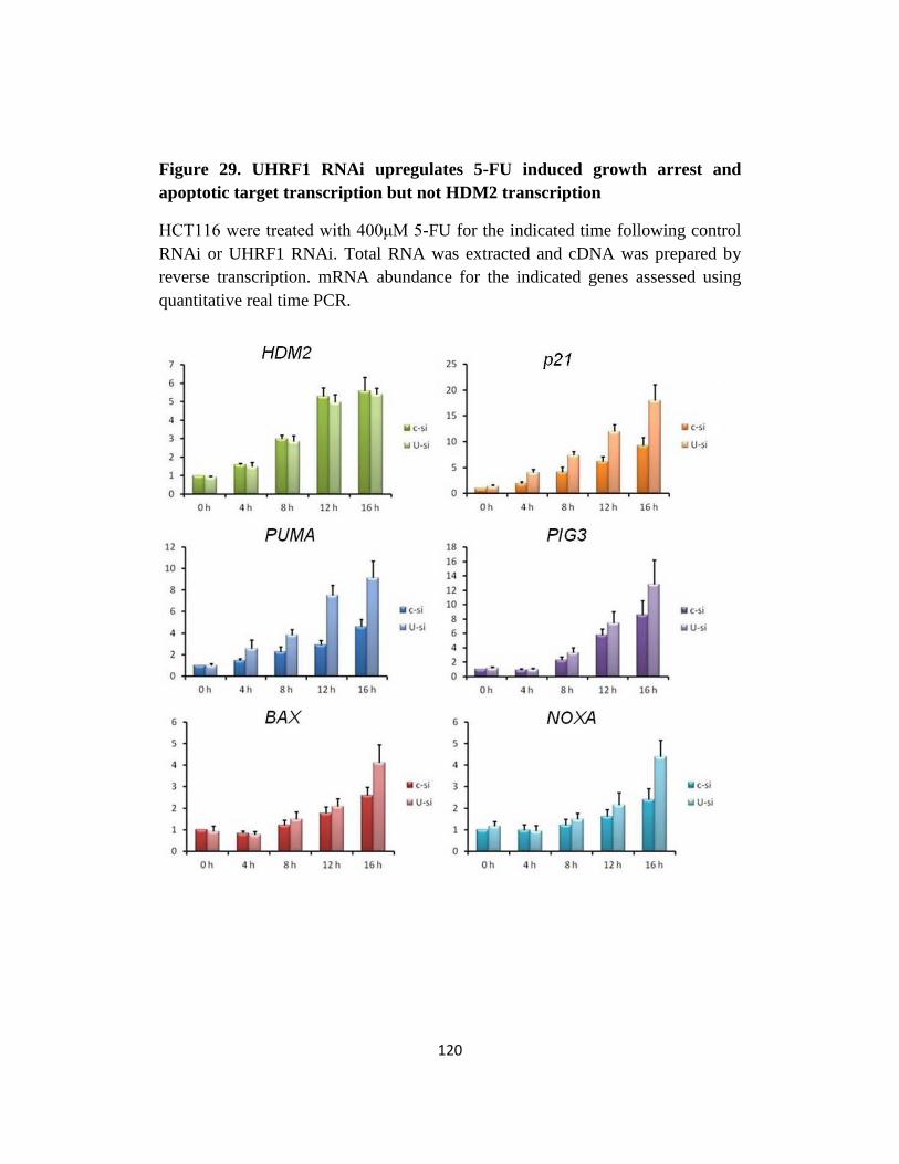
Fig 29. UHRF1 RNAi upregulates 5-FU induced growth arrest and apoptotic
target transcription but not HDM2 transcription…..…….…....120
Fig 30. UHRF1 modulation of damage-induced PUMA and p21 activation in a
p53-dependent manner…..………..………...………..…122
Page 15
x
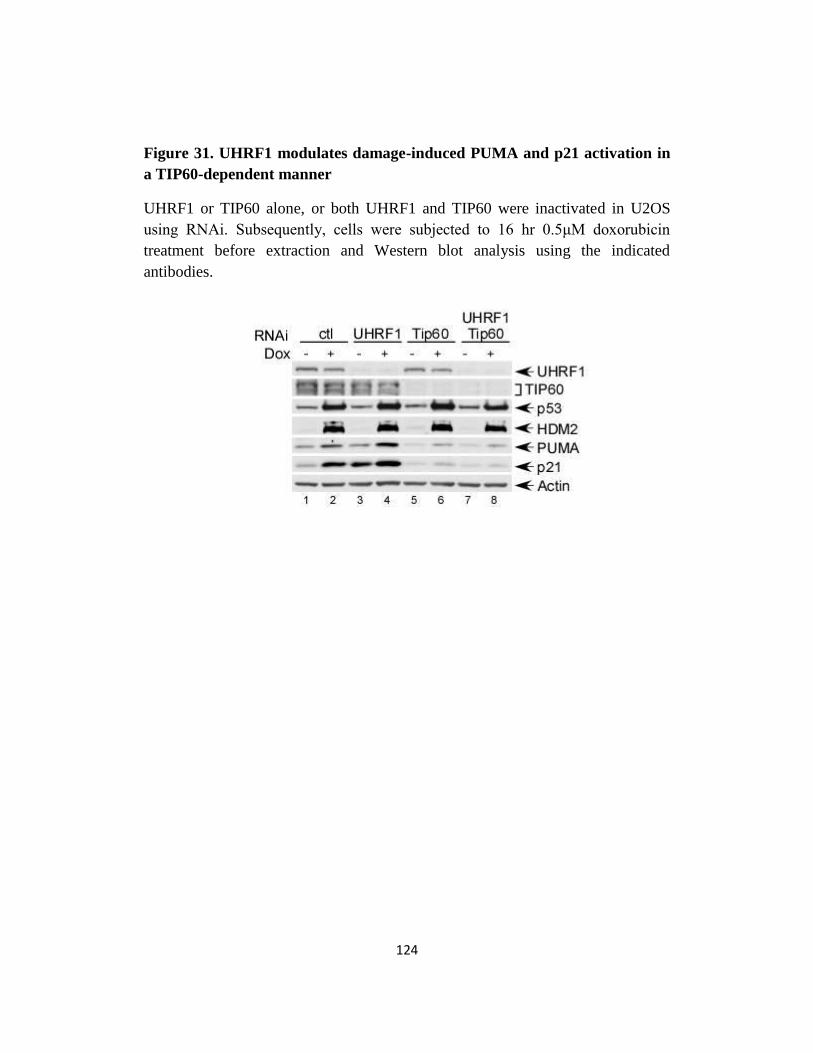
Fig 31. UHRF1 modulates damage-induced PUMA and p21 activation in a
TIP60-dependent manner…..………..………..….……...124
Fig 32. UHRF1 depletion upregulates damage-induced growth arrest…...126
Fig 33. A model for tumorigenesis/tumor progression in cells with UHRF1
overexpression…..………..………..………..….…….129
Chapter 4
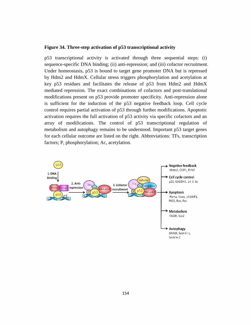
Fig 34. Three-step activation of p53 transcriptional activity…..……….154
Page 16
xi
ACKNOWLEDGEMENTS
This work could not have been accomplished without the support of many
people. First, I owe my deepest gratitude to my mentor Dr. Wei Gu for his endless
guidance, support, patience and encouragement throughout my entire Ph.D. study.
He has been a wonderful and insightful mentor. I have benefited a lot and will
continue to benefit from what I have learnt from him.
I would also like to thank my research committee members Dr. Jean Gautier
and Dr. Songtao Jia for taking time out of their busy schedules to offer insightful
scientific inputs, valuable suggestions and support. Special thanks to Dr. Stuart
Aaronson and Dr. Ming-Ming Zhou for agreeing to be my outside thesis
committee members.
It has been a great pleasure to work in the Gu lab. I am deeply thankful to Dr.
Muyang Li, Dr. Yi Tang, and Dr. Wenhui Zhao who patiently helped me with
techniques and offered inputs to my projects during the early stage of my Ph.D.
study, and Dr. Jing Shan who, as a senior student in the lab, guided me during my
rotation and eased my transition into the Gu lab. I am also thankful to Dr.
Dingding Shi, Dr. James Lee and Dr. Omid Tavana, who shared with me many of
their own experiences and stories of being a Ph.D. student and a fresh graduate.
Dr. Tongyuan Li, thank you for being the greatest bay-mate, encouraging me,
Page 17
xii
giving inputs, and keeping our shared workspace mess-free when I get lazy.
Special thanks to Miss Jiayun Zhong, for your pleasant personality and great lab
management. I also thank all the rest of the Gu lab for being the friendly,
supportive and fun lab mates that you are, making my entire graduate study a
pleasant and memorable experience.
I also thank all my classmates who entered in 2007 and shared this same
experience with me. Kehui Xiang, and Ruixue Fan, I will never forget the many
evenings we spent together in the library studying the first year Biology Core.
Finally, my particular appreciation goes to my family. I am forever grateful to
my parents, who love and support me unconditionally. I would be nothing without
you. To my dear husband, thank you for being so tremendously loving, patient,
and understanding. I am the luckiest person to have you in my life.
Page 18
xiii
DEDICATION
I dedicate my thesis and the last six years of work that went into creating it to
my husband, Lin, who has given me unconditional love and support throughout
this entire process. I couldn’t have done this without him. I also dedicate it to my
parents, who have helped make me the person I am today and have given me the
love and guidance needed to accomplish this task.
Page 19
xiv
COPYRIGHT NOTICE
Portions of this thesis appeared in:
Dai, C., and Gu, W. (2010) p53 post-translational modification: deregulated in
tumorigenesis. Trends Mol. Med. 16(11): 528-536.
Dai, C., Tang, Y., Jung, S.Y., Qin, J., Aaronson, S.A., and Gu, W. (2011)
Differential effects on p53-mediated cell cycle arrest vs. apoptosis by p90. Proc.
Natl. Acad. Sci. USA 108(47):18937-18942.
Dai, C., Shi, D., and Gu, W., Negative Regulation of the Acetyltransferase
TIP60-p53 Interplay by UHRF1. (2013) J Biol. Chem. 288(27): 19581-19592.
Page 20
1
CHAPTER 1
INTRODUCTION
Page 21
2
1.1 p53 is a tumor suppressor
p53, encoded by the TP53 gene, is often regarded as “guardian of the genome”
because of its pivotal role in tumor suppression [1]. p53 was initially discovered
independently by David Lane and Arnold Levine in 1979 as a simian virus 40
(SV40) large T antigen interacting partner that migrates at 53 kDa on sodium
dodecyl sulfate polyacrylamide gel electrophoresis (SDS-PAGE) [2,3]. Early
work revealing excessive p53 production in transformed and cancer cells and
work demonstrating that p53 cooperates with the Ras oncoprotein to transform or
immortalize cells led to the classification of p53 as an oncoprotein [4-7]. It was
later found that the TP53 cDNA initially cloned from tumor cell mRNA was a
dominant negative allele containing a valine (V) to alanine (A) mutation at codon
135 that activates transforming properties. In 1989, work by the Oren group and
Levine group showing that wild type p53 could suppress oncogene driven
transformation and work by the Vogelstein group demonstrating frequent
mutations of the TP53 gene in human colorectal carcinomas collectively
characterized p53 as a tumor suppressor [8-10].
Following these initial observations, TP53 mutations were reported in a wide
spectrum of human cancers, with mutation rates ranging from ~10% in
hematopoietic malignancies to ~50%-70% in ovarian, colorectal, lung, and head
and neck malignancies [11]. Trp53 (encoding mouse p53) deficient mice are
Page 22
3
susceptible to early onset spontaneous tumorigenesis [12], and germline mutation
of p53 in humans which leaves only one functional allele of the TP53 gene is
associated with the Li-Fraumeni syndrome characterized by a 25-fold increase in
cancer susceptibility and early onset of a wide range of malignancies such as
breast cancer, brain tumors, and soft tissue sarcomas [13,14]. It is now known that
p53 mutations or perturbation of the p53 regulatory network exist in over half of
all human cancer cases [15-17].
1.2 p53 functions as a sequence-specific transcription factor
The p53 protein comprises several domains: an amino (N-) terminal
transactivation domain (TAD; consisting of two transactivation subdomains,
TAD-I, residues 1-42, and TAD-II, residues 43-62) [18-20], a proline rich domain
(PRD; residues 63-97), a central DNA binding core domain (DBD; residues 100-
300) [21,22], a tetramerization domain (TD; residues 307-355) [23,24], and a
carboxyl (C-) terminal regulatory domain (CTD; residues 356-393) [25].
Soon after its characterization as a bona fide tumor suppressor, p53 was
identified to possess binding affinity, through its central domain, to specific DNA
sequences termed “the p53 consensus binding site” or “the p53 response element”
[26]. The consensus sequence consists of two 5’-PuPuPuC(A/T)(T/A)GPyPyPy-3’
Page 23
4
decameric palindromes called “p53 binding half-sites” separated by 0-13
basepairs [26].
The presence of p53 response elements in the regulatory regions (promoters,
introns, and upstream sequences) of genes predicts transcription regulation by p53.
A combination of gene expression microarrays, chromatin-immunoprecipitation-
based microarrays (ChIP-chip) and ChIP sequencing analysis have to date
identified at least 500 p53 binding loci throughout the human genome [27-30]. At
least 100 genes have been identified to possess p53 response elements and are
experimentally validated as p53 target genes [31,32].
DNA sequencing of tumor samples bearing mutant p53 revealed that the vast
majority of p53 mutations are missense mutations within the DNA binding
domain, resulting in mutant p53 proteins with altered conformation and attenuated
sequence-specific binding to DNA [33]. The significance of p53 mutations in
tumorigenesis is 3-fold: (i) they abolish wild type p53 function, (ii) they create
dominant negative activity through tetramer formation with wild type p53, and (iii)
they convey “oncogenic” function through the selective growth advantages of
cells with the mutations, the transactivation of new target genes or via
inappropriate interaction with other cellular proteins [34].
Page 24
5
1.3 p53 centrally coordinates cellular responses to a wide range of stresses
p53 exerts tumor suppressive capacities by centrally coordinating a regulatory
circuit that monitors and responds to a variety of stress signals. Under
homeostasis, both p53 abundance and p53 transcription activity is kept low by its
primary negative regulators Human Double Minute 2 (HDM2, mouse ortholog is
Mdm2) and Human Double Minute X (HDMX, mouse ortholog is MdmX). In
the event of genotoxic stresses such as DNA damage, abnormal oncogene
activation, telomere erosion, hypoxia etc, p53 is rapidly stabilized and activated to
transcribe target genes that mediate cell cycle arrest, apoptosis, DNA repair,
senescence, energy metabolism, or autophagy. Through executing and balancing
these cellular responses, p53 ultimately protects cellular and genomic stability,
preventing the propagation of genetic lesions and tumor formation.
A multitude of chemo-reagents converge onto the activation of p53. The
cytotoxic agent etoposide forms a ternary complex with DNA and the
topoisomerase II enzyme, thus preventing re-ligation of the DNA strand and
causing DNA strand breaks in cancerous cells that undergo rapid DNA replication
and cell division [35]. The anthracyclines (doxorubicin, daunorubicin, and their
derivatives) work by intercalating DNA as well as undergoing redox reactions
that generate reactive oxygen species (ROS) [36]. DNA strand breaks are
recognized by the MRN complex (consisting of three proteins Mre11, RAD50,
Page 25
6
and NBS1) which in turn activates ATM-CHK1 or ATM-CHK2 kinase cascades
that transmit this information to p53 through phosphorylating both p53 and
HDM2, ultimately inhibiting their association and stabilizing p53 [37]. Reagents
that disrupt rRNA biogenesis, such as actinomycin D, increase ribosomal stress
and release ribosomal proteins from the nucleoli, which in turn bind to HDM2 and
result in p53 stabilization [38]. The uracil analogue 5-fluorouracil (5-FU)
antimetabolite functions through misincorporation into nascent RNA and
irreversibly blocking thymidylate synthase, causing dTMP depletion in rapidly
dividing cells [39], as well as triggering a ribosomal stress response that releases
ribosomal proteins to activate p53 by ablating the HDM2-p53 feedback loop [40].
Although the p53 effects are predominantly exerted through its transcription
activation of target genes, our knowledge of p53 functions have been expanded
into transcription repression [41], regulation of translation [42] and homologous
recombination [43], and the induction of a transcription-independent apoptotic
response [44].
1.4 p53 and the “big three”: growth arrest, apoptosis and senescence
Growth arrest, apoptosis, and senescence are the most well characterized
cellular responses following p53 activation and thought of as major mediators of
Page 26
7
the tumor suppressive function of p53. The ability of p53 to remove damaged
cells through apoptosis is a more evolutionarily conserved function: in lower
eukaryotes, including D. melanogaster and C. elegans, p53 is critical for
eliminating damaged cells to preserve germline and tissue integrity [45]. In higher
eukaryotes, genotoxic stresses activate p53, leading to cell cycle pauses allowing
time for damage repair or the irreversible cellular senescence or apoptosis in the
event of prolonged damage as safeguards against neoplasia [31].
Here I will briefly revisit the means by which p53 regulates each of these
pathways, and discuss their roles in tumor suppression.
1.4.1 Growth arrest
Cell cycle checkpoint is a common theme of regulation in eukaryotes to
ensure fidelity of DNA replication and mitosis, thus protecting from propagation
of genetic lesions and progressive accumulation of genomic changes that
eventually leads to neoplastic transformation. Halting the cell cycle at checkpoints
presumably permits repair of damage before the cell reinitiates DNA replication
(G1 arrest) or enters mitosis (G2 arrest).
The first line of evidence suggesting p53 control of cell cycle progression
comes from the work from Kastan and colleagues demonstrating that ataxia
telangiectasia mutated (ATM), p53 and GADD45 comprise a signal transduction
Page 27
8
pathway that controls the mitotic checkpoint upon DNA damage [46]. Soon
afterwards, p53 was shown to be required for G1 checkpoint arrest following
DNA damage, primarily through transcription activation of one of the best
characterized p53 target genes CDKN1A encoding p21CIP1/WAF1
, a cyclin-
dependent kinase (CDK) inhibitor [47,48]. Elevated p21, through binding to and
inactivating cyclin/CDK complexes required for the G1/S transition, arrests cells
in the G1 phase to allow time for DNA damage repair.
Mouse embryonic fibroblasts (MEFs) derived from mice lacking p21 are
almost entirely deficient in G1 arrest following DNA damage, underlying the
significance of p21 in the G1 checkpoint arrest [49]. However, unlike Trp53-/-
mice, p21-/-
mice are not susceptible to early onset of spontaneous tumor
development [49]. Nevertheless, loss of p21 promotes tumor initiation,
progression or metastasis in some mouse tumor models driven by carcinogens,
activated oncogene or γ-irradiation [50,51], suggesting that p21 deficiency
promotes tumorigenesis in certain settings.
1.4.2 Apoptosis
The finding of p53 regulation of apoptosis comes from work by Oren and
colleagues utilizing a temperature-sensitive p53 mutant that behaved like wild
type p53 at the permissive temperature. Re-introduction of p53 into p53-deficient
Page 28
9
myeloid leukemia cells potently induced apoptosis that could be counteracted by a
pro-survival cytokine [52].
It is now known that at least three apoptotic pathways exist (the mitochondrial
pathway, the death receptor pathway, and the endoplasmic reticulum pathway)
and they cross-communicate with each other and converge to a common
downstream caspase activation that eventually leads to programmed cell death
[53].
p53 can transactivate a wide array of downstream death effectors including
the pro-apoptotic Bcl-2 family members Bax [54], Bid [55], PUMA [56] and
NOXA [57] involved in the mitochondrial apoptotic pathway, Killer/Dr5 and Fas
(also called CD95 and Apo-1) of the death-receptor pathway [58-61], and Scotin
of the endoplasmic reticulum pathway [62]. In addition to transactivating death
effectors, p53 can mediate transcription repression of anti-apoptotic proteins (Bcl-
2, Bcl-XL, and survivin) [63-65], or cytoplasmic p53 can translocate to the
mitochondria and directly interact with pro- and anti-apoptotic Bcl-2 family
members to induce mitochondrial outer membrane permeabilization (MOMP)
[66-71].
Despite the many p53-activated death effectors and the transcription-
independent apoptotic function of cytoplasmic p53, p53-induced apoptosis in vivo
is mediated predominantly by PUMA and to a lesser extent by NOXA [57,72-75],
Page 29
10
because Bbc3 (puma) knockout mice recapitulates nearly all apoptotic
deficiencies in Trp53 knockout mice, although in a tissue specific manner further
loss of Pmaip1 (noxa) was required for the complete abolishment of apoptosis
following whole body gamma-irradiation [75].
p53 mediated apoptosis undoubtedly plays an important role in suppressing
tumor growth and progression in response to oncogenic events or DNA damage.
Using a brain cancer mouse model in which the pRB tumor suppressor is
perturbed, Dyke and colleagues showed the first evidence that apoptosis
contributes to p53 tumor suppression function in vivo: tumors develop
aggressively in the absence of p53 but grow slowly in the presence of p53, and
that this is attributed to high levels of p53-dependent apoptosis [76]. In addition,
in Eµ-Myc transgenic mice, a model for B-cell lymphoma, disruption of apoptosis
downstream of p53 through Bcl-2 or dominant negative caspase 9 expression,
recapitulates the tumor growth advantage observed for loss of p53 [77].
Furthermore, lymphoma development driven by c-Myc or low dose γ-irradiation
is significantly accelerated by loss of puma and/or noxa [78-80].
1.4.3 Senescence
Cellular senescence is the process of irreversible cell-cycle arrest in spite of
mitogenic signals, and was first described almost fifty years ago by Hayflick and
colleagues when they showed that normal cells had a finite proliferative capacity
Page 30
11
in culture [81]. Senescent cells manifest phenotypic changes including a
flattened/enlarged morphology, increased adherence, and the expression of
senescence-associated β-galactosidase (SA-β-GAL), the staining of which is a
common and reliable method for detection of senescence [82,83]. Senescent cells
also acquire an altered gene expression profile, including upregulation of
inflammatory cytokines and other immune modulators [84].
Although replicative senescence is triggered by telomere erosion, premature
senescence can also be acutely achieved through oncogene activation, oxidative
stress, DNA damage and treatment with anticancer drugs [85-87], all of which
seemingly converge upon the activation of the DNA damage response (DDR).
Both telomere- and damage-initiated cellular senescence depend strongly on
p53 mediated induction of the pleiotropic CDK inhibitor p21. In many cases, this
is followed by a delayed stable activation of p16Ink4A
(encoded by CDKN2a) CDK
inhibitor [88], which itself is a tumor suppressor frequently mutated in cancer
[89,90]. It is believed that p53 acts to initiate senescence through the induction of
p21, while the subsequent increase in p16 level then acts to maintain senescence.
The fact that cancer cells are immortal and can proliferate indefinitely
suggests that cellular senescence needs to be bypassed at some point prior to
malignant transformation. One such example would be the common benign
human tumor melanocytic naevi (moles), which frequently possess oncogenic
Page 31
12
mutations but typically remain in a growth arrested state for decades and only
rarely progress to malignant melanomas [91]. Indeed, senescence markers are
expressed by nevi in vivo [91], demonstrating that cellular senescence efficiently
suppresses malignant transformation of benign tumors.
Studies with oncogene driven tumor mouse models support the role of p53-
mediated cellular senescence in suppressing tumor in vivo. Expression of
oncogenic Eµ-N-Ras in p53 knockout mice readily drives aggressive T cell
lymphomas, whereas in the presence of wild type p53 Eµ-N-Ras transgenic mice
developed nonlymphoid neoplasia with prevalent signs of senescence [92]. An
oncogenic K-RasG12V
transgenic mouse model also demonstrates senescence in
the early stages of lung and pancreatic tumors [93].
Importantly, reactivation of p53 in p53-deficient liver carcinoma induced the
cellular senescence program, in turn triggering tumor clearance in vivo through
the innate immune system [94], highlighting the potential of tailored pro-
senescence therapies in cancer treatment.
1.4.4 Tumor suppression: the “big three” and beyond
Over the past 30 years of p53 research, the tumor suppressive capacity of p53
cell-cycle arrest, apoptosis, and senescence targets have been rigorously tested
using target gene knockout mouse models, and numerous studies have
Page 32
13
demonstrated the importance of several key targets in suppressing tumor in the
context of oncogene activation, tumor suppressor deficiency, irradiation, and
DNA damage. However, increasing evidence suggests that p53-mediated tumor
suppression is more complex than just the “big three”.
A number of knockout mice lacking single p53 target genes have been
generated, and none of these could recapitulate the dramatic and completely
penetrant phenotype of spontaneous tumor predisposition observed in Trp53 null
mice [95]. This then led to generation of compound mouse knockouts such as p21-
/-puma
-/- , puma
-/-noxa
-/- and p21
-/-puma
-/-noxa
-/- mice, which again were not prone
to spontaneous early onset of tumorigenesis [78,96,97].
A recent p533KR/3KR
knockin mouse generated by the Gu team, in which three
lysine (K) acetylation sites (K117, K161, and K162; human counterparts are
K120 and K164) were collectively mutated to the non-acetylable arginine (R)
residue, was still resistant to spontaneous tumorigenesis despite loss of DNA-
damage induced growth arrest, apoptosis, and senescence [98], suggesting that
loss of all three functions is insufficient for abrogating p53 tumor suppression in
vivo. Strikingly, p533KR/3KR
retains regulation of non-conventional target genes
involved in energy metabolism (upregulation of GLS2, encoding a mitochondrial
glutaminase that modulates mitochondrial respiration and ATP generation;
downregulation of GLUT3, encoding a glucose transporter) and reactive oxygen
Page 33
14
species production (upregulation of TIGAR, encoding a a fructose bisphosphatase
that downregulates glycolysis by reducing cellular levels of fructose-2, 6,-
bisphosphate), implying that these non-canonical p53 functions may be more
relevant to suppression of early onset tumorigenesis in vivo.
Interestingly, loss of growth arrest, apoptosis, and senescence seems to confer
a certain degree of genome instability. Indeed, in the p533KR/3KR
background, de
novo mutation of Trp533KR
gene is observed in several animals, contributing to
latent spontaneous tumorigenesis [98]. Similarly, taking advantage of the
hypomorphic p53R172P
mutant that delays spontaneous tumor onset due to
complete loss of apoptosis and partial loss of cell cycle arrest, the Lozano team
showed that Trp53R172P/R172P
p21-/-
mice display accelerated tumor onset compared
to Trp53R172P/R172P
mice due to genome instability as demonstrated by aneuploidy
and chromosomal aberrations that were absent in Trp53R172P/R172P
malignancies
[99].
1.4.5 Summary
It is probable that different p53-dependent response pathways are
differentially required for tumor suppression under different biological settings or
tumor types, and the composite loss of several effector pathways (coordination of
cell cycle arrest, apoptosis, senescence, DNA repair, energy metabolism, etc.)
Page 34
15
collectively accounts for the high penetrance and early onset of tumors when p53
is mutated in mice [12] and in humans (Li-Fraumeni syndrome) [14].
For instance, in an unchallenged state, cell cycle arrest, apoptosis and
senescence may keep damaged cells in check; however these may not be the rate
limiting step in protecting from tumor formation. Instead, loss of apoptosis and
temporary or permanent growth arrest allows proliferation of damaged cells and
accumulation of genome instability, eventually leading to surpassing certain
thresholds in energy metabolism, allowing for selective growth advantage of
cancer cells.
Importantly, unlike laboratory animals, humans are frequently challenged by
environmental insults, increasing chances for acquiring carcinogen or oncogene-
driven mutation. Under these stress conditions, cell cycle, apoptosis, and
senescence may be of particular importance for p53 tumor suppressor function.
Page 35
16
1.5 Regulation of p53 function
In order to coordinate a wide variety of cellular processes, p53 demands a
refined and complicated regulatory network consisting of many positive and
negative regulators. At homeostasis, the steady state level of p53 is kept low and
p53 function is repressed mainly by the negative regulators HDM2 and HDMX.
Under stress conditions, however, p53 is stabilized, translocated to the nucleus,
released from repression, and its transcription activity is further activated in a
promoter-specific manner.
Significantly, covalent post-translational modifications play a pivotal role in
the regulation of p53 under homeostasis and every aspect of the stress induced
p53 response. p53 harbors many conserved amino acid residues that can be
regulated by a multitude of post-translational modifications, including
ubiquitination, phosphorylation, acetylation, methylation, sumoylation and
neddylation (Fig. 1). Interestingly, a single enzyme may target several p53 sites
for modification, and a single site may be targeted for multiple modifications.
Many modifications display dramatic regulatory effects on p53 function as
demonstrated in various in vitro and cell culture based studies, however
transgenic mice expressing mutant p53 deficient in a single residue modification
often show modest phenotypes, supporting a certain degree of regulatory
redundancy.
Page 36
17
Here I will revisit some of the most important modes of regulation of p53
stability, localization, DNA binding, cofactor recruitment and promoter-specific
transcription activity, highlighting recent advances in our understanding of post-
translational modifications with key roles in modulating these aspects of p53
regulation, their regulatory effects in vivo, and how deregulated p53 modifications
contribute to tumorigenesis.
Page 37
18
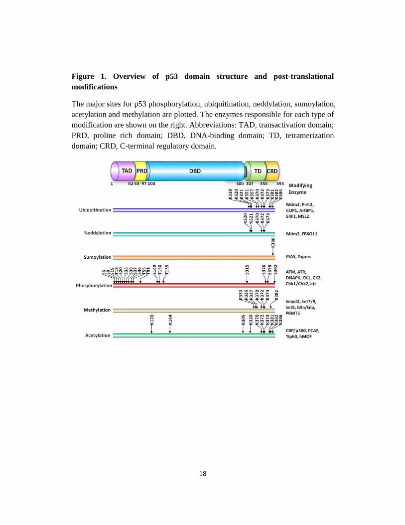
Figure 1. Overview of p53 domain structure and post-translational
modifications
The major sites for p53 phosphorylation, ubiquitination, neddylation, sumoylation,
acetylation and methylation are plotted. The enzymes responsible for each type of
modification are shown on the right. Abbreviations: TAD, transactivation domain;
PRD, proline rich domain; DBD, DNA-binding domain; TD, tetramerization
domain; CRD, C-terminal regulatory domain.
Page 38
19
1.5.1 Regulation of p53 stability
The cellular protein stability of p53 is tightly controlled: p53 has a very short
half-life in normal unstressed cells, ranging from 5-30 min [100], and the rapid
stabilization of p53 following stress stimuli allows for exertion of diverse
response pathways, such as the halter of cell cycle, the activation of the DNA
damage repair response, and the induction of the apoptotic response, to combat
theses stresses and protect cellular and genomic stability. The tight control of p53
stability is made possible by the ubiquitin-dependent proteasome degradation
pathway, with HDM2 being the chief mediator of p53 ubiquitination and
degradation.
1.5.1.1 Ubiquitination overview
Ubiquitination refers to the covalent conjugation of one or more ~8 kDa
ubiquitin molecules to a protein substrate, and requires the consecutive function
of three enzymes. The E1 ubiquitin-activating enzyme links the C-terminal
glycine of the ubiquitin molecule to its own active site cysteine through the
formation of a thioester bond; the ubiquitin molecule is then transferred to the
active site cysteine of the E2 ubiquitin-conjugating enzyme; and finally an E3
ubiquitin-ligating enzyme transfers the ubiquitin molecule to the protein
susbstrate and directing it to rapid degradation by the 26S proteasome [101].
Page 39
20
E1 and E2 enzymes have low substrate specificity: a single E1 can bind to
dozens of E2s, and a single E2 can bind to hundreds of E3s in a hierarchical way.
Unlike E1 and E2, the E3 ubiquitin ligase displays high target specificity, usually
through a specific substrate recognition domain or through other cofactors in the
case of multi-subunit E3 ubiquitin complexes.
E3 ligases can be divided into two types: those that harbor a Really Interesting
New Gene (RING) domain and those with a Homologous to the E6-AP Carboxyl
Terminus (HECT) domain [102].
1.5.1.2 p53 ubiquitination by HDM2
HDM2 is the pivotal E3 ubiquitin ligase and negative regulator of p53
[103,104]. HDM2 targets six p53 lysine (K) residues within the C-terminal
regulatory domain (K370, K372, K373, K381, K382, and K386; Fig. 2), promotes
p53 degradation by its E3 ubiquitin ligase function and ultimately inhibits p53
transcription activity. p53 is poly-ubiquitinated by high levels of HDM2 and
mono-ubiquitinated by low levels of HDM2 [105]. HDM2-mediated suppression
of p53 is 2-fold: (i) as an E3 ubiquitin ligase it targets p53 for ubiquitin-dependent
proteasomal degradation and (ii) it inhibits p53 transcriptional activation by
directly binding to and repressing p53 [106] (discussed in more detail in Chapter
1.5.3).
Page 40
21
Importantly, the gene encoding HDM2 is a p53 transcription target, therefore
the stress-induced increase in p53 levels leads to the expression of its own
negative regulator HDM2, which in turn downregulates p53, creating an
autoregulatory feedback loop [107]. The stabilization and activation of p53 go
hand in hand with the inhibition of HDM2 E3 ubiquitin ligase function [108,109].
The p53/HDM2 feedback loop is regulated by multiple factors including the
Alternate Reading Frame of the INK4a/ARF locus (ARF) tumor suppressor [110],
the E3-ligase activity-lacking HDM2 homolog HDMX (also known as HDM4)
[106] , the deubiquitinating enzyme ubiquitin specific protease 7 (USP7, also
known as herpesvirus associated ubiquitin specific protease (HAUSP)) [111], and
post-translational modifications of HDM2 such as phosphorylation and
acetylation [112-114].
The critical role for HDM2 suppression of p53 is best illustrated by the
overactivation of p53 in mdm2 null mice leading to embryonic lethality, which
can be rescued by the loss of p53 [115]. Furthermore, mice expressing a cysteine
(C) 462A mutated version of mdm2 (equivalent to C464A in HDM2), which loses
its E3 ligase activity but retains p53 binding capacity, die during embryogenesis
but can be rescued by the loss of p53 [116], demonstrating that the E3 ligase
activity of mdm2 is indispensable for the repression of p53.
Page 41
22
1.5.1.3 p53 ubiquitination by HDM2-independent E3 ubiquitin ligases
Despite the elevated p53 level and the spontaneous activation of p53 function
in mdm2 null mice [117], supporting that HDM2 is the principal endogenous E3
ubiquitin ligase targeting p53 with high specificity [118-120], p53 still undergoes
degradation in the absence of mdm2 [117], suggesting the existence of HDM2-
independent degradation pathways.
Indeed, several other E3 ligases have been shown to regulate p53 degradation
and localization independent of HDM2. In cell culture, the RING domain
containing p53-Induced protein with a RING-H2 domain (PIRH2) [121],
Constitutively Photomorphogenic 1 (COP1) [122], Carboxy terminus of Hsp70p-
Interacting Protein (CHIP) [123], Caspase 8/10-Associated RING Proteins
(CARPs) and SYNOVIOLIN [124,125], the HECT domain containing ARF-
Binding Protein 1 (ARF-BP1) [126] as well as Ubiquitin-Conjugating enzyme 13
(UBC13) (containing neither domain) [127] poly-ubiquitinate p53 and target it for
proteolysis. Whether these E3 ligases regulate p53 stability in vivo needs further
genetic validation.
Recent studies also support the existence of E4 ubiquitin ligases that
specifically target mono-ubiquitinated p53 in the cytosol for homeostatic
proteolytic degradation [128], possibly antagonizing the transcription-independent
Page 42
23
apoptotic functions of cytosolic p53, which requires mono-ubiquitinated p53 in
the mitochondria.
The presence of multiple ubiquitin ligases that control p53 stability suggests a
“fail-proof” redundancy in negative regulation. The capacity of these ligases to
repress p53 function predicts that these p53-specific E3 ubiquitin ligases could be
oncogenes. Indeed PIRH2, COP1 and WWP1 are amplified or overexpressed in
certain cancers [129-131].
1.5.1.4 p53 deubiquitination by USP7
The ubiquitination of p53 is counteracted mainly by the USP7
deubiquitinating enzyme. USP7 deubiquitinates p53, auto-ubiquitinated HDM2
and ubiquitinated HDMX [132], and changes in USP7 levels produce non-linear
effects on the p53-HDM2/HDMX pathway, therefore USP7 plays a dynamic role
in tumorigenesis.
Moderate down regulation of USP7 preferably stabilizes HDM2, therefore
leading to p53 destabilization and favors cell proliferation [133]. These data lend
support to the finding in a study of patient samples of Non-small Cell Lung
Cancer (NSCLC) that nearly 50% of NSCLC samples with wild-type p53 display
reduced USP7 mRNA expression [134]. In contrast, complete loss of USP7
function through a robust small interfering (si)RNA knockdown or knockout of
Page 43
24
the usp7 gene destabilizes HDM2 and HDMX, therefore stabilizing p53 and
would inhibit tumor growth [132]. This is consistent with the observation that no
USP7 mutation was identified in the TP53+/+
NSCLC samples [134].
Inhibition of USP7, therefore, presents a promising therapeutic approach for
treating cancers that retain wild-type p53. Indeed, a small molecule inhibitor
HBX41108 identified for USP7 by high-throughput screening stabilizes p53 in
tissue culture and inhibits tumor cell growth [135], warranting further studies to
confirm the anti-tumor effect in vivo.
1.5.2 Regulation of p53 localization
In normal cells under homeostasis, p53 is shuttled between the nucleus and
the cytoplasm [136]. In response to stress, however, p53 is rapidly translocated to
the nucleus to exert its biological function as a transcription factor. The recent
discovery of transcription-independent functions of p53 in the cytoplasm,
including direct activation of apoptosis at the mitochondria and inhibition of
autophagy, further underscore the importance of regulation of p53 localization
[137-139]. Indeed, interference with p53 localization has detrimental effects in
vivo: constitutive cytoplasmic localization of p53 has been linked to poor
response to chemotherapy, tumor metastasis and poor prognosis [140-142].
1.5.2.1 Cytoplasmic targeting of p53 by ubiquitination
Page 44
25
p53 ubiquitination not only targets it for proteasomal degradation, but also
plays a key role in regulating the cellular localization of p53 (Fig. 2). p53 is
polyubiquitinated when HDM2 levels are high and monoubiquitinated when
HDM2 levels are low [105]. Poly-ubiquitination primarily targets p53 for
proteasomal degradation, while mono-ubiquitination facilitates p53 cytoplasmic
translocation through exposing a C-terminal nuclear export signal and promoting
dissociation from HDM2 [103,143,144].
Several other E3 ubiquitin ligases also preferentially target p53 for nuclear
export independent of HDM2. WW domain-containing Protein 1 (WWP1)
mediates p53 ubiquitination and, unlike HDM2, stabilizes p53 at the protein level
and causes cytoplasimc accumulation of p53 [145]. Male-Specific Lethal-2
(MSL2) also promotes p53 ubiquitination but does not regulate p53 protein level;
instead it preferentially targets p53 for nuclear export [146].
1.5.2.2 Nuclear import of p53 through deubiquitination by USP10
Another member of the large deubiquitinase (DUB) family [147], USP10, has
been shown to remove ubiquitin chains from p53. However, unlike USP7, USP10
does not deubiquitinate HDM2 or HDMX. Rather, USP10 reverses HDM2-
induced p53 nuclear export, thereby recycling cytoplasmic p53 back to the
nucleus [148]. Thus, although both USP7 and USP10 target p53 for
deubiquitination, they function in different compartments: USP7 deubiquitinates
Page 45
26
and stabilizes p53 primarily in the nucleus [111], whereas USP10 largely
deubiquitinates cytoplasmic p53 during homeostasis, although it retains
deubiquitinase activity upon translocation to the nucleus following DNA damage
[148].
Using human Renal Cell Carcinoma (RCC) cell lines, Yuan and colleagues
showed that USP10 is capable of stabilizing both wild-type and mutant p53;
therefore USP10 might have different roles in tumorigenesis depending on the
p53 status [148]. In RCC cell lines that retain wild-type p53, USP10 behaves like
a tumor suppressor and upregulation of USP10 is favorable for repression of
cancer growth. In RCC cell lines that have mutant p53, USP10 promotes cancer
cell proliferation, and downregulation of USP10 would be beneficial for the
inhibition of cancer growth. usp10 knockout mice studies would facilitate our
understanding of the physiological role of USP10 in tumorigenesis. It is
perceivable that discovery of USP10-activating or -inhibiting drugs would offer
promising treatments for cancers with wild-type or mutant p53.
Page 46
27
Figure 2. Regulation of p53 stability and localization by ubiquitination
Nuclear p53 is targeted by HDM2 for monoubiquitination promoting cytoplasmic
translocalization or polyubiquitination promoting proteosomal degradation. The
abundance of HDM2 and HDMX are also regulated by ubiquitination and
deubiquitination. USP7 stabilizes p53, HDM2, and HDMX through
deubiquitination. In the cytoplasm, USP10 deubiquitinates monoubiquitinated p53,
reversing nuclear export and recycling p53 into the nucleus. Monoubiquitinated
p53 in the cytoplasm can possibly be further ubiquitinated by E4 ubiquitin ligases
and targeted for degradation. Cytoplasmic p53 also has transcription-independent
roles in activating apoptosis through permeabilization of the mitochondrial outer
membrane and the inhibition of autophagy through mechanisms yet to be
discovered. Abbreviations: U, Ubiquitination.
Page 47
28
1.5.3 p53 repression on promoters by HDM2 and HDMX
It was originally believed that p53 exists in a DNA-free form until cells
encounter stress stimuli, which in turn stabilizes and activates sequence-specific
DNA binding. However, increasing evidence now supports p53 basal binding to
DNA in a non sequence-specific manner, and the presence of p53 repressors at
target gene promoters prevent transcription activation until a stress stimuli occurs.
1.5.3.1 p53 is bound to DNA at homeostasis
Early studies focusing on the sequence specific DNA binding capacity of p53,
often utilizing in vitro assays such as Electrophoretic Mobility Shift Assay
(EMSA), led to the presumption that p53 exists in a DNA-free form under
homeostasis and that only stress-activated p53 could bind to DNA. DNA binding
was also thought to be mediated exclusively by the p53 central core domain and
requires stringent conformity to the consensus p53 response element. However
global Chromatin immunoprecipitation (ChIP) and microarray analysis of p53
binding to genomic DNA reveal considerable divergence from the consensus p53
binding response element [29,149]. Instead, the majority of p53-binding events in
vivo were found at non sequence-specific regions.
It is now understood that both the p53 central core domain and the C-terminal
regulatory domain possess DNA binding capacities [150,151], with the former
Page 48
29
providing primarily sequence-specific DNA binding and the latter recognizing
DNA structure and topology [150,152], thereby enabling DNA binding within the
vicinity of canonical p53 binding sites and providing a basis for sliding and
searching for specific sequences.
Additionally, although promoter-binding of p53 is increased in response to
genotoxic stress, quantitative ChIP assays reveal disproportionality to the fold
induction of target gene mRNA; instead a portion of p53 is bound to target gene
promoters in the absence of stress, and genotoxic stresses further enhances
promoter binding [153]. These studies support a model in which p53 is bound to
DNA but under constant repression.
1.5.3.2 Repression of p53 at promoters by HDM2/HDMX
Both HDM2 and HDMX interact directly with p53 and are recruited to p53
response elements in a p53-dependent manner. HDM2, HDMX and p53 form a
protein complex on target gene promoters and repress p53 function by preventing
access to the general transcriptional machinery [154,155].
The repression of p53 by HDM2 and HDMX is non-overlapping, because
neither regulator can compensate for the embryonic lethality caused by the loss of
the other [33]. The importance of HDM2 and HDMX in repressing p53 tumor
suppressor function is further supported by the prevalence (around 1/3 of human
Page 49
30
tumors) of HDM2 or HDMX gene amplification or overexpression in human
tumors retaining wild-type p53 [33,156].
1.5.3.3 De-repression of p53 is required for transcription activation
While DNA binding alone may be sufficient for p53 to maintain basal level
transcription of p53 negative regulators such as HDM2 and Pirh2 [106], in order
to induce a stress response through transactivating distinct subsets of target genes
p53 must first be released from HDM2/HDMX mediated repression. The
necessity for disrupting HDM2 mediated repression is highlighted by the Nutlin-
3A small molecule HDM2 antagonist, currently in phase I clinical trial, that is
sufficient to restore p53 transcription activity in cells with a wild type TP53 gene
[157].
De-repression of p53 from HDM2 and HDMX can be achieved through
several mechanisms. Post translational modifications on certain p53 residues
facilitate the dissociation of p53 from HDM2 (discussed in detail in Chapter
1.5.4). In response to DNA damage, HDM2 and HDMX also undergo a number
of post-translational modifications that either decrease protein stability or disrupt
interaction with p53 [112-114,158-160]. One such example is the ATM-
dependent phosphorylation of HDM2 and HDMX, which reduces their affinity for
the USP7 deubiquitinase and therefore accelerates HDM2 and HDMX
degradation [112]. Furthermore, in response to oncogenic activation, p14ARF
Page 50
31
directly interacts with the central region of HDM2, thereby antagonizing its
activity toward p53 [110]. Several nucleolar or ribosomal proteins also interact
directly with HDM2 and prevent its negative regulation of p53 [38,161,162].
1.5.4 Regulation of p53 transcription activity by post-translational
modifications
p53 is subject to a diverse and complex array of post-translational
modifications that influence its transcription activity at specific target gene
promoters. The most commonly reported post-translational modifications
affecting p53 transcription activity include phosphosphorylation of serines and/or
threonines and acetylation, sumoylation, neddylation and methylation of lysine
residues (Fig. 1). The presence of multiple p53 residues targeted by a single
enzyme and multiple modification possibilities on C-terminal lysines allows for a
multitude of combinations of post translational modifications that can be
conferred on the p53 protein. These serve as a “histone-like” code to dictate
correct and differentiated activation of certain sets of downstream targets
involving different cellular responses.
1.5.4.1 Phosphorylation
Human p53 harbors an array of serine (S)/threonine (T) phosphorylation sites
that span the entire protein but are concentrated in the N-terminal transactivation
Page 51
32
domain and the C-terminal regulatory domain (Fig. 1). The majority of these sites
are rapidly phosphorylated following cellular stress, although a few (e.g. T55 and
S376) are constitutively phosphorylated in unstressed cells and dephosphorylated
following stress [163,164]. p53 phosphorylation at the N terminus shows
significant redundancy; a single site can be phosphorylated by multiple kinases
and a single kinase can phosphorylate multiple sites [106].
1.5.4.1a Phorphorylation at Ser15/Ser20
The most extensively studied N-terminal p53 phosphorylation sites are S15
and S20 (S18 and S23 in mice). S15/S20 phosphorylation reduces p53 affinity for
its primary negative regulator HDM2, and promotes the recruitment of
transcriptional co-activators p300 and CBP on p53 target gene promoters [33].
Studies with mice containing single and double S to alanine (A) mutations reveal
a certain level of redundancy in the physiological importance of these two
phosphorylation sites. Although the individual mutations in gene knock-in
experiments in mice only marginally change p53 stability and transactivation
activity, the mice bearing p53 with both S15A and S20A mutations display a
more severe phenotype including tissue-specific deficiency in pro-apoptotic
capacity, mildly compromised replicative senescence and a latent development of
a spectrum of tumors [165].
1.5.4.1b Phosphorylation at Ser46
Page 52
33
S46 phosphorylation has recently attracted much attention. Phosphorylation of
S46 is critical for p53-mediated induction of pro-apoptotic genes such as p53-
regulated Apoptosis-Inducing Protein 1 (p53AIP1) but is not required for the
induction of cell cycle arrest targets [166,167]. Indeed, the resistance of a human
oral squamous cell carcinoma cell line HSC-3 to p53 is attributed to deficiency in
S46 phosphorylation, and the introduction of the exogenous phospho-mimic
p53S46D (aspartic acid) mutant enhanced transcription of the pro-apoptotic target
Noxa and restored apoptosis in HSC-3 cells [168]. A study with knock-in mice
expressing the human TP53 gene with the S46A mutation partially supports the
idea that S46 has a physiological role in differentially regulating cell cycle arrest
and apoptosis. The mutant mice, compared to knock-in mice expressing the wild-
type human TP53 gene, displayed modestly reduced p53 transcription of some
pro-apoptotic targets and compromised apoptosis but not cell cycle arrest,
although the effects were tissue-specific [169].
1.5.4.1c Phosphorylation at Ser392
Phosphorylation of C-terminal S392 in response to Ultra-Violet (UV) light
activates specific DNA binding through the stabilization of the p53 tetramer [34].
Knock-in mice with a S389A (human S392A) mutation displayed normal p53
stability but an increased predisposition to UV-induced skin cancer as well as
altered expression of p53 target genes compared to wild-type mice [170-172],
Page 53
34
supporting a physiological role for S392 phosphorylation in the tumor suppressive
responses of p53 to UV. However, some studies report a correlation between
S392 hyper-phosphorylation and poor prognosis, advanced tumor stage and tumor
grade in p53-positive cancers [173,174]. How does a tumor-suppressive
modification acquire tumor-promoting functions? Perhaps S392 phosphorylation
enhances the tetramer formation of certain gain-of-function p53 mutants, turning
these mutants into more potent oncoproteins. Further investigation is needed to
determine whether S392 phosphorylation is common to both wild-type and
mutant p53, and if so, how it might contribute to tumor progression.
1.5.4.2 Ubiquitin-like modifications
p53 is targeted by two ubiquitin-like proteins, Small Ubiquitin-like Modifier
(SUMO) and Neural precursor cell Expressed Developmentally Down-regulated
protein 8 (NEDD8), both of which are evolutionarily conserved in eukaryotes and
resemble ubiquitin in both their three-dimensional structure and their mechanism
of conjugation through lysines [175-177]. p53 is sumoylated at a single site K386
by members of the Protein Inhibitor of Activated Stat (PIAS) family and Topors
[178,179]. Neddylation of p53 is mediated by HDM2 and F-box protein 11
(FBXO11): HDM2 catalyzes the neddylation of three C-terminal lysines (K370,
K372 and K373) that are also targeted for ubiquitination [180], FBXO11
neddylates two lysines (K320 and K321) [181]. Unlike ubiquitination,
Page 54
35
neddylation and sumoylation have not been demonstrated to affect p53 stability or
localization. Neddylation inhibits p53 transcriptional activation activity [180,181],
whereas the functional consequences of K386 sumoylation is interesting, albeit
not well-defined; some reports have linked it to increased p53 transcriptional
activity and premature senescence [178,182-184].
It is noteworthy, that the low abundance of SUMO- or NEDD-8 modified p53
in vivo, normally less than 5% of total cellular p53, poses a challenge for defining
the cellular roles of these modifications. Reconstituted systems allow robust
testing of the roles of these ubiquitin-like modifications in vitro, but are unlikely
to recapitulate the physiological conditions in which these modifications occur. It
remains to be determined under what circumstances sumoylation and neddlyation
might affect p53 function.
1.5.4.3 Methylation
The large number of lysine and arginine residues in p53 presents the potential
for regulation by methylation (Figure 1). Arginine methylation has only been
shown for one methyltransferase, Protein Arginine N-Methyl Transferase 5
(PRMT5) [185,186], which targets R333, R335 and R337 in the tetramerization
domain, and methylation of these residues differentially affect the target gene
specificity of p53 [186]. p53 lysine methylation is better understood: p53 is
monomethylated by three different Lysine Methyl Transferases (KMTs) and
Page 55
36
dimethylated by at least two KMTs [187]. The functional consequences of p53
lysine methylation can be either activating or repressive, depending on the
location of the modification and the number of methyl groups attached.
Monomethylation at K372 is mediated by SET7/9 (also known as KMT7) and
this modification promotes the transactivation of target genes [188]. SET8 (also
known as KMT5A)-mediated K382 monomethylation and SMYD2 (also known
as KMT3C)-mediated K370 monomethylation repress p53 transcriptional activity
[189,190]. G9A (also known as KMT1C) and G9A-like Protein (GLP, also known
as KMT1D) dimethylate p53 at K373, thereby negatively regulating p53-mediated
apoptosis [191]. Interestingly, however, conjugation of a second methyl group to
K370 (K370me2), by a currently unknown enzyme, leads to a distinct functional
consequence from monomethylation. K370me2 increases in response to DNA
damage and promotes p53 function by facilitating the association of p53 with the
coactivator p53 Binding Protein 1 (53BP1) [192]. Lysine Specific Demethylase 1
(LSD1, also known as KDM1) preferentially removes this positive-acting second
methyl group thereby repressing p53 function by inhibiting the association of p53
with 53BP1 [192]. These findings suggest that p53 methylation and
demethylation dynamically regulate p53 function, at least in part by allowing or
disallowing p53 binding to coactivators.
Page 56
37
Interestingly, there appears to be crosstalk between p53 methylation at
different sites and between p53 methylation and acetylation. Activating
methylation of K372 inhibits the repressive methylation of K370 by preventing
SMYD2 binding to p53 [189]. Moreover, the repressive methylation of K382
normally prevents acetylation at this same site by CBP/p300 [190]. Upon DNA
damage, the level of methylation at K382 decreases, reversing its inhibitory effect
and allowing CBP/p300 acetylation of K382 and thereby promoting p53 activity.
Together, the interplay between p53 methylation sites as well as between p53
methylation and acetylation provide mechanisms for triggering a rapid increase in
p53 transcriptional activity in response to stress.
The presence of negatively acting lysine methylation sites and KMTs that
normally maintain p53 in an inactive state suggests the possibility that abnormally
high levels of KMTs could be oncogenic. Indeed, the SET domain containing
methyltransferase G9A is upregulated in many cancer cell types and its homolog
GLP is also overexpressed in brain tumors and multiple myeloma [191].
1.5.4.4 Acetylation
The acetylation of p53 is a powerful mechanism for activating function. The
significance of p53 acetylation is three-fold: (i) it promotes p53 stabilization by
excluding ubiquitination on the same site; (ii) it inhibits the formation of
Page 57
38
HDM2/HDMX repressive complexes on target gene promoters; and (iii) it recruits
cofactors for the promoter specific activation of p53 transcriptional activity.
Ten acetylation sites have been identified for p53, and the Histone Acetyl
Transferases (HATs) responsible for these modifications include the structurally
related p300 (also known as K(lysine) acetyltransferase 3B (KAT3B)) and CREB-
Binding Protein (CBP, also known as KAT3A), P300/CBP-Associated Factor
(PCAF, also known as KAT2B) and the MYST (named for members MOZ,
Ybf2/Sas3, Sas2 and Tip60) family HATs, Tat-Interactive Protein of 60 kDa
(TIP60, also known as KAT5) and human Males absent On the First (hMOF, also
known as MYST1/KAT8) [25,193-195] (Fig. 1).
1.5.4.4a Acetylation at the C-terminus
Six lysine residues (K370, K372, K373, K381, K382 and K386) in the C-
terminal regulatory domain are acetylated by CBP/p300 and ubiquitinated by
HDM2 [193] (Fig. 1). Acetylation in tissue culture systems activates sequence-
specific binding of p53 to DNA and its transcriptional activation activity and
enhances the stability of p53, owing to the mutual exclusion of acetylation and
ubiquitination. Nevertheless, despite some cell type-specific differences in
transcriptional profiles, mice expressing C-terminal acetylation-deficient p53
(p536KR
and p537KR
knock-in mice) generally exhibited no major difference in cell
cycle control, apoptosis or tumor suppression [196,197], which is in line with the
Page 58
39
fact that mutation in the p53 C-terminal regulatory domain is rarely found in
human cancers (UMD_TP53 Mutation database http://p53.free.fr/).
1.5.4.4b Acetylation at Lys320
K320 in the tetramerization domain is acetylated by PCAF [198]. It has been
reported that the competition between the mutually exclusive ubiquitination and
acetylation of K320 tips the cell fate balance. The atypical E3 ubiquitin ligase
E4F1 mediates non-degraded K48-linked oligo-ubiquitination of p53 on K320,
and competes with PCAF mediated acetylation [199]. High levels of K320
ubiquitination resulting from E4F1 overexpression specifically favors cell
survival by promoting p53-mediated induction of p21 [199]. This is supported by
studies using K317R (equivalent to human K320R) knock-in mice, showing
increased expression of pro-apoptotic target genes and enhanced p53-dependent
apoptosis upon irradiation [200], suggesting apoptotic repression by K320
acetylation.
1.5.4.4c Acetylation in the DNA binding domain
Two additional acetylation sites, K120 (K117 in mice, acetylation mediated
by TIP60/hMOF) [194,201] and K164 (K161 and K162 in mice, acetylation by
CBP/p300) [155] were discovered in the DNA binding domain. Importantly, both
K120 and K164 are recurrently mutated in cancer (UMD_TP53 Mutation
Page 59
40
database http://p53.free.fr/), implying that these two modifications might have
profound and nonredundant effects on p53 function.
K120 acetylation is indispensable for the activation of target genes involved in
apoptosis but not cell cycle arrest [194,201], suggesting a means for controlling
promoter specificity and hence cell fate. Indeed, in p53K117R
knock-in mice p53-
dependent cell cycle arrest and senescence remain intact but apoptotic induction
following ionizing radiation is completely abrogated [98], confirming the
indispensability of K120 acetylation to p53-mediated apoptosis. Additionally,
K120 acetylation might be required for p53 to effectively displace the
proapoptotic protein BCL2-Antagonist/Killer 1 (BAK) from the oncoprotein
Myeloid Cell Leukemia sequence 1 (MCL-1) at the mitochondria [202].
Therefore, it is probable that K120 acetylation by TIP60 contributes to both
transcription-dependent and transcription-independent apoptotic functions of p53.
In cell culture based assays using human p53, individual K to R mutation can
be compensated for by acetylation at other sites; however the collective mutation
of eight acetylation sites (p538KR
: mutation at K120, K164, and six CBP/p300-
targeted C-terminal sites) completely abolishes p53-mediated cell cycle arrest and
apoptosis [155], demonstrating that acetylation is indispensible for the canonical
p53 functions. Mechanistically, acetylation allows p53 to evade HDM2 and
Page 60
41
HDMX repression by blocking recruitment of HDM2 and HDMX to target gene
promoters [155].
In mice, however, the collective loss of acetylation at K117 (human K120)
and K161/K162 (human K164) seems sufficient to recapitulate the phenotypes
seen with human p538KR
. p533KR
knock-in mice are completely deficient in
eliciting growth arrest, apoptosis, or senescence in vivo [98], confirming the
physiological importance of acetylation in the transcription activation of
canonical p53 targets.
1.5.4.4d Deacetylation by HDACs and SIRT1
Equilibrium in the acetylation of p53 is maintained by the Histone
Deacetylases (HDACs), HDAC1 and Sirtuin 1 (SIRT1) [203,204]. SIRT1
preferentially deactylates p53 at K382 and has a profound negative impact on the
capacity of p53 to induce the expression of target genes involved in apoptosis,
such as PUMA and BAX. Thymocytes of Sirt1-deficient mice exhibit p53
hyperacetylation and increased radiation-induced apoptosis compared to wild-
type thymocytes [205]. SIRT1 is negatively regulated at the transcriptional level
by Hypermethylated In Cancer 1 (HIC1) and at the translational level by the
microRNA (miR)-34a [206,207], both of which are targets of p53 [208-212].
SIRT1 expression is elevated in leukemia [213], prostate cancer [214] and skin
cancer [215], and it is negatively regulated by Deleted in Breast Cancer 1 (DBC1)
Page 61
42
[216,217], supporting a role for SIRT1 in tumorigenesis. However, the
suppression of intestinal tumorigenesis and colon cancer growth in a β-catenin-
driven mouse model of colon cancer by ectopic induction of Sirt1 [218] suggests
that it also has tumor-suppressive properties.
The evidence that SIRT1 harbors both tumor-promoting and tumor-
suppressing functions generates interest in developing SIRT1-targeted drug
therapies for cancer treatment [219]. The most promising SIRT1 inhibitors
discovered to date are tenovin-1 and its more water-soluble derivative, tenovin-6
[220]. At low micromolar concentrations, tenovins potently inhibit the
deacetylase activities of SIRT1 and SIRT2, significantly increase the level of p53
K382 acetylation in tissue culture and decrease tumor growth in xenograft mouse
tumor models. Studies on activators of SIRT1 focus on resveratrol, which is
abundant in grapes. Although dietary intake of resveratrol delays aging in mice
[221], more studies are needed to assure that resveratrol activation of Sirt1 does
not impose cancer susceptibility.
1.5.4.5 Concluding remarks
Although biochemical and cell culture based studies have highlighted the
crucial role of a number of post-translational modifications in the activation of
p53 transcription activity, the relatively mild and tissue/cell type-specific
phenotypes of many knock-in mice with a single point mutation that abolishes a
Page 62
43
certain modification suggest functional redundancy, perhaps important for the
“fail-proof” regulation of p53 considering its central role in tumor suppression.
Although each site/modification might only fine-tune p53 function, the numerous
possible combinations of different modifications could dictate p53 activity in a
promoter-specific manner, allowing p53 to exert a spectrum of functions.
The striking phenotype of the p533KR
mice, however, undeniably underscores
the absolute requirement for p53 acetylation in activating the transcription of
canonical targets involved in the classic growth arrest, apoptosis and senescence
response pathways.
1.6 Summary
Although accumulating evidence supports the indispensability of acetylation
in the activation of p53 function and indicates cell fate modulation, the underlying
mechanisms are not completely understood. The experiments in this study were
designed to identify novel regulators of p53 acetylation and to study the
mechanisms modulating p53-mediated cell fate decision.
In this study we identify p90 and UHRF1 as two novel members of the p53
regulatory network. Although both function upstream of the TIP60-p53 interplay,
Page 63
44
they act through distinct and opposing mechanisms to dynamically modulate
TIP60-mediated effects on p53 in vivo.
Page 64
45
CHAPTER 2
DIFFERENTIAL EFFECTS ON p53-MEDIATED CELL
CYCLE ARREST VS. APOPTOSIS BY p90
Page 65
46
2.1 Introduction
p53 was the first nonhistone protein known to be regulated by acetylation and
deacetylation [25,203]. There is accumulating evidence indicating that acetylation
of p53 plays a major role in activating p53 function during stress responses
[222,223]. Following early findings of C terminus p53 acetylation [25], the Gu
team and others recently showed that p53 is also acetylated by TIP60 (also known
as KAT5)/MOF (human ortholog of males absent on the first) at residue Lys120
(K120) within the DNA-binding domain [194,201,224]. K120 acetylation is
crucial for p53-mediated apoptosis but has no obvious effect on p21 expression,
an essential target of p53-mediated growth arrest [98]. Notably, although TIP60 is
required for K120 acetylation of p53 in vivo, the levels of K120 acetylation are
dynamically regulated in vivo and the interaction between p53 and TIP60 is not
very stable, indicating that additional regulators may play a role in controlling
K120 acetylation and subsequent p53-mediated apoptotic response [225-227].
Through biochemical purification, we identified p90 as a unique regulator for
p53. p90, also called CCDC8 (coiled-coil domain containing 8), which was
previously found down-regulated in human cancer cells [228,229], interacts with
p53 both in vitro and in vivo. Knockdown of p90 has no obvious effect on p53-
mediated activation of p21 but specifically abrogates its effect on p53 upregulated
modulator of apoptosis, also known as Bbc3 (PUMA) activation. Moreover, p90
Page 66
47
also interacts with TIP60 and promotes TIP60-dependent Lys120 acetylation of
p53, therefore enhancing the apoptotic response of p53. These data reveal p90 as
an upstream regulator of the TIP60-p53 interaction and demonstrate that p90 is
specifically required for p53-mediated apoptosis upon DNA damage.
Page 67
48
2.2 Results
2.2.1 Identification of p90 as a unique component of p53-associated
complexes
To further elucidate the mechanisms of p53-mediated promoter-specific
activation in vivo, p53-associated protein complexes were isolated from human
cells. Attempts to purify p53-containing protein complexes were hindered in the
past because cells cannot tolerate expressing even low levels of wild-type p53.
Interestingly, recent studies by the Gu team indicate that p538KR
, in which all
eight p53 acetylation sites are mutated to arginine, is inactive in inducing cell
cycle arrest or apoptosis [155]. Moreover, p538KR
retains the capacity to bind
target gene promoters as well as to activate the p53-HDM2 feedback loop,
suggesting that p538KR
, unlike the hot spot tumor mutant p53H175R
, may retain a
similar conformation as wild-type p53 in human cells. Therefore we have utilized
an H1299 p53-null lung carcinoma cell line that stably expresses a double tagged
human p538KR
mutant protein with N-terminal FLAG and C-terminal HA epitopes
(FLAG-p538KR
-HA) (Fig. 3A).
To ensure physiological interactions, H1299 derivatives were selected such
that the expression level of the ectopic p538KR
protein is not much higher than
endogenous p53 in HCT116 colon cancer cells upon DNA damage treatment. As
Page 68
49
expected, HDM2 is activated in the p538KR
stable line to a similar level compared
to that induced by DNA damage in HCT116 cells. Consistent with previous
findings, pro-apoptotic and growth arrest targets such as PUMA and p21 are not
activated in the p538KR
stable line (Fig. 3B).
Page 69
50
Figure 3. Construction of p538KR
stable line in H1299
(A) Schematic representation of the p538KR
protein used for protein complex
purification. Mutations of acetylation sites are indicated. TAD,
transcription activation domain; PRD, proline rich domain; DBD, DNA-
binding domain; TD, tetramerization domain; CTD, C-terminal regulatory
domain.
(B) Characterization of H1299 cells stably expressing p538KR
. Total cell
extracts from FLAG-p538KR
HA/H1299 stable cell line and HCT116 cells
with or without 8hr treatment with 20 μM etoposide were assayed by
Western blot analysis using antibodies against p53, β-actin, Mdm2,
PUMA, and p21.
A
B
Page 70
51
To isolate p538KR
-containing complexes, cell extracts from the stable line
were subjected to a two-step affinity chromatography previously described [126].
The tandem affinity-purified p53-associated proteins were analyzed by liquid
chromatography (LC) MS/MS. As expected, known p53 binding proteins such as
HDM2, tumor protein 53 binding protein 1 (p53BP1), USP7, and the CREB
binding protein (CBP) were identified as specific components of the p53 complex
(Fig. 4A). In addition, MS analysis of a protein band p90 (with the apparent size
at approximately 90 kDa molecular mass) revealed six peptide sequences matched
with a signal cDNA sequence in the database, which is also named CCDC8 (Fig.
4B). Because none of the peptide sequences of p90 were identified from the
control complexes purified in parental H1299 cells, p90 is likely a unique binding
partner of p53.
The cDNA of p90/CCDC8 encodes a 538 amino acid protein possessing no
known functional domains other than two small coiled-coil regions that are likely
to mediate protein–protein interactions (Fig. 4B). Although p90/CCDC8 has been
reported as a candidate tumor suppressor gene in renal cell carcinoma (RCC), the
molecular function of this protein is unclear [230].
Page 71
52
Figure 4. Identification of p90 as a component of a p53-containing protein
complex
(A) Silver staining of purified p538KR
containing protein complex. Peptide
sequences identified from the mass spectrometric analysis are presented.
(B) Schematic representation of the p90 protein. p90 contains two coiled-coil
regions and two potential ATM/ATR phosphorylation sites.
A
B
Page 72
53
2.2.2 p90 is a bona fide p53 interacting protein
To investigate a role for p90 in regulating p53 function in vivo, the interaction
between p90 and p53 was first tested. Thus, H1299 cells were transfected with
expression vectors for FLAG-tagged p53 and HA-tagged p90, and Western blot
analysis revealed that p90 is readily detected in p53-associated
immunoprecipitates (Fig. 5A).
To further elucidate this interaction under physiological settings, a polyclonal
antiserum was then raised against the full-length p90 protein. Upon Western blot
analysis, the affinity-purified antibody specifically detected in human cells an
approximately 90 kDa polypeptide, the level of which decreases significantly
after treatment with p90-specific siRNA oligos (Fig. 5B). To investigate the
interaction between endogenous p90 and p53 proteins, extracts from U2OS
osteosarcoma cells were immunoprecipitated with α-p53 antibody or with the
control IgG. As expected, the α-p53 antibody immunoprecipitated endogenous
p53; more importantly, p90 is easily detected in the immunoprecipitates obtained
with the α-p53 antibody but not the control IgG (Fig. 5C, lanes 2 and 3),
confirming that p90 and p53 interact endogenously.
Page 73
54
Figure 5. p90 interacts with p53 in vivo
(A) p90 coimmunoprecipitates with p53 in an overexpression system. H1299
cells were transiently transfected with plasmid DNA expressing HA-p90
or/and FLAG-p53. The cell extracts and the M2 immunoprecipitates (IP)
were analyzed by Western blot analysis using α-HA and α-p53 antibodies.
(B) Purified p90 antisera specifically recognizes p90 protein. U2OS cells
were transiently transfected with either control siRNA or p90-specific
siRNA. Whole cell lysates were analyzed with α-90 antisera.
(C) p90 interacts with p53 endogenously. Total cells extracts from U2OS cells
were immunoprecipitated with α-p53 (DO-1) antibody or a control mouse
IgG . Extracts and immunoprecipitates were assayed by Western blot
analysis using α-p90 and α-p53 antibodies.
A B
C
Page 74
55
An in vitro GST-pulldown assay was performed to further assess direct
interaction. p53 can be divided into an N-terminal (NT) fragment containing the
transactivation domain, a middle fragment (M) containing the DNA-binding
domain, and a C-terminal (CT) fragment containing the tetramerization domain as
well as the regulatory domain. Purified recombinant GST-tagged p53 full-length
and fragment proteins were incubated with in vitro translated 35
S-methione-
labeled HA-p90. Following immobilization with GST resins and recovery of
captured complexes using reduced glutathione, the eluted complexes were
resolved by SDS-PAGE and analyzed by autoradiography. 35
S- methionine-
labeled HA-p90 strongly bound immobilized GST-tagged full length and CT
fragment of p53 (Fig. 6, lanes 2 and 5), but not the NT and middle fragments of
p53 or GST alone (Fig. 6, lanes 3, 4, and 6). These data demonstrate that p90
interacts with p53 in vitro through binding directly to the C-terminal portion of
p53.
Page 75
56
Figure 6. p90 interacts with the C-terminal domain of p53 in vitro
The bacteria purified full-length and fragmented GST-p53 fusion proteins or GST
were used in the GST-pulldown assay with in vitro translated 35
S-methionine
labeled HA-p90 protein. The complexes were captured with GST sepharose beads
and eluted with reduced glutathione. The eluted complexes were resolved by
SDS-PAGE and analyzed by autoradiography. The levels of the GST fusion
proteins are shown in the bottom panel stained by Coomassie blue.
Page 76
57
We further mapped the binding fragment on p90. The N-terminal and C-
terminal fragments of p90 were subcloned into HA expression vector, and in vitro
translated 35S-methionine labeled HA-p90 fragments were incubated with GST-
p53 or GST. Following capture and elution, the complexes were resolved by SDS-
PAGE and analyzed by autoradiography. The full length p90 protein and the N-
terminal fragment, but not the C-terminal fragment of p90 bound GST-p53 (Fig.
7). These data demonstrate that the N-terminal fragment of p90 protein and the C-
terminal portion of p53 interact directly in vitro.
Page 77
58
Figure 7. p90 interacts with p53 through its N-terminal fragment
The N-terminal and the C-terminal fragments of p90 are subcloned into HA-
tagged expression vector. The bacteria purified GST-p53 or GST were used in the
GST-pulldown assay with in vitro translated 35
S-methionine labeled HA-p90
fragments, essentially as in Fig. 7. The eluted complexes were resolved by SDS-
PAGE and analyzed by autoradiography.
Page 78
59
Because p90 was identified in the p53 complex purified from whole cell
extracts, we assessed the cellular localization of the p90–p53 interaction. To this
end, U2OS cell line stably expressing FLAG and HA double tagged p90 was
established. Using parental U2OS cells as control, cell extracts from the p90
stable line were fractionated, and both nuclear and cytoplasmic fractions were
immunoprecipitated with M2/FLAG agarose beads. Western blot analysis showed
that p53, as expected, localizes mainly in the nuclear fraction, whereas p90 is
present in both fractions but more so in the cytoplasmic fraction (Fig. 8A).
Furthermore, in the M2 immunoprecipitate, p53 is primarily found in the nuclear
fraction (Fig. 8A). The difference in p90/p53 ratio in the cytoplasmic and nuclear
fractions as well as the p53 abundance in the nuclear M2 immunoprecipitate
indicate that, although p90 is present in both the cytoplasm and the nucleus, it
interacts with p53 predominantly in the nucleus. In agreement with the
fractionation experiment, immunostaining of HA-p90 transfected U2OS cells
revealed that p90 is localized to both the nucleus and the cytoplasm, regardless of
etoposide challenge. p53 was stabilized following etoposide treatment and co-
localized with p90 in the nucleus. (Fig. 8B). These data demonstrate that p90
interacts with p53 primarily in the nucleus and probably regulates p53 nuclear
activities.
Page 79
60
Figure 8. p90 interacts with p53 in the nucleus
(A) p90 localizes in both the cytoplasm and nucleus but interacts with p53
predominantly in the nucleus. Parental U2OS or FLAG-HA-p90/U2OS
stable cell line were fractionated. Extracts and M2 immunoprecipitates
from the cytoplasmic and nuclear fractions were assayed by Western blot
analysis using α-HA and α-p53 antibodies. β-tubulin and proliferating cell
nuclear antigen (PCNA) were used as cytoplasmic and nuclear protein
markers, respectively.
(B) p90 localization remains unchanged regardless of DNA damage and p90
colocalizes with activated p53. U2OS transiently transfected with
expression plasmid for HA-p90 was subjected to 8hr of etoposide
treatment and immunostained with α-HA and α-p53 (DO-1) antibody.
A
B
Page 80
61
2.2.3 Inactivation of p90 attenuates p53-mediated activation of PUMA but
not p21
To understand the physiological role of p90, we examined whether
inactivation of endogenous p90 has any effect on the stability and functions of
p53. To this end, U2OS cells were transfected with a p90-specific (p90-RNAi#1)
siRNA oligo or a control (control-RNAi) siRNA oligo. As shown in Fig. 9A,
lanes 1 and 2, the level of endogenous p90 polypeptides was severely re- duced
after transfection with p90-RNAi. p53 protein level was unaffected by p90
ablation, suggesting that p90 does not regulate p53 stability. We then assessed the
effect of p90 inactivation on the level of two important p53 downstream targets:
the growth arrest target p21 and the apoptotic target PUMA. Surprisingly, p90
ablation displayed differential effects on the two different endogenous targets: the
level of PUMA was significantly reduced, whereas p21 expression remained
unchanged. To exclude off-target effects, cells were treated with three additional
p90 siRNAs (p90-RNAi#2, p90-RNAi#3; p90-RNAi#4) that target different
regions of the p90 mRNA. Again, the levels of PUMA were decreased by p90
knockdown, although there was no significant change for the levels of p53 and
p21 (Fig. 9A, lanes 3–5).
Because p53 is strongly activated upon DNA damage and regulates
downstream targets, we wanted to assess whether p90 affects p53 and
Page 81
62
downstream target activation upon DNA damage. U2OS cells were transfected
with p90-specific siRNA oligos followed by treatment with the DNA damage
reagent etoposide. As expected, p53 levels increased drastically upon DNA
damage (Fig. 9B, lane 2 vs. lane 1), and notably, RNAi-mediated ablation of p90
displays no effect on p53 accumulation following etoposide treatment (Fig. 9B,
lane 4, 6, 8, and 10 vs. lane 2). p21 was strongly induced upon treatment, however,
damage-induced PUMA expression was severely attenuated in the cells treated
with p90-RNAi (Fig. 9B, lanes 4, 6, 8, 10 vs. lane 2).
Page 82
63
Figure 9. p90 inactivation reduces basal PUMA level and differentially
affects PUMA and p21 induction upon DNA damage
(A) p90 RNAi does not affect p53 stability or p21 basal level but reduces basal
PUMA expression. U2OS cells were transiently transfected with either
control siRNA or four different p90-specific siRNA oligos. Cell extracts
were assayed by Western blot analysis using the indicated antibodies.
(B) p90 RNAi attenuates PUMA but not p21 activation upon DNA damage.
U2OS cells transfected with the indicated siRNAs were treated with or
without 20 μM etoposide for 8hr. Total cell extracts were assayed by
Western blot analysis using the indicated antibodies.
A
B
Page 83
64
To validate that the differential effect of p90 on PUMA and p21 is p53
dependent, we inactivated both p53 and p90 in U2OS using RNAi prior to
etoposide treatment. In cells transfected with the control siRNA, p53 accumulates
and both PUMA and p21 are activated significantly upon treatment (Fig. 10, lanes
1 and 2). In a p53-deficient background, DNA damage fails to activate PUMA
and p21 (lanes 3 and 4), and more importantly p90 ablation displayed no effect on
PUMA and p21 in the absence of p53 (lanes 5 and 6). Taken together, these data
demonstrate that p90 inactivation differentially affects PUMA and p21 induction
in a p53-dependent manner.
Page 84
65
Figure 10. Differential regulation of PUMA and p21 activation by p90 is
dependent on p53
p53 alone, or both p53 and p90 were inactivated in U2OS cells using RNAi.
Subsequently, cells were treated with or without 20 μM etoposide for 8 hr before
extraction and Western blot analysis using the indicated antibodies.
Page 85
66
2.2.4 p90 is required for p53-mediated apoptosis upon DNA damage
To further confirm the differential effects on p53-mediated activation of p21
versus PUMA, we collected the cells at different time points following treatment
with etoposide. At all time points, p53 accumulation and p21 activation were
unaffected by p90 ablation but PUMA induction was severely attenuated (Fig.
11A, lanes 5 and 6 vs. lanes 2 and 3).We further confirmed that p90 ablation
affected p53-dependent activation of p21 and PUMA at the transcription level by
examining the mRNA levels of these targets. Indeed, basal PUMA mRNA was
reduced in samples treated with p90-RNAi, consistent with our finding that p90
ablation reduces basal PUMA protein level (Fig. 11A). PUMA activation was
attenuated at the mRNA level following p90 ablation, whereas p21 mRNA level
increased upon etoposide treatment at all time points and remained unaffected in
samples treated with p90-RNAi.
To further confirm these differential effects of p90 in p53 responses, we
repeated these experiments in the cells treated with another DNA damage reagent
doxirubicin. Again, we observed the differential effects of p90 on p53-dependent
p21 and PUMA activation upon doxirubicin treatment (Fig. 11B, lanes 6–8 vs.
lanes 2–4). These results suggest that p90 is crucial for p53-dependent activation
of PUMA, which is a very important mediator of p53-mediated apoptosis
[56,57,231].
Page 86
67
Figure 11. Inactivation of p90 attenuates p53-dependent PUMA activation in
time point experiments
(A and B) p90 RNAi reduces PUMA but not p21 activation upon DNA damage.
U2OS cells transiently transfected with either control siRNA or p90-specific
siRNA were treated with 20 μM etoposide or 0.34 μM doxirubicin for the
indicated time. Cell extracts were analyzed by Western blot analysis using the
indicated antibodies. Total RNA were isolated from the same experiment.
Followng reverse transcription, PCR was performed to detect the PUMA and p21
transcripts.
A
B
Page 87
68
We therefore speculated whether the attenuation of PUMA activation by loss
of p90 can be translated into a phenotypic effect on apoptosis. To this end, we
transfected U2OS cells with either control siRNA or p90 siRNA prior to
etoposide treatment. Cells were collected at different timepoints, stained with
propidium iodide (PI), and analyzed by flow cytometry for apoptotic cells
according to DNA content. As shown in Fig. 12A, basal level sub-G1 content is
minimally affected by inactivation of p90. However, following 18hr or 24hr of
etoposide treatment, an average of 18.67% or 26.84% of cells transfected with
control siRNA were apoptotic, whereas only 8.04% or 11.49% of cells transfected
with p90 siRNA were apoptotic (Fig. 12B). These data demonstrate that p90 is
crucial for p53-mediated apoptosis.
Page 88
69
Figure 12. Inactivation of p90 impairs p53-mediated apoptosis upon damage
(A) FACS analysis of p90-inactivated U2OS cells treated with etoposide.
U2OS cellstransiently transfected with either control siRNA or p90 siRNA
were treated with 20 μM etoposide for the indicated time. Cells were fixed
in cold methanol, stained with propidium iodide and subjected to DNA
content analysis by flow cytometry.
(B) p90 RNAi attenuates apoptosis. Percentages of apoptotic cells from (A)
are presented. Values are an average of three independent experiments.
Error bars, 1 standard deviation.
A
B
Page 89
70
2.2.5 Mechanistic insights into p90-mediated effect on p53-dependent
apoptotic responses
Previous studies demonstrated that p53 acetylation at Lys120 (p53 AcK120)
by TIP60 is indispensable for apoptosis but not required for growth arrest
[194,201], leading to our speculation that p90 may regulate p53-mediated PUMA
activation through promoting TIP60-dependent acetylation of p53 at K120. To
investigate the mechanism underlying the effect of p90 on the apoptotic target
PUMA, we first assessed the interaction between p90 and TIP60. To this end,
H1299 cells were transfected with expression vectors for TIP60 and FLAG/HA
double-tagged p90. Western blot analysis revealed that TIP60 is readily detected
in p90 associated immunoprecipitates (Fig. 13A). Using a GST-pulldown assay,
we further tested the in vitro interaction of TIP60 and p90. As shown in Fig. 13B,
TIP60 bound to immobilized GST-tagged p90 but not GST alone, demonstrating
that p90 and TIP60 interacts directly. Further, p90, TIP60 and p53 form a ternary
complex (Fig. 13C).
Page 90
71
Figure 13. p90 interacts with TIP60 and forms a ternery complex with TIP60
and p53
(A) TIP60 coimmunoprecipitates with p90 in an overexpression system.
H1299 cells were transiently transfected with the plasmid DNA expressing
TIP60 or/and FLAG-HA-p90. Cell extracts and M2 immunoprecipitates
were assayed by Western blot analysis using α-TIP60 and α-HA
antibodies.
(B) p90 interacts with TIP60 in vitro. The GST-p90 fusion protein or GST
alone was used in the GST-pulldown assay with in vitro translated 35
S-
methione labeled FLAG-HA-TIP60 protein. The immobilized complexes
were resolved by SDS-PAGE and analyzed by autoradiography.
(C) Total cell extracts from H1299 cells transfected with HA-p0, F-p53 and
Tip60 are subjected to a 2-step immunoprecipitation using M2/Flag and
HA agarose beads. Extracts and eluates are assayed by Western Blot using
antibodies against HA, p53, and TIP60.
A B
C
Page 91
72
To investigate the role of p90 in p53 K120 acetylation by Tip60, we examined
whether TIP60-mediated p53 acetylation is modulated by p90 status. As expected,
in a cotransfection system, p53 was readily acetylated by TIP60. Notably,
although p90 itself does not acetylate p53, p53 acetylation by TIP60 was
significantly enhanced upon p90 expression (Fig 14A). These data demonstrate
that p90 promotes the aceylation of p53.
In order to confirm the effect of p90 on Tip60-mediated p53 K120 acetylation
under physiological settings, we inactivated p90 in U2OS cells via RNAi and
assessed endogenous acetylation of p53 at K120. Because the steady-state levels
of K120 acetylation are dynamically regulated by both acetylases and
deacetylases, in order to exclude the potential effect on p53 acetylation levels by
deacetylases, cells were treated with deacetylase inhibitors trichostatin A (for
inhibiting histone deacetylase 1/histone deacetylase 2-mediated deacetylation of
p53) and nicotinamide (for inhibiting Sirt1-mediated deacetylation of p53) prior
to harvesting [201,225,227]. Cell extracts were immunoprecipitated with α-Ac-
p53K120 or control IgG. As shown in Fig. 14B, p53 acetylation at K120 was
easily detected in the cells with the deacetylase inhibitor treatment; however, the
levels of p53 acetylation at K120 were significantly reduced upon p90
knockdown (Fig. 14B, lane 4 vs. lane 2). Taken together, these data indicate that
Page 92
73
p90 is a critical cofactor for p53-mediated apoptosis through promoting K120
acetylation of p53.
Page 93
74
Figure 14. p90 promotes TIP60-mediated p53 acetylaiton at K120
(A) p90 promotes p53 acetylation by TIP60 at K120. H1299 cells were
transiently transfected with plasmid DNA expressing FLAG-p53, Tip60,
and/or HA-p90. Cell extracts and M2 immunoprecipitates were assayed by
Western blot analysis using antibodies against HA, p53, TIP60, and p53-
AcK120.
(B) Inactivation of p90 significantly reduces p53 acetylation at K120. U2OS
cells were transiently transfected with either control siRNA or p90 siRNA,
and treated with 1 μM trichostatin A (TSA) and 5 mM nicotinamide (NTA)
6 h prior to harvesting. Cell extracts and immunoprecipitates obtained
with α-Acp53K120 or control IgG were analyzed by Western blot analysis
using antibodies against p90, p53, and β-actin.
A
B
Page 94
75
2.3 Discussion
Our findings reveal that p90 is a p53 interacting protein with differential
effects on p53-mediated activation of target genes. Here, we have demonstrated
that p90 is a bona fide p53 interacting protein and that this interaction primarily
occurs in the nucleus. Inactivation of p90 attenuates apoptosis due to
downregulation of p53-mediated PUMA activation upon DNA damage. However,
p90 does not appear to affect growth-arrest targets such as p21. To dissect the
molecular mechanism underlying this differential regulation, we found that p90
interacts with the TIP60 acetyltransferase and promotes TIP60-mediated
acetylation of p53 at K120, a posttranslational modification that has previously
been reported to modulate the decision between cell cycle arrest and apoptosis
[194,201,224]. Thus, p90 likely serves as an upstream regulator of the p53-TIP60
interplay that is required for apoptotic signaling and allows for transcription
induction of PUMA in cells at risk of DNA damage.
K120 is located within the p53 DNA-binding domain and is recurrently
mutated in cancer (UMD_TP53 mutation database http://p53.free.fr/). Acetylation
at K120 is indispensable for activation of pro-apoptotic targets but is not required
for activation of growth-arrest targets [194,201]. Although the mechanism
underlying this target specificity remains to be elucidated, it is possible that
acetylation at K120 may impose specificity through altering the p53 quaternary
Page 95
76
structure and thus endowing p53 binding to low-affinity response elements that
are found on pro-apoptotic promoters [232,233].
We also noticed a small amount of cytoplasmic p53–p90 interaction.
Cytoplasmic localization of p53 was originally thought to passively block
transactivation in the nucleus. However increasing evidence suggests cytoplasmic
p53 has important roles in regulating apoptosis and autophagy. Cytoplasmic p53
promotes apoptosis through increasing mitochondrial outer-membrane
permeabilization and release of cytochrome c [66,69,70]. Basal levels of wild-
type p53 in the cytoplasm also inhibits autophagy, although the exact mechanism
remains to be understood [138]. It will be interesting to explore the possibilities of
p90 regulating transcription-independent functions of p53 in the cytoplasm.
It is noteworthy that p90 itself is underexpressed in human tumors, including
kidney cancer and myeloma, based on the cancer gene expression profile database
from Oncomine Research [228,229]. In this regard, p90 has also been identified
as a candidate tumor suppressor gene as hypermethylation and transcriptional
silencing of the p90 promoter was found in 35% of primary RCC tumor samples
[230]. It will be interesting to test whether p53-mediated apoptosis is abrogated in
the human tumors lacking p90 expression and whether reactivation of silenced
p90 promotes apoptosis thereby contributing to tumor suppression.
Page 96
77
Finally, protein modifications of the components in the p53 pathway are well
accepted as the key mechanisms for controlling p53 function during stress
responses [146,177](2, 35). Interestingly, p90 contains two potential ataxia
telangiectasia mutated/ataxia telangiectasia and Rad3 related (ATM/ATR)
phosphorylation sites at Ser-199 and Ser-302 (Fig. 4B). Indeed, in a screen assay
performed by the Elledge Group [234], a phosphorylated peptide derived from
p90 was identified as an ATM/ATR substrate. Future investigations are required
to validate if p90 undergoes damage-induced phosphorylation by ATM/ATR and
dissect whether p90 phosphorylation modulates its interaction with p53 and Tip60
as well as p53-mediated apoptotic responses. It is possible that p90 is functionally
regulated by ATM/ATR mediated phosphorylation during the DNA damage
response to control the decision between cell cycle arrest and apoptosis mediated
by p53.
Page 97
78
2.4 Materials and methods
Plasmids
The full-length p90 cDNA was PCR-amplified from Human MGC Verified
FL cDNA (Open Biosystems) and subcloned into pcDNA3.1/V5-His-Topo vector
(Invitrogen), pCIN4-FLAG-HA, or pCIN4-HA expression vector [235]. To
construct the GST-p90 plasmid, cDNA sequences corresponding to the full-length
p90 were amplified by PCR from other expression vectors and subcloned into
pGEX-2T (GE Healthcare) vector for expression in bacteria.
Cell culture
H1299 and U2OS cells were maintained in DMEM (Cellgro) and HCT116
cells in McCoy’s 5A medium (Cellgro). All media were supplemented with 10%
fetal bovine serum (Gibco), 100 I.U./mL penicillin and 100 ug/mL streptomycin
(Cellgro). The stable cell lines were established by transfecting H1299 or U2OS
cells with the plasmids pCIN4-FLAG-p538KR-HA and pCIN4-FLAG-HA-p90,
respectively, followed by selection with 1 mg/mL or 0.5 mg/mL G418 (EMD
Biosciences). Independent clones were selected and evaluated for expression by
immunoblot. Transfections with plasmid DNA were performed using the calcium
phosphate method and siRNA transfections by Lipofectamine2000 (Invitrogen)
according to the manufacturer’s protocol.
Page 98
79
Antibodies
The rabbit polyclonal antibody specific for p90 was generated by Covance.
Rabbits were immunized with purified full-length GST-p90 protein. Antisera
from the immunized rabbits were first depleted with a GST-affinity column, then
affinity purified by use of a GST-p90 affinity column using the Aminolink Plus
Immobilization kit (Thermo Scientific). Antibodies used for immunoprecipitation
are p53 (DO-1) from Santa Cruz, α-FLAG M2 affinity gel from Sigma, and α-HA
affinity gel from Roche. Antibodies used for Western blot analysis are p53 (DO-
1), p53 (FL-393), β-tubulin (D-10), p21 (C-19 and SX118), and PCNA (PC10)
from Santa Cruz, β-actin (AC-15), PUMA (NT), and FLAG M2 from Sigma,
Mdm2 (Ab-5) from EMD Biosciences, HA (3F10) from Roche Applied Science.
α-Acp53K120 antibody has been described [201]. α-TIP60 (CLHF) was a gift
from Chiara Gorrini and Bruno Amati (European Institute of Oncology, Milan,
Italy).
Protein complex purification from H1299/FLAG-p538KR-HA stable cell line
To purify p53 containing protein complexes, a large scale two-step affinity
purification was performed using H1299/FLAG-p538KR
-HA stable cell line. Cells
were lysed for 2 hr in cold BC300 buffer [20 mM Tris, (pH 7.9), 300mM NaCl,
10% glycerol, 0.2 mM EDTA, 0.4% Triton X-100, and freshly supplemented
Page 99
80
protease inhibitor], then for another 2 hr following addition of equal volume of
BC0 buffer [20 mM Tris, (pH 7.9), 10% glycerol, 0.2 mM EDTA, and freshly
supplemented protease inhibitor]. Following high-speed centrifugation (21,885 xg
for 15 min), the cleared extract was then subjected to overnight
immunoprecipitation with α-FLAG M2 affinity gel at 4 °C. After five washes
with BC150 buffer [20 mM Tris, (pH 7.9), 150mM NaCl, 10% glycerol, 0.2 mM
EDTA, and freshly supplemented protease inhibitor], the bound proteins were
eluted twice using FLAG-peptide (Sigma) in BC150 buffer for 2 hr each at 4 °C.
The eluted material was subjected to a second round of immunoprecipitation with
α-HA affinity gel (Roche). After five washes with BC150 buffer, the bound
proteins were eluted with 0.1% trifluoroacetic acid in 50% acetonitrile. The eluted
complexes were then lyophilized using a freeze dryer (FreeZone 2.5Plus,
Labconco), resuspended in SDS sample loading buffer and assayed by SDS-
PAGE.
Western blot analysis and immunoprecipitation
For Western blot analysis, immunoprecipitation of ectopically expressed
FLAG-tagged proteins, or from the U2OS FH-p90 stable line, cells were lysed in
cold FLAG lysis buffer [50 mM Tris-HCl (pH 7.9), 137 mM NaCl, 10 mM NaF,
1 mM EDTA, 1% Triton X-100, 0.2% Sarkosyl, 10% glycerol, and freshly
supplemented protease inhibitor cocktail]. For immunoprecipitation, extracts were
Page 100
81
incubated with the α-FLAG M2 affinity gel (Sigma) at 4 °C overnight. After five
washes with the lysis buffer, the bound proteins were eluted using FLAG-peptide
(Sigma) in BC100 for 2 h at 4 °C. The eluted material was resolved by SDS-
PAGE and immunoblotted with antibodies as indicated. To immunoprecipitate
endogenous p53, cells were lysed in BC100 buffer and cell lysates were pre-
cleared by incubating with 20 uL protein A/G agarose beads (Santa Cruz) for 2
hours with gentle rotation. The cleared supernatants were incubated with α-p53
(DO-1) antibody at 4 °C overnight before addition of 20ul of protein A/G agarose
beads for 4 hours. After five washes with the lysis buffer, the immunoprecipitated
materials were eluted with the SDS sample buffer with boiling, resolved by SDS-
PAGE and detected with antibodies as indicated.
Preparation of cytoplasmic and nuclear fractions
Cytoplasmic extracts were prepared by resuspension of pelleted cells in
hypotonic buffer [10 mM Tris-HCl (pH 7.9), 10 mM KCl, 1.5 mM MgCl2,
supplemented with fresh protease inhibitor] followed by Dounce homogenization
(six strokes with Type A pestle) and subsequent low-speed pelleting of nuclei
(600 xg for 10 min). The supernatant was removed for use as cytoplasmic extract.
The pellet from the low-speed spin was washed once with hypotonic buffer
containing 0.1% Nonidet P-40, and further extracted with BC200 [20 mM Tris
(pH 7.9), 200 mM NaCl, 10% glycerol, 0.2 mM EDTA, 0.4% Triton X-100,
Page 101
82
supplemented with fresh protease inhibitor]. The nuclear extract was then
clarified by high-speed centrifugation (21,885 xg for 15 min). For subsequent
immunoprecipitation, both fractions were adjusted to a final concentration of 150
mM NaCl and 0.2% Triton X-100.
GST-pulldown assay
GST and GST-tagged protein fragments were purified as described
previously [236]. 35
S-methione labeled proteins were prepared by in vitro
translation using the TNT Coupled Reticulocyte Lysate System (Promega). GST
or GST-tagged fusion proteins were incubated with in vitro translated 35
S-
methione-labeled proteins overnight at 4 °C in BC100 buffer containing 0.2%
Triton X-100 and 0.2% BSA. GST resins (Novagen) were then added, and the
solution was incubated at 4 °C for 3 h. After five washes, the bound proteins were
eluted for 1.5 h at 4 °C in BC100 buffer containing 0.2% Triton X-100 and 20mM
reduced glutathione (Sigma), and resolved by SDS-PAGE. The presence of 35
S-
labeled protein was detected by autoradiography.
siRNA-mediated ablation of p90 and p53
Ablation of p90 was performed by transfection of U2OS cells with siRNA
duplex oligonucleotides synthesized by Dharmacon:
p90-RNAi-1(5’-GGACUUGACAACUGACGAA-3’);
Page 102
83
p90-RNAi-2(5’-GGCAAGAAGGUGCGCAAAA-3’);
p90-RNAi-3(5’-CAGAUAAUCAGAGGGCGG-3’);
p90-RNAi-4(5’-ACACAAUGGGGUUGCGUCA-3’).
Ablation of p53 was performed by transfection of U2OS cells with siRNA
duplex oligoset (On-Target-Plus Smartpool L00332900, Dharmacon). Control
RNAi (On-Target-Plus siControl nontargeting pool D00181010, Dharmacon) was
also used for transfection. RNAi transfections were performed two times with
Lipofectamine 2000 at a final concentration of 100 pM according to the
manufacturer’s protocol (Invitrogen). 72hr after the first transfection, cells were
either harvested for Western blotting or subjected to drug treatment.
Immunofluorescent staining
Cells were washed with lukewarm phosphate buffered saline solution (PBS),
and fixed with 4% paraformaldehyde in PBS at 37 °C for 30 min, rehydrated for 5
min in serum-free DMEM, and permeabilized with 0.1% Triton X-100 in PBS for
10 min at room temperature. Cells were incubated in blocking buffer (1% bovine
serum albumin (BSA), 5% goat serum in PBS) for 45 min. Primary antibodies (as
indicated) were added in blocking buffer for 1 hr at room temperature. After three
washes with 1% BSA/PBS, Alexa Fluor conjugated secondary antibodies were
added and incubated for 30 min at room temperature. Finally, cells were
counterstained with DAPI to visualize the nuclei.
Page 103
84
CHAPTER 3
NEGATIVE REGULATION OF THE TIP60-P53 INTERPLAY
BY UHRF1
Page 104
85
3.1 Introduction
TIP60 is a ubiquitously expressed and evolutionarily conserved founding
member of the MYST family of lysine acetyltranferases. It was originally isolated
in a yeast-2-hybrid screen as a protein interacting with and augmenting the
function of the potent HIV gene transactivator HIV-1-Tat [237]. Tip60 possesses
acetyltranferase activity, through acting in multiprotein complexes that are
structurally and functionally conserved from yeast to human, with specificity for
histones (core histones H2A, H3 and H4) [238-241] and non histone proteins
[241-244].
Histone acetylation occurs at the N-terminal tail and on the surface of the
nucleosome core. Acetylation removes the positive charge on histones, thereby
decreasing the affinity of histones to the negatively charged phosphate groups of
DNA. As a consequence, the local chromatin structure becomes more relaxed,
often leading to greater levels of gene transcription. Apart from acetylating
histones, TIP60 can also acetylate transcription factors and directly affect DNA
binding, or recruit other coactivators/corepressors. TIP60 can positively or
negatively regulate transcription activation depending on the specific transcription
factor with which it is complexed. For example, TIP60 promotes the transcription
activity of HIV-1-Tat [237] and nuclear hormone receptors such as the androgen
receptor, the estrogen receptor and progesterone receptor [245,246], but promotes
Page 105
86
transcription repression when complexed with Signal transducer and activator of
transcription 3 (STAT3) [247] or TEL [248], which frequently translocates and
fuses to Acute Myeloid Leukemia protein (AML) in pediatric leukemia.
In addition to its function in transcriptional regulation, TIP60 plays important
roles in the DNA damage response pathway: activation of the ataxia telangiectasia
mutated (ATM) DNA damage sensor is dependent on the acetyltransferase
activity of TIP60, and TIP60 is recruited to sites of DNA lesions in Drosophila to
facilitate DNA repair [249,250]. A key study of large-scale inhibitory RNA
(RNAi) screening identified TIP60 to be essential for p53-dependent cell growth
arrest, thereby suggesting TIP60 as a component of the p53 pathway [251]. This
was soon confirmed by studies demonstrating TIP60 interaction with p53 and
TIP60-mediated p53 acetylation at K120, which specifically favors expression of
p53-dependent apoptotic targets [194,201]. Although TIP60 is a potent positive
regulator of p53 activation, the dynamically regulated levels of TIP60-mediated
K120 acetylation and the unstable interaction between TIP60 and p53 suggests
additional players in regulating the TIP60-p53 interplay.
Recent studies have identified Tip60 to be a new member in the
macromolecular epigenetic regulating protein complex that contains UHRF1
(Ubituitin-like containing PHD Ring Finger 1, also known as ICBP90 in humans
and Np95 in mice), the maintenance DNA methyltransferase DNMT1, the de
Page 106
87
novo DNMT3a/3b, the histone deacetylase 1 (HDAC1), ubiquitin specific
protease 7 (USP7, also known as HAUSP), proliferating cell nuclear antigen
(PCNA), and the euchromatic histone lysine N methyltransferase 2 (EHMT2, also
called G9a) [252-255].
UHRF1 was originally isolated in a yeast-1-hybrid screen to enhance
expression of topoisomerase IIα by binding to the CCAAT box of its promoter
region [256,257]. UHRF1 is a key component and critical coordinator of the
epigenetic regulating complex. This is made possible by the multiple protein
modalities of UHRF1 (Fig. 15) that facilitate the coordination of other epigenetic
regulators through linking DNA methylation and histone modifications. UHRF1
interacts with PCNA at the replication fork and, through its Set and Ring
associated (SRA) domain that is distinctive to the UHRF family, recognizes
hemimethylated DNA [258-260] and tethers DNMT1 to methylate the newly-
synthesized DNA strand [261], thereby maintaining genomic DNA methylation
patterns. In addition, UHRF1 also recruits G9a to methylate Histone H3K9.
Trimethylated histone H3K9 is then read by UHRF1 through its tudor domain
[262], and HDAC1 is recruited through the SRA domain [263]. HDAC1 in turn
deacetylates histones, causing them to become positively charged and tightly
bound to the negatively charged DNA, causing heterochromatin formation to
perpetuate transcription repression of certain tumor suppressor genes.
Page 107
88
The implication of UHRF1 involvement in tumorigenesis stems from studies
reporting high UHRF1 expression level in actively proliferating tissues and low
expression level in quiescent cells and highly-differentiated tissues [256,264].
Indeed, UHRF1 was found upregulated in numerous cancers, including breast
cancer [263,265], pancreatic cancer [266], brain tumor [267], lung cancer
[268,269], bladder and kidney cancer [270], cervical cancer [271], and colon
cancer [263,265-267,269-272].
The carboxyl (C-) terminal RING domain of UHRF1 confers intrinsic E3
ligase activity towards histones and non-histone proteins [262,269,273,274]. It
has been shown that UHRF1 is capable of mediating ubiquitination-dependent
degradation of the promyelocytic leukemia protein (PML) [274], a tumor
suppressor protein capable of promoting apoptosis, inhibiting cell proliferation,
inducing senescence, and suppressing cell migration [275]. Further, a recent
study reported that UHRF1 ubiquitinates and targets DNMT1 for proteasomal
degradation through coordinating other DNMT1-associated proteins including
TIP60 [276].
Although coexistence of TIP60 and UHRF1 in the same macro-molecular
protein complex is indicated, direct interaction has not been reported [254,276].
Here we have identified UHRF1 as a direct interacting partner of TIP60 and a
unique negative regulator of the TIP60-p53 interplay. UHRF1 expression induces
Page 108
89
TIP60 ubiquitination, which does not trigger proteolysis but partially contributes
to marked suppression of p53 K120 acetylation mediated by TIP60. Ablation of
UHRF1 promotes K120 acetylation and p53-mediated apoptosis. Through its
SRA and RING domains UHRF1 binds to TIP60 and severely inhibits TIP60-p53
interaction, thereby modulating transcription of K120 acetylation-dependent and -
independent p53 targets PUMA and p21. These data reveal that UHRF1
negatively regulates TIP60 and modulates TIP60 function in the p53 response
pathway both dependent and independent of K120 acetylation.
Page 109
90
Figure 15. Schematic representation of UHRF1 domain structure
UHRF1 possesses multiple protein modalities. NIRF_N: ubiquitin-like domain;
TTD: tandem tudor domain; PHD: plant homeo domain; SRA: set and ring
associated domain; RING: really interesting new gene domain.
Page 110
91
3.2 Results
3.2.1. UHRF1 interacts with TIP60 both in vitro and in vivo
Recent studies demonstrated correlation of UHRF1 overexpression with tumor
growth and aggressiveness and poor prognosis in prostate cancer and colorectal
cancer [277,278]. The oncogenic role of UHRF1 has long been implicated
through epigenetic regulation, however recent indication of coexistence of
UHRF1 in the same multi-protein complex with the TIP60 acetyltransferase
[254,276] that modulates p53- dependent growth arrest and apoptosis leads to the
attractive hypothesis that UHRF1 may be linked to the TIP60-p53 interplay and
modulate the p53-dependent damage response pathway.
We first sought to confirm interaction between UHRF1 and TIP60 by
performing coimmunoprecipitation experiments in H1299 p53 null lung
carcinoma cells transfected with FLAG-Tip60 and HA-UHRF1 expression
vectors. Cell extracts were subjected to immunoprecipitation with M2/FLAG
antibody. Western blot analysis of M2 eluates revealed that TIP60 was effectively
precipitated and that UHRF1 is readily detected in TIP60-associated
immunoprecipitates (Fig. 16A). To further elucidate this interaction under
physiological settings, we made use of a 3xFLAG-tagged TIP60 knock-in
HCT116 colon carcinoma cell line. As expected, these cells express endogenous
Page 111
92
3xFLAG-TIP60 protein that can be specifically recognized by α-FLAG antibody,
and the level of UHRF1 protein expressed is comparable to that in control
HCT116 cells (Fig. 16B, lanes 1 and 2). When we subjected extracts from TIP60
knock-in cells or control HCT116 cells to immunoprecipitation with M2/FLAG
antibody, TIP60 was specifically immunoprecipitated from the knock-in cells;
more importantly UHRF1 is easily detected in the immunoprecipitates obtained
from the knock-in cells but not the control cells (Fig. 16B, lane 2 vs. lane 1),
suggesting that UHRF1 interacts with TIP60 endogenously. Inversely, extracts
from U2OS osteosarcoma cells were immunoprecipitated with the α-UHRF1
antibody or with the control IgG. As expected, endogenous UHRF1 of ~95 kD
was specifically immunoprecipitated with the α-UHRF1 antibody; more
importantly, TIP60 is readily detected in the immunoprecipitates obtained with
the α-UHRF1 antibody but not the control IgG (Fig. 16C, lanes 2 and 3). Thus,
reciprocal immunoprecipitation confirms that UHRF1 and TIP60 interact
endogenously.
Page 112
93
Figure 16. UHRF1 coimmunoprecipitates with TIP60 exogenously and
endogenously
(A) UHRF1 coimmunoprecipitates with TIP60 in an overexpression system.
Whole cell extracts or immunoprecipitates with M2/FLAG antibody from
H1299 cells transiently transfected with plasmid DNA expressing HA-
UHRF1 or/and FLAG Tip60 were subjected to Western blot with α-
FLAG and α-HA antibodies.
(B) UHRF1 interacts with TIP60 endogenously in 3xFLAG-TIP60 knock-in
cells. Whole cell extracts or M2/FLAG immunoprecipitates from control
HCT116 cells or HCT116 3xFLAG-Tip60 knock-in cells were subjected
to Western blot with α-UHRF1 and α-FLAG antibodies.
(C) TIP60 interacts with UHRF1 endogenously in U2OS cells. U2OS-derived
nuclear extracts or immunoprecipitates with a control IgG or α-UHRF1
antibody were subjected to Western blot with α-TIP60 and α-UHRF1
antibodies.
A B
C
Page 113
94
An in vitro GST-pulldown assay was performed to further assess direct
interaction. Purified GST or GST-tagged TIP60 protein was incubated with in
vitro translated 35
S-methione labeled FLAG-UHRF1 protein. Following capture
with GST resins and recovery of immobilized complexes, the eluted complexes
were resolved by SDS-PAGE and analyzed by autoradiography. 35
S-UHRF1
strongly bound immobilized GST-TIP60, but not GST alone (Fig. 17),
demonstrating direct UHRF1-TIP60 binding in vitro. Taken together, these data
confirm that UHRF1 is a bone fide interacting partner of TIP60.
Page 114
95
Figure 17. UHRF1 interacts with TIP60 directly in vitro
In vitro translated 35
S-methione labelled 3xFLAG-UHRF1 protein was
incubated with purified GST-TIP60 or GST alone. Complexes
immobilized with GST resins and recovered using reduced glutathione
were subjected to SDS-PAGE and analyzed by autoradiography. The
levels of purified GST- TIP60 and GST are shown in the bottom panel
stained by Coomassie blue.
Page 115
96
3.2.2 UHRF1 induces degradation-independent ubiquitination of TIP60
The C-terminal RING domain endows UHRF1 with intrinsic E3 ubiquitin
ligase activity. Recent studies have identified PML (promyelocytic leukemia
protein) and DNMT1 to be substrates for UHRF1-mediated ubiquitin-dependent
proteolysis [274,276]. We therefore first tested the possibility of UHRF1-
mediated Tip60 ubiquitination.
To test this hypothesis, a cell-based ubiquitination assay was performed where
H1299 cells were transfected with expression vectors for FLAG-Tip60 and His-
ubiquitin alone or in combination with HA-UHRF1. Ubiquitinated proteins were
captured with nickel-nitrilotriacetic acid (Ni-NTA) affinity chromatography and
analyzed by Western blot. Immunoblotting with α-FLAG antibody revealed that
coexpression of UHRF1 and TIP60 produced significant levels of ubiquitinated
TIP60 (Fig. 18A, lane 4).
Page 116
97
Figure 18. UHRF1 promotes ubiquitination of TIP60
UHRF1 induces ubiquitination of TIP60 in vivo. H1299 cells were cotransfected
with expression vectors encoding FLAG-Tip60 or/and HA-UHRF1 in
combination with His-ubiquitin. Whole cell extracts and Ni-NTA affinity-purified
fractions were analyzed by Western blot with α-FLAG and α-HA antibodies. GFP
was used as a control to confirm equal transfection.
Page 117
98
To confirm that UHRF1 induces TIP60 ubiquitination through its E3 ubiquitin
ligase activity, we further made use of C724A UHRF1, a RING finger point
mutant that retains TIP60 binding affinity (Fig. 19A) but was previously reported
deficient in in vitro autoubiquitination [269]. In a cell-based ubiquitination assay,
wild-type UHRF1 displays robust autoubiquitination while the C724A UHRF1
mutant lack E3 ligase activity (Fig. 19B, middle panel, lane 3 vs. lane 4). More
importantly, coexpression of wild-type UHRF1 strongly induced TIP60
ubiquitination, while in the absence of UHRF1 E3 ligase activity TIP60
ubiquitination was undetectable (Fig. 19B, upper panel, lane 3 vs. lane 4).
Together these data demonstrate that UHRF1 induces TIP60 ubiquitination
directly via its E3 ligase activity conferred by the RING domain.
Page 118
99
Figure 19. UHRF1 induces TIP60 ubiquitination directly through its E3
ligase activity
(A) C724A UHRF1 mutant retains interaction with TIP60. H1299 were
transiently transfected with FLAG-Tip60 in combination with Myc-
UHRF1 or Myc-C724A UHRF1. Whole cell extracts or
immunoprecipitates with M2/FLAG antibody were analyzed by Western
blot with α-MYC and α-FLAG antibodies.
(B) UHRF1 directly ubiquitinates TIP60 through its E3 ubiquitin ligase
activity. H1299 cells were cotransfected with FLAG-Tip60 and His-
ubiquitin alone or together with either HA-UHRF1 or HA-C724A UHRF1
expression vectors. Whole cell extracts and Ni-NTA affinity purified
fractions were analyzed by Western blot with α-FLAG and α-HA
antibodies. GFP was used as control to confirm equal transfection.
A B
Page 119
100
Because ubiquitination is most frequently associated with proteosomal
degradation, we sought to test whether UHRF1 promotes Tip60 degradation. To
this end, H1299 cells were transfected with FLAG-HA-Tip60 in combination of
increasing amounts of HA-UHRF1 expression vector. Western blot analysis of
total cell extracts revealed that exogenous TIP60 protein levels remain unchanged
in the presence of increasing levels of UHRF1 (Fig. 20A). To further confirm that
UHRF1 is incapable of regulating Tip60 stability, we inactivated endogenous
UHRF1 in 3xFLAG-TIP60 knock-in HCT116 cells. Following two rounds of
transfection with either the control siRNA or UHRF1-specific siRNA, total cell
extracts were analyzed by Western blot and untreated parental HCT116 was used
as a control to confirm expression of 3xFLAG tagged TIP60. As expected, the
level of endogenous UHRF1 protein was severely ablated after transfection with
UHRF1-RNAi (Fig. 20B, lane 2 vs. lane 1). More importantly, TIP60 protein
level was unaffected by UHRF1 ablation (Fig. 20B, lanes 1 and 2 vs. lane 3),
suggesting that UHRF1 does not regulate TIP60 protein stability. Furthermore, we
assessed Tip60 mRNA level in HCT116 cells that were transfected with UHRF1-
specific siRNA oligos or a control siRNA oligo. Quantitative real time PCR
analysis reveals that the level of Tip60 mRNA remains unaffected by UHRF1
ablation (Fig. 20C). Taken together, these data suggest that UHRF1 does not
promote degradation of Tip60 or regulate Tip60 at the transcription level.
Page 120
101
Figure 20. UHRF1 does not promote TIP60 degradation or affect Tip60 at
the transcription level
(A) UHRF1 expression does not induce TIP60 degradation. H1299 cells were
transfected with FLAG-HA-Tip60 in combination with increasing levels
of HA-UHRF1. Whole cell extracts were analyzed by Western blot with
α-HA antibody.
(B) UHRF1 depletion does not affect TIP60 protein level. HCT116 cells were
treated with 2 rounds of knock-down with either control RNAi, UHRF1
RNAi, or TIP60 RNAi. Whole cell extracts were analyzed by Western blot
with α-UHRF1 and α- TIP60 antibodies.
(C) UHRF1 depletion does not affect Tip60 mRNA level. Total RNA was
extracted from control RNAi or UHRF1 RNAi treated HCT116.
Following reverse transcription, the abundance of Tip60 mRNA was
assessed using quatitative real time PCR.
A B
C
Page 121
102
Our finding that the TIP60-conjugated polyubiquitin chains generated by
UHRF1 do not serve as a targeting signal for proteolysis predicts that they may
not be of the typical K48-linked ubiquitin chains. Chains that conjugate through
K63 of ubiquitin, for example, have been reported to involve protein trafficking or
DNA repair [279,280]. In a cell-based ubiquitination assay performed with either
wild-type ubiquitin (WT-Ub), mutant ubiquitin with arginine (R) replacing
specific lysine sites (KR-Ub), or a mutant ubiquitin with all lysines replaced (K0-
Ub) (Fig. 21A), TIP60 ubiquitination was retained in the presence of K48R-Ub.
In contrast, K27R-Ub diminished TIP60 ubiquitination level to almost that
obtained with K0-Ub; K6R-, K29R- or K63R-Ub also attenuated TIP60
ubiquitination to different extents (Fig. 21A). Conversely, by utilizing mutant
ubiquitin containing only one unaltered lysine (K-Ub) in the same assay, K27-Ub
was most readily incorporated by UHRF1, and K6-, K29-, and K63 ubiquitin
could all be incorporated to a lesser extent (Fig. 21B). Together these data
confirm that K48-dependent linkage, which is characteristic of ubiquitin-mediated
proteolysis, is excluded and suggest that TIP60 ubiquitination by UHRF1 relies
on K27 as a major site for conjugation but likely involves a combination of K6,
K29 and K63 linkages as well.
Page 122
103
Figure 21. UHRF1 ubiquitinates TIP60 through atypical ubiquitin lysine
linkages
(A and B) UHRF1 mediates K48-independent polyubiquitin chain conjugation on
TIP60. H1299 cells were cotransfected with expression vectors for FLAG-HA-
Tip60 and HA-UHRF1, in combination with His/HA double tagged ubiquitin
mutants. Ni-NTA purified fractions were immunoblotted with α-FLAG antibody;
whole cell lysates were immunoblotted with α-HA and α-FLAG antibodies. GFP
was used as a control for equal transfection.
A
B
Page 123
104
3.2.3 UHRF1 depletion increases TIP60-mediated p53 acetylation at K120
and enhances apoptosis
Because TIP60 is a known regulator of p53 acetylation at the K120 residue,
we first assessed whether UHRF1 modulates TIP60-dependent acetylation of p53.
M2/FLAG immunoprecipitation of extracts from H1299 cells transfected with
FLAG-p53, Tip60, and HA-UHRF1, revealed that p53 is easily acetylated by
TIP60 and acetylation was markedly attenuated upon UHRF1 expression (Fig.
22A).
UHRF1 was further ablated in U2OS cells via RNAi and endogenous p53
acetylation was assessed by immunoprecipitating cell extracts with the α-Ac-p53-
K120 antibody. Prior to harvesting, cells were subjected to trichostatin A (for
inhibiting HDAC1/HDAC2-mediated p53 deacetylation) and nicotinamide (for
inhibiting SIRT1-mediated p53 deacetylation) treatment to enrich acetylated
endogenous p53. As shown in Fig. 22B, p53 K120 acetylation was readily
detected with treatment of deacetylase inhibitors, and significantly enhanced upon
UHRF1 ablation, suggesting that UHRF1 is a potent suppressor of TIP60-
mediated acetylation of p53 at K120.
Page 124
105
Figure 22. UHRF1 suppresses TIP60-mediated p53 acetylation at K120
(A) UHRF1 expression inhibits p53 acetylation by TIP60 at K120. H1299
cells were transiently transfected with plasmid DNA expressing FLAG-
p53, Tip60 and HA-UHRF1. Total cell extracts and M2
immunoprecipitates were assayed by Western blot using antibodies against
HA, p53 and p53-AcK120.
(B) UHRF1 inactivation significantly increases p53 acetylation at K120.
U2OS cells were transiently transfected with either control siRNA or
UHRF1 siRNA, and treated for 6 hr with 1µM trichostatin A (TSA) and
5mM nicotinamde (NTA) prior to harvesting. Cell extracts and
immunoprecipitates obtained with α-Ac-p53K120 or control IgG were
analyzed with Western blot using α-UHRF1, α-p53 and α-Ac-p53K120
antibodies.
A
B
Page 125
106
The Gu team and others previously demonstrated that p53 K120 acetylation is
indispensable for apoptosis, leading to our speculation that UHRF1 might
modulate apoptosis in cells at risk of DNA damage. Apoptosis was therefore
assessed by flow cytometric analysis of DNA fragmentation in UHRF1-ablated
U2OS cells treated with etoposide and stained with propidium iodide (PI). As
shown in Fig. 23A, UHRF1 inactivation minimally affected basal level sub-G1
content but markedly increased apoptosis following etoposide challenge.
Quantitative analysis revealed that following 36 or 44 hr of etoposide treatment,
an average of 10.21% and 21.58% of control RNAi-treated cells were apoptotic,
whereas a dramatically elevated 26.91% and 34.96% of UHRF1 RNAi-treated
cells underwent apoptosis (Fig. 23B). Collectively, these data demonstrate that
UHRF1 negatively regulates damage-induced apoptosis through attenuating
TIP60-mediated p53 K120 acetylation.
Page 126
107
Figure 23. UHRF1 depletion augments damage-induced apoptosis
(A) FACS analysis of UHRF1-inactivated U2OS cells treated with etoposide.
U2OS cells transiently transfected with either control siRNA or UHRF1
siRNA were treated with 20uM etoposide for the indicated time. Cells
were subsequently fixed in 80% cold methanol, stained with propidium
iodide and subjected to DNA content analysis by flow cytometry.
(B) UHRF1 RNAi increases apoptosis. Apoptosis was assessed as in (A) and
percentages of apoptotic cells are presented as average values of three
independent experiments. Error bars, ±1 standard deviation.
A
B
Page 127
108
3.2.4 UHRF1 inhibits TIP60-p53 interaction
We further assessed UHRF1 regulation of TIP60-p53 interaction by
transfecting H1299 with increasing amounts of Myc-UHRF1 in the presence of
p53 and FLAG-Tip60 and subjecting cell extracts to M2/FLAG
immunoprecipitation. p53 was readily detected in TIP60-associated
immunoprecipitates; however, upon UHRF1 expression the amount of TIP60-
bound p53 decreased drastically (Fig. 24A), indicating that UHRF1 inhibits TIP60
interaction with p53.
Having established that UHRF1 is capable of attenuating the TIP60-p53
interplay, we next sought to test whether this is achieved through TIP60
ubiquitination. Coimmunoprecipitation of p53 and TIP60 was performed in
extracts of H1299 cells transfected with p53, FLAG-Tip60 and Myc-tagged
UHRF1 or C724A UHRF1. As shown in Fig. 24B, expression of wild-type
UHRF1 diminished the amount of p53 detected in TIP60-associated
immunoprecipitates, whereas the ligase activity deficient C724A mutant only
partially inhibited TIP60 interaction with p53. Furthermore, while wild-type
UHRF1 completely abolished p53 K120 acetylation by TIP60, the C724A mutant
mildly diminished p53 acetylation (Fig. 24C). Taken together, these results
demonstrate that UHRF1 inhibits TIP60-p53 interaction and TIP60-mediated p53
acetylation, and that UHRF1-induced TIP60 ubiquitination may contribute
partially to the suppression of the TIP60-p53 interplay.
Page 128
109
Figure 24. UHRF1 suppresses TIP60-p53 interaction partially through
promoting TIP60 ubiquitination
(A) UHRF1 expression inhibits TIP60 interaction with p53. H1299 cells were
transiently transfected with FLAG-Tip60, p53 and HA-UHRF1 expression
vectors. Cell extracts and M2 immunoprecipitates were assayed by
Western blot using α-HA, α-FLAG and α-p53 antibodies.
(B) Loss of UHRF1-mediated TIP60 ubiquitination partially suppresses TIP60
interaction with p53. Total cell extracts and M2 immunoprecipitates from
H1299 transiently transfected with FLAG-Tip60, p53 and Myc-tagged
UHRF1 or C724A UHRF1 expression vectors were assayed by Western
blot using antibodies against MYC, FLAG, and p53.
(C) Loss of UHRF1-mediated TIP60 ubiquitination partially inhibits p53
acetylation by TIP60 at K120. Total cell extracts and M2
immunoprecipitates from H1299 transiently transfected with FLAG-p53,
Tip60 and Myc-tagged UHRF1 or C724A UHRF1 expression vectors
were assayed by Western blot using antibodies against MYC, TIP60, p53
and p53-AcK120.
A
B C
Page 129
110
3.2.5 SRA and RING domains of UHRF1 are indispensable for UHRF1
suppression of TIP60-p53 interaction
The partial suppression of the TIP60-p53 interplay by the ligase activity
deficient UHRF1 mutant suggests that UHRF1 may elicit inhibitory effects
through mechanisms including but not limited to TIP60 ubiquitination. It would
therefore be interesting to elucidate what other functional domains might be
responsible for exerting the inhibition. To this end, we generated a series of Myc-
tagged UHRF1 truncation mutants as diagrammed in Fig. 25A with deletion of
one or more functional domains.
UHRF1-TIP60 interaction was determined by transfecting H1299 with F-
Tip60 and Myc-tagged mutant UHRF1, followed by Western blot analysis of
M2/FLAG immunoprecipitates. Mutant UHRF1 lacking the N-terminal ubiquitin-
like domain (ΔNIRF), the Plant Homeo domain (ΔPHD), or the truncation mutant
possessing only the SRA (Set and Ring Associated) and RING domains (S+R)
bound strongly to TIP60 (Fig. 25B, lanes 3, 6, and 7); mutant UHRF1 that lacked
either the SRA domain (ΔSRA) or the RING domain (ΔRING) showed weaker
interaction with TIP60 (Fig. 25B, lanes 4 and 8); whereas loss of both SRA and
RING domains (ΔS+R) completely abolished TIP60 interaction (Fig. 25B, lane 5),
indicating that the SRA and RING domains are indispensable for UHRF1
interaction with TIP60.
Page 130
111
Figure 25. The SRA and RING domains of UHRF1 confer interaction with
TIP60
(A) Schematic representation of UHRF1 deletion mutants used in interaction
domain mapping. Full-length UHRF1 and all deletion mutants were
subcloned into pCMV-Myc expression vector.
(B) UHRF1 interacts with TIP60 through its SRA and RING domains. H1299
cells were transiently transfected with expression vectors for FLAG-Tip60
and MYC-tagged UHRF1 deletion contructs. Total cell extracts and M2
immunoprecipitates were assayed by Western blot using antibodies against
MYC and FLAG.
A
B
Page 131
112
To test whether the inhibition of TIP60-p53 interaction requires UHRF1
affinity for TIP60, coimmunoprecipitation of p53 with TIP60 was performed in
extracts of H1299 cells transfected with p53, FLAG-Tip60, and Myc-UHRF1 or
Myc-ΔS+R UHRF1. As expected, Western blot analysis of M2/FLAG eluates
revealed that the level of TIP60-associated p53 was severely ablated upon
UHRF1 expression (Fig. 26A, lane 3 vs. lane 2). Strikingly, the ΔS+R mutant,
which lacks binding affinity or TIP60, was incapable of suppressing TIP60-p53
interaction (Fig. 26A, lane 4 vs. lane 2).
In an effort to test whether p53 K120 acetylation is affected by loss of
UHRF1-TIP60 interaction, the level of K120 acetylation was assessed by
M2/FLAG immunoprecipitation of total p53 from H1299 cells transfected with
FLAG-p53, Tip60, and Myc-UHRF1 or Myc-ΔS+R UHRF1 expression vectors
and immunoblotting with α-Ac-p53K120 antibodies. Consistent with Fig. 24C,
wild-type UHRF1 significantly diminished p53 acetylation by TIP60 (Fig. 26B,
lane 2 and lane 3). However the ΔS+R mutant completely lost inhibition of p53
K120 acetylation (Fig. 26B, lane 4).
Page 132
113
Figure 26. The SRA and RING domains of UHRF1 are indispensable for
inhibition of TIP60-p53 interaction and p53 acetylation
(A) The SRA and RING domains of UHRF1 are indispensable for inhibition
of TIP60-p53 interaction. Total cell extracts and M2 immunoprecipitate
from H1299 transiently transfected with FLAG-Tip60, p53 and Myc-
tagged full-length UHRF1 or ΔS+R UHRF1 expression vectors were
analyzed by Western blot using antibodies against MYC, FLAG, and p53.
(B) The UHRF1ΔSRA+RING mutant loses inhibition of TIP60-mediated p53
acetylation at K120. Total cell extracts and M2 immunoprecipitates from
H1299 transiently transfected with FLAG-p53, Tip60 and MYC-tagged
full-length UHRF1 or ΔS+R UHRF1 expression vectors were analyzed by
Western blot using α-Myc, α- TIP60, α-p53 and α-Acp53K120 antibodies.
A B
Page 133
114
We then sought to validate that the loss of TIP60 affinity is conferred through
the deletion of SRA and RING domains rather than altered protein localization.
Therefore cellular localization of full-length and mutant UHRF1 was assessed by
immunostaining Myc-UHRF1 or Myc-ΔS+R UHRF1 transfected U2OS cells. Co-
staining with the α-Myc antibody and the nuclei labeling reagent 4,6-diamidino-2-
phenylindole (DAPI) demonstrated that both the full-length UHRF1 protein and
the ΔS+R mutant localize to the nucleus (Fig. 27). These data collectively suggest
that UHRF1 inhibits TIP60-p53 interaction through competitively binding to
TIP60 via its SRA and RING domains, and ablation of UHRF1-TIP60 binding
completely loses inhibition of the TIP60-p53 interplay.
Page 134
115
Figure 27. The UHRF1ΔSRA+RING mutant retains nuclear localization
The UHRF1ΔSRA+RING mutant retains nuclear localization. U2OS cells were
transiently transfected with expression vectors for Myc-tagged full-length UHRF1
or ΔS+R UHRF1. 24h posttransfection, cells were fixed with paraformaldehyde,
immunostained with α-Myc antibody and counterstained with 4,6-diamidino-2-
phenylindole (DAPI). Subcellular localization was assessed by fluorescent
microscopy.
Page 135
116
3.2.6 UHRF1 depletion upregulates activation of PUMA and p21 but not
HDM2
Previous studies demonstrate that apart from promoting p53 acetylation at the
K120 site, which is specifically required for the activation of apoptotic targets,
TIP60 also regulates p53 transcription of p21 in the absence of K120 acetylation,
through p53-dependent recruitment to the p21 promoter and modulation of
histone H4 acetylation [201]. In contrast, TIP60 is not recruited to the HDM2
promoter and activation of the p53 feedback target HDM2 is not dependent on
TIP60 [201]. Therefore our finding that UHRF1 inhibits not only p53 acetylation
but also TIP60-p53 interaction predicts that UHRF1 ablation should result in
increased activation of both PUMA and p21 in response to DNA damage because
an increased amount of TIP60 now becomes available for promoter co-
recruitment and/or co-activation of p53. Conversely, HDM2 induction by p53,
which is independent of TIP60, should remain unaffected regardless of UHRF1
status.
To test this hypothesis, we performed RNAi-mediated inactivation of UHRF1
in HCT116 cells followed by treatment with the 5-fluorouracil (5-FU)
antimetabolite that strongly activates p53 and induces p53-dependent growth
arrest and apoptosis in HCT116 [281]. As expected, upon 5-FU treatment p53
levels increased drastically; and notably UHRF1 inactivation affected neither
Page 136
117
basal p53 level (Fig. 28A, lane 6 vs. lane 1) nor the accumulation of p53 protein.
Furthermore, at all time points assessed, HDM2 induction was unaffected by
UHRF1 depletion but PUMA and p21 activation was significantly increased in
the UHRF1-depleted group (Fig. 28A, lanes 7-10 vs. lanes 2-5).
To exclude off-target effects of RNAi and validate that our finding was not
specific to HCT116 cells or the 5-FU drug, we ablated UHRF1 in U2OS using
three siRNA oligos targeting different regions of the UHRF1 mRNA, and further
treated these cells with another DNA damage reagent doxorubicin. UHRF1 was
effectively ablated by all three siRNA oligos and doxorubicin-induced HDM2
induction remain unaffected by UHRF1 depletion, however PUMA and p21
activation was significantly increased by UHRF1 ablation using all three oligos
(Fig. 28B).
Page 137
118
Figure 28. UHRF1 depletion upregulates activation of PUMA and p21 but
not HDM2 following DNA damage
(A) UHRF1 RNAi in HCT116 cells upregulates 5-FU induced PUMA and
p21 activation but not HDM2 activation. HCT1116 cells were treated
with 2 rounds of knock-down with either control RNAi or UHRF1
RNAi. Following treatment with 400μM 5-FU for the indicated time,
whole cell extracts were analyzed by Western blot with the indicated
antibodies.
(B) UHRF1 RNAi upregulates doxorubicin induced PUMA and p21 but
not MDM2 activation in U2OS cells. U2OS cells were transiently
transfected with control siRNA or three different U2OS-specific
siRNA oligos and treated with or without 0.5μM doxorubicin for 16 hr.
Total cell extracts were analyzed by Western blot using the indicated
antibodies.
A
B
Page 138
119
We further confirmed that UHRF1 ablation affected p53-dependent
transcription of PUMA and p21 but not HDM2 by examining the mRNA levels of
these targets using qRT-PCR (Fig. 29). Indeed, basal HDM2, PUMA and p21
mRNA levels were unaffected by UHRF1 depletion. Upon 5-FU treatment,
PUMA and p21 transcription was considerably augmented following UHRF1-
RNAi, whereas HDM2 mRNA level increased upon 5-FU treatment and remained
unaffected in the UHRF1-RNAi treated group (Fig. 29). mRNA levels of other
p53 apoptotic targets such as BAX, PIG3, and NOXA were also assessed; and
UHRF1 ablation augmented the mRNA induction of these targets, especially at
later time points (Fig. 29). These data confirm that UHRF1 antagonizes p53-
dependent transcription of growth arrest and apoptotic targets, but not the HDM2
feedback target.
Together, these data validate UHRF1 ablation does not affect p53-mediated
transcription of HDM2, but increases transcription of apoptotic and growth arrest
targets.
Page 139
120
Figure 29. UHRF1 RNAi upregulates 5-FU induced growth arrest and
apoptotic target transcription but not HDM2 transcription
HCT116 were treated with 400μM 5-FU for the indicated time following control
RNAi or UHRF1 RNAi. Total RNA was extracted and cDNA was prepared by
reverse transcription. mRNA abundance for the indicated genes assessed using
quantitative real time PCR.
Page 140
121
To verify that the effect of UHRF1 on PUMA and p21 activation is p53-
dependent, we ablated UHRF1 in the HCT116 p53+/+
and p53-/-
pair prior to 5-FU
treatment. Following damage, p53 accumulated in p53+/+
HCT116 and PUMA
and p21 were activated, the extent to which were significantly increased by
UHRF1 depletion (Fig. 30A, lanes 1-4). In a p53-deficient background 5-FU
failed to activate PUMA and p21; and UHRF1 ablation did not increase PUMA
and p21 level upon 5-FU treatment in the absence of p53 (Fig. 30A, lanes 5-8). 5-
FU induced HDM2 only in the presence of p53, and no difference in HDM2
activation was observed upon UHRF1 RNAi.
Alternatively, p53-dependency was confirmed in U2OS by double
inactivation of UHRF1 and p53 followed by doxorubicin treatment (Fig. 30B).
Inactivating UHRF1 alone strongly increased doxorubicin-induced PUMA and
p21 activation, when combined with p53 depletion UHRF1 ablation could not
affect PUMA and p21 levels before and after damage, suggesting that the effect of
UHRF1 on PUMA and p21 activation upon damage treatment is p53-dependent.
Page 141
122
Figure 30. UHRF1 modulation of damage-induced PUMA and p21 activation
in a p53-dependent manner
(A) p53 dependency in HCT pair. UHRF1 is inactivated by RNAi in either
p53+/+ or p53-/- HCT116 cells. Subsequently, cells were treated with or
without 400μM 5-FU for 8 hr before extraction and Western blot analysis
using the indicated antibodies.
(B) p53 dependency using double knocking down in U2OS. UHRF1 alone, or
both p53 and UHRF1 were ablated in U2OS cells using RNAi.
Subsequently, cells were treated with or without 1µM doxorubicin for 16
hr before extraction and Western blot analysis with the indicated
antibodies.
A
B
Page 142
123
To further validate that these effects are also dependent on TIP60, we
inactivated UHRF1 or TIP60 alone, or in combination in U2OS cells using RNAi.
p53 levels accumulated and HDM2 was activated normally following doxorubicin
treatment in samples that were depleted of either UHRF1, TIP60 or both.
However the induction of PUMA and p21 expression was severely diminished by
TIP60 ablation (Fig. 31, lanes 5 and 6); and more importantly, in the TIP60-
deficient background UHRF1 depletion displayed no effect on PUMA and p21
levels before and after doxorubicin treatment (Fig. 31, lanes 5-8).
Page 143
124
Figure 31. UHRF1 modulates damage-induced PUMA and p21 activation in
a TIP60-dependent manner
UHRF1 or TIP60 alone, or both UHRF1 and TIP60 were inactivated in U2OS
using RNAi. Subsequently, cells were subjected to 16 hr 0.5μM doxorubicin
treatment before extraction and Western blot analysis using the indicated
antibodies.
Page 144
125
Because UHRF1 ablation increases p53-mediated damage-induced p21
expression, the physiological role of UHRF1 in regulating p53-mediated cell
growth arrest was also tested. To this end, the effect of UHRF1 ablation on cell
growth was examined by monitoring BrdU incorporation, a marker for
proliferating cells, following etoposide treatment. U2OS cells were transfected
with either control siRNA or UHRF1 siRNA prior to treatment with etoposide.
Following 16 or 24 hr of etoposide treatment, these cells were then labeled for 1
hr with BrdU, fixed in paraformaldehyde and double stained with the α-BrdU
antibody and 4,6-diamidino-2-phenylindole (DAPI). As shown in Fig. 32, at 0
timepoint, U2OS cells transfected with UHRF1 siRNA showed a similar level of
BrdU incorporation (~50%) compared to cells transfected with control siRNA. In
contrast, following 16 or 24 hr of etoposide treatment, only 24.37% and 16.5% of
cells transfected with UHRF1 siRNA were detected BrdU-positive, whereas 44.74%
and 31.23% of cells transfected with control siRNA were BrdU-positive. These
data validate that UHRF1 negatively regulates p53-mediated growth arrest.
Together our data suggest that UHRF1 modulates p53 activity through
negatively regulating TIP60-mediated functions in both K120 acetylation-
dependent and -independent manners.
Page 145
126
Figure 32. UHRF1 depletion upregulates damage-induced growth arrest
(A) BrdU incorporation of UHRF1-inactivated U2OS cells treated with
etoposide. U2OS cells were transiently transfected with either control
siRNA or UHRF1 siRNA. 72 hr post-transfection, cells were treated 1 hr
with 10µM BrdU and immunostained with the α-BrdU antibody. The
nuclei are in blue (DAPI), and BrdU-positive nuclei are shown in red.
(B) UHRF1 RNAi decreases cel proliferation. BrdU positive cells in (A) were
counted in 6-8 microscopic fields and percentages of BrdU positive cells
are presented as average values from 3 independent experiments. Error
bars, ±1 standard deviation.
A
B
Page 146
127
3.3 Discussion
Our findings identify that UHRF1 is a direct interacting partner of TIP60 and
a potent negative regulator of the TIP60-p53 interplay. UHRF1 expression
induces degradation-independent ubiquitination of TIP60, which partially
contributes to the marked suppression of TIP60-mediated p53 acetylation at K120.
In contrast, UHRF1 ablation significantly increases p53 K120 acetylation, upon
which p53-mediated apoptosis is dependent. Further elucidating the underlying
mechanism, we found that UHRF1 severely inhibits TIP60 interaction with p53,
leading to UHRF1 modulation of TIP60 function both dependent and independent
of its ability to acetylate p53 at K120. Upon DNA damage, UHRF1 inactivation
augments PUMA and p21 transcription, both of which rely on TIP60 but are
differentially dependent on p53 K120 acetylation; in contrast, UHRF1 depletion
does not affect stressed induced HDM2 transcription by p53, which is
independent of TIP60 status. Therefore, our findings suggest that UHRF1 acts as
a critical negative regulator of TIP60 upstream of the p53 pathway, thereby
negatively regulating TIP60-dependent transcription of key targets involved in
growth arrest and apoptosis in cells at risk of DNA damage.
Based on our observation, we propose a model of tumorigenesis and/or tumor
progression in the presence of high levels of cellular UHRF1 (Fig. 33). In normal
cells, p53 is stabilized and activated upon DNA damage and induces transcription
of the HDM2 feedback regulator in the absence of TIP60 recruitment and histone
Page 147
128
H4 acetylation in the vicinity of the HDM2 promoter. TIP60-p53 interaction is
required for TIP60 recruitment to p53 target promoters and the induction of
histone H4 acetylation, leading to p21 transactivation and growth arrest. Finally in
cells that have undergone excessive damage, TIP60 acetylates p53 at K120,
resulting in induction of PUMA and activation of the irreversible apoptotic
pathway, thereby maintaining cellular and genomic stability and suppressing
tumorigenesis (Fig. 33A). In contrast, UHRF1 overexpression leads to excessive
UHRF1-TIP60 interaction and sequestration of TIP60 from associating with p53.
This suppresses stress-induced TIP60 recruitment to p53 target promoters,
acetylation of histone H4, and p53 actylation at K120. As a result, p53-dependent
p21 and PUMA transactivation are abolished, and proprogation of the damaged
genome leads to tumorigenesis and/or tumor progression (Fig. 33B).
Page 148
129
Figure 33. A model for tumorigenesis/tumor progression in cells with UHRF1
overexpression
See text for details.
A
B
Page 149
130
TIP60 is a haplo-insufficient tumor suppressor with well-documented
functions in regulating transcription, DNA damage repair, and p53-mediated
growth arrest and apoptosis [282]. Therefore TIP60 function should require tight
regulation, which to date has been shown achievable through post-translational
modification or protein-protein interaction. Post-translationally, phosphorylation
and autoacetylation of TIP60 upon DNA damage are required for TIP60 HAT
activity and p53-mediated apoptosis [227,283,284]; whereas the E3 ligases
HDM2 and CUL3 have been reported to target TIP60 directly for ubiquitin-
dependent proteolysis [285,286]. With regards to protein-protein interaction, a
few studies have implicated that interaction with viral transforming proteins
attenuates TIP60 HAT activity, de-stabilizes TIP60, or abrogates p53- dependent
apoptosis [226,287]. Recently, we have identified the p90 protein (also known as
CCDC8) to specifically enhance p53-dependent apoptotic response through
binding to TIP60 and promoting TIP60-mediated p53 acetylation [288]. Our
current study identifies UHRF1 to be another upstream regulator but functions to
repress p53-dependent damage response through binding to TIP60, inhibiting
TIP60-p53 interaction and TIP60-mediated p53 acetylation. Thus p90 and
UHRF1, while both regulating upstream of TIP60 through protein-protein
interaction, controls p53 function via distinct and opposing mechanisms.
At present, it is unclear how UHRF1 binding to TIP60 releases p53 from
TIP60 interaction and renders TIP60 inactive in acetylating p53. TIP60 comprises
Page 150
131
an N-terminal chromodomain and a catalytic MYST domain. It is possible that
UHRF1 and p53 compete for the same binding site within TIP60, or that UHRF1
binding induces a conformation change that makes TIP60 inaccessible for p53
binding. Further mapping of UHRF1-TIP60 and TIP60-p53 interaction domains
and structural analysis of binding pockets may shed light in this respect. In
addition to directly inhibiting TIP60-p53 binding, UHRF1 may change TIP60
conformation to compromise its HAT activity towards p53 and histone H4, the
acetylation of which is required on p21 and PUMA promoters but not HDM2
promoter for transcription activation of respective gene targets [201].
Our results also reveal a previously unrecognized mechanism of TIP60
regulation through degradation-independent ubiquitination. Two previous studies
report regulation of TIP60 stability by E3 ubiquitin ligases [285,286], which
likely contribute to maintenance of low TIP60 protein level in the absence of
damage. Here we show that UHRF1 mediates TIP60 ubiquitination, which does
not affect protein stability but rather negatively regulates TIP60-p53 interaction
and TIP60 acetyltransferase activity. Although ectopic expression of E3 ligase-
deficient mutant UHRF1 mildly suppresses the TIP60-p53 interplay, this could be
owing to limited amount of cellular TIP60 being ubiquitinated by UHRF1. While
physiological functions of degradation independent ubiquitination are not entirely
understood, there have been a few studies implicating signal transduction,
recruitment of interacting partners and regulation of enzymatic activities [289].
Page 151
132
That ubiquitinated TIP60 loses acetyltransferase activity as suggested by our data
is an interesting hypothesis and needs to be investigated further. It is also possible
that UHRF1-mediated ubiquitination of TIP60 decreases its affinity for p53 or
compromises recruitment to chromatin.
Overexpression of UHRF1 is found in a wide array of human tumors,
including breast cancer, pancreatic cancer, brain tumor, lung cancer, bladder and
kidney cancer, cervical cancer, and colon cancer [263,265-267,269-271].
Recently UHRF1 overexpression has been linked to tumor progression and poor
prognosis in prostate cancer and colorectal cancer [277,278]. UHRF1 function in
heterochromatin formation and inheritance of genomic DNA methylation patterns
has long been implicated as its major oncogenic role. A number of anticancer
drugs have been developed to target UHRF1 complex members such as HDAC1
and DNMT1 [270,290], both of which are upregulated in tumors [255,291-296].
However, the presence of multiple members of the HDAC family and the
ubiquitous basal expression of HDACs and DNMT1 in normal cells create
significant challenge for high specificity and low side effect [270,296]. In contrast
to HDAC1 and DNMT1, the basal expression of UHRF1 in normal tissues is
significantly lower and almost non-detectable in differentiated tissues [255,297],
making UHRF1 a very attractive therapeutic target and suggests that UHRF1
inhibitors, if available, could have fewer side effects than current drugs.
Page 152
133
Our study suggests that apart from epigenetic regulation, the oncogenic
functions of UHRF1 may also be conferred through inhibition of the TIP60-p53
interplay and p53-dependent damage-induced apoptosis and growth arrest. It will
be interesting to test whether UHRF1 overexpression and p53 mutation are
mutually exclusive in human tumors, and in tumors with wild-type p53 whether
downregulation of UHRF1 or treatment with small molecule inhibitors targeting
UHRF1-TIP60 interaction would de-repress the TIP60-p53 interplay and
reactivate p53-dependent growth arrest and apoptosis, thereby inhibiting tumor
growth.
Page 153
134
3.4 Materials and methods
Plasmids
All UHRF1 expression vectors were constructed by PCR amplification from a
pET28a-UHRF1 expression vector that was generously gifted from Dr. Zhenghe
Wang at Case Western Reserve University. UHRF1 was subcloned into either the
pCMV-Myc-N or pCMV-HA expression vectors (Clontech). The 3xFLAG-
UHRF1 construct used for in vitro translation is also a gift from Dr. Zhenghe
Wang. Deletion mutants of UHRF1 were further constructed by PCR
amplification from the full length expression plasmids and subcloning into
respective vectors. Point mutants of UHRF1 were introduced using the
Quikchange site-directed mutagenesis kit (Stratagene) according to the
manufacturer’s protocol.
Cell culture
H1299 and U2OS cells were maintained in DMEM (Cellgro) and HCT116
cells in McCoy’s 5A medium (Cellgro). All media were supplemented with 10%
fetal bovine serum (Gibco), 100 I.U./mL penicillin and 100 ug/mL streptomycin
(Cellgro). Transfections with plasmid DNA and siRNA were performed using
Lipofectamine2000 (Invitrogen) according to the manufacturer’s protocol.
Page 154
135
Antibodies
Antibodies used in this study include UHRF1 (H-65 and H-8), p53 (DO-1),
p21 (SX118), PUMA (H-136), TIGAR (E-2) and Myc (9E10) from Santa Cruz, β-
actin (AC-15) and Flag (M2) from Sigma, MDM2 (Ab-5) from EMD Biosciences,
HA (3F10) from Roche Applied Science, GFP (JL-8) from Clontech, and α-Ac-
p53K120 antibody [201]. α-TIP60 (CLHF) antibody was a generous gift from Dr.
Bruno Amati at European Institute of Oncology.
Western blot and immunoprecipitation
For Western blot analysis, cells were lysed in cold RIPA buffer (20mM Tris-
HCl, pH8.0, 150mM NaCl, 1% Triton-X100, 1% DOC, 1mM EDTA, 0.05% SDS
and freshly supplemented protease inhibitor cocktail).
Co-immunoprecipitation assays were performed as decribed previously (Dai
et al., 2011). In brief, cells were lysed in cold BC100 bufffer (20mM Tris-HCl,
pH7.9, 100mM NaCl, 10% glycerol, 0.2mM EDTA, 0.2% Triton X-100 and
freshly supplemented protease inhibitor) with mild sonication. 5% of cell extracts
were saved for input, and the rest was first pre-cleared with A/G PLUS agarose
beads (Santa Cruz Biotechnology) at 4ºC with rotation, then incubated with the
antibody or control IgG at 4ºC overnight. A/G PLUS agarose beads were then
Page 155
136
added for 3 hr incubation at 4ºC. After five washes with the lysis buffer, the
bound proteins were eluted by boiling with SDS sample buffer.
For immunoprecipitation of ectopically expressed FLAG-tagged proteins,
cells were lysed in Flag lysis buffer (50mM Tris-HCl, pH7.9, 137mM NaCl,
10mM NaF, 1mM EDTA, 1% Triton X-100, 0.2% sarkosyl, 10% glycerol and
freshly supplemented protease inhibitor). When detection of acetylated proteins
was desired, lysis buffer was freshly supplemented with 2µM trichostatin A and
10mM nicotinamide. Cell extracts were incubated with the monoclonal M2/FLAG
agarose beads (Sigma) at 4ºC overnight. After five washes with the lysis buffer,
the bound proteins were eluted with FLAG-peptide (Sigma) in BC100 for 2 hr at
4ºC.
siRNA-mediated Ablation of UHRF1, TIP60 and p53
Ablation of UHRF1 was performed by transfection of HCT116 cells or U2OS
cells with siRNA duplex oligonucleotides (Silencer Select S26553, S26554,
S26555) synthesized by Ambion. Ablation of TIP60 was performed by
transfection with a siRNA duplex: 5’-ACGGAAGGUGGAGGUGGUUdTdT-3’
and 5’-AACCACCUCCACCUUCCGUdTdT-3’ synthesized by Dharmacon.
Ablation of p53 was performed by transfection with siRNA duplex oligoset (On-
Target-Plus Smartpool L00332900, Dharmacon). Control siRNA (On-Target-Plus
Page 156
137
siControl nontargeting pool D00181010, Dharmacon) was also used for
transfection.
RNAi transfections were performed 2 times in HCT116 cells and 3 times in
U2OS cells with Lipofectamine2000 according to the manufacturer’s protocol
(Invitrogen).
Apoptosis
Apoptosis was measured by FACs analysis of DNA content. After treatment
with DNA damage reagents for the indicated time, dead and live cells were
collected by mild trypsinization, washed with PBS, fixed in cold 80% methanol,
and stored at -20ºC until stained. After fixation, cells were washed twice with cold
PBS and incubated 20 min at room temperature with 50µg/ml of RNaseA in PBS
and stained 5min with 25µg/ml of propidium iodide. Flow cytometry was
performed with a FACScalibur flow cytometer (BD Biosciences). Analysis was
performed using the CellQuest software.
BrdU incorporation
To examine growth arrest, cells were treated with 10 µM BrdU for 1 hr. Cells
were then fixed in 4% paraformaldehyde, and costained with the anti-BrdU
antibody and DAPI. Cells were then visualized with a fluorescent microscope.
Page 157
138
Cell-based Ubiquitination Assay
Cell-based ubiquitination assays were performed essentially as described [105]
with some modifitions. H1299 cells were transfected with FLAG-Tip60, HA-
UHRF1 and His-ubiquitin. 24 hr post transfection, 10% of the cells were lysed
with FLAG lysis buffer and extracts were saved as input. The rest of the cells
were lysed with phosphate/guanidine buffer (6M guanidin-HCl, 0.1M Na2HPO4,
6.8mM NaH2PO4, 10mM TrisHCl, pH8.0, 0.2% Triton-X100, freshly
supplemented with 10mM β-mercaptoethanol and 5mM imidazole) with mild
sonication and subjected to Ni-NTA (Qiagen) pulldown overnight. Ni-NTA
captured fractions were then washed with phosphate/guanidine buffer and urea
wash buffer (8M urea, 0.1M Na2HPO4, 6.8mM NaH2PO4, 10mM Tris-HCl,
pH8.0, 0.2% Triton-X100, freshly supplemented with 10mM β-mercaptoethanol
and 5mM imidazole) once each, and further washed 3 times with buffer (8M urea,
18mM Na2HPO4, 80mM NaH2PO4, 10mM TrisHCl, pH6.3, 0.2% Triton-X100,
freshly supplemented with 10mM β-mercaptoethanol and 5mM imidazole).
Precipitates were eluted by 30 min incubation with Elution buffer (0.5M
imidazole, 0.125M DTT) and resolved by SDS-PAGE.
RNA Isolation and Quantitative RT-PCR
Page 158
139
Total RNA was isolated from cells using TRIzol (Invitrogen) and treated with
DNase I (Ambion). 2µg of total RNA was reverse-transcribed using SuperScript
III First-Strand Synthesis Supermix (Invitrogen) and random primers following
the manufacturer’s protocol. PCR was performed in triplicates using SYBR green
mix (Applied Biosystems) with a 7500 Fast Real Time PCR System (Applied
Biosystems). The relative amount of specific mRNA was first normalized to β-
actin and then to control sample (ctl RNAi, 0 hr).
For the qRT-PCR analysis of human transcripts the following primers were
used: HDM2 forward 5’-CGATGAATCTACAGGGACGCCATCG-3’,
HDM2 reverse 5’-TCCTGATCCAACCAATCACCTG-3’;
p21 forward 5’-CCATGTGGACCTGTCACTGTCTT-3’,
p21 reverse 5’-CGGCCTCTTGGAGAAGATCAGCCG-3’;
PUMA forward 5’-GGTCCTCAGCCCTCGCTCTC-3’,
PUMA reverse 5’-GTACGACTTGTCTCCGCCGCTCGTAC-3’.
GST-Pulldown Assay
GST and GST-tagged protein fragments were purified as described previously
[236]. 35
S-methione labeled proteins were prepared by in vitro translation using
Page 159
140
the TNT Coupled Reticulocyte Lysate System (Promega). GST or GST-tagged
fusion proteins were incubated with in vitro translated 35
S-methione-labeled
proteins overnight at 4 °C in BC100 buffer containing 0.2% Triton X-100 and 0.2%
BSA. GST resins (Novagen) were then added, and the solution was incubated at
4 °C for 3 h. After five washes, the bound proteins were eluted for 1.5 hr at 4 °C
in BC100 buffer containing 0.2% Triton X-100 and 20mM reduced glutathione
(Sigma), and resolved by SDS-PAGE. The presence of 35
S-labeled protein was
detected by autoradiography.
Immunofluorescent Staining
Cells were washed with lukewarm phosphate buffered saline solution (PBS),
and fixed with 4% paraformaldehyde in PBS at 37 °C for 30 min, rehydrated for 5
min in serum-free DMEM, and permeabilized with 0.1% Triton X-100 in PBS for
10 min at room temperature. Cells were incubated in blocking buffer (1% bovine
serum albumin (BSA), 5% goat serum in PBS) for 45 min. Primary antibodies (as
indicated) were added in blocking buffer for 1 hr at room temperature. After three
washes with 1% BSA/PBS, Alexa Fluor conjugated secondary antibodies were
added and incubated for 30 min at room temperature. Finally, cells were
counterstained with DAPI to visualize the nuclei.
Page 160
141
CHAPTER 4
CONCLUDING REMARKS AND FUTURE DIRECTIONS
Page 161
142
The critical role of p53 in tumor suppression is underscored by the high
mutation rate of the Tp53 gene or the inactivation of the p53 regulatory pathway
in human cancers. As a central coordinator of cellular responses to various stress
types, p53 represses the propagation of damaged cells by transcription activation
of specific target gene sets that transmit to the appropriate responses, including
cell cycle arrest, apoptosis, cellular senescence, DNA repair, energy metabolism,
and autophagy. The mechanisms defining which specific target gene sets are
activated and which cellular outcomes are adopted upon p53 activation are not
fully understood. This ignorance hampers the development of therapies that could
employ the apoptotic potential of p53 for the selective elimination of cancer cells.
Interestingly, the recently identified evolutionarily conserved acetylation site
within the central DNA binding domain of p53 plays a key role in the selective
activation of pro-apoptotic target genes and the apoptotic cell fate. Following
acute DNA damage, as is often the case with chemotherapy and radiotherapy,
Lys120 undergoes rapid acetylation mediated by the TIP60 acetyltransferase.
Abrogation of this modification alone, as seen in the tumor-derived Arginine (R)
mutant, completely abolishes the p53-dependent apoptotic response but retains the
capactity to induce transient or permanent growth arrest. Although K120
acetylation is indispensable for p53-dependnet apoptosis in vivo, the levels of
K120 acetylation are dynamically regulated and the interaction between p53 and
Page 162
143
TIP60 is quite unstable, indicating that additional regulators may play a role in
controlling K120 acetylation and subsequent p53-mediated apoptotic response
[225-227].
The identification of p90 and UHRF1 in this study as novel regulators of the
TIP60-p53 interplay and TIP60-mediated p53 acetylation shed light on our
understanding of how p53 acetylation at the K120 key site is achieved, and also
provide insight on the regulation of the p53-mediated apoptotic response.
4.1 p90 and renal cell carcinoma
The differential effect of p90 on p53-mediated cell cycle arrest and apoptosis
is of particular interest because it suggests that the level of cellular p90 could
dictate the successful elimination of cancer cells through chemo- and/or
radiotherapy induced apoptosis in wild type p53 retaining cancers. Importantly,
p90 is one of several candidate tumor suppressor genes identified to be
epigenetically inactivated in more than 30% of primary renal cell carcinomas
(RCC) using the technique of methylated DNA immunoprecipitation (MeDIP)
combined with high-density whole-genome microarray analysis [230].
Inactivation of p90 through RNAi also conferred anchorage-independent growth
advantage in the kidney cancer derived HEK293 cells, dubbing p90 as a potential
tumor suppressor whose inactivation may play in a role in kidney tumorigenesis.
Interestingly, RCC is one of several tumor types that is highly resistant to chemo-
Page 163
144
and radiotherapy, probably because RCC commonly retains wild type yet
functionally inactive p53 [298]. In fact, in response to genotoxic stress, p53
showed intact protein stabilization, nuclear translocation, and specific DNA
binding in RCC [299], but fail to activate transcription. Functional and expression
analysis of well known and commonly inactivated p53 regulators reveal that the
observed defects of p53 function in RCC are not attributed to HDM2, HDMX or
ARF mutation and/or inactivation. This suggests that the repression of p53
transcription activation and the lack of apoptotic response to therapies in RCC are
probably conferred through a less well-characterized p53 regulator.
Future investigations are needed to elucidate if inactivation of p90 in RCC
contributes, at least in part, to the repression of therapy induced p53-dependent
apoptosis. Efforts should also be directed at reactivating p90 in RCC and
determining if this may sensitize wild type p53-bearing tumors to chemo- and/or
radiotherapies, although the selective reactivation of specific genes without
impacting the global chromatin structure and epigenetic profile remains to be a
major challenge.
4.2 p90 and post-translational modifications
p90 contains two sites (Ser 199 and Ser 302) (Fig. 5B) that can be potentially
modified by the ATM/ATR DNA damage checkpoint protein kinases. ATM/ATR
Page 164
145
substrates often contain serine or thereonine residues with glutamine (Q) at the +1
position (the SQ/TQ motif) and immediately preceded by a hydrophobic or
negatively charged amino acid [300]. In a large-scale proteomic screen assay
performed by the Elledge Group utilizing SQ/TQ peptide immunoprecipitation,
phosphorylated S302 peptide derived from p90 was identified as an ATM/ATR
substrate [234]. Future efforts are needed to develop phospho-specific antibodies
to p90 and validate if p90 undergoes DNA damage induced phosphorylation
mediated by the ATRM/ATR kinases. Further investigations are also required to
dissect whether p90 phosphorylation plays a modulating role on the p90-p53
interaction, the p90-TIP60-p53 ternary complex formation, or the p53-mediated
apoptotic response. It is possible that following DNA damage ATM/ATR
mediated phosphorylation functionally regulates p90 to control the decision
between cell cycle arrest and apoptosis mediated by p53.
Bioinformatic analysis of p90 reveals a number of additional sites that are
potentially modified through amidation, glycosylation, phosphorylation, and
myristalation [301]. The specific enzymes catalyzing these modifications and
their potential functional roles in regulating the p90-p53 interplay remain to be
understood.
4.3 p90 as a potential promoter specific cofactor for p53
Page 165
146
Given that p90, TIP60 and p53 can form a ternary protein complex, whether
this complex exists on chromatin is an interesting question. In the absence of
TIP60 recruitment and histone H4 acetylation, transcription activation is limited
to the HDM2 feedback target. When TIP60 is recruited to p53 responsive gene
promoters and acetylates histone H4 in the vicinity of the promoter, transcription
activation of growth arrest targets such as p21 is enabled; however the full
activation of p53 transcription activity and the induction of apoptotic targets such
as PUMA require not only histone H4 acetylation but also p53 K120 acetylation
mediated by TIP60. It is possible that p90 coexists with TIP60 and p53 at
apoptotic target promoters, facilitates K120 acetylation, and serve as a
transcription coactivator. Although owing to antibody limitation, we did not
detect co-recruitment of p90 at the PUMA promoter, this is an interesting
hypothesis that should be further tested should a ChIP quality antibody for p90
becomes available.
4.4 The regulation of TIP60
Downregulation of TIP60 expression has been observed in colon and lung
cancers [302], and the HTATIP gene encoding TIP60 is a frequent target of mono-
allelic loss in human carcinomas, including lymphomas, head-and-neck
carcinomas and mammary carcinomas [282]. Because TIP60 can broadly regulate
transcription, DNA damage repair, growth arrest and apoptosis, TIP60
Page 166
147
acetyltransferase activity should require tight regulation, since even a 2-fold
reduction of its activity would result in accelerated lymphomagenesis driven by
Myc activation [282] and excessive activation of TIP60 would result in apoptosis
[303].
Several studies have demonstrated that TIP60 activity is regulated by
acetylation and phosphorylation. The autoacetylation of TIP60, which is
augmented by UV radiation and negatively regulated by SIRT1, activates TIP60
HAT activity through facilitating the dissociation of TIP60 oligomers and
enhancing substrate binding [227]. The glycogen synthase kinase-3 (GSK-3)
phosphorylates TIP60 at S86, and S86 phosphorylation of Tip60 is required for
TIP60-mediated acetylation of p53 at K120, histone H4 acetylation at the PUMA
promoter, and the induction of PUMA but not p21 transcription [283]. A recent
study identified that TIP60 is phosphorlated at Y44 by the c-Abl proto-oncogene
in response to damage, and Y44 phosphorylation of TIP60 triggers its
acetyltransferase activity towards ATM [304]. c-Abl was previously shown to
modulate p53 functions through phosphorylating HDM2 and impairing the
inhibition of p53 by HDM2 [305,306]. It would be interesting to test if p53
acetylation by TIP60 is also dependent on Y44 phosphorylation by c-Abl, as this
would add another layer of regulation to p53 activation by c-Abl.
Page 167
148
The identification of UHRF1 as a p53 regulator upstream of TIP60 also
broadens our understanding of TIP60 regulation through ubiquitination. UHRF1
targets TIP60 ubiquitination through atypical ubiquitin lysine linkages and TIP60
ubiquitination mediated by UHRF1 does not promote protein turnover;
nevertheless, the ubiquitinated form of TIP60 partially loses its acetyltransferase
activity. Interestingly, two recent studies implicate that the USP7 deubiquitinase
targets both UHRF1 and TIP60 for removal of ubiquitin chains [252,307]. Given
that UHRF1 inhibits TIP60 function through both ubiquitination and direct
impairment of the TIP60-p53 interaction, future investigations are required to
determine the conditions that favor UHRF1 or TIP60 deubiquitination by USP7. It
is likely that the modulation of the UHRF1-TIP60 interplay by USP7 is complex
and non-linear, as in the case with USP7 modulation of HDM2/HDMX and p53.
4.5 UHRF1 and cancer therapy
UHRF1 expression is virtually non-detectable in terminally differentiated
tissues, but detected exclusively in actively proliferating and/or undifferentiated
cells and tissues, such as hematopoietic stem cells, germinal center B cells, and
endometrial lining of the uterus [256,297,308,309]. UHRF1 expression is
downregulated accompanied by differentiation [256,264,308,310].
Overexpression of UHRF1 is found in a wide array of human tumors, including
breast cancer, pancreatic cancer, brain tumor, lung cancer, bladder and kidney
Page 168
149
cancer, cervical cancer, and colon cancer [263,265-267,269-271], and the extent
of UHRF1 overexpression has been linked to tumor progression and poor
prognosis [270,277,278].
The oncogenic role of UHRF1 has traditionally been attributed solely to its
function in causing heterochromatin formation and transcription repression at
promoters of certain tumor suppressor genes, including CDKN2A (encoding
p16INK4A
) [311], Human Mutant L homologue 1 (hMLH1) [312], Breast Cancer
Eearly Onset (BRCA1) [313] and Retinoblastoma 1 (RB1 encoding pRB) [314].
The current study adds another layer to the oncogenic role of UHRF1: UHRF1
promotes tumorigenesis and/or tumor progression through inhibition of the
TIP60-p53 interplay and p53-dependent damage-induced apoptosis and growth
arrest. Future efforts are needed to investigate whether UHRF1 overexpression
and p53 mutation are mutually exclusive in human tumors, identify wild type p53
retaining tumors in which UHRF1 is upregulated, and test in these tumors
whether downregulation of UHRF1 or treatment with small molecule inhibitors
targeting UHRF1-TIP60 interaction would de-repress the TIP60-p53 interplay and
reactivate p53-dependent growth arrest and apoptosis, thereby inhibiting tumor
growth.
Unlike genetic mutations which are irreversible, epigenetic alterations are
reversible and sensitive to environmental conditions including nutritional changes
Page 169
150
[315,316], making them interesting therapeutic targets. To date two large families
of specific inhibitors have been developed to target DNMT1 and HDAC1 [290],
key partners of UHRF1 and components of the epigenetic regulating complex that
are also upregulated in cancer [255,291-293,295]. For instance, the HDAC
inhibitor Vorinostat (ZolinzaTM
, Merck, NJ) received approval by the US Food
and Drug Administration (FDA) in 2006 and have shown efficacy for treatment of
cutaneous T-cell lymphoma (CTCL) [290]; several other HDAC inhibitors are in
clinical trial and may follow soon. However the presence of 11 classical members
of the HDAC family and the basal expression of HDACs in normal cells and
tissues create significant challenge for high specificity: the currently available
HDAC inhibitors target all or at least several of the 11 classical HDAC family
members [291,296]. Cytosine analogues are another type of chemotherapeutic
drugs targeting the UHRF1 complex. They work through occupying the cytosine
recognition pocket of DNMT1, thereby inhibiting DNMT1 activity and promoting
proteasome degradation [255]. Two such commercially available DNMT1
inhibitors are azacitidine (VidazaTM
, Phamion, CO) and decitabine (DacogenTM
,
SuperGen, CA, and MGI Pharma, MN). However because DNMT1 is
ubiquitously expressed, including in vital organs such as the heart and kidney,
cytosine analogues causes various adverse reactions and impose risks for causing
genome-wide hypomethylation that contributes to further aggravation of cancer
[255].
Page 170
151
Considering that UHRF1 is required for DNMT1 and HDAC1 to fully exert
their effects, and that UHRF1 has the additional function of suppressing the
TIP60-p53 interplay, inhibition of UHRF1 would theoretically not only mimic the
cumulative effects of HDAC1 and DNMT1 inhibitors, but also reactivate the p53
pathway to achieve killing of cancer cells. Furthermore, in contrast to HDAC1
and DNMT1, the basal expression of UHRF1 is almost non-detectable in normal
tissues [255,297] and no expression of UHRF1 is observed at the protein level in
vital organs including heart, lungs, liver, kidneys, and bladder [270]. This offers a
foreseeable advantage that UHRF1 inhibitors may display a higher selectivity for
tumor cells and have fewer side effects than currently available drugs targeting
HDAC1 or DNMT1. One of the potential strategies for interfering with UHRF1-
TIP60 binding would be the utilization of a permeable dominant negative peptide
that is a partial region of UHRF1 or TIP60 in their respective binding motifs.
Similar approaches have proven successful in the treatment of breast cancer: a
peptide derived from AMAP1 specifically blocked AMAP1-cortactin binding and
effectively inhibited breast cancer invasion and metastasis [317].
4.6 Acetylation is required for all major steps of p53 activation
Based on current knowledge of p53 research reviewed in this thesis and the
results in this study, we propose that the key steps in p53 transcription activity are
Page 171
152
sequence specific DNA binding, de-repression from its key inhibitor HDM2, and
promoter specific recruitment of corepressors/coactivators.
Importantly acetylation is required for all the major steps of p53 activation: (i)
p53 acetylation at the C-terminus enhances sequence-specific binding to DNA at
target gene promoters [318]; (ii) C-terminal and K164 acetylation disrupts the
repressive HDM2-p53-DNA complex on chromatin; and (iii) K120 acetylation is
required for the full induction of transcription activity of p53 on pro-apoptotic
targets and acetylated p53 recruits additional cofactors that are required for
promoter specific transcription activation.
The importance of p53 cofactors in regulating target specificity has been
implicated by several studies. For example, the ankyrin-repeat, SH3-domain- and
proline-rich-region-containing proteins (ASPP) family, consisting of two pro-
apoptotic mediators (ASPP1 and ASPP2) and one anti-apoptotic mediator iASPP,
specifically regulates the p53-mediated apoptotic response. ASPP1 and ASPP2
selectively stimulates the apoptotic function of p53 by enhancing p53 binding to
BAX, PUMA and PIG3 promoters [319], whereas iASPP counteracts the effects of
ASPP1 and ASPP2 and inhibits p53-mediated apoptosis [320]. Interestingly the
expression ratio of ASPP1/2 to iASPP correlates with the cellular sensitivity to
apoptosis-inducing drugs [320]. In contrast, the hematopoietic zinc finger (HZF)
protein, through direct interaction with the p53 DNA binding domain, enhances
Page 172
153
p53 binding to p21 and 14-3-3σ promoters and attenuates PUMA and BAX
expression, thereby favoring the pro-survival cell fate [321]. This current study
identifying p90 to be another cofactor of p53 that stimulates TIP60-dependent
acetylation at K120 and is specifically required for p53-mediated apoptosis
broadens our knowledge of p53 cofactors and supports the importance of
cofactor-determined promoter specificity.
Page 173
154
Figure 34. Three-step activation of p53 transcriptional activity
p53 transcriptional activity is activated through three sequential steps: (i)
sequence-specific DNA binding; (ii) anti-repression; and (iii) cofactor recruitment.
Under homeostasis, p53 is bound to target gene promoter DNA but is repressed
by Hdm2 and HdmX. Cellular stress triggers phosphorylation and acetylation at
key p53 residues and facilitates the release of p53 from Hdm2 and HdmX
mediated repression. The exact combinations of cofactors and post-translational
modifications present on p53 provide promoter specificity. Anti-repression alone
is sufficient for the induction of the p53 negative feedback loop. Cell cycle
control requires partial activation of p53 through further modifications. Apoptotic
activation requires the full activation of p53 activity via specific cofactors and an
array of modifications. The control of p53 transcriptional regulation of
metabolism and autophagy remains to be understood. Important p53 target genes
for each cellular outcome are listed on the right. Abbreviations: TFs, transcription
factors; P, phosphorylation; Ac, acetylation.
Page 174
155
4.7 Other modifications/cofactors for p53 regulation of metabolism,
antioxidant defense and autophagy
It is noteworthy that although the best studied function of p53 is its control of
temporary and permanent growth arrest and apoptotic cell death, ever-increasing
evidence demonstrates that p53 regulates cellular metabolism, antioxidant defense,
and autophagy [322-326]. A complete review of p53 regulation of target genes
involved in these processes is beyond the scope of this thesis; instead, a few
examples would be visited briefly.
Cancer cells adopt a fundamentally different metabolic profile to sustain their
rapid growth and survival in the stressful and dynamic microenvironment of the
solid tumor [327]. One of the best characterized metabolic phenotypes of cancer
cells is the “Warburg effect”, which describes the shift of energy generation from
oxidative phosphorylation to aerobic glycolysis [328]. In normal cells, incoming
glucose is converted through glycolysis to pyruvate, which then enters the
mitochondrial tricarboxylic acid (TCA) cycle for maximal ATP generation
through oxidative phosphorylation. Cancer cells, however, convert most of the
incoming glucose to lactate even when oxygen is plentiful (thus termed “aerobic
glycolysis”); this provides a high flux of biosynthetic substrates for
macromolecule building, therefore offering a biosynthetic advantage to cancer
cells [327]. Because ATP generation from aerobic glycolysis is less efficient in
Page 175
156
terms of molecule of ATP generated per unit of glucose consumed, this must be
compensated by a higher rate of glycolysis. Indeed cancer cells demand a
significantly higher rate of glucose uptake and proliferating cancer cells typically
have glycolytic rates up to 200 times higher than cells derived from their normal
tissue of origin.
p53 is known to downregulate glucose metabolism via transcription of the
TP53-Induced Glycolysis and Apoptosis Regulator (TIGAR) [324]. TIGAR is an
inhibitor of fructose-2,6-bisphosphate (Fru-2,6-P2), which strongly stimulates
glycolysis at the third step of glucose breakdown through allosteric modulation of
phosphofructokinase 1 (PFK-1) [329]. TIGAR directs glucose metabolism away
from glycolysis and towards the pentose phosphate shunt, generating maximal
levels of NADPH thus causing an increase in cellular glutathione (GSH), which
acts as Reactive Oxygen Species (ROS) scavengers. ROS quenching protects the
cell from p53-mediated apoptosis as a result of genotoxic stress [324].
Furthermore, increased NADPH inhibits apoptotic effector caspases, thus also
contributing to the neutralization of the p53 apoptotic response [329].
Glutaminase 2 (GLS2) is a p53 transcription target that plays important roles
in the antioxidant defense mechanism. As a key enzyme in the conversion of
glutamine to glutamate [326,330], GLS2 facilitates glutamine metabolism and
synthesis of the glutathione antioxidant, thus lowering the levels of intracellular
Page 176
157
ROS and decreasing overall DNA oxidation. By transcriptionally activating GLS2,
p53 fights intracellular ROS, and prevents the accumulation of genomic damage,
allowing cell survival after mild and repairable genotoxic stress [326].
Autophagy allows recycling of intracellular constituents as an alternative
energy source during periods of metabolic stress, thereby enabling homeostasis
and viability [331]. Nuclear p53 induces autophagy following genotoxic stress by
transcriptionally upregulating the mammalian Target Of Rapamycin (mTOR)
inhibitors, Phosphatase and Tensin homolog (PTEN) and Tuberous Sclerosis 1
(TSC1), or the p53-regulated autophagy and cell death gene Damage-Regulated
Autophagy Modulator (DRAM) [323,325], whereas basal levels of cytoplasmic
p53 inhibit autophagy through transcription-independent mechanisms such as
AMP-Activated 10 Protein Kinase (AMPK) activation and mTOR inhibition
[138,325,332].
Interestingly, although loss of acetylation abolished p53-mediated
transcription of canonical target genes involved in cell cycle arrest, apoptosis and
senescence, the activation of metabolic and antioxidant defense targets such as
TIGAR and GLS2 is retained by the p533KR
mice [98], likely contributing to the
suppression of early onset spontaneous tumorigenesis. Because the role of p53 in
regulating metabolism, antioxidant defense and autophagy has only recently
begun to be appreciated, in vitro and in vivo studies of p53 post-translational
Page 177
158
modifications and their mediators have generally overlooked these aspects of p53
function. Future efforts are required to further elucidate the exact combinations of
post-translational modifications and the cofactors recruited for the transcription
activation of such targets. Furthermore, previously generated p53 mutant mice
and any future mouse models should also be carefully investigated for p53
transcription dependent and -independent functions on metabolism, antioxidant
defense and autophagy.
Page 179
160
1. Lane DP. (1992). Cancer. p53, guardian of the genome. Nature 358:15-6.
2. Lane DP and LV Crawford. (1979). T antigen is bound to a host protein in
SV40-transformed cells. Nature 278:261-3.
3. Linzer DI and AJ Levine. (1979). Characterization of a 54K dalton
cellular SV40 tumor antigen present in SV40-transformed cells and
uninfected embryonal carcinoma cells. Cell 17:43-52.
4. DeLeo AB, G Jay, E Appella, GC Dubois, LW Law and LJ Old. (1979).
Detection of a transformation-related antigen in chemically induced
sarcomas and other transformed cells of the mouse. Proc Natl Acad Sci U
S A 76:2420-4.
5. Rotter V. (1983). p53, a transformation-related cellular-encoded protein,
can be used as a biochemical marker for the detection of primary mouse
tumor cells. Proc Natl Acad Sci U S A 80:2613-7.
6. Eliyahu D, A Raz, P Gruss, D Givol and M Oren. (1984). Participation of
p53 cellular tumour antigen in transformation of normal embryonic cells.
Nature 312:646-9.
7. Parada LF, H Land, RA Weinberg, D Wolf and V Rotter. (1984).
Cooperation between gene encoding p53 tumour antigen and ras in
cellular transformation. Nature 312:649-51.
8. Eliyahu D, D Michalovitz, S Eliyahu, O Pinhasi-Kimhi and M Oren.
(1989). Wild-type p53 can inhibit oncogene-mediated focus formation.
Proc Natl Acad Sci U S A 86:8763-7.
9. Finlay CA, PW Hinds and AJ Levine. (1989). The p53 proto-oncogene
can act as a suppressor of transformation. Cell 57:1083-93.
10. Baker SJ, ER Fearon, JM Nigro, SR Hamilton, AC Preisinger, JM Jessup,
P vanTuinen, DH Ledbetter, DF Barker, Y Nakamura, R White and B
Page 180
161
Vogelstein. (1989). Chromosome 17 deletions and p53 gene mutations in
colorectal carcinomas. Science 244:217-21.
11. Brosh R and V Rotter. (2009). When mutants gain new powers: news from
the mutant p53 field. Nat Rev Cancer 9:701-13.
12. Donehower LA, M Harvey, BL Slagle, MJ McArthur, CA Montgomery,
Jr., JS Butel and A Bradley. (1992). Mice deficient for p53 are
developmentally normal but susceptible to spontaneous tumours. Nature
356:215-21.
13. Malkin D, FP Li, LC Strong, JF Fraumeni, Jr., CE Nelson, DH Kim, J
Kassel, MA Gryka, FZ Bischoff, MA Tainsky and et al. (1990). Germ line
p53 mutations in a familial syndrome of breast cancer, sarcomas, and
other neoplasms. Science 250:1233-8.
14. Srivastava S, ZQ Zou, K Pirollo, W Blattner and EH Chang. (1990).
Germ-line transmission of a mutated p53 gene in a cancer-prone family
with Li-Fraumeni syndrome. Nature 348:747-9.
15. Hollstein M, D Sidransky, B Vogelstein and CC Harris. (1991). p53
mutations in human cancers. Science 253:49-53.
16. Levine AJ, J Momand and CA Finlay. (1991). The p53 tumour suppressor
gene. Nature 351:453-6.
17. Soussi T and C Beroud. (2001). Assessing TP53 status in human tumours
to evaluate clinical outcome. Nat Rev Cancer 1:233-40.
18. Fields S and SK Jang. (1990). Presence of a potent transcription activating
sequence in the p53 protein. Science 249:1046-9.
19. Raycroft L, HY Wu and G Lozano. (1990). Transcriptional activation by
wild-type but not transforming mutants of the p53 anti-oncogene. Science
249:1049-51.
Page 181
162
20. Candau R, DM Scolnick, P Darpino, CY Ying, TD Halazonetis and SL
Berger. (1997). Two tandem and independent sub-activation domains in
the amino terminus of p53 require the adaptor complex for activity.
Oncogene 15:807-16.
21. Bargonetti J, JJ Manfredi, X Chen, DR Marshak and C Prives. (1993). A
proteolytic fragment from the central region of p53 has marked sequence-
specific DNA-binding activity when generated from wild-type but not
from oncogenic mutant p53 protein. Genes Dev 7:2565-74.
22. Wang Y, M Reed, P Wang, JE Stenger, G Mayr, ME Anderson, JF
Schwedes and P Tegtmeyer. (1993). p53 domains: identification and
characterization of two autonomous DNA-binding regions. Genes Dev
7:2575-86.
23. Clore GM, JG Omichinski, K Sakaguchi, N Zambrano, H Sakamoto, E
Appella and AM Gronenborn. (1994). High-resolution structure of the
oligomerization domain of p53 by multidimensional NMR. Science
265:386-91.
24. Jeffrey PD, S Gorina and NP Pavletich. (1995). Crystal structure of the
tetramerization domain of the p53 tumor suppressor at 1.7 angstroms.
Science 267:1498-502.
25. Gu W and RG Roeder. (1997). Activation of p53 sequence-specific DNA
binding by acetylation of the p53 C-terminal domain. Cell 90:595-606.
26. el-Deiry WS, SE Kern, JA Pietenpol, KW Kinzler and B Vogelstein.
(1992). Definition of a consensus binding site for p53. Nat Genet 1:45-9.
27. Zhao R, K Gish, M Murphy, Y Yin, D Notterman, WH Hoffman, E Tom,
DH Mack and AJ Levine. (2000). Analysis of p53-regulated gene
expression patterns using oligonucleotide arrays. Genes Dev 14:981-93.
Page 182
163
28. Hoh J, S Jin, T Parrado, J Edington, AJ Levine and J Ott. (2002). The
p53MH algorithm and its application in detecting p53-responsive genes.
Proc Natl Acad Sci U S A 99:8467-72.
29. Cawley S, S Bekiranov, HH Ng, P Kapranov, EA Sekinger, D Kampa, A
Piccolboni, V Sementchenko, J Cheng, AJ Williams, R Wheeler, B Wong,
J Drenkow, M Yamanaka, S Patel, S Brubaker, H Tammana, G Helt, K
Struhl and TR Gingeras. (2004). Unbiased mapping of transcription factor
binding sites along human chromosomes 21 and 22 points to widespread
regulation of noncoding RNAs. Cell 116:499-509.
30. Wei CL, Q Wu, VB Vega, KP Chiu, P Ng, T Zhang, A Shahab, HC Yong,
Y Fu, Z Weng, J Liu, XD Zhao, JL Chew, YL Lee, VA Kuznetsov, WK
Sung, LD Miller, B Lim, ET Liu, Q Yu, HH Ng and Y Ruan. (2006). A
global map of p53 transcription-factor binding sites in the human genome.
Cell 124:207-19.
31. Vousden KH and C Prives. (2009). Blinded by the Light: The Growing
Complexity of p53. Cell 137:413-31.
32. Riley T, E Sontag, P Chen and A Levine. (2008). Transcriptional control
of human p53-regulated genes. Nat Rev Mol Cell Biol 9:402-12.
33. Toledo F and GM Wahl. (2006). Regulating the p53 pathway: in vitro
hypotheses, in vivo veritas. Nat Rev Cancer 6:909-23.
34. Matsumoto M, M Furihata and Y Ohtsuki. (2006). Posttranslational
phosphorylation of mutant p53 protein in tumor development. Med Mol
Morphol 39:79-87.
35. Hande KR. (1998). Etoposide: four decades of development of a
topoisomerase II inhibitor. Eur J Cancer 34:1514-21.
Page 183
164
36. Pommier Y, E Leo, H Zhang and C Marchand. (2010). DNA
topoisomerases and their poisoning by anticancer and antibacterial drugs.
Chem Biol 17:421-33.
37. Deisenroth C and Y Zhang. (2011). The Ribosomal Protein-Mdm2-p53
Pathway and Energy Metabolism: Bridging the Gap between Feast and
Famine. Genes Cancer 2:392-403.
38. Zhang Y and H Lu. (2009). Signaling to p53: ribosomal proteins find their
way. Cancer Cell 16:369-77.
39. Longley DB, DP Harkin and PG Johnston. (2003). 5-fluorouracil:
mechanisms of action and clinical strategies. Nat Rev Cancer 3:330-8.
40. Sun XX, MS Dai and H Lu. (2007). 5-fluorouracil activation of p53
involves an MDM2-ribosomal protein interaction. J Biol Chem 282:8052-
9.
41. Ho J and S Benchimol. (2003). Transcriptional repression mediated by the
p53 tumour suppressor. Cell Death Differ 10:404-8.
42. Ewen ME and SJ Miller. (1996). p53 and translational control. Biochim
Biophys Acta 1242:181-4.
43. Bertrand P, Y Saintigny and BS Lopez. (2004). p53's double life:
transactivation-independent repression of homologous recombination.
Trends Genet 20:235-43.
44. Vaseva AV, ND Marchenko and UM Moll. (2009). The transcription-
independent mitochondrial p53 program is a major contributor to nutlin-
induced apoptosis in tumor cells. Cell Cycle 8:1711-9.
45. Lu WJ and JM Abrams. (2006). Lessons from p53 in non-mammalian
models. Cell Death Differ 13:909-12.
Page 184
165
46. Kastan MB, Q Zhan, WS el-Deiry, F Carrier, T Jacks, WV Walsh, BS
Plunkett, B Vogelstein and AJ Fornace, Jr. (1992). A mammalian cell
cycle checkpoint pathway utilizing p53 and GADD45 is defective in
ataxia-telangiectasia. Cell 71:587-97.
47. el-Deiry WS, T Tokino, VE Velculescu, DB Levy, R Parsons, JM Trent, D
Lin, WE Mercer, KW Kinzler and B Vogelstein. (1993). WAF1, a
potential mediator of p53 tumor suppression. Cell 75:817-25.
48. Harper JW, GR Adami, N Wei, K Keyomarsi and SJ Elledge. (1993). The
p21 Cdk-interacting protein Cip1 is a potent inhibitor of G1 cyclin-
dependent kinases. Cell 75:805-16.
49. Deng C, P Zhang, JW Harper, SJ Elledge and P Leder. (1995). Mice
lacking p21CIP1/WAF1 undergo normal development, but are defective in
G1 checkpoint control. Cell 82:675-84.
50. Jones JM, XS Cui, D Medina and LA Donehower. (1999). Heterozygosity
of p21WAF1/CIP1 enhances tumor cell proliferation and cyclin D1-
associated kinase activity in a murine mammary cancer model. Cell
Growth Differ 10:213-22.
51. Jackson RJ, RW Engelman, D Coppola, AB Cantor, W Wharton and WJ
Pledger. (2003). p21Cip1 nullizygosity increases tumor metastasis in
irradiated mice. Cancer Res 63:3021-5.
52. Yonish-Rouach E, D Resnitzky, J Lotem, L Sachs, A Kimchi and M Oren.
(1991). Wild-type p53 induces apoptosis of myeloid leukaemic cells that
is inhibited by interleukin-6. Nature 352:345-7.
53. Hainaut P and K Wiman. 25 years of p53 research. (2005). Springer,
Dordrecht.
54. Miyashita T, S Krajewski, M Krajewska, HG Wang, HK Lin, DA
Liebermann, B Hoffman and JC Reed. (1994). Tumor suppressor p53 is a
Page 185
166
regulator of bcl-2 and bax gene expression in vitro and in vivo. Oncogene
9:1799-805.
55. Sax JK, P Fei, ME Murphy, E Bernhard, SJ Korsmeyer and WS El-Deiry.
(2002). BID regulation by p53 contributes to chemosensitivity. Nat Cell
Biol 4:842-9.
56. Nakano K and KH Vousden. (2001). PUMA, a novel proapoptotic gene, is
induced by p53. Mol Cell 7:683-94.
57. Villunger A, EM Michalak, L Coultas, F Mullauer, G Bock, MJ
Ausserlechner, JM Adams and A Strasser. (2003). p53- and drug-induced
apoptotic responses mediated by BH3-only proteins puma and noxa.
Science 302:1036-8.
58. Vousden KH and X Lu. (2002). Live or let die: the cell's response to p53.
Nat Rev Cancer 2:594-604.
59. Takimoto R and WS El-Deiry. (2000). Wild-type p53 transactivates the
KILLER/DR5 gene through an intronic sequence-specific DNA-binding
site. Oncogene 19:1735-43.
60. Wu GS, TF Burns, ER McDonald, 3rd, W Jiang, R Meng, ID Krantz, G
Kao, DD Gan, JY Zhou, R Muschel, SR Hamilton, NB Spinner, S
Markowitz, G Wu and WS el-Deiry. (1997). KILLER/DR5 is a DNA
damage-inducible p53-regulated death receptor gene. Nat Genet 17:141-3.
61. Bennett M, K Macdonald, SW Chan, JP Luzio, R Simari and P Weissberg.
(1998). Cell surface trafficking of Fas: a rapid mechanism of p53-
mediated apoptosis. Science 282:290-3.
62. Bourdon JC, J Renzing, PL Robertson, KN Fernandes and DP Lane.
(2002). Scotin, a novel p53-inducible proapoptotic protein located in the
ER and the nuclear membrane. J Cell Biol 158:235-46.
Page 186
167
63. Miyashita T, M Harigai, M Hanada and JC Reed. (1994). Identification of
a p53-dependent negative response element in the bcl-2 gene. Cancer Res
54:3131-5.
64. Hoffman WH, S Biade, JT Zilfou, J Chen and M Murphy. (2002).
Transcriptional repression of the anti-apoptotic survivin gene by wild type
p53. J Biol Chem 277:3247-57.
65. Sugars KL, V Budhram-Mahadeo, G Packham and DS Latchman. (2001).
A minimal Bcl-x promoter is activated by Brn-3a and repressed by p53.
Nucleic Acids Res 29:4530-40.
66. Mihara M, S Erster, A Zaika, O Petrenko, T Chittenden, P Pancoska and
UM Moll. (2003). p53 has a direct apoptogenic role at the mitochondria.
Mol Cell 11:577-90.
67. Marchenko ND, S Wolff, S Erster, K Becker and UM Moll. (2007).
Monoubiquitylation promotes mitochondrial p53 translocation. EMBO J
26:923-34.
68. Chipuk JE, U Maurer, DR Green and M Schuler. (2003). Pharmacologic
activation of p53 elicits Bax-dependent apoptosis in the absence of
transcription. Cancer Cell 4:371-81.
69. Chipuk JE, T Kuwana, L Bouchier-Hayes, NM Droin, DD Newmeyer, M
Schuler and DR Green. (2004). Direct activation of Bax by p53 mediates
mitochondrial membrane permeabilization and apoptosis. Science
303:1010-4.
70. Leu JI, P Dumont, M Hafey, ME Murphy and DL George. (2004).
Mitochondrial p53 activates Bak and causes disruption of a Bak-Mcl1
complex. Nat Cell Biol 6:443-50.
71. Vaseva AV and UM Moll. (2009). The mitochondrial p53 pathway.
Biochim Biophys Acta 1787:414-20.
Page 187
168
72. Jeffers JR, E Parganas, Y Lee, C Yang, J Wang, J Brennan, KH MacLean,
J Han, T Chittenden, JN Ihle, PJ McKinnon, JL Cleveland and GP
Zambetti. (2003). Puma is an essential mediator of p53-dependent and -
independent apoptotic pathways. Cancer Cell 4:321-8.
73. Erlacher M, EM Michalak, PN Kelly, V Labi, H Niederegger, L Coultas,
JM Adams, A Strasser and A Villunger. (2005). BH3-only proteins Puma
and Bim are rate-limiting for gamma-radiation- and glucocorticoid-
induced apoptosis of lymphoid cells in vivo. Blood 106:4131-8.
74. Naik E, EM Michalak, A Villunger, JM Adams and A Strasser. (2007).
Ultraviolet radiation triggers apoptosis of fibroblasts and skin
keratinocytes mainly via the BH3-only protein Noxa. J Cell Biol 176:415-
24.
75. Michalak EM, A Villunger, JM Adams and A Strasser. (2008). In several
cell types tumour suppressor p53 induces apoptosis largely via Puma but
Noxa can contribute. Cell Death Differ 15:1019-29.
76. Symonds H, L Krall, L Remington, M Saenz-Robles, S Lowe, T Jacks and
T Van Dyke. (1994). p53-dependent apoptosis suppresses tumor growth
and progression in vivo. Cell 78:703-11.
77. Schmitt CA, JS Fridman, M Yang, E Baranov, RM Hoffman and SW
Lowe. (2002). Dissecting p53 tumor suppressor functions in vivo. Cancer
Cell 1:289-98.
78. Michalak EM, ES Jansen, L Happo, MS Cragg, L Tai, GK Smyth, A
Strasser, JM Adams and CL Scott. (2009). Puma and to a lesser extent
Noxa are suppressors of Myc-induced lymphomagenesis. Cell Death
Differ 16:684-96.
79. Garrison SP, JR Jeffers, C Yang, JA Nilsson, MA Hall, JE Rehg, W Yue,
J Yu, L Zhang, M Onciu, JT Sample, JL Cleveland and GP Zambetti.
(2008). Selection against PUMA gene expression in Myc-driven B-cell
lymphomagenesis. Mol Cell Biol 28:5391-402.
Page 188
169
80. Michalak EM, CJ Vandenberg, AR Delbridge, L Wu, CL Scott, JM
Adams and A Strasser. (2010). Apoptosis-promoted tumorigenesis:
gamma-irradiation-induced thymic lymphomagenesis requires Puma-
driven leukocyte death. Genes Dev 24:1608-13.
81. Hayflick L. (1965). The Limited in Vitro Lifetime of Human Diploid Cell
Strains. Exp Cell Res 37:614-36.
82. Dimri GP, X Lee, G Basile, M Acosta, G Scott, C Roskelley, EE Medrano,
M Linskens, I Rubelj, O Pereira-Smith and et al. (1995). A biomarker that
identifies senescent human cells in culture and in aging skin in vivo. Proc
Natl Acad Sci U S A 92:9363-7.
83. Matsumura T, Z Zerrudo and L Hayflick. (1979). Senescent human
diploid cells in culture: survival, DNA synthesis and morphology. J
Gerontol 34:328-34.
84. Shelton DN, E Chang, PS Whittier, D Choi and WD Funk. (1999).
Microarray analysis of replicative senescence. Curr Biol 9:939-45.
85. Chang BD, EV Broude, M Dokmanovic, H Zhu, A Ruth, Y Xuan, ES
Kandel, E Lausch, K Christov and IB Roninson. (1999). A senescence-like
phenotype distinguishes tumor cells that undergo terminal proliferation
arrest after exposure to anticancer agents. Cancer Res 59:3761-7.
86. Di Leonardo A, SP Linke, K Clarkin and GM Wahl. (1994). DNA damage
triggers a prolonged p53-dependent G1 arrest and long-term induction of
Cip1 in normal human fibroblasts. Genes Dev 8:2540-51.
87. Serrano M, AW Lin, ME McCurrach, D Beach and SW Lowe. (1997).
Oncogenic ras provokes premature cell senescence associated with
accumulation of p53 and p16INK4a. Cell 88:593-602.
88. Campisi J. (2001). Cellular senescence as a tumor-suppressor mechanism.
Trends Cell Biol 11:S27-31.
Page 189
170
89. Chang BD, ME Swift, M Shen, J Fang, EV Broude and IB Roninson.
(2002). Molecular determinants of terminal growth arrest induced in tumor
cells by a chemotherapeutic agent. Proc Natl Acad Sci U S A 99:389-94.
90. Itahana K, G Dimri and J Campisi. (2001). Regulation of cellular
senescence by p53. Eur J Biochem 268:2784-91.
91. Michaloglou C, LC Vredeveld, MS Soengas, C Denoyelle, T Kuilman,
CM van der Horst, DM Majoor, JW Shay, WJ Mooi and DS Peeper.
(2005). BRAFE600-associated senescence-like cell cycle arrest of human
naevi. Nature 436:720-4.
92. Braig M, S Lee, C Loddenkemper, C Rudolph, AH Peters, B
Schlegelberger, H Stein, B Dorken, T Jenuwein and CA Schmitt. (2005).
Oncogene-induced senescence as an initial barrier in lymphoma
development. Nature 436:660-5.
93. Collado M, J Gil, A Efeyan, C Guerra, AJ Schuhmacher, M Barradas, A
Benguria, A Zaballos, JM Flores, M Barbacid, D Beach and M Serrano.
(2005). Tumour biology: senescence in premalignant tumours. Nature
436:642.
94. Xue W, L Zender, C Miething, RA Dickins, E Hernando, V Krizhanovsky,
C Cordon-Cardo and SW Lowe. (2007). Senescence and tumour clearance
is triggered by p53 restoration in murine liver carcinomas. Nature
445:656-60.
95. Bieging KT and LD Attardi. (2012). Deconstructing p53 transcriptional
networks in tumor suppression. Trends Cell Biol 22:97-106.
96. Leibowitz BJ, W Qiu, H Liu, T Cheng, L Zhang and J Yu. (2011).
Uncoupling p53 functions in radiation-induced intestinal damage via
PUMA and p21. Mol Cancer Res 9:616-25.
Page 190
171
97. Valente LJ, DH Gray, EM Michalak, J Pinon-Hofbauer, A Egle, CL Scott,
A Janic and A Strasser. (2013). p53 Efficiently Suppresses Tumor
Development in the Complete Absence of Its Cell-Cycle Inhibitory and
Proapoptotic Effectors p21, Puma, and Noxa. Cell Rep 3:1339-45.
98. Li T, N Kon, L Jiang, M Tan, T Ludwig, Y Zhao, R Baer and W Gu.
(2012). Tumor suppression in the absence of p53-mediated cell-cycle
arrest, apoptosis, and senescence. Cell 149:1269-83.
99. Barboza JA, G Liu, Z Ju, AK El-Naggar and G Lozano. (2006). p21
delays tumor onset by preservation of chromosomal stability. Proc Natl
Acad Sci U S A 103:19842-7.
100. Moll UM and O Petrenko. (2003). The MDM2-p53 interaction. Mol
Cancer Res 1:1001-8.
101. Pickart CM. (2001). Mechanisms underlying ubiquitination. Annu Rev
Biochem 70:503-33.
102. Lee JT and W Gu. (2010). The multiple levels of regulation by p53
ubiquitination. Cell Death Differ 17:86-92.
103. Brooks CL and W Gu. (2006). p53 ubiquitination: Mdm2 and beyond.
Mol Cell 21:307-15.
104. Michael D and M Oren. (2003). The p53-Mdm2 module and the ubiquitin
system. Semin Cancer Biol 13:49-58.
105. Li M, CL Brooks, F Wu-Baer, D Chen, R Baer and W Gu. (2003). Mono-
versus polyubiquitination: differential control of p53 fate by Mdm2.
Science 302:1972-5.
106. Kruse JP and W Gu. (2009). Modes of p53 regulation. Cell 137:609-22.
Page 191
172
107. Wu X, JH Bayle, D Olson and AJ Levine. (1993). The p53-mdm-2
autoregulatory feedback loop. Genes Dev 7:1126-32.
108. Alarcon-Vargas D and Z Ronai. (2002). p53-Mdm2--the affair that never
ends. Carcinogenesis 23:541-7.
109. Haupt S, I Louria-Hayon and Y Haupt. (2003). P53 licensed to kill?
Operating the assassin. J Cell Biochem 88:76-82.
110. Sherr CJ. (2006). Divorcing ARF and p53: an unsettled case. Nat Rev
Cancer 6:663-73.
111. Brooks CL, M Li, M Hu, Y Shi and W Gu. (2007). The p53--Mdm2--
HAUSP complex is involved in p53 stabilization by HAUSP. Oncogene
26:7262-6.
112. Meulmeester E, Y Pereg, Y Shiloh and AG Jochemsen. (2005). ATM-
mediated phosphorylations inhibit Mdmx/Mdm2 stabilization by HAUSP
in favor of p53 activation. Cell Cycle 4:1166-70.
113. Lu X, O Ma, TA Nguyen, SN Jones, M Oren and LA Donehower. (2007).
The Wip1 Phosphatase acts as a gatekeeper in the p53-Mdm2
autoregulatory loop. Cancer Cell 12:342-54.
114. Wang X, J Taplick, N Geva and M Oren. (2004). Inhibition of p53
degradation by Mdm2 acetylation. FEBS Lett 561:195-201.
115. Jones SN, AE Roe, LA Donehower and A Bradley. (1995). Rescue of
embryonic lethality in Mdm2-deficient mice by absence of p53. Nature
378:206-8.
116. Itahana K, H Mao, A Jin, Y Itahana, HV Clegg, MS Lindstrom, KP Bhat,
VL Godfrey, GI Evan and Y Zhang. (2007). Targeted inactivation of
Mdm2 RING finger E3 ubiquitin ligase activity in the mouse reveals
mechanistic insights into p53 regulation. Cancer Cell 12:355-66.
Page 192
173
117. Ringshausen I, CC O'Shea, AJ Finch, LB Swigart and GI Evan. (2006).
Mdm2 is critically and continuously required to suppress lethal p53
activity in vivo. Cancer Cell 10:501-14.
118. Haupt Y, R Maya, A Kazaz and M Oren. (1997). Mdm2 promotes the
rapid degradation of p53. Nature 387:296-9.
119. Honda R, H Tanaka and H Yasuda. (1997). Oncoprotein MDM2 is a
ubiquitin ligase E3 for tumor suppressor p53. FEBS Lett 420:25-7.
120. Kubbutat MH, SN Jones and KH Vousden. (1997). Regulation of p53
stability by Mdm2. Nature 387:299-303.
121. Leng RP, Y Lin, W Ma, H Wu, B Lemmers, S Chung, JM Parant, G
Lozano, R Hakem and S Benchimol. (2003). Pirh2, a p53-induced
ubiquitin-protein ligase, promotes p53 degradation. Cell 112:779-91.
122. Dornan D, I Wertz, H Shimizu, D Arnott, GD Frantz, P Dowd, K
O'Rourke, H Koeppen and VM Dixit. (2004). The ubiquitin ligase COP1
is a critical negative regulator of p53. Nature 429:86-92.
123. Esser C, M Scheffner and J Hohfeld. (2005). The chaperone-associated
ubiquitin ligase CHIP is able to target p53 for proteasomal degradation. J
Biol Chem 280:27443-8.
124. Yang W, LM Rozan, ER McDonald, 3rd, A Navaraj, JJ Liu, EM Matthew,
W Wang, DT Dicker and WS El-Deiry. (2007). CARPs are ubiquitin
ligases that promote MDM2-independent p53 and phospho-p53ser20
degradation. J Biol Chem 282:3273-81.
125. Yamasaki S, N Yagishita, T Sasaki, M Nakazawa, Y Kato, T Yamadera, E
Bae, S Toriyama, R Ikeda, L Zhang, K Fujitani, E Yoo, K Tsuchimochi, T
Ohta, N Araya, H Fujita, S Aratani, K Eguchi, S Komiya, I Maruyama, N
Higashi, M Sato, H Senoo, T Ochi, S Yokoyama, T Amano, J Kim, S Gay,
A Fukamizu, K Nishioka, K Tanaka and T Nakajima. (2007). Cytoplasmic
Page 193
174
destruction of p53 by the endoplasmic reticulum-resident ubiquitin ligase
'Synoviolin'. EMBO J 26:113-22.
126. Chen D, N Kon, M Li, W Zhang, J Qin and W Gu. (2005). ARF-
BP1/Mule is a critical mediator of the ARF tumor suppressor. Cell
121:1071-83.
127. Laine A, I Topisirovic, D Zhai, JC Reed, KL Borden and Z Ronai. (2006).
Regulation of p53 localization and activity by Ubc13. Mol Cell Biol
26:8901-13.
128. Shi D, MS Pop, R Kulikov, IM Love, AL Kung and SR Grossman. (2009).
CBP and p300 are cytoplasmic E4 polyubiquitin ligases for p53. Proc Natl
Acad Sci U S A 106:16275-80.
129. Duan W, L Gao, LJ Druhan, WG Zhu, C Morrison, GA Otterson and MA
Villalona-Calero. (2004). Expression of Pirh2, a newly identified ubiquitin
protein ligase, in lung cancer. J Natl Cancer Inst 96:1718-21.
130. Dornan D, S Bheddah, K Newton, W Ince, GD Frantz, P Dowd, H
Koeppen, VM Dixit and DM French. (2004). COP1, the negative regulator
of p53, is overexpressed in breast and ovarian adenocarcinomas. Cancer
Res 64:7226-30.
131. Chen C, X Sun, P Guo, XY Dong, P Sethi, W Zhou, Z Zhou, J Petros, HF
Frierson, Jr., RL Vessella, A Atfi and JT Dong. (2007). Ubiquitin E3
ligase WWP1 as an oncogenic factor in human prostate cancer. Oncogene
26:2386-94.
132. Meulmeester E, MM Maurice, C Boutell, AF Teunisse, H Ovaa, TE
Abraham, RW Dirks and AG Jochemsen. (2005). Loss of HAUSP-
mediated deubiquitination contributes to DNA damage-induced
destabilization of Hdmx and Hdm2. Mol Cell 18:565-76.
Page 194
175
133. Li M, CL Brooks, N Kon and W Gu. (2004). A dynamic role of HAUSP
in the p53-Mdm2 pathway. Mol Cell 13:879-86.
134. Masuya D, C Huang, D Liu, T Nakashima, H Yokomise, M Ueno, N
Nakashima and S Sumitomo. (2006). The HAUSP gene plays an important
role in non-small cell lung carcinogenesis through p53-dependent
pathways. J Pathol 208:724-32.
135. Colland F, E Formstecher, X Jacq, C Reverdy, C Planquette, S Conrath, V
Trouplin, J Bianchi, VN Aushev, J Camonis, A Calabrese, C Borg-Capra,
W Sippl, V Collura, G Boissy, JC Rain, P Guedat, R Delansorne and L
Daviet. (2009). Small-molecule inhibitor of USP7/HAUSP ubiquitin
protease stabilizes and activates p53 in cells. Mol Cancer Ther 8:2286-95.
136. Hayon IL and Y Haupt. (2002). p53: an internal investigation. Cell Cycle
1:111-6.
137. Green DR and G Kroemer. (2009). Cytoplasmic functions of the tumour
suppressor p53. Nature 458:1127-30.
138. Tasdemir E, MC Maiuri, L Galluzzi, I Vitale, M Djavaheri-Mergny, M
D'Amelio, A Criollo, E Morselli, C Zhu, F Harper, U Nannmark, C
Samara, P Pinton, JM Vicencio, R Carnuccio, UM Moll, F Madeo, P
Paterlini-Brechot, R Rizzuto, G Szabadkai, G Pierron, K Blomgren, N
Tavernarakis, P Codogno, F Cecconi and G Kroemer. (2008). Regulation
of autophagy by cytoplasmic p53. Nat Cell Biol 10:676-87.
139. Marchenko ND and UM Moll. (2007). The role of ubiquitination in the
direct mitochondrial death program of p53. Cell Cycle 6:1718-23.
140. Bosari S, G Viale, M Roncalli, D Graziani, G Borsani, AK Lee and G
Coggi. (1995). p53 gene mutations, p53 protein accumulation and
compartmentalization in colorectal adenocarcinoma. Am J Pathol
147:790-8.
Page 195
176
141. Moll UM, M LaQuaglia, J Benard and G Riou. (1995). Wild-type p53
protein undergoes cytoplasmic sequestration in undifferentiated
neuroblastomas but not in differentiated tumors. Proc Natl Acad Sci U S A
92:4407-11.
142. Schlamp CL, GL Poulsen, TM Nork and RW Nickells. (1997). Nuclear
exclusion of wild-type p53 in immortalized human retinoblastoma cells. J
Natl Cancer Inst 89:1530-6.
143. Carter S, O Bischof, A Dejean and KH Vousden. (2007). C-terminal
modifications regulate MDM2 dissociation and nuclear export of p53. Nat
Cell Biol 9:428-35.
144. Lee JT and W Gu. (2009). The multiple levels of regulation by p53
ubiquitination. Cell Death Differ 17:86-92.
145. Laine A and Z Ronai. (2007). Regulation of p53 localization and
transcription by the HECT domain E3 ligase WWP1. Oncogene 26:1477-
83.
146. Kruse JP and W Gu. (2009). MSL2 promotes Mdm2-independent
cytoplasmic localization of p53. J Biol Chem 284:3250-63.
147. Nijman SM, MP Luna-Vargas, A Velds, TR Brummelkamp, AM Dirac,
TK Sixma and R Bernards. (2005). A genomic and functional inventory of
deubiquitinating enzymes. Cell 123:773-86.
148. Yuan J, K Luo, L Zhang, JC Cheville and Z Lou. (2010). USP10 regulates
p53 localization and stability by deubiquitinating p53. Cell 140:384-96.
149. Liu Y, JP Lagowski, GE Vanderbeek and MF Kulesz-Martin. (2004).
Facilitated search for specific genomic targets by p53 C-terminal basic
DNA binding domain. Cancer Biol Ther 3:1102-8.
Page 196
177
150. Kim E and W Deppert. (2006). The versatile interactions of p53 with
DNA: when flexibility serves specificity. Cell Death Differ 13:885-9.
151. Ahn J and C Prives. (2001). The C-terminus of p53: the more you learn
the less you know. Nat Struct Biol 8:730-2.
152. Liu Y and MF Kulesz-Martin. (2006). Sliding into home: facilitated p53
search for targets by the basic DNA binding domain. Cell Death Differ
13:881-4.
153. Kaeser MD and RD Iggo. (2002). Chromatin immunoprecipitation
analysis fails to support the latency model for regulation of p53 DNA
binding activity in vivo. Proc Natl Acad Sci U S A 99:95-100.
154. Ohkubo S, T Tanaka, Y Taya, K Kitazato and C Prives. (2006). Excess
HDM2 impacts cell cycle and apoptosis and has a selective effect on p53-
dependent transcription. J Biol Chem 281:16943-50.
155. Tang Y, W Zhao, Y Chen, Y Zhao and W Gu. (2008). Acetylation is
indispensable for p53 activation. Cell 133:612-26.
156. Marine JC, MA Dyer and AG Jochemsen. (2007). MDMX: from bench to
bedside. J Cell Sci 120:371-8.
157. Vassilev LT, BT Vu, B Graves, D Carvajal, F Podlaski, Z Filipovic, N
Kong, U Kammlott, C Lukacs, C Klein, N Fotouhi and EA Liu. (2004). In
vivo activation of the p53 pathway by small-molecule antagonists of
MDM2. Science 303:844-8.
158. Marine JC and G Lozano. (2010). Mdm2-mediated ubiquitylation: p53
and beyond. Cell Death Differ 17:93-102.
159. Wade M, YV Wang and GM Wahl. (2010). The p53 orchestra: Mdm2 and
Mdmx set the tone. Trends Cell Biol 20:299-309.
Page 197
178
160. Okamoto K, K Kashima, Y Pereg, M Ishida, S Yamazaki, A Nota, A
Teunisse, D Migliorini, I Kitabayashi, JC Marine, C Prives, Y Shiloh, AG
Jochemsen and Y Taya. (2005). DNA damage-induced phosphorylation of
MdmX at serine 367 activates p53 by targeting MdmX for Mdm2-
dependent degradation. Mol Cell Biol 25:9608-20.
161. Colombo E, JC Marine, D Danovi, B Falini and PG Pelicci. (2002).
Nucleophosmin regulates the stability and transcriptional activity of p53.
Nat Cell Biol 4:529-33.
162. Dai MS, XX Sun and H Lu. (2008). Aberrant expression of nucleostemin
activates p53 and induces cell cycle arrest via inhibition of MDM2. Mol
Cell Biol 28:4365-76.
163. Li HH, X Cai, GP Shouse, LG Piluso and X Liu. (2007). A specific PP2A
regulatory subunit, B56gamma, mediates DNA damage-induced
dephosphorylation of p53 at Thr55. EMBO J 26:402-11.
164. Bode AM and Z Dong. (2004). Post-translational modification of p53 in
tumorigenesis. Nat Rev Cancer 4:793-805.
165. Chao C, D Herr, J Chun and Y Xu. (2006). Ser18 and 23 phosphorylation
is required for p53-dependent apoptosis and tumor suppression. EMBO J
25:2615-22.
166. Taira N, K Nihira, T Yamaguchi, Y Miki and K Yoshida. (2007). DYRK2
is targeted to the nucleus and controls p53 via Ser46 phosphorylation in
the apoptotic response to DNA damage. Mol Cell 25:725-38.
167. Olsson A, C Manzl, A Strasser and A Villunger. (2007). How important
are post-translational modifications in p53 for selectivity in target-gene
transcription and tumour suppression? Cell Death Differ 14:1561-75.
168. Ichwan SJ, S Yamada, P Sumrejkanchanakij, E Ibrahim-Auerkari, K Eto
and MA Ikeda. (2006). Defect in serine 46 phosphorylation of p53
Page 198
179
contributes to acquisition of p53 resistance in oral squamous cell
carcinoma cells. Oncogene 25:1216-24.
169. Feng L, M Hollstein and Y Xu. (2006). Ser46 phosphorylation regulates
p53-dependent apoptosis and replicative senescence. Cell Cycle 5:2812-9.
170. Johnson TM and LD Attardi. (2006). Dissecting p53 tumor suppressor
function in vivo through the analysis of genetically modified mice. Cell
Death Differ 13:902-8.
171. Bruins W, O Bruning, MJ Jonker, E Zwart, TV van der Hoeven, JL
Pennings, H Rauwerda, A de Vries and TM Breit. (2008). The absence of
Ser389 phosphorylation in p53 affects the basal gene expression level of
many p53-dependent genes and alters the biphasic response to UV
exposure in mouse embryonic fibroblasts. Mol Cell Biol 28:1974-87.
172. Bruins W, MJ Jonker, O Bruning, JL Pennings, MM Schaap, EM
Hoogervorst, H van Steeg, TM Breit and A de Vries. (2007). Delayed
expression of apoptotic and cell-cycle control genes in carcinogen-
exposed bladders of mice lacking p53.S389 phosphorylation.
Carcinogenesis 28:1814-23.
173. Bar JK, I Slomska, J Rabczynki, L Noga and M Grybos. (2009).
Expression of p53 protein phosphorylated at serine 20 and serine 392 in
malignant and benign ovarian neoplasms: correlation with
clinicopathological parameters of tumors. Int J Gynecol Cancer 19:1322-8.
174. Matsumoto M, M Furihata, A Kurabayashi, S Sasaguri, K Araki, H
Hayashi and Y Ohtsuki. (2004). Prognostic significance of serine 392
phosphorylation in overexpressed p53 protein in human esophageal
squamous cell carcinoma. Oncology 67:143-50.
175. Rabut G and M Peter. (2008). Function and regulation of protein
neddylation. 'Protein modifications: beyond the usual suspects' review
series. EMBO Rep 9:969-76.
Page 199
180
176. Geiss-Friedlander R and F Melchior. (2007). Concepts in sumoylation: a
decade on. Nat Rev Mol Cell Biol 8:947-56.
177. Carter S and KH Vousden. (2008). p53-Ubl fusions as models of
ubiquitination, sumoylation and neddylation of p53. Cell Cycle 7:2519-28.
178. Bischof O, K Schwamborn, N Martin, A Werner, C Sustmann, R
Grosschedl and A Dejean. (2006). The E3 SUMO ligase PIASy is a
regulator of cellular senescence and apoptosis. Mol Cell 22:783-94.
179. Stehmeier P and S Muller. (2009). Regulation of p53 family members by
the ubiquitin-like SUMO system. DNA Repair (Amst) 8:491-8.
180. Xirodimas DP, MK Saville, JC Bourdon, RT Hay and DP Lane. (2004).
Mdm2-mediated NEDD8 conjugation of p53 inhibits its transcriptional
activity. Cell 118:83-97.
181. Abida WM, A Nikolaev, W Zhao, W Zhang and W Gu. (2007). FBXO11
promotes the Neddylation of p53 and inhibits its transcriptional activity. J
Biol Chem 282:1797-804.
182. Melchior F and L Hengst. (2002). SUMO-1 and p53. Cell Cycle 1:245-9.
183. Li T, R Santockyte, RF Shen, E Tekle, G Wang, DC Yang and PB Chock.
(2006). Expression of SUMO-2/3 induced senescence through p53- and
pRB-mediated pathways. J Biol Chem 281:36221-7.
184. Yates KE, GA Korbel, M Shtutman, IB Roninson and D DiMaio. (2008).
Repression of the SUMO-specific protease Senp1 induces p53-dependent
premature senescence in normal human fibroblasts. Aging Cell 7:609-21.
185. Scoumanne A and X Chen. (2008). Protein methylation: a new mechanism
of p53 tumor suppressor regulation. Histol Histopathol 23:1143-9.
Page 200
181
186. Jansson M, ST Durant, EC Cho, S Sheahan, M Edelmann, B Kessler and
NB La Thangue. (2008). Arginine methylation regulates the p53 response.
Nat Cell Biol 10:1431-9.
187. Sims RJ, 3rd and D Reinberg. (2008). Is there a code embedded in
proteins that is based on post-translational modifications? Nat Rev Mol
Cell Biol 9:815-20.
188. Chuikov S, JK Kurash, JR Wilson, B Xiao, N Justin, GS Ivanov, K
McKinney, P Tempst, C Prives, SJ Gamblin, NA Barlev and D Reinberg.
(2004). Regulation of p53 activity through lysine methylation. Nature
432:353-60.
189. Huang J, L Perez-Burgos, BJ Placek, R Sengupta, M Richter, JA Dorsey,
S Kubicek, S Opravil, T Jenuwein and SL Berger. (2006). Repression of
p53 activity by Smyd2-mediated methylation. Nature 444:629-32.
190. Shi X, I Kachirskaia, H Yamaguchi, LE West, H Wen, EW Wang, S Dutta,
E Appella and O Gozani. (2007). Modulation of p53 function by SET8-
mediated methylation at lysine 382. Mol Cell 27:636-46.
191. Huang J, J Dorsey, S Chuikov, L Perez-Burgos, X Zhang, T Jenuwein, D
Reinberg and SL Berger. (2010). G9a and Glp methylate lysine 373 in the
tumor suppressor p53. J Biol Chem 285:9636-41.
192. Huang J, R Sengupta, AB Espejo, MG Lee, JA Dorsey, M Richter, S
Opravil, R Shiekhattar, MT Bedford, T Jenuwein and SL Berger. (2007).
p53 is regulated by the lysine demethylase LSD1. Nature 449:105-8.
193. Kruse JP and W Gu. (2008). SnapShot: p53 posttranslational
modifications. Cell 133:930-30 e1.
194. Sykes SM, HS Mellert, MA Holbert, K Li, R Marmorstein, WS Lane and
SB McMahon. (2006). Acetylation of the p53 DNA-binding domain
regulates apoptosis induction. Mol Cell 24:841-51.
Page 201
182
195. Allis CD, SL Berger, J Cote, S Dent, T Jenuwien, T Kouzarides, L Pillus,
D Reinberg, Y Shi, R Shiekhattar, A Shilatifard, J Workman and Y Zhang.
(2007). New nomenclature for chromatin-modifying enzymes. Cell
131:633-6.
196. Krummel KA, CJ Lee, F Toledo and GM Wahl. (2005). The C-terminal
lysines fine-tune P53 stress responses in a mouse model but are not
required for stability control or transactivation. Proc Natl Acad Sci U S A
102:10188-93.
197. Feng L, T Lin, H Uranishi, W Gu and Y Xu. (2005). Functional analysis
of the roles of posttranslational modifications at the p53 C terminus in
regulating p53 stability and activity. Mol Cell Biol 25:5389-95.
198. Liu L, DM Scolnick, RC Trievel, HB Zhang, R Marmorstein, TD
Halazonetis and SL Berger. (1999). p53 sites acetylated in vitro by PCAF
and p300 are acetylated in vivo in response to DNA damage. Mol Cell
Biol 19:1202-9.
199. Le Cam L, LK Linares, C Paul, E Julien, M Lacroix, E Hatchi, R Triboulet,
G Bossis, A Shmueli, MS Rodriguez, O Coux and C Sardet. (2006). E4F1
is an atypical ubiquitin ligase that modulates p53 effector functions
independently of degradation. Cell 127:775-88.
200. Chao C, Z Wu, SJ Mazur, H Borges, M Rossi, T Lin, JY Wang, CW
Anderson, E Appella and Y Xu. (2006). Acetylation of mouse p53 at
lysine 317 negatively regulates p53 apoptotic activities after DNA damage.
Mol Cell Biol 26:6859-69.
201. Tang Y, J Luo, W Zhang and W Gu. (2006). Tip60-dependent acetylation
of p53 modulates the decision between cell-cycle arrest and apoptosis.
Mol Cell 24:827-39.
202. Sykes SM, TJ Stanek, A Frank, ME Murphy and SB McMahon. (2009).
Acetylation of the DNA binding domain regulates transcription-
independent apoptosis by p53. J Biol Chem 284:20197-205.
Page 202
183
203. Luo J, F Su, D Chen, A Shiloh and W Gu. (2000). Deacetylation of p53
modulates its effect on cell growth and apoptosis. Nature 408:377-81.
204. Luo J, AY Nikolaev, S Imai, D Chen, F Su, A Shiloh, L Guarente and W
Gu. (2001). Negative control of p53 by Sir2alpha promotes cell survival
under stress. Cell 107:137-48.
205. Cheng HL, R Mostoslavsky, S Saito, JP Manis, Y Gu, P Patel, R Bronson,
E Appella, FW Alt and KF Chua. (2003). Developmental defects and p53
hyperacetylation in Sir2 homolog (SIRT1)-deficient mice. Proc Natl Acad
Sci U S A 100:10794-9.
206. Yamakuchi M, M Ferlito and CJ Lowenstein. (2008). miR-34a repression
of SIRT1 regulates apoptosis. Proc Natl Acad Sci U S A 105:13421-6.
207. Chen WY, DH Wang, RC Yen, J Luo, W Gu and SB Baylin. (2005).
Tumor suppressor HIC1 directly regulates SIRT1 to modulate p53-
dependent DNA-damage responses. Cell 123:437-48.
208. Wales MM, MA Biel, W el Deiry, BD Nelkin, JP Issa, WK Cavenee, SJ
Kuerbitz and SB Baylin. (1995). p53 activates expression of HIC-1, a new
candidate tumour suppressor gene on 17p13.3. Nat Med 1:570-7.
209. Guerardel C, S Deltour, S Pinte, D Monte, A Begue, AK Godwin and D
Leprince. (2001). Identification in the human candidate tumor suppressor
gene HIC-1 of a new major alternative TATA-less promoter positively
regulated by p53. J Biol Chem 276:3078-89.
210. Bommer GT, I Gerin, Y Feng, AJ Kaczorowski, R Kuick, RE Love, Y
Zhai, TJ Giordano, ZS Qin, BB Moore, OA MacDougald, KR Cho and ER
Fearon. (2007). p53-mediated activation of miRNA34 candidate tumor-
suppressor genes. Curr Biol 17:1298-307.
211. He L, X He, LP Lim, E de Stanchina, Z Xuan, Y Liang, W Xue, L Zender,
J Magnus, D Ridzon, AL Jackson, PS Linsley, C Chen, SW Lowe, MA
Page 203
184
Cleary and GJ Hannon. (2007). A microRNA component of the p53
tumour suppressor network. Nature 447:1130-4.
212. Raver-Shapira N, E Marciano, E Meiri, Y Spector, N Rosenfeld, N
Moskovits, Z Bentwich and M Oren. (2007). Transcriptional activation of
miR-34a contributes to p53-mediated apoptosis. Mol Cell 26:731-43.
213. Bradbury CA, FL Khanim, R Hayden, CM Bunce, DA White, MT
Drayson, C Craddock and BM Turner. (2005). Histone deacetylases in
acute myeloid leukaemia show a distinctive pattern of expression that
changes selectively in response to deacetylase inhibitors. Leukemia
19:1751-9.
214. Huffman DM, WE Grizzle, MM Bamman, JS Kim, IA Eltoum, A
Elgavish and TR Nagy. (2007). SIRT1 is significantly elevated in mouse
and human prostate cancer. Cancer Res 67:6612-8.
215. Hida Y, Y Kubo, K Murao and S Arase. (2007). Strong expression of a
longevity-related protein, SIRT1, in Bowen's disease. Arch Dermatol Res
299:103-6.
216. Kim JE, J Chen and Z Lou. (2008). DBC1 is a negative regulator of
SIRT1. Nature 451:583-6.
217. Zhao W, JP Kruse, Y Tang, SY Jung, J Qin and W Gu. (2008). Negative
regulation of the deacetylase SIRT1 by DBC1. Nature 451:587-90.
218. Firestein R, G Blander, S Michan, P Oberdoerffer, S Ogino, J Campbell, A
Bhimavarapu, S Luikenhuis, R de Cabo, C Fuchs, WC Hahn, LP Guarente
and DA Sinclair. (2008). The SIRT1 deacetylase suppresses intestinal
tumorigenesis and colon cancer growth. PLoS One 3:e2020.
219. Deng CX. (2009). SIRT1, is it a tumor promoter or tumor suppressor? Int
J Biol Sci 5:147-52.
Page 204
185
220. Lain S, JJ Hollick, J Campbell, OD Staples, M Higgins, M Aoubala, A
McCarthy, V Appleyard, KE Murray, L Baker, A Thompson, J Mathers,
SJ Holland, MJ Stark, G Pass, J Woods, DP Lane and NJ Westwood.
(2008). Discovery, in vivo activity, and mechanism of action of a small-
molecule p53 activator. Cancer Cell 13:454-63.
221. Barger JL, T Kayo, JM Vann, EB Arias, J Wang, TA Hacker, Y Wang, D
Raederstorff, JD Morrow, C Leeuwenburgh, DB Allison, KW Saupe, GD
Cartee, R Weindruch and TA Prolla. (2008). A low dose of dietary
resveratrol partially mimics caloric restriction and retards aging
parameters in mice. PLoS One 3:e2264.
222. Berger SL. (2010). Keeping p53 in check: a high-stakes balancing act.
Cell 142:17-9.
223. Loewer A, E Batchelor, G Gaglia and G Lahav. (2010). Basal dynamics of
p53 reveal transcriptionally attenuated pulses in cycling cells. Cell 142:89-
100.
224. Li X, L Wu, CA Corsa, S Kunkel and Y Dou. (2009). Two mammalian
MOF complexes regulate transcription activation by distinct mechanisms.
Mol Cell 36:290-301.
225. Mellert HS, TJ Stanek, SM Sykes, FJ Rauscher, 3rd, DC Schultz and SB
McMahon. (2011). Deacetylation of the DNA-binding domain regulates
p53-mediated apoptosis. J Biol Chem 286:4264-70.
226. Jha S, S Vande Pol, NS Banerjee, AB Dutta, LT Chow and A Dutta.
(2010). Destabilization of TIP60 by human papillomavirus E6 results in
attenuation of TIP60-dependent transcriptional regulation and apoptotic
pathway. Mol Cell 38:700-11.
227. Wang J and J Chen. (2010). SIRT1 regulates autoacetylation and histone
acetyltransferase activity of TIP60. J Biol Chem 285:11458-64.
Page 205
186
228. Yusenko MV, RP Kuiper, T Boethe, B Ljungberg, AG van Kessel and G
Kovacs. (2009). High-resolution DNA copy number and gene expression
analyses distinguish chromophobe renal cell carcinomas and renal
oncocytomas. BMC Cancer 9:152.
229. Zhan F, B Barlogie, V Arzoumanian, Y Huang, DR Williams, K Hollmig,
M Pineda-Roman, G Tricot, F van Rhee, M Zangari, M Dhodapkar and JD
Shaughnessy, Jr. (2007). Gene-expression signature of benign monoclonal
gammopathy evident in multiple myeloma is linked to good prognosis.
Blood 109:1692-700.
230. Morris MR, CJ Ricketts, D Gentle, F McRonald, N Carli, H Khalili, M
Brown, T Kishida, M Yao, RE Banks, N Clarke, F Latif and ER Maher.
(2011). Genome-wide methylation analysis identifies epigenetically
inactivated candidate tumour suppressor genes in renal cell carcinoma.
Oncogene 30:1390-401.
231. Yu J, Z Wang, KW Kinzler, B Vogelstein and L Zhang. (2003). PUMA
mediates the apoptotic response to p53 in colorectal cancer cells. Proc Natl
Acad Sci U S A 100:1931-6.
232. Weinberg RL, DB Veprintsev, M Bycroft and AR Fersht. (2005).
Comparative binding of p53 to its promoter and DNA recognition
elements. J Mol Biol 348:589-96.
233. Arbely E, E Natan, T Brandt, MD Allen, DB Veprintsev, CV Robinson,
JW Chin, AC Joerger and AR Fersht. (2011). Acetylation of lysine 120 of
p53 endows DNA-binding specificity at effective physiological salt
concentration. Proc Natl Acad Sci U S A 108:8251-6.
234. Matsuoka S, BA Ballif, A Smogorzewska, ER McDonald, 3rd, KE Hurov,
J Luo, CE Bakalarski, Z Zhao, N Solimini, Y Lerenthal, Y Shiloh, SP
Gygi and SJ Elledge. (2007). ATM and ATR substrate analysis reveals
extensive protein networks responsive to DNA damage. Science
316:1160-6.
Page 206
187
235. Abida WM and W Gu. (2008). p53-Dependent and p53-independent
activation of autophagy by ARF. Cancer Res 68:352-7.
236. Li M, D Chen, A Shiloh, J Luo, AY Nikolaev, J Qin and W Gu. (2002).
Deubiquitination of p53 by HAUSP is an important pathway for p53
stabilization. Nature 416:648-53.
237. Kamine J, B Elangovan, T Subramanian, D Coleman and G Chinnadurai.
(1996). Identification of a cellular protein that specifically interacts with
the essential cysteine region of the HIV-1 Tat transactivator. Virology
216:357-66.
238. Ikura T, VV Ogryzko, M Grigoriev, R Groisman, J Wang, M Horikoshi, R
Scully, J Qin and Y Nakatani. (2000). Involvement of the TIP60 histone
acetylase complex in DNA repair and apoptosis. Cell 102:463-73.
239. Fuchs M, J Gerber, R Drapkin, S Sif, T Ikura, V Ogryzko, WS Lane, Y
Nakatani and DM Livingston. (2001). The p400 complex is an essential
E1A transformation target. Cell 106:297-307.
240. Doyon Y, W Selleck, WS Lane, S Tan and J Cote. (2004). Structural and
functional conservation of the NuA4 histone acetyltransferase complex
from yeast to humans. Mol Cell Biol 24:1884-96.
241. Yamamoto T and M Horikoshi. (1997). Novel substrate specificity of the
histone acetyltransferase activity of HIV-1-Tat interactive protein Tip60. J
Biol Chem 272:30595-8.
242. Gaughan L, IR Logan, S Cook, DE Neal and CN Robson. (2002). Tip60
and histone deacetylase 1 regulate androgen receptor activity through
changes to the acetylation status of the receptor. J Biol Chem 277:25904-
13.
Page 207
188
243. Frank SR, T Parisi, S Taubert, P Fernandez, M Fuchs, HM Chan, DM
Livingston and B Amati. (2003). MYC recruits the TIP60 histone
acetyltransferase complex to chromatin. EMBO Rep 4:575-80.
244. Patel JH, Y Du, PG Ard, C Phillips, B Carella, CJ Chen, C Rakowski, C
Chatterjee, PM Lieberman, WS Lane, GA Blobel and SB McMahon.
(2004). The c-MYC oncoprotein is a substrate of the acetyltransferases
hGCN5/PCAF and TIP60. Mol Cell Biol 24:10826-34.
245. Brady ME, DM Ozanne, L Gaughan, I Waite, S Cook, DE Neal and CN
Robson. (1999). Tip60 is a nuclear hormone receptor coactivator. J Biol
Chem 274:17599-604.
246. Gaughan L, ME Brady, S Cook, DE Neal and CN Robson. (2001). Tip60
is a co-activator specific for class I nuclear hormone receptors. J Biol
Chem 276:46841-8.
247. Xiao H, J Chung, HY Kao and YC Yang. (2003). Tip60 is a co-repressor
for STAT3. J Biol Chem 278:11197-204.
248. Nordentoft I and P Jorgensen. (2003). The acetyltransferase 60 kDa trans-
acting regulatory protein of HIV type 1-interacting protein (Tip60)
interacts with the translocation E26 transforming-specific leukaemia gene
(TEL) and functions as a transcriptional co-repressor. Biochem J 374:165-
73.
249. Sun Y, X Jiang, S Chen, N Fernandes and BD Price. (2005). A role for the
Tip60 histone acetyltransferase in the acetylation and activation of ATM.
Proc Natl Acad Sci U S A 102:13182-7.
250. Kusch T, L Florens, WH Macdonald, SK Swanson, RL Glaser, JR Yates,
3rd, SM Abmayr, MP Washburn and JL Workman. (2004). Acetylation by
Tip60 is required for selective histone variant exchange at DNA lesions.
Science 306:2084-7.
Page 208
189
251. Berns K, EM Hijmans, J Mullenders, TR Brummelkamp, A Velds, M
Heimerikx, RM Kerkhoven, M Madiredjo, W Nijkamp, B Weigelt, R
Agami, W Ge, G Cavet, PS Linsley, RL Beijersbergen and R Bernards.
(2004). A large-scale RNAi screen in human cells identifies new
components of the p53 pathway. Nature 428:431-7.
252. Ma H, H Chen, X Guo, Z Wang, ME Sowa, L Zheng, S Hu, P Zeng, R
Guo, J Diao, F Lan, JW Harper, YG Shi, Y Xu and Y Shi. (2012). M
phase phosphorylation of the epigenetic regulator UHRF1 regulates its
physical association with the deubiquitylase USP7 and stability. Proc Natl
Acad Sci U S A 109:4828-33.
253. Kim JK, PO Esteve, SE Jacobsen and S Pradhan. (2009). UHRF1 binds
G9a and participates in p21 transcriptional regulation in mammalian cells.
Nucleic Acids Res 37:493-505.
254. Achour M, G Fuhrmann, M Alhosin, P Ronde, T Chataigneau, M Mousli,
VB Schini-Kerth and C Bronner. (2009). UHRF1 recruits the histone
acetyltransferase Tip60 and controls its expression and activity. Biochem
Biophys Res Commun 390:523-8.
255. Unoki M. (2011). Current and potential anticancer drugs targeting
members of the UHRF1 complex including epigenetic modifiers. Recent
Pat Anticancer Drug Discov 6:116-30.
256. Hopfner R, M Mousli, JM Jeltsch, A Voulgaris, Y Lutz, C Marin, JP
Bellocq, P Oudet and C Bronner. (2000). ICBP90, a novel human CCAAT
binding protein, involved in the regulation of topoisomerase IIalpha
expression. Cancer Res 60:121-8.
257. Hopfner R, M Mousli, P Oudet and C Bronner. (2002). Overexpression of
ICBP90, a novel CCAAT-binding protein, overcomes cell contact
inhibition by forcing topoisomerase II alpha expression. Anticancer Res
22:3165-70.
Page 209
190
258. Arita K, M Ariyoshi, H Tochio, Y Nakamura and M Shirakawa. (2008).
Recognition of hemi-methylated DNA by the SRA protein UHRF1 by a
base-flipping mechanism. Nature 455:818-21.
259. Hashimoto H, JR Horton, X Zhang, M Bostick, SE Jacobsen and X Cheng.
(2008). The SRA domain of UHRF1 flips 5-methylcytosine out of the
DNA helix. Nature 455:826-9.
260. Avvakumov GV, JR Walker, S Xue, Y Li, S Duan, C Bronner, CH
Arrowsmith and S Dhe-Paganon. (2008). Structural basis for recognition
of hemi-methylated DNA by the SRA domain of human UHRF1. Nature
455:822-5.
261. Sharif J, M Muto, S Takebayashi, I Suetake, A Iwamatsu, TA Endo, J
Shinga, Y Mizutani-Koseki, T Toyoda, K Okamura, S Tajima, K Mitsuya,
M Okano and H Koseki. (2007). The SRA protein Np95 mediates
epigenetic inheritance by recruiting Dnmt1 to methylated DNA. Nature
450:908-12.
262. Karagianni P, L Amazit, J Qin and J Wong. (2008). ICBP90, a novel
methyl K9 H3 binding protein linking protein ubiquitination with
heterochromatin formation. Mol Cell Biol 28:705-17.
263. Unoki M, T Nishidate and Y Nakamura. (2004). ICBP90, an E2F-1 target,
recruits HDAC1 and binds to methyl-CpG through its SRA domain.
Oncogene 23:7601-10.
264. Fujimori A, Y Matsuda, Y Takemoto, Y Hashimoto, E Kubo, R Araki, R
Fukumura, K Mita, K Tatsumi and M Muto. (1998). Cloning and mapping
of Np95 gene which encodes a novel nuclear protein associated with cell
proliferation. Mamm Genome 9:1032-5.
265. Mousli M, R Hopfner, AQ Abbady, D Monte, M Jeanblanc, P Oudet, B
Louis and C Bronner. (2003). ICBP90 belongs to a new family of proteins
with an expression that is deregulated in cancer cells. Br J Cancer 89:120-
7.
Page 210
191
266. Crnogorac-Jurcevic T, R Gangeswaran, V Bhakta, G Capurso, S Lattimore,
M Akada, M Sunamura, W Prime, F Campbell, TA Brentnall, E Costello,
J Neoptolemos and NR Lemoine. (2005). Proteomic analysis of chronic
pancreatitis and pancreatic adenocarcinoma. Gastroenterology 129:1454-
63.
267. Oba-Shinjo SM, MH Bengtson, SM Winnischofer, C Colin, CG Vedoy, Z
de Mendonca, SK Marie and MC Sogayar. (2005). Identification of novel
differentially expressed genes in human astrocytomas by cDNA
representational difference analysis. Brain Res Mol Brain Res 140:25-33.
268. Unoki M, Y Daigo, J Koinuma, E Tsuchiya, R Hamamoto and Y
Nakamura. (2010). UHRF1 is a novel diagnostic marker of lung cancer. Br
J Cancer 103:217-22.
269. Jenkins Y, V Markovtsov, W Lang, P Sharma, D Pearsall, J Warner, C
Franci, B Huang, J Huang, GC Yam, JP Vistan, E Pali, J Vialard, M
Janicot, JB Lorens, DG Payan and Y Hitoshi. (2005). Critical role of the
ubiquitin ligase activity of UHRF1, a nuclear RING finger protein, in
tumor cell growth. Mol Biol Cell 16:5621-9.
270. Unoki M, JD Kelly, DE Neal, BA Ponder, Y Nakamura and R Hamamoto.
(2009). UHRF1 is a novel molecular marker for diagnosis and the
prognosis of bladder cancer. Br J Cancer 101:98-105.
271. Lorenzato M, S Caudroy, C Bronner, G Evrard, M Simon, A Durlach, P
Birembaut and C Clavel. (2005). Cell cycle and/or proliferation markers:
what is the best method to discriminate cervical high-grade lesions? Hum
Pathol 36:1101-7.
272. Kofunato Y, K Kumamoto, K Saitou, S Hayase, H Okayama, K Miyamoto,
Y Sato, K Katakura, I Nakamura, S Ohki, Y Koyama, M Unoki and S
Takenoshita. (2012). UHRF1 expression is upregulated and associated
with cellular proliferation in colorectal cancer. Oncol Rep 28:1997-2002.
Page 211
192
273. Citterio E, R Papait, F Nicassio, M Vecchi, P Gomiero, R Mantovani, PP
Di Fiore and IM Bonapace. (2004). Np95 is a histone-binding protein
endowed with ubiquitin ligase activity. Mol Cell Biol 24:2526-35.
274. Guan D, D Factor, Y Liu, Z Wang and HY Kao. (2012). The epigenetic
regulator UHRF1 promotes ubiquitination-mediated degradation of the
tumor-suppressor protein promyelocytic leukemia protein. Oncogene.
275. Reineke EL and HY Kao. (2009). PML: An emerging tumor suppressor
and a target with therapeutic potential. Cancer Ther 7:219-226.
276. Du Z, J Song, Y Wang, Y Zhao, K Guda, S Yang, HY Kao, Y Xu, J Willis,
SD Markowitz, D Sedwick, RM Ewing and Z Wang. (2010). DNMT1
stability is regulated by proteins coordinating deubiquitination and
acetylation-driven ubiquitination. Sci Signal 3:ra80.
277. Babbio F, C Pistore, L Curti, I Castiglioni, P Kunderfranco, L Brino, P
Oudet, R Seiler, GN Thalman, E Roggero, M Sarti, S Pinton, M Mello-
Grand, G Chiorino, CV Catapano, GM Carbone and IM Bonapace. (2012).
The SRA protein UHRF1 promotes epigenetic crosstalks and is involved
in prostate cancer progression. Oncogene 31:4878-87.
278. Sabatino L, A Fucci, M Pancione, V Carafa, A Nebbioso, C Pistore, F
Babbio, C Votino, C Laudanna, M Ceccarelli, L Altucci, IM Bonapace
and V Colantuoni. (2012). UHRF1 coordinates peroxisome proliferator
activated receptor gamma (PPARG) epigenetic silencing and mediates
colorectal cancer progression. Oncogene 31:5061-72.
279. Lauwers E, C Jacob and B Andre. (2009). K63-linked ubiquitin chains as
a specific signal for protein sorting into the multivesicular body pathway. J
Cell Biol 185:493-502.
280. Walters KJ and X Chen. (2009). Measuring ubiquitin chain linkage:
Rap80 uses a molecular ruler mechanism for ubiquitin linkage specificity.
EMBO J 28:2307-8.
Page 212
193
281. Bunz F, PM Hwang, C Torrance, T Waldman, Y Zhang, L Dillehay, J
Williams, C Lengauer, KW Kinzler and B Vogelstein. (1999). Disruption
of p53 in human cancer cells alters the responses to therapeutic agents. J
Clin Invest 104:263-9.
282. Gorrini C, M Squatrito, C Luise, N Syed, D Perna, L Wark, F Martinato,
D Sardella, A Verrecchia, S Bennett, S Confalonieri, M Cesaroni, F
Marchesi, M Gasco, E Scanziani, M Capra, S Mai, P Nuciforo, T Crook, J
Lough and B Amati. (2007). Tip60 is a haplo-insufficient tumour
suppressor required for an oncogene-induced DNA damage response.
Nature 448:1063-7.
283. Charvet C, M Wissler, P Brauns-Schubert, SJ Wang, Y Tang, FC Sigloch,
H Mellert, M Brandenburg, SE Lindner, B Breit, DR Green, SB McMahon,
C Borner, W Gu and U Maurer. (2011). Phosphorylation of Tip60 by
GSK-3 determines the induction of PUMA and apoptosis by p53. Mol Cell
42:584-96.
284. Gupta A, S Jha, DA Engel, DA Ornelles and A Dutta. (2012). Tip60
degradation by adenovirus relieves transcriptional repression of viral
transcriptional activator EIA. Oncogene.
285. Legube G, LK Linares, C Lemercier, M Scheffner, S Khochbin and D
Trouche. (2002). Tip60 is targeted to proteasome-mediated degradation by
Mdm2 and accumulates after UV irradiation. EMBO J 21:1704-12.
286. Bhoumik A, N Singha, MJ O'Connell and ZA Ronai. (2008). Regulation
of TIP60 by ATF2 modulates ATM activation. J Biol Chem 283:17605-14.
287. Creaven M, F Hans, V Mutskov, E Col, C Caron, S Dimitrov and S
Khochbin. (1999). Control of the histone-acetyltransferase activity of
Tip60 by the HIV-1 transactivator protein, Tat. Biochemistry 38:8826-30.
288. Dai C, Y Tang, SY Jung, J Qin, SA Aaronson and W Gu. (2011).
Differential effects on p53-mediated cell cycle arrest vs. apoptosis by p90.
Proc Natl Acad Sci U S A 108:18937-42.
Page 213
194
289. Ikeda F and I Dikic. (2008). Atypical ubiquitin chains: new molecular
signals. 'Protein Modifications: Beyond the Usual Suspects' review series.
EMBO Rep 9:536-42.
290. Batty N, GG Malouf and JP Issa. (2009). Histone deacetylase inhibitors as
anti-neoplastic agents. Cancer Lett 280:192-200.
291. Witt O and R Lindemann. (2009). HDAC inhibitors: magic bullets, dirty
drugs or just another targeted therapy. Cancer Lett 280:123-4.
292. Weichert W. (2009). HDAC expression and clinical prognosis in human
malignancies. Cancer Lett 280:168-76.
293. Etoh T, Y Kanai, S Ushijima, T Nakagawa, Y Nakanishi, M Sasako, S
Kitano and S Hirohashi. (2004). Increased DNA methyltransferase 1
(DNMT1) protein expression correlates significantly with poorer tumor
differentiation and frequent DNA hypermethylation of multiple CpG
islands in gastric cancers. Am J Pathol 164:689-99.
294. Peng DF, Y Kanai, M Sawada, S Ushijima, N Hiraoka, T Kosuge and S
Hirohashi. (2005). Increased DNA methyltransferase 1 (DNMT1) protein
expression in precancerous conditions and ductal carcinomas of the
pancreas. Cancer Sci 96:403-8.
295. Sawada M, Y Kanai, E Arai, S Ushijima, H Ojima and S Hirohashi.
(2007). Increased expression of DNA methyltransferase 1 (DNMT1)
protein in uterine cervix squamous cell carcinoma and its precursor lesion.
Cancer Lett 251:211-9.
296. Witt O, HE Deubzer, T Milde and I Oehme. (2009). HDAC family: What
are the cancer relevant targets? Cancer Lett 277:8-21.
297. Bronner C, M Achour, Y Arima, T Chataigneau, H Saya and VB Schini-
Kerth. (2007). The UHRF family: oncogenes that are drugable targets for
cancer therapy in the near future? Pharmacol Ther 115:419-34.
Page 214
195
298. Gurova KV, JE Hill, C Guo, A Prokvolit, LG Burdelya, E Samoylova, AV
Khodyakova, R Ganapathi, M Ganapathi, ND Tararova, D Bosykh, D
Lvovskiy, TR Webb, GR Stark and AV Gudkov. (2005). Small molecules
that reactivate p53 in renal cell carcinoma reveal a NF-kappaB-dependent
mechanism of p53 suppression in tumors. Proc Natl Acad Sci U S A
102:17448-53.
299. Gurova KV, JE Hill, OV Razorenova, PM Chumakov and AV Gudkov.
(2004). p53 pathway in renal cell carcinoma is repressed by a dominant
mechanism. Cancer Res 64:1951-8.
300. Kim ST, DS Lim, CE Canman and MB Kastan. (1999). Substrate
specificities and identification of putative substrates of ATM kinase
family members. J Biol Chem 274:37538-43.
301. Hanson D, PG Murray, J O'Sullivan, J Urquhart, S Daly, SS Bhaskar, LG
Biesecker, M Skae, C Smith, T Cole, J Kirk, K Chandler, H Kingston, D
Donnai, PE Clayton and GC Black. (2011). Exome sequencing identifies
CCDC8 mutations in 3-M syndrome, suggesting that CCDC8 contributes
in a pathway with CUL7 and OBSL1 to control human growth. Am J Hum
Genet 89:148-53.
302. ME LL, F Vidal, D Gallardo, M Diaz-Fuertes, F Rojo, M Cuatrecasas, L
Lopez-Vicente, H Kondoh, C Blanco, A Carnero and S Ramon y Cajal.
(2006). New p53 related genes in human tumors: significant
downregulation in colon and lung carcinomas. Oncol Rep 16:603-8.
303. Dai C, D Shi and W Gu. (2013). Negative Regulation of the
Acetyltransferase TIP60-p53 Interplay by UHRF1 (ubiquitin-like with
PHD and RING finger domains 1). J Biol Chem.
304. Kaidi A and SP Jackson. (2013). KAT5 tyrosine phosphorylation couples
chromatin sensing to ATM signalling. Nature 498:70-4.
Page 215
196
305. Goldberg Z, R Vogt Sionov, M Berger, Y Zwang, R Perets, RA Van Etten,
M Oren, Y Taya and Y Haupt. (2002). Tyrosine phosphorylation of Mdm2
by c-Abl: implications for p53 regulation. EMBO J 21:3715-27.
306. Sionov RV, S Coen, Z Goldberg, M Berger, B Bercovich, Y Ben-Neriah,
A Ciechanover and Y Haupt. (2001). c-Abl regulates p53 levels under
normal and stress conditions by preventing its nuclear export and
ubiquitination. Mol Cell Biol 21:5869-78.
307. Dar A, E Shibata and A Dutta. (2013). Deubiquitination of Tip60 by USP7
Determines the Activity of the p53-Dependent Apoptotic Pathway. Mol
Cell Biol.
308. Muto M, M Utsuyama, T Horiguchi, E Kubo, T Sado and K Hirokawa.
(1995). The characterization of the monoclonal antibody Th-10a, specific
for a nuclear protein appearing in the S phase of the cell cycle in normal
thymocytes and its unregulated expression in lymphoma cell lines. Cell
Prolif 28:645-57.
309. Mori T, DD Ikeda, Y Yamaguchi and M Unoki. (2012). NIRF/UHRF2
occupies a central position in the cell cycle network and allows coupling
with the epigenetic landscape. FEBS Lett 586:1570-83.
310. Pichler G, P Wolf, CS Schmidt, D Meilinger, K Schneider, C Frauer, K
Fellinger, A Rottach and H Leonhardt. (2011). Cooperative DNA and
histone binding by Uhrf2 links the two major repressive epigenetic
pathways. J Cell Biochem 112:2585-93.
311. Daskalos A, U Oleksiewicz, A Filia, G Nikolaidis, G Xinarianos, JR
Gosney, A Malliri, JK Field and T Liloglou. (2011). UHRF1-mediated
tumor suppressor gene inactivation in nonsmall cell lung cancer. Cancer
117:1027-37.
312. Alhosin M, T Sharif, M Mousli, N Etienne-Selloum, G Fuhrmann, VB
Schini-Kerth and C Bronner. (2011). Down-regulation of UHRF1,
associated with re-expression of tumor suppressor genes, is a common
Page 216
197
feature of natural compounds exhibiting anti-cancer properties. J Exp Clin
Cancer Res 30:41.
313. Jin W, L Chen, Y Chen, SG Xu, GH Di, WJ Yin, J Wu and ZM Shao.
(2010). UHRF1 is associated with epigenetic silencing of BRCA1 in
sporadic breast cancer. Breast Cancer Res Treat 123:359-73.
314. Jeanblanc M, M Mousli, R Hopfner, K Bathami, N Martinet, AQ Abbady,
JC Siffert, E Mathieu, CD Muller and C Bronner. (2005). The
retinoblastoma gene and its product are targeted by ICBP90: a key
mechanism in the G1/S transition during the cell cycle. Oncogene
24:7337-45.
315. Burdge GC and KA Lillycrop. (2010). Nutrition, epigenetics, and
developmental plasticity: implications for understanding human disease.
Annu Rev Nutr 30:315-39.
316. Jackson AA, GC Burdge and KA Lillicrop. (2010). Diet, nutrition and
modulation of genomic expression in fetal origins of adult disease. J
Nutrigenet Nutrigenomics 3:192-208.
317. Hashimoto S, M Hirose, A Hashimoto, M Morishige, A Yamada, H
Hosaka, K Akagi, E Ogawa, C Oneyama, T Agatsuma, M Okada, H
Kobayashi, H Wada, H Nakano, T Ikegami, A Nakagawa and H Sabe.
(2006). Targeting AMAP1 and cortactin binding bearing an atypical src
homology 3/proline interface for prevention of breast cancer invasion and
metastasis. Proc Natl Acad Sci U S A 103:7036-41.
318. Luo J, M Li, Y Tang, M Laszkowska, RG Roeder and W Gu. (2004).
Acetylation of p53 augments its site-specific DNA binding both in vitro
and in vivo. Proc Natl Acad Sci U S A 101:2259-64.
319. Samuels-Lev Y, DJ O'Connor, D Bergamaschi, G Trigiante, JK Hsieh, S
Zhong, I Campargue, L Naumovski, T Crook and X Lu. (2001). ASPP
proteins specifically stimulate the apoptotic function of p53. Mol Cell
8:781-94.
Page 217
198
320. Bergamaschi D, Y Samuels, NJ O'Neil, G Trigiante, T Crook, JK Hsieh,
DJ O'Connor, S Zhong, I Campargue, ML Tomlinson, PE Kuwabara and
X Lu. (2003). iASPP oncoprotein is a key inhibitor of p53 conserved from
worm to human. Nat Genet 33:162-7.
321. Das S, L Raj, B Zhao, Y Kimura, A Bernstein, SA Aaronson and SW Lee.
(2007). Hzf Determines cell survival upon genotoxic stress by modulating
p53 transactivation. Cell 130:624-37.
322. Ide T, L Brown-Endres, K Chu, PP Ongusaha, T Ohtsuka, WS El-Deiry,
SA Aaronson and SW Lee. (2009). GAMT, a p53-inducible modulator of
apoptosis, is critical for the adaptive response to nutrient stress. Mol Cell
36:379-92.
323. Crighton D, S Wilkinson, J O'Prey, N Syed, P Smith, PR Harrison, M
Gasco, O Garrone, T Crook and KM Ryan. (2006). DRAM, a p53-induced
modulator of autophagy, is critical for apoptosis. Cell 126:121-34.
324. Bensaad K, A Tsuruta, MA Selak, MN Vidal, K Nakano, R Bartrons, E
Gottlieb and KH Vousden. (2006). TIGAR, a p53-inducible regulator of
glycolysis and apoptosis. Cell 126:107-20.
325. Feng Z, H Zhang, AJ Levine and S Jin. (2005). The coordinate regulation
of the p53 and mTOR pathways in cells. Proc Natl Acad Sci U S A
102:8204-9.
326. Suzuki S, T Tanaka, MV Poyurovsky, H Nagano, T Mayama, S Ohkubo,
M Lokshin, H Hosokawa, T Nakayama, Y Suzuki, S Sugano, E Sato, T
Nagao, K Yokote, I Tatsuno and C Prives. (2010). Phosphate-activated
glutaminase (GLS2), a p53-inducible regulator of glutamine metabolism
and reactive oxygen species. Proc Natl Acad Sci U S A 107:7461-6.
327. Cairns RA, IS Harris and TW Mak. (2011). Regulation of cancer cell
metabolism. Nat Rev Cancer 11:85-95.
Page 218
199
328. Warburg O. (1956). On the origin of cancer cells. Science 123:309-14.
329. Madan E, R Gogna, M Bhatt, U Pati, P Kuppusamy and AA Mahdi.
(2011). Regulation of glucose metabolism by p53: emerging new roles for
the tumor suppressor. Oncotarget 2:948-57.
330. Hu W, C Zhang, R Wu, Y Sun, A Levine and Z Feng. (2010).
Glutaminase 2, a novel p53 target gene regulating energy metabolism and
antioxidant function. Proc Natl Acad Sci U S A 107:7455-60.
331. Mathew R, V Karantza-Wadsworth and E White. (2007). Role of
autophagy in cancer. Nat Rev Cancer 7:961-7.
332. Tasdemir E, M Chiara Maiuri, E Morselli, A Criollo, M D'Amelio, M
Djavaheri-Mergny, F Cecconi, N Tavernarakis and G Kroemer. (2008). A
dual role of p53 in the control of autophagy. Autophagy 4:810-4.