Acta Pathol. Jpn. 32(3): 513-526, 1982 PAGETOID RETICULOSIS (WORINGER-KOLOPP DISEASE) An Ultrastructural and Immunocytological Study Hiroshi TAKAHASHI, Kiyoshi TAKAHASHI*, Kazuho TANNO**, and Susumu IIJIMA** Dermatology Division, Oota General Hospital, Koriyama * Second Departmnt of Pathology, K u m m t o University Medical School, Kunzamto ** Department of Demzatology, Fuhshima Medical College, Fuku-shhima Histopathological, immunocytological and ultrastructural observations are reported in the first case of pagetoid reticulosis (Woringer-Kolopp disease) in Japan. The patient was a 61-year-old woman with multiple skin lesions running a chronic and apparently benign clinical course. Histology of the skin biopsies revealed typical pagetoid appearance of the epidermis due to intraepidermal infiltration of abnormal cells. Ultrastructural investiga- tion showed that the intraepidermal abnormal cells were classified into mycosis fungoides cells, SBzary cells, lymphoblast-me cells, and large blastoid cells and that the mycosis fungoides cells were a major cell population. Inter mediate or transitional cells were found between these cells and large blastoid cells were mostly situated in the basal cell layer. By the rosetthg assays of the free cell suspensions prepared from the epidermis of the biopsied skin lesions, 93% of the suspended cells were positive for spontaneous rosette forma- tion with sheep erythrocytes. The immunoperoxidase technique demonstrated no cytoplasmic immunoglobulins in almost all the intraepidermal abnormal cells. These results indicate that the intraepidermal abnormal cells are T- lymphocytes. Thus, it is concluded that the present case is a cutaneous T-cell lymphoma of low-grade malignancy showing prominent epidermotropism. This case is the first description of the disease in Japan. ACTA PATHOL. JPN. 32: 513- 526, 1982. Introduction Although over 40 years have passed since the first description by KETRON and GOOD MAN^^ and subsequently by WORINGER and KOLOPP,~~ pagetoid reticulosis (PR) is a rare cutaneous disorder defined as a single nosological entity by BRAUN-FALCO et al. in 19733 and characterized by histopathological resemblance to Paget's disease of the breast and by benign clinical behavior and evolution of the skin lesions. It is otherwise called Woringer-Kolopp disease. Searching the literature, there are only 21 case reports,l-5J9-15,1*-23,26-29,34,36,37~39-42 including those described under synonyms, such as cutaneous reticulosis,l* epidermotropic r e t i c u l o ~ i s , 1 ~ ~ ~ ~ ~ ~ ~ cutaneous reticulohistiocyto- sis,27 solitaryz1 or localized mycosis f u n g o i d e ~ . ~ ~ These previously reported cases are classified according to a striking difference in development and distribution of the skin lesions into two major types; localized and disseminated (generalized or Ketron-Goodman5) type. In the disseminated type, two variants have been Accepted for publication June 17, 1981. ,M%i @, ,hR tsvl, mt, a Mailing address : 26 Naka-machi, Koriyama-shi Fukushima-ken, 963 JAPAN. Hiroshi TAEAHASHI, M.D., Dermatology Division, Oota General Hospital, 5-
Transcript
Acta Pathol. Jpn. 32(3): 513-526, 1982
PAGETOID RETICULOSIS (WORINGER-KOLOPP DISEASE)
An Ultrastructural and Immunocytological Study
Hiroshi TAKAHASHI, Kiyoshi TAKAHASHI*, Kazuho TANNO**, and Susumu IIJIMA**
Dermatology Division, Oota General Hospital, Koriyama * Second Departmnt of Pathology, K u m m t o University Medical School, Kunzamto
** Department of Demzatology, Fuhshima Medical College, Fuku-shhima
Histopathological, immunocytological and ultrastructural observations are reported in the first case of pagetoid reticulosis (Woringer-Kolopp disease) in Japan. The patient was a 61-year-old woman with multiple skin lesions running a chronic and apparently benign clinical course. Histology of the skin biopsies revealed typical pagetoid appearance of the epidermis due to intraepidermal infiltration of abnormal cells. Ultrastructural investiga- tion showed that the intraepidermal abnormal cells were classified into mycosis fungoides cells, SBzary cells, lymphoblast-me cells, and large blastoid cells and that the mycosis fungoides cells were a major cell population. Inter mediate or transitional cells were found between these cells and large blastoid cells were mostly situated in the basal cell layer. By the rosetthg assays of the free cell suspensions prepared from the epidermis of the biopsied skin lesions, 93% of the suspended cells were positive for spontaneous rosette forma- tion with sheep erythrocytes. The immunoperoxidase technique demonstrated no cytoplasmic immunoglobulins in almost all the intraepidermal abnormal cells. These results indicate that the intraepidermal abnormal cells are T- lymphocytes. Thus, it is concluded that the present case is a cutaneous T-cell lymphoma of low-grade malignancy showing prominent epidermotropism. This case is the first description of the disease in Japan. ACTA PATHOL. JPN. 32: 513- 526, 1982.
Introduction
Although over 40 years have passed since the first description by KETRON and GOOD MAN^^ and subsequently by WORINGER and KOLOPP,~~ pagetoid reticulosis (PR) is a rare cutaneous disorder defined as a single nosological entity by BRAUN-FALCO et al. in 19733 and characterized by histopathological resemblance to Paget's disease of the breast and by benign clinical behavior and evolution of the skin lesions. It is otherwise called Woringer-Kolopp disease. Searching the literature, there are only 21 case reports,l-5J9-15,1*-23,26-29,34,36,37~39-42 including those described under synonyms, such as cutaneous reticulosis,l* epidermotropic r e t i c u l o ~ i s , 1 ~ ~ ~ ~ ~ ~ ~ cutaneous reticulohistiocyto- sis,27 solitaryz1 or localized mycosis f u n g o i d e ~ . ~ ~ These previously reported cases are classified according to a striking difference in development and distribution of the skin lesions into two major types; localized and disseminated (generalized or Ketron-Goodman5) type. In the disseminated type, two variants have been
Accepted for publication June 17, 1981. ,M%i @, ,hR tsvl, m t , a Mailing address : 26 Naka-machi, Koriyama-shi Fukushima-ken, 963 JAPAN.
Hiroshi TAEAHASHI, M.D., Dermatology Division, Oota General Hospital, 5-
514 PAGETOTI) RETICULOSIS Aeta Pathol. Jpn.
recently recognized ; one progresses a faster fatal course (Dupont-Vandaele type4") and another runs a long-lasting and apparently benign course (intermediate ~ a r i a n t l ~ ~ ~ " ) . Although i t is still in debate whether all these clinical types of PR are classifiable as a single nosological entity, the pagetoid appearance of the skin lesions is histopathologically common to all of the previously reported cases and is thought to he pathognomonic for the disease, Recent electron microscopic and immunological studies have provided evidence that intraepidermal infiltrating abnormal cells are T- lyniphocytes.1~ 413PV3139-41 Thus, i t has been recently acknowledged that this disease is a cutaneous T-cell lymphonia.1~13,~,23 along with mycosis fungoides (ME') and SBzary's syndrome (SS). Against such a view, REVUZ, et uZ.28,28 considered it to he a prolifera- tive disorder of Merkel cells, while a few groups of r e ~ e a r c h e r A ~ ~ 1 ~ ~ have asserted that it is intraepidermal proliferation of histiocytes or nionocyte-inacrophage systeni.
Recently, we have examined a case of PR with niultiple skin lesions exhibiting chronic and henign clinical course. As this case was already reported mainly froin the clinicopathological aspect by TANNO et al.38 as the first case in Japan, the present paper deals with the histopathological, irnniunocytological and electron niicroscopical investigations of the case in an attempt to clarify further the nature of the disorder.
Case Report
A 61-year-old woman was seen in the Department of Dermatology, Fnkushinia Medical College, because of her cutaneous lesions having started as a slightly pruritic,
brown patch on the right upperarin 5 years before and subsequently developed on the chest. back. and hip. Family history was not contril)ntory and personal niedical history revealed essential hypertension of 5 year duration, for which she had received treat- ment.
Physical examination showed neither hepatoqplenoniegaly nor lymphadenopathy. The multiple skin lesions were niostly ex- teiisive or large and irregular-shaped, erythe- niatous plaques with slightly infiltrative, distinct horders and with reticular patterns of brownish to dark brown piginentation in their center (Fig. 1). Poikiloderma-like picture was accompanied in some of the lesions. Furthermore, several hrown papular lesions were found on the left side of the back and a few areas of sharply-outlined, light brown pigmentation, niostly about a hen's egg in size, were present on the breast.
Fig. 1 . Skin lesions in the left lumbar, gluteal and femoral regions.
32(3): 1982 H. TARAHASHI et al. 515
On laboratory examination, the results of blood cell counts, blood chemicals and enzyme determinations, hepatic function tests, and urinalysis were almost all normal. However, differential cell count of peripheral blood hemogram disclosed relative lymphocytosis with predominance of medium-sized lymphoid cells. As for lymphocyte subpopulation, T-cells were 59% and B cells 24%. Erythrocyte sedimentation rate was elevated to 38 mm/hr and plasma immunoglobulin levels were slightly increased, mainly of IgG and IgA. Skin tests with a purified protein derivative of tuberculin were intensely positive with formation of vesicles but DNCB sensitization test yielded a negative result.
The patient is still alive without signs and symptoms and the cutaneous lesions have little changed.
Materials and Methods
Punch biopsy specimens obtained from the hip lesions of the patient were used for light- and electron microscopy and for detection of membrane markers. For light microscopy, tissue specimens were fixed in 10% neutral formalin, embedded in paraffi and routinely sectioned. These sections were stained with hematoxylin and eosin (HE), periodic acid-Schiff (PAS) method and peroxidase-nntiperoxidase (PAP) method. For electron microscopy, other specimens were cut into small pieces of blocks and were fixed for 2 hours in 2.5% gultaraldehyde diluted in 0.1 M cacodylate buffer solution (CBS) pH 7.4. After washing overnight with CBS, the blocks were refixed for 2 hours in l”/b osmic acid-0.1 M CBS, dehydrated in a graded series of ethanol and embedded in Epon-araldite. The Epon-embedded blocks were sectioned by LKB ultrotome and ultrathin sections were double stained with uranyl acetate and lead citrate and examined in Hitachi JOL 8 electron microscope. Additionally, other thin sections were cut from the Epon-embedded blocks and stained with 1% toluidine blue for light microscopical observation.
For thr demonstration of surface markers, skin biopsy tissue specimens and peripheral blood samples were used. Immediately after the skin biopsy, the tissue specimens were dipped in cold RPMI 1640 and sliced into small pieces of blocks, 1 mm in thickness. From each block, the epidermis was separated carefully, mashed by a grater and filtered through fine meshes. By adding phosphate buffer solution (PBS) to the filtrate, free cell suspension of intraepidermal cells was made and washed twice with PBS. The number of suspended cells was adjusted to 5 x 10e/ml. On the other hand, from 15ml of the patient’s heparinized peripheral blood, mononuclear cell population including lymphocytes, were separated by Ficoll-Hypaque gradient method and washed twire with PBS, and the number of cells was adjusted to 5 x 10e/ml. According to the method described in our previous paper,8s the cell suspensions of intraepidermal cells and peripheral blood were assayed by rosetting with E, IgM-EAC and IgG-EA, using sheep red cells (SRC). As to E rosetting assay, neuraminidase-treated SRC were used. Cytocentrifuge preparations of rosette- forming cells were prepared and stained with May-Giemsa-Griinwald (MGG) stain for light mirroscopic evaluation and those made from peripheral blood were submitted to electron microscopy.
As ultrastructural comparison, tissue specimens biopsied from the skin lesions of four patients with MF were subjected to electron microscopy.
Results
Light Microscopic Observation
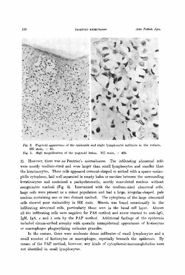
The striking features of skin lesions were infiltration of abnormal cells in the epidermis, particularly from the basal cell layer to the prickle cell layer, and clear halo formation around single or clustered, infiltrating cells between the surrounding keratinocytes, imparting a pagetoid appearance to the epidermis a t the first sight (Pig.
516 PAGETOID RETICULOSIS Acta Pathol. Jpia.
Fig. 2.
Pig. 3. High magnification of tho pngetoid lesion. HE stain, x 425.
Ptlgetoid iippearmce of the epidermis and slight lymphocytic infiltrate in the corium. HE stain, x 85.
2). However, there was no Pautrier’s niicroabscess. The infiltrating abnormal cells were mostly niediuni-sized and were larger than small lymphocytes and snialler than the keratinocytes. These cells appeared crescent-shaped or arched with a sparse eosino- philic cytoplasm, laid well separated in empty halos or cavities between the surrounding keratinocytes and contained a pachychromatic, mostly convoluted nucleus without recognizable nucleoli (Fig. 3). Intermixed with the medium-sized abnormal cells, large cells were present as a minor population and had a large, irregular-shaped, pale nucleus containing one or two distinct nucleoli. The cytoplasm of the large abnormal cells showed poor stainability in HE stain. Mitosis was found occasionally in the infiltrating abnormal cells, particularly those seen in the basal cell layer. Almost all the infiltrating cells were negative for PAS method and never reacted to anti-IgG, IgM, IgA, K and h sera by the PAP method. Additional findings of the epiderniis included circuniscrihed atrophy with sporadic intrapidernial appearance of histiocytes or niacrophages phagocytizing nielanine granules.
In the corimn, there were moderate dense infiltrates of small lymphocytes and a small numher of histiocytes or macrophages, especially beneath the epiderniis. By means of the PAP method, however, any kinds of cytoplasmic immunoglobulins were not identified in small lymphocytes.
32(3): 1982 H. T a A H b S H I el al. 517
Electron Microscopic Observation
Epidermis : The abnormal cells infiltrating in the epidermis varied in size, of which the majority were medium-sized, measuring about 10-11 p on an average (Fig. 4). In the medium-sized cells, the major cell population was round or ovoid in morphology and characterized by an irregular-shaped, mostly lobulated or convoluted nucleus with heterochromatin margination, containing small indistinct nucleoli. In the relatively copious cytoplasm, Golgi complexes were modestly developed often with centrioles or microtubules and several clustered dense bodies, numerous small mitochondria with either dark or clear matrix, and occasional multivesicular bodies were localized unilater- ally. A small amount of cytoplasmic microfilaments were seen in the paranuclear areas. Abundant free ribosomes distributed throughout the cytoplasm but rough endoplasmic reticulum was poorly developed. These cells resembled mycosis fungoides (MF) cells (Fig. 5). From the cell surfaces, however, numerous short microvilli were projected more markedly than those of atypical lymphoid cells in MF.
Among the medium-sized lymphoid cell population, cells typical of SBzary cells were present as a minor cell population. These cells were characterized by a cerebri- form nucleus and scant cytoplasm with a modest number of small dark mitochondria and poor other intracellular organelles. Projection of numerous short microvilli from the cells was striking as compared to SBzary cells in SS (Fig. 6).
Besides, there appeared infrequently medium-sized round cells containing a round nucleus with scant chromatin, a distinct nucleolus and slightly undulated nuclear membrane. Abundant polyribosomes and increased amounts of microfilaments were found in the cell cytoplasm. Together with certain numbers of microvilli, pseudopodia were extended from the cell surfaces. These cells reminded us of lymphoblasts though smaller than the usual ones (Fig. 7).
Between the above-mentioned cell types, we could find out variable transitional cells or intermediate forms. Almost all the cells were striking in that they projected much more prominent and numerous microvilli as compared to atypical lymphoid cells infiltrating in the epidermal and dermal tissues of four patients with MF examined in the present investigation. These cells put the tip of their microvilli on the smooth cell surfaces of surrounding keratinocytes and left a wide distance between their cell body and the keratinocytes.
In addition to the medium-sized lymphoid cell population, large blastoid cells were observed mostly confined to the basal cell layer (Fig. 8). They measured 15 to 20 p in diameter, seldoni reaching 25 p in the largest, and were nearly round with a copious cytoplasm containing abundant polysomes, clustered dense bodies, large clear mitochondria, and bundles of microfilaments. The nucleus was large and euchromatic, with one or two large distinct nucleoli and uneven or serrated contour of nuclear membrane. Pseudopodia and a few short microvilli were found to be projected from the cell surfaces. Mitosis was seldom encountered in the large blastoid cells (Fig. 9).
No desinosonies or junctional complexes were seen between the intraepidermal Small lymphocytes were infrequent in the epidermis.
516 PAOETOII) RETIOULOSIS Actcc Pathol. Jpn.
Pig. 4. Infiltration of atypical lymphoid cells in the epidermis. Infiltrating (*ells vary in morphology and their surrounding keratinocytes appear markedly drformcd. / 3,400. C
Pig. 5. Intraepidermal lymphoid cell resembling mycosis fungoides cell projects niimcroiix short microvilli from the cell surface. x 8,925.
32(3): 1982 H. TAKAHASHI et af. 519
Fig. 6.
Fig. 7. lntraepidermal lymphoblast-like cell. x 8,330.
1nt.raepidermttl lymphoid cell similar to SBzary cell. It has a characteristic cerebriform or serpentine nucleus and projects numerous microvilli from the cytoplasm. x 10,625.
520 PAGETOID RETICULOSIS Actu I’athol. Jpn.
Fig. 8. Fig. 9. Mitosis of a large blastoid cell. x 5,780
Large blastoid cell in the basal cell layer of the epidermis. x 5,780.
32(3): 1982 H. T W S F U ct al. 521
lymphoid cells or between them and their surrounding keratinocytes. In the cytoplasm of the infiltrating lymphoid cells, melanosomes, tonofilaments, Langerhans cell granules or Merkel cell granules were absent.
As for other cell components of the epidermis, Langerhans cells were decreased in number and contained decreased numbers of Langerhans cell granules in the cytoplasm. Conversely, indeterminate dendritic cells were encountered frequently. However, these cells revealed no cellular damage or disruption, and their specific interaction or apposition t o any intraepidermal infiltrating lymphoid cells was not observed. In the pagetoid lesions, keratinocytes compressed by the infiltrating lymphoid cells displayed deformity or disruption of desmosomes without any signs of cellular damage or degeneration. Disruption of the basement membrane and desmosomes of basal keratinocytes occurred occasionally in the basal cell layer where large blastoid cells proliferated.
Corium : Small lymphocytes, mostly several micra in diameter, predominated and However, medium-sized or large lymphoid cells as had a slightly indented nucleus.
seen in the epiderms were small in number.
Immunocytological Study with Rosettinq Assays
Rosetting assays of the free cell suspension prepared from the epidermis of the skin lesions demonstrated that 93% of the freely suspended cells formed spontaneous rosettes with SRC, of which the majority were confirmed in MGG stained preparations to correspond to atypical lymphoid cells. Among the free suspended cells, EAC rosette- and EA rosette-forming cells were counted 1.4% and S.9yo, respectively. All of the atypical lymphoid cells showed no immune phagocytosis. These results indicate that the intraepidermally infiltrating abnormal cells are T-lymphocytes.
In the free cell suspensions sampled from the patient’s peripheral blood, percentages of mononuclear cells positive for E, EA, and EAC rosette formations were 59, 10.5, and 13.5%, respectively. In the E-rosetting cell population, medium-sized, atypical lymphoid cells occasionally containing a markedly indented or convoluted nucleus were observed light microscopically and displayed almost the same fine structure as MP cells or SQzary cells (Pig. 10).
In the mononuclear cell population of peripheral blood, plasmocytes were encountered infrequently among lymphocytes.
Discussiov~
Although in the early stage of the present case, the clinical diagnosis of MP was suspected because of the development of multiple skin lesions, its benign clinical course and histological confirmation of pagetoid lesions by skin biopsies confirmed the diagnosis of PR or Woringer-Kolopp disease. As mentioned previously, this disorder is classified into the localized type and disseminated type, of which the latter is further divided into two subtypes ; Dupont-Vandaele type and intermediate variant. Because of benign clinical evolution and multiple cutaneous lesions, the present case is
I'AUETOIU UETICULOSIS dct r r Pcrthol. Jpn.
Pig. 10. E-rosetting lymphoid cell in the free cell suspension prepared frnrn pwiphcral blood shows the same finr structure as SBzary cell. / l l ,O , j~ .
regarded as corresponding to the intermediate variant of the disseniinated type of PR. The pagetoid appearance of skin lesions pathognonionic for this disease is due
principally to intraepidermal infiltration and proliferation of ahnornial cells and resultant halo or space formation around the abnornial cells hetween their surrounding keratinocytes. In the previous reports, there is a great difference in views as to the nature and origin of the intraepidernial abnornial cells in the pagetoid lesions of the disorder. In the early descriptions,3~~0~~4~~~1~~ the authors believed it to be a peculiar cutaneous reticulosis from the viewpoint that the intraepiderriial abnormal cells were reticuluni cells. However, this view has recently lost support, as the ultrastructure of the abnormal cells was pointed out by several groups of 4( i n agree- ment with the results of the present investigation, to he different from that of reticuluni cells.
Supporting the suggestion by BRAUN-FALCO et nl. of the possibility that infiltrating ahnormal cells in the epiderniis of pagetoid lesions were not lymphoid cells hut cells pre- sumably of nionocyte-macrophage system. CHU and associates**~*~ in their ultrastructural and immunohistochemical studies of a case of PR have more recently emphasized that the majority of the infiltrating cells in the epiderniis are of histiocytic origin. In the present case, however, the great majority of the intraepidernial abnornial cells were proved by rosetting assays to hear no Fc receptor nor C3 receptor and to exhibit no immune phagocytosis and were shown to differ from histiocytes or niacrophages in fine
32(3): 1982 H. TAKAHASHI el al. 523
structure . On the other hand, REVUZ et ~ 1 . ~ 2 ~ ~ regarded PR as a unique disorder of Merkel cell
proliferation on the basis of the results of their electron microscopic observation that cytoplasmic granules with a round electron-opaque core and neurite-like structures, which resembled those seen in Merkel cells of normal human epidermis, were present in the intraepidermal abnormal cells. Agreeing with the results of observations by BRAUN-FALCO et u Z . , ~ , ~ ALIAGA et a1.'j2 and TANNO et al.,39 however, the present observa- tion could not confirm any Merkel cell granules or neurite-like structures in the in- traepidermal infiltrating cells.
Recent electron microscopic studies by several groups of researcher~1,3,5,20,23,~6,
3°-41 have provided evidences that the intraepidermal abnormal cells of PR are rather atypical lymphoid cells, though the ultrastructure of the cells has been described variably in the previous reports. especially with respect to their intracellular organellar com- ponents. Generally speaking, most of investigators stated that the abnormal cells were typical SBzary ce11s15~20~39~40 or showed a striking analogy to those seen in MF or SS,4,13,41 whereas MEDENICA and L O R I N Z ~ ~ and BRAUN-FALCO et aL5 took note of differences in volume of the cytoplasm and in kinds or morphology of intracellular organelles between the abnormal cells and SBzary cells or MF cells and called the abnormal cells stimulated lymphocytes. In the present case, the major cell population of the intraepidermal infiltrating abnormal cells is characterized fine structurally by irregular-shaped, lobulated or convoluted nuclei, clustered dense bodies, numerous mitochondria, some niultivesicular bodies, and a small number of niicrofilaments. These ultrastructural characteristics of the infiltrating cells are consistent with those which have been established as MF cells in the literat~re.8,17,2~,25,30,3~ Besides the MF cells, SBzary cells with cerebriforni nuclei and poor intracellular organelles, medium- sized, lymphoblast-like cells and large blastoid cells were found as minor cell popula- tions. ALIAGA et al.1 in their membrane marker identification of lymphocytes in tissue specimens demonstrated that the intraepidermal cells were T-lymphocytes. The present investigation with rosetting assays has likewise demonstrated that the majority of suspended cells prepared from the epidermis of skin lesions of this case were T- lymphocytes. From these findings, it is concluded that the present case is a cutaneous T-cell lymphoma of low-grade malignancy.
Based on recently accumulating data of ultrastructural and immunological studies, it has been generally accepted that MF and SS are categorized in cutaneous T-cell lymphoma. Most researchers have agreed that MF demonstrates a wide clinical spectrum with variable courses and durations and that SS is a leukemic variant of MF.16~24 In spite of a striking difference in prognosis and clinical evolution between PR and MF or SS, some investigators regarded the former as a variant of the latterl3tZ3J3 or pre- niycosis f ~ n g o i d e s ~ ~ from the ultrastructural similarities of atypical lymphoid cells between both disorders. Against this view, WOOD et aL41 and JONES and C H U ~ ~ offered that the dermal infiltrate of PR could be pathohistologically differentiated from that of MF. Anyway, as stated in a few previous reports,13,40,41 the disseminated type with
524 PAOPTOIU RETIOULOSIS Acto Pathol. J p i ~ .
faster fatal course (Dupont-Vandaele type) seems to be clinically separable from other benign types of P R and the possibility that this type is a variant of MF is undeniable. Excepting this problem, it may be a t present reasonable to understand that P R is a single nosological entity defined by benign clinical behavior and histological features of the pagetoid appearance and closely related to MF or SS.
Among various disorders of cutaneous T-cell lymphomas, PR reveals the most prominent epidermotropisni, though its mechanism is still unclear. Based on the findings of their electron niicroscopic autoradiography of skin biopsies from three patients wi th SS, SAGLIER-GUEDON et al.32 proposed the speculation that lymphocytes entering the cpiderniis initiated blastogenesis by a certain stimulation, multiplied actively arid were transformed into SBzary cells and maintained preferential replication of the SQzary cells in the epidermis. Lending support to this speculation, the present investigation has shown that large blastoid cells presumably derived froni T- lymphocytes infiltrating in the coriuni multiply mostly in the hasal cell layer and arc transformed principally into M F cells and partly into SBzary cells within the epidermis. Interestingly enough, unlike adult T-cell leukemia, the present case showed no leukemic nianifestations but atypical lymphoid cells with convoluted or cerebriform nuclei, which resembled SBzary cells or MF cells, were found infrequently in peripheral blood. Accordingly, some relationship between the intraepidermal infiltration of atypical lym- phoid cells and the appearance of similar cells in peripheral circulation must be considered in the present case.
As mentioned repeatedly, the pagetoid appearance of skin lesions is pathognomonic for PH. However, its pathogenesis remains unsolved. Electron microscopic observa- tions by ROWDEN et aL3I and by SCHMITT and T H I V O L T ~ ~ with regard to pathogenesis of Pautrier’s microabscess in MF demonstrated interactions between T-lymp hocytes, Langerhans cells and keratinocytes, disappearance of the Langerhans cells and disruption of the keratinocytes, and pointed out increased turnover of Langerhans cell population. In the present case, decrease in number of Langerhans cells and frequent appearance of indeterminate dendritic cells found in the pagetoid lesions may reflect enhanced turnover of this cell population. In spite of our detailed observation, however, we could not find out any particular interactions or appositions of intraepidernial lymphoid cells to Langerhans cells and any pictures of degeneration or necrosis in keratinocytes, Langerhans cells and indeterminate dendritic cells. Moreover, i t is worthy of notice that the intraepidernial lymphoid cells projected numerous short microvilli from the cytoplasm and brought the tip of niocrivlli into contact with the surrounding keratino- cytes. This suggests interaction between T-lyniphocytes and keratiriocytcs and niay contribute to the pagetoid appearance of skin lesions in this disorder. In this connec- tion, further detailed ultrastructural and inmiunological studies will he required to clarify precisely the pathogenesis of pagetoid lesions in this disorder.
References
1. ALLAUA, A, BOMBI, J.A., BAKB~:HA, E., iind FORTEA, J.M.: Wonugrr-Kolopp clisenue. Dermntologicai 160: 45-66, 1980.
32(3): 1982 H. TAEAEKASHI el al. 525
2.
3.
4.
5.
6.
7.
8.
9.
10.
11 .
12.
13.
14.
15.
16.
17.
18.
19.
20.
21.
22.
23.
24.
25.
ALIAQA, A., BOMBI, J.A., FORTEA, J.M., HERNANDEZ, B. and OLIVER, V.: Maladie de Wor- inger-Kolopp. Bull. Soc. franv. Derm. Syph. 83: 282-283, 1976. BRAUN-FALCO, O., MARQHESCU, S., and WOLFF, H.H. : Pagetoide Reticulose. Morbus Woringer-Kolopp. Hautarzt. 24: 11-21, 1973. BRAUN-FALCO, O., SCHMOECREL, C., and WOLFF, H.H. : The ultrastructure of mycosis fung- oides, of SBzary’s syndrome, and of Woringer-Kolopp’s disease (pagetoid reticulosis). Bull. Cancer 64: 191-208, 1977. BRAUN-FALCO, O., SCHMOECKEL, C., BURQ, G., and RYCKMANS, F.: Pagetoid reticulosis. A further case report with a review of the literature. Acta Dermatovener. 59 (Suppl. 89): 11-20, 1979. BRODER, S., EDELSON, R.L., LUTZNER, M.A., HELSON, D.L., MACDERYOTT, R.P., DURM, M.E., GOLDMAN, C.K., MEADE, B.D., and WALDMANN, Th. A.: The SBzary syndrome. A malignant proliferation of helper T cells. J. Clin. Invest. 58: 1297-1306, 1976. BRODER, S., UCHIYAMA, T., and WALDMANN, Th. A.: Neoplasm of immunoregulatory cells. Amer. J. Clin. Pathol. 72: 724,731, 1979. BROWNLEE, Th. R. and MURAD, T.: Cancer 26: 686- 698. 1970 BURU, G., WOLFF, H.H., BRAUN-FALCO, O., and MARQHESCU, S.: Pagetoid reticulosis - a cutaneous T-cell lymphoma. J. Invest. Dermatol. 68: 249, 1978. CASTERMANS-ELIAS, S. : Rdticulose Bpidermotrope (maladie de Woringer-Kolopp). Argu- ments enfavenr d’un rattachement an mycosis fungoide. Arch. belg. Derm. 30: 187-194, 1974. CHU, A.C. and MACDONALD, D.M.: Pagetoid reticulosis: a disease of histiocytic origin. Brit. J. Derm. 103: 147-157, 1980. COHEN, E.L. : Woringer-Kolopp disease (pagetoid reticulosis). Clin. Exp. Dermatol. 3 : 447450, 1978. DEQREEF, H., HOLVOET, C., VAN VLOTEN, W.A., DESMET, V., and de WOLF-PEETERS. C.: Woringer-Kolopp disease. An epidermotropic variant of mycosis fungoides. Cancer 38 :
DUPONT, A. and VANDAELE, R. : Bull. SOC. franp. Derm. Syph. 66: 178-181, 1959. DUPRI?, A., BOCAF~, J.-L., CARR~RE, S., BONISSON, H., and GROZDA, J.: Lymphoma cutanbe complexe avec aspect histologique de ((maladie)) de Woringer-Kolopp. Ann. Derm . VBnBrol. (Paris) 104: 321-323, 1977. EDELSON, R.L. : Cutaneous T-cell lymphoma: mycosis fungoides, SBzary syndrome, and other variants. J. Amer. Acad. Dermatol. 2: 89-106, 1980. EDESLON, R.L., KIRKPATRICK, C.H., SEEVACH, E.M., SCHEIN, P.S., SMITH, R.W., GREEN, I., and LUTZNER, M. : Preferential cutaneous infiltration by neoplastic thymus-derived lympho- cytes. GISIQER, 0. : Zur Differentialdiagnose der von Woringer und Kolopp beschriebenen Reticulose. Dermatologica 140 (Suppl. 11): 19-34, 1970. GROSSHANS, E., HEE, P., BASSET, A., and MALEVLLLE, J.: La maladie de Woringer e t Kolopp (rBticulose cutanhe Bpidermotrope (r6ticulose pagbtoide)). Arch. belg. Derm. 29: 195-203, 1973. HANESE, E., TULUSAN, A.H., and WEIDNER, F. : Histological features of “pagetoid reticulosis” (Woringer-Kolopp) in pre-mycosis fungoides. JONES, R.R. and CHU, R. : Distinct clinicopathological entities. KETRON, L.W. and GOODMAN, M.H.: Multiple lesions of the skin apparently of epithelial origin resembling clinically mycosis fungoides. Arch. Derm. Syph. 24: 758-777, 1931. LEVER, W.F. : Localized mycosis fungoides with prominent epidermotropism. Woringer- Kolopp disease. LUTZNER, M., EDELSON, R., SCHEIN, P., GREEN, I., KIREPATRICK, C.H. and ~ E D , A.: Cutaneous T-cell lymphomas: The SBzary syndrome, mycosis fungoides, and related disorders. LUTZNER, M.A., HOBBS, J.W., and HORVATH, P.: Ultrastructure of abnormal cells in SBzary’s
Ultrastructure of mycosis fungoides.
2 154-2165, 1976. RBticulose epidermotrope et IBsions gastriques.
Morphologic and functional studies. Ann. Intern. Med. 80: 685-692, 1974.
syndrome, mycosis fungoidcs and parapsoriasis en plaques. MEDENICA, M. and LORINZ, A.L. : Pngetoid reticulosis (Woringer-Kolopp disease). Arch. Ikrm.
MEZZADRA, G. and SAPUPP, A. : I-leticiilo-istiocitosi cutanea subacutocronirn con piirt,icolnri aspeti clinico-istologi. Giorn. Ital. Derm. 107: 323-336, 1966.
La rkticulosc pagktoide de Woringer et. Kolopp (une mnladie de In cellule dr Merkel). Ann. Derm. VBnkr. 104: 312-320, 1977. R ~ v u z , J. and TOURAINE, R.: Maltitdie de Woringer et Kolopp. Dermatologicti 157: 377-- 385, 1978. ROSAS-URIBE, A., VARIAKOJIS, D., MOLNER, Z., and PAPPORT, H.: Mycosis fungoides: An ultrastructural study. Cancer 34: Ci3P645, 1974. ROWDEN, G., PHILLLPY, T.M., LEWIS, M.G., and WILKINYON, R.D.: Target role of Langerhans cells in mycosis fiingoides: transmission and immunoelectron microscopic studies. J. Cutan. Pat,hol. 6: 364-382, 1979. SAQLIER-GUEDON, I., PKUNLER, M., DUREPAIRE, R., and GRUPPER, Ch. : t’referential replication of SBenry cells in the epidermis. SANCHEZ, J.L. and ACKERMAN, A.B. : The patch stage of mycosis fungoides. Criteria for histologir diagnosis. Amer. .J. Dermatopathol. 1 : 5-26, 1979. S A N ~ I I , l’., CARDENAY, F., and HUMBOLT, F. : Enfermedad de Woringer-Kolopp do localiztrtion cntaneo-visceral. SCHMITT, D. and THIVOLET, J. : Lymphocyte-epidermis interactions in mslignant epider- motropic lymphomas: SCIIMOECREL, Ch., BRAUN-FALCO, 0.. and RURQ, G. : Die pagetoide Itetikulose - ein T-Zell Lymphom? SMOES-CHARLES, J. iind DUPONT, A.: A propos dune forme part)iculiere gBnkralisBe de rBti-
I. Ultrastrnctural aspects. ActiL Dermtztovener. 60: 1-1 1 , 1980.
Hautsrzt 29 (Suppl. Ill) : 57-60, 1978.
cnlose kpidermotrope (mctladie de Woringer-Kolopp). 1!)73.
Arch. belg. Derm. Syph. 29 : 205-211,
TAKAHASHI, K., SAKUMA, H., NAITO, M., YAQINUMA, Y., TAKAHASHI, H., ASANO, S., HOJO, H., and KOJIMA, M. : Cytological characters, transformation and ontogenesis of dermal histiocytes nnd fibroblasts of rats. Acta Pathol. Jpn. 30: 743-766, 1980. TANNO, K., TAICAHASRI, H., NAQAO, S., IIJLMA, S., and ToaAsHr, R.: Pagetoid reticolosis (Woringer-Kolopp disease). TORIBIO, J . , Q U ~ O N E S , P.A., and VIGIL, T.R. : Woringer-Kolopp disease. Pagetoid retirulosis. D~rmntologica 159: 283-291, 1978. WOOD, W.S., KILLBY, V.A.A., and STEWART, Wm. D.: Pngetoid reticulosis (Woringcr-Kolopp disease). WORINQER, M.M.Fr. and KOLOPP, P. : LBsion ErythBmato-squiimeusc polycycliynr de I’avant-bras evoluant depuis 6 iins chez un garponnet de 1 3 am. Histologiquemrnt in- filtrat intrtz-kpidrrmique d’apparence tumorale.