20/ CURRENT CONCEPTS REVIEW SOUBORNÝ REFERÁT ACTA CHIRURGIAE ORTHOPAEDICAE ET TRAUMATOLOGIAE ČECHOSL., 78, 2011, p. 20–26 Painful Flatfoot Deformity Bolestivé plochonoží M. WIEWIORSKI, V. VALDERRABANO Orthopaedic Department University Hospital Basel, Basel, Switzerland SUMMARY The posterior tibial muscle is the key dynamic support of the medial longitudinal arch of the foot. When it fails –typical- ly in women older than 40 years of age- progressively, the arch slowly collapses, the heel drifts into valgus, the midfoot flattens, and the forefoot gradually abducts and supinates, resulting in painful acquired pes planovalgus abductus et supi- natus. Posterior tibial tendon insufficiency (PTTI) is often misdiagnosed as a chronic ankle sprain, osteoarthritis, or col- lapsed arch as a result of aging or obesity, and it leaves the patient debilitated. Prompt diagnosis prevents frustration for the patient and allows treatment to be started at an earlier, more easily managed stage. The diagnosis of PTTI is largely a clinical one. An increased awareness of the existence of PTTI should serve to help patients with earlier referral and treat- ment and by limiting the amount of disability. INTRODUCTION Posterior tibial tendon insufficiency (PTTI) is accep- ted as the most common etiology of the acquired adult flatfoot deformity. In spite increased awareness and intensive research over the past decade, PTTI is still fre- quently missed. Because this slowly progressing condi- tion is best treated at early, easier manageable stage, prompt diagnosis is necessary. PTTI is characterized by hindfoot valgus, flattening of the longitudinal foot arch, and abduction and supination of the forefoot (29, 38). This occurs most commonly in women older than 40 year of age. Since the first description of a posterior ti- bial tendon rupture by Key et al. in 1953 (15), methods of treatment have evolved and the pathology and func- tion of the tendon have been extensively investigated. This article will review the current concepts with regard to the pathomechanism, diagnostics, and treatment stra- tegy of this debilitating condition. Anatomy and Biomechanics of the posterior tibial muscle The posterior tibial muscle arises from the posterior surface of the interosseous membrane, the lateral por- tion of the posterior surface of the body of the tibia, and from the upper medial surface of the fibula. It is the most deeply seated muscle of the deep posterior compartment in the back of the leg. In the lower fourth of the leg its tendon passes in front of that of the flexor digitorum lon- gus and lies with it in a groove behind the medial mal- leolus in a separate tendon sheath. It then passes under the flexor retinaculum of lower limb and over the del- toid ligament into the foot, and then beneath the spring ligament (calcaneonavicular ligament) inserting into the tuberosity of the navicular bone. Finally, it inserts with multiple expansions spreading out across the plantar aspect of the hind- and midfoot (sustentaculum tali of the calcaneus, forward to the medial cuneiform and first metatarsal bone, and lateralwards to the other cuneiform bones and the bases of the second, third, and fourth meta- tarsal bones). Since it lies posterior to the axis of the ankle and medial to the axis of the subtalar joint, its func- tion is to plantar flex the ankle, while it inverts the sub- talar and oblique axis of the midtarsal joints (2). Due to its anatomical location it provides dynamic support along the plantar aspect of the foot. By passing under- neath the spring ligament it stabilizes the osseous con- figuration at the talonavicular joint und prevents collap- sing of the medial longitudinal arch. Loss of the medial longitudinal arch results in dorsiflexion of the first meta- tarsal. Insufficiency of the first ray may be found in ear- ly stages of PTTI (16). The foot is a complex mechanism acting as a mobile adapter during weight acceptance and a rigid lever arm during propulsion (4). The talonavicular and calcaneo- cuboid joints (the midtarsal joint) seem to play an impor- tant role in this transition (20). Blackwood et al. demon- strated significantly less range of motion in the midtarsal joint when the calcaneus was maximally inverted com- pared to when the calcaneus was maximally everted (4). During normal gait, contraction of posterior tibial muscle limits subtalar eversion caused by the gastroc- soleus complex. Without the inverting force of the poste- rior tibial muscle in the stance phase there is less int- rinsic osseus stability at the midtarsal joint and the forward propulsive force of the complex of gastrocsole- us acts at the midfoot instead of at the metatarsal heads (push-off phase) (31). Due to its large cross-sectional area the relative strength of posterior tibial muscle is more than twice
Transcript
20/ CURRENT CONCEPTS REVIEWSOUBORNÝ REFERÁT
ACTA CHIRURGIAE ORTHOPAEDICAEET TRAUMATOLOGIAE ČECHOSL., 78, 2011, p. 20–26
Painful Flatfoot Deformity
Bolestivé plochonoží
M. WIEWIORSKI, V. VALDERRABANO
Orthopaedic Department University Hospital Basel, Basel, Switzerland
SUMMARY
The posterior tibial muscle is the key dynamic support of the medial longitudinal arch of the foot. When it fails –typical-ly in women older than 40 years of age- progressively, the arch slowly collapses, the heel drifts into valgus, the midfootflattens, and the forefoot gradually abducts and supinates, resulting in painful acquired pes planovalgus abductus et supi-natus. Posterior tibial tendon insufficiency (PTTI) is often misdiagnosed as a chronic ankle sprain, osteoarthritis, or col-lapsed arch as a result of aging or obesity, and it leaves the pa tient debilitated. Prompt diagnosis prevents frustration forthe patient and allows treatment to be started at an earlier, more easily managed stage. The diagnosis of PTTI is largelya clinical one. An increased awareness of the existence of PTTI should serve to help patients with earlier referral and treat -ment and by limiting the amount of disability.
INTRODUCTION
Posterior tibial tendon insufficiency (PTTI) is accep-ted as the most common etiology of the acquired adultflatfoot deformity. In spite increased awareness andintensive research over the past decade, PTTI is still fre-quently missed. Because this slowly progressing condi-tion is best treated at early, easier manageable stage,prompt diagnosis is necessary. PTTI is characterized byhindfoot valgus, flattening of the longitudinal foot arch,and abduction and supination of the forefoot (29, 38).This occurs most commonly in women older than 40year of age. Since the first description of a posterior ti-bial tendon rupture by Key et al. in 1953 (15), methodsof treatment have evolved and the pathology and func-tion of the tendon have been extensively investigated.This article will review the current concepts with regardto the pathomechanism, diagnostics, and treatment stra-tegy of this debilitating condition.
Anatomy and Biomechanics of the posteriortibial muscle
The posterior tibial muscle arises from the posteriorsurface of the interosseous membrane, the lateral por-tion of the posterior surface of the body of the tibia, andfrom the upper medial surface of the fibula. It is the mostdeeply seated muscle of the deep posterior compartmentin the back of the leg. In the lower fourth of the leg itstendon passes in front of that of the flexor digitorum lon-gus and lies with it in a groove behind the medial mal-leolus in a separate tendon sheath. It then passes underthe flexor retinaculum of lower limb and over the del-toid ligament into the foot, and then beneath the springligament (calcaneonavicular ligament) inserting into thetuberosity of the navicular bone. Finally, it inserts with
multiple expansions spreading out across the plantaraspect of the hind- and midfoot (sustentaculum tali ofthe calcaneus, forward to the medial cuneiform and firstmetatarsal bone, and lateralwards to the other cuneiformbones and the bases of the second, third, and fourth meta-tarsal bones). Since it lies posterior to the axis of theankle and medial to the axis of the subtalar joint, its func-tion is to plantar flex the ankle, while it inverts the sub-talar and oblique axis of the midtarsal joints (2). Due toits anatomical location it provides dynamic supportalong the plantar aspect of the foot. By passing under-neath the spring ligament it stabilizes the osseous con-figuration at the talonavicular joint und prevents collap-sing of the medial longitudinal arch. Loss of the me diallongitudinal arch results in dorsiflexion of the first meta-tarsal. Insufficiency of the first ray may be found in ear-ly stages of PTTI (16).
The foot is a complex mechanism acting as a mobileadapter during weight acceptance and a rigid lever armduring propulsion (4). The talonavicular and calcaneo-cuboid joints (the midtarsal joint) seem to play an impor-tant role in this transition (20). Blackwood et al. demon-strated significantly less range of motion in the midtarsaljoint when the calcaneus was maximally inverted com-pared to when the calcaneus was maximally everted (4).During normal gait, contraction of posterior tibialmuscle limits subtalar eversion caused by the gastroc-soleus complex. Without the inverting force of the poste-rior tibial muscle in the stance phase there is less int-rinsic osseus stability at the midtarsal joint and theforward propulsive force of the complex of gastrocsole-us acts at the midfoot instead of at the metatarsal heads(push-off phase) (31).
Due to its large cross-sectional area the relativestrength of posterior tibial muscle is more than twice
that of peroneus brevis, its primary antagonist (21). Withloss of an antagonist force due to PTTI, the unopposedpull from the peroneal tendons forces the heel into ever-sion.
Epidemiology The prevalence of PTTI and posterior tibial tendon
rupture parallel the degenerative processes of aging.Hypertension, diabetes mellitus, seronegative arthropat-hies and obesity have all been identified as risk factorsfor PTTI (18). Additionally, the effects of corticoste-roids and local surgical procedures may further be asso-ciated with local vascular impairment and eventual rup-ture (13).
Pathomechanism of the adult flatfootA posterior tibial tendon dysfunction evolves from
degeneration of the tendon due to repetitive microtrau-ma or chronic overuse (22).
The posterior tibial tendon is acutely angled where itpasses posterior to the medial malleolus. This area ofincreased stress on the tendon is the most common siteof degeneration of the posterior tibial tendon. The blo-od supply of the tendon can be divided into the proxi-mal and distal areas. The proximal tendon is suppliedby branches of the posterior tibial artery and the distal,which is at the bone-tendon interface, by branches of theposterior tibial and dorsalis pedis arteries (10). Whetherdegeneration of the PTTI is due to physiological hypo-vascularity of the tendon at the degeneration site rema-ins controversial. Frey et al showed that there is an areaof hypovascularity in the tendon about 1 cm distal to themedial malleolus (10). Contrary to these findings Pradoet al. did not find decreased vascularization in the mid-portion of the tendon (26). Histologic examinations ofsurgical posterior tibial tendons from patients with PTTIrevealed a degenerative tendinosis characterized by inc-reased mucin content, fibroblast hypercellularity, chond-roid metaplasia, and neovascularization. Those result indisruption of the linear orientation of the collagen bund-les (9, 22). Although PTTI is widely accepted as a sig-nificant contributor to this deformity, the pathology anddeformity involve more than the tendon itself. Basma-jian and Stecko concluded from their electromyographicmeasurement that the ligaments and osseus configura-tion of the foot form the primary stabilizers of the foot
arch (3). Muscular involvement in stabilization is me-rely called upon during increased load (take off phase inwalking). MRI studies confirmed the association ofspring ligament failure with PTTI (30, 37). The supero-medial calcaneonavicular ligament is most commonlyinvolved, followed by the inferomedial calcaneonavicu-lar and talocalcaneal interosseous ligaments. In patientswith surgically proven spring ligament tear, Toye and al.demonstrated an abnormal spring ligament thickening,a fullthickness gap and an abnormal increased signal onpreoperative T2-weighted MRI images (30).
The involvement of the deltoid ligament is being dis-cussed (for clinical differentiation see Diagnostics). Hin-termann and al. found PTTI in 22% of patients withmedial ankle instability (11). However, it remains unc-lear if medial ankle instability may cause a secondaryPTTI with elongation and/or rupture of the tendon, orvice versa 11. Patients with a preexisting flatfoot defor-mity show increased gliding resistance at the tendon-sheath interface of the posterior tibial tendon. The fin-dings indicate a possible vicious circle of deformity andtendon dysfunction (32).
Clinical examinationThe diagnosis of PTTI is essentially clinical. Syste-
matic examination of the soft tissues needs to considerthe potentially unaffected contralateral side. Both feetare inspected in a standing position with parallel feet,shoulder width apart. Inspection includes hindfootalignment, deformity, and swelling. The medial longi-tudinal arch is inspected for presence of flattening(Fig. 1B). Hindfoot alignment is best inspected frombehind the patient. Valgus hindfoot 11. Valgus angula-tion of more than 10° (or valgus in bilateral compari-son) is typically found in PTTI in stage II and above(Fig. 1A). Fore foot abduction is indicated by the “too-many-toes sign” (Fig. 1A). The test is positive whenmore of the toes are visible lateral to the ankle joint ofthe involved side then on the contralateral side, whenviewed from behind 14. The dynamic function of theposterior tibial tendon is determined by the single-limbheel-rise test. malalignement presents as an asymmetri-cal planus and pronation deformity of the affected Thepatient is asked to rise onto the ball of one foot whilethe other foot is suspended in the air. As the patient risesoff the floor, the posterior tibial tendon inverts and sta-
Fig. 1. PTTI stage III. Two patients withstage III posterior tibial tendon insuffi-ciency. Viewed from behind, a distinctvalgus of the hindfoot with a positive too-many-toes-sign (→) is revealed (A). Theside view shows a collapse of the me diallongitudinal arch and a protruding talushead due to subluxation at the talonavi-cular joint (B).
the posterior tibial tendon the pain maximum may shiftto the lateral ankle due to fibulo-calcanear impingement.Retromalleolar pain can be a sign of posterior ankle jointimpingement. Other differential diagnosis include: tar-sal coalition, osteoarthritis, and other posttraumatic,neurologic, diabetogenic or iatrogenic pathologies (19).
RadiologyPosterior tibial tendon dysfunction is essentially a cli-
nical diagnosis. Plain radiography helps confirming theextent of the deformity and presence of osteoarthritis.Conventional radiologic imaging consists of antero -posterior and lateral weigth-bearing radiographs of thewhole foot and an anteroposterior radiograph of theankle joint. If the deformity is present, the anteroposte-rior radiograph shows a pathological a.p. talar-first meta-tarsal angle (normal angle, 0 to 10 degrees) with andabduction of the forefoot at the transverse tarsal joint,with the navicular sliding laterally on the talar head(Fig 3A). In patients who have an advanced deformity,subluxation or dislocation of the talonavicular joint mayoccur in association with degenerative osteoarthritis ofthe posterior facet of the subtalar joint. The lateral radio -graph shows a decrease in the lateral talar-first metatar-sal angle (normal angle, 0 to 10 degrees) and flatteningof the longitudinal arch (Fig 3B). The anteroposteriorradiograph of the ankle joint reveals potential deformity
bilizes the hindfoot (Fig. 2A). In patients with PTTI theheel fails to invert and remains in valgus (Fig. 2B). Ifunable to achieve the tip toe position on one foot, thepatient should perform a bilateral heel-rise. In late sta-ge PTTI patients often will not be able to perform neit-her test. This test is also helpful to differentiate PTTIfrom elongation or rupture of the deltoid ligament (me-dial ankle ligament instability). In cases of isolatedmedial ankle instability without involvement of theposterior tibial tendon, the hindfoot valgus deformity iscorrected by activity of the poste-rior tibial muscle whenin tiptoe position. Ankle joint stability must be assessed(drawer test and talar tilt test). Still, clinical differentia-tion between PTTI and medial ankle instability is chal-lenging and diagnosis frequently needs to be further cla-rified by MRI or arthroscopy 12. Progressing PTTI isoften associated with an abduction and supination defor-mity of the forefoot. In a patient standing fully weight-bearing on both feet, external rotation of the tibia is app-lied to provoke varus movement of the hindfoot. Ina patient with supination deformity, the first metatarsalwill rise off the floor (first metatarsal rise sign).
In flexible stage I and II of PTTI the patients showpain tenderness in the course of the posterior tibial ten-don, especially at the medial malleolar groove, the springligament and the tarsal insertion. In later stages withfixed deformity and frequently found chronic rupture of
Fig. 2. Heel-rise test. Physiologicalinversion of the hindfoot in tip-toe posi-tion (A). In patients with posterior tibi-al tendon insufficiency, the hindfoot failsto invert and remains in eversion (B).Patients with late stage PTTI often willnot be able to perform this test.
Fig. 3. Conventional radiographs of stage IV posterior tibialtendon isufficiency. The anteroposterior view shows abductionof the forefoot at the transverse tarsal joint, with the navicularsliding laterally on the talar head (A). The lateral radiographshows a decrease in the talometatarsal angle (B). Pain at thelateral ankle is often due to fibulo-calcanear impingement (C).Additional slight valgus tilt of the ankle joint is present (––).
(talar tilt), degeneration and narrowing of the fibulo-cal-canear space (Fig. 3C). Several authors emphasize thatmagnetic resonance imaging (MRI) is not required tomake the diagnosis and does not assist in the planning oftreatment. Some authors have suggested MRI to be use-ful in the evaluation of PTTI (27, 28). Contrary other aut-hors state that it is used too frequently in clinical practi-ce and that its clinical usefulness is questionable (13, 23).
Classification There is a continuum of PTTI ranging from tenosy-
novitis to fixed deformity. In 1989 Johnson and Stromdescribed three clinical stages of dysfunction (14).Myerson et al. added a fourth to describe the most seve-re deformity with valgus collapse of the talus within theankle (23) (Table 1). Stage I incorporates tenosynovitis.In this stage, the tendon is of normal length and symp-toms are usually mild to moderate. Pain and swellingare present on the medial aspect of the foot. Mild weak-ness and minimal deformity are present. In stage II the-re is elongation or tearing of the tendon. The limb isweak and the patient is unable to stand on tiptoe on theaffected side. There is secondary deformity as the mid-foot pronates and the forefoot abducts at the transversetarsal joint. The subtalar joint remains mobile. Stage IIIis characterized by a more severe deformity and a fixedhindfoot. Stage IV involves a valgus deformity and dege-neration of the ankle joint.
TreatmentTreatment of PTTI intends to stop progression of the
tendon dysfunction and to protect the longitudinal arch sta-bilizing soft tissues (e.g. spring ligament, deltoid). Thiscan be achieved by reconstruction of the anatomic align-ment and recovery of physiological biomechanics. PTTIwith no or beginning deformity characterized by a flexib-le hindfoot foot can be treated joint-preserving by conse-rvative and tendon reconstructive methods. Treatment ofrigid hindfoot deformities in later stage III and IV intendsto reconstruct the painful deformity. Osseous repositionand subsequent arthrodesis are often inevitable.
Stages I and II – The flexible foot
Stage IA period of four to eight weeks of immobilisation in
a plaster cast below the knee or a walking boot may berequired to control accompanying inflammation. Com-plemental measures are RICE (rest, ice, compression,and elevation) and anti-inflammatory drugs. Footwearplays an important role, and patients should be encou-raged to wear flat lace-up shoes, or even lace-up boots,which accommodate orthoses. Stage I patients may beable to manage with a casted insoles. The various cas-ted, semirigid insoles support the medial longitudinalarch of the foot and either hold the heel in a neutral align-ment (stage I) or correct the outward bent heel to a neu-tral alignment (stage II). This approach is meant to ser-ve several functions: to alleviate stress on the posteriortibial tendon and muscle; to make gait more efficient byholding the hindfoot fixed; and thirdly, to prevent pro-gression of deformity. When this approach has beenused, two thirds of patients have good to excellent results(18). However, from our experience, conservative the-rapy shows poor results in the long-term. Some authorspropose tenosynovectomy for patients who have advan-ced stage I disfunction. Good results have been repor-ted for either open or tendoscopic technique (5, 34).
Stage IINo soft-tissue reconstructive surgical technique on its
own can sufficiently contain the forces of a malalignedhindfoot. Therefore, consensus is growing that surgicaltreatment of stage II PTTI should include a tendon tran-sfer in combination with corrective osteotomy (23, 25).The rationale behind this approach is that the osteoto-my is required to correct the bony architecture of thefoot in order to optimize the biomechanics of the recon-structed posterior tibial tendon and protect other foot sta-bilizing ligaments and tendons (11). According to Val-derrabano et al., only about 60% of force regenerationcan be expected after surgical reconstruction of a dys-functional or ruptured posterior tibial tendon. The mag-
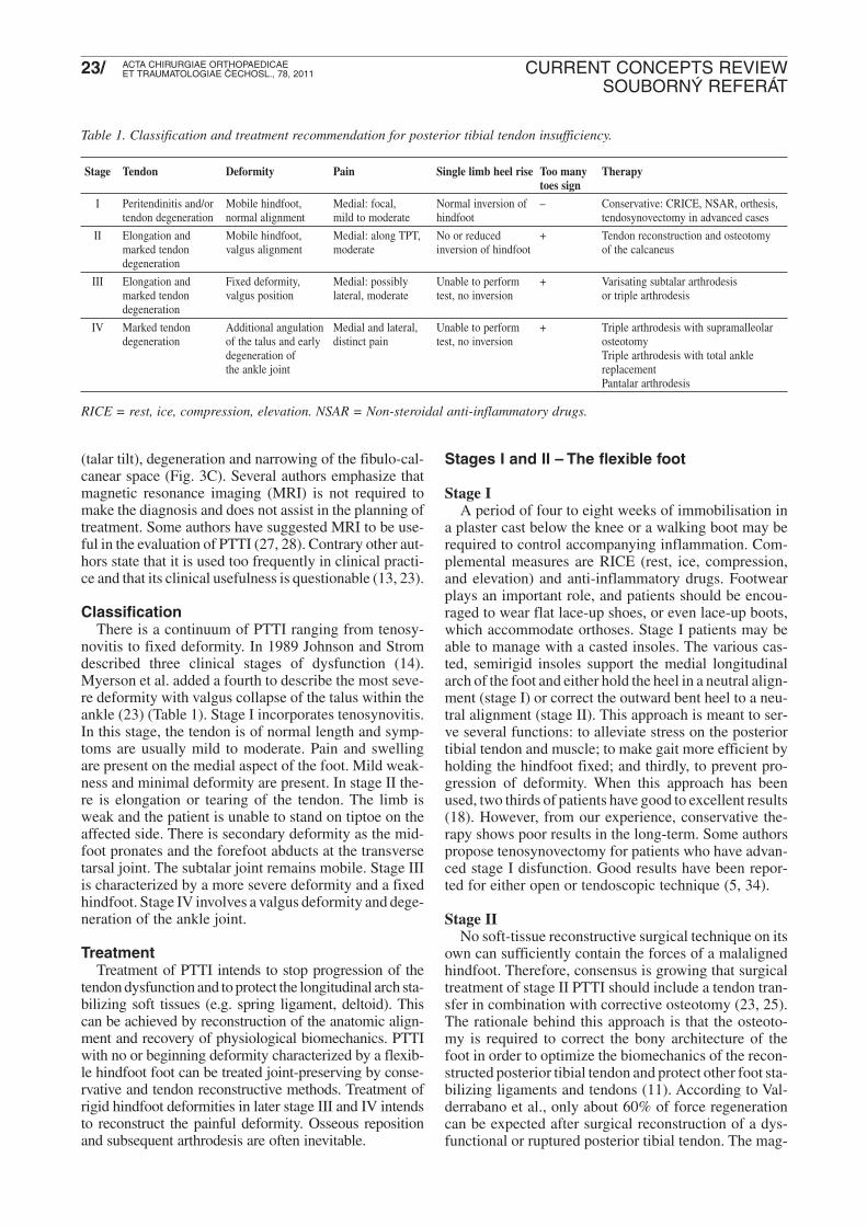
Stage Tendon Deformity Pain Single limb heel rise Too many Therapy toes sign
I Peritendinitis and/or Mobile hindfoot, Medial: focal, Normal inversion of – Conservative: CRICE, NSAR, orthesis,tendon degeneration normal alignment mild to moderate hindfoot tendosynovectomy in advanced cases
II Elongation and Mobile hindfoot, Medial: along TPT, No or reduced + Tendon reconstruction and osteotomymarked tendon valgus alignment moderate inversion of hindfoot of the calcaneusdegeneration
III Elongation and Fixed deformity, Medial: possibly Unable to perform + Varisating subtalar arthrodesismarked tendon valgus position lateral, moderate test, no inversion or triple arthrodesisdegeneration
IV Marked tendon Additional angulation Medial and lateral, Unable to perform + Triple arthrodesis with supramalleolar degeneration of the talus and early distinct pain test, no inversion osteotomy
degeneration of Triple arthrodesis with total ankle the ankle joint replacement
nitude of regained strength is primarily dependent on theduration of the dysfunction and amount of atrophy (33).Therefore PTTI needs to be surgically addressed at anearly after failure of conservative treatment.
The two recommended tendon reconstruction techni-ques are flexor digitorum longus (FDL) tendon transferand transfer of a split anterior tibial tendon (Cobb pro-cedure). Rerouting a part of the anterior tibial tendon tothe plantar aspect of the cuneiform allows the posteriortibial tendon to pull at its physiological insertion site.Additionaly the Cobb procedure decreases the tensionof the anterior tibial tendon (16), thus preventing occa-sionally anterior tibial tendon ruptures. This may add tocorrect the deformity, by reduction of the pull of the ante-rior tibial tendon, which is usually increased in PTTI.This dynamic correction may allow the patient to adaptthe forefoot to the ground as required. Additionally, thisprocedure does not sacrifice the FDL tendon.
Another method is the transfer of flexor digitorumlongus tendon. The FDL tendon is detached proximal ofthe juncture with the flexor hallucis longus (Henry’sknot). The periosteum over the navicular is then dissec-ted and a drill hole is made in the tuberosity from thedorsal to plantar aspects. The tendon is sutured side-to-side to the posterior tibial tendon and passed through thedrill hole from plantar to dorsal. The use of the flexorhallucis longus tendon is not recommended due to itsimportant role in the push-off phase off the foot (31).Intraoperative exploration of the spring ligament inPTTI is mandatory, because of its frequent concomitantdegeneration or rupture (37). If found ruptured, recon-struction has to consider both components of the springligament complex (6). The author suggest that ifa accompanying medial ankle instability is suspectedupon clinical examination, an initial ankle arthroscopyneeds to be performed to rule out deltoid ligament invol-vement. If ligament instability is found, it needs to beaddressed during the following surgery.
Various osteotomies of the calcaneus can correct thepathological bony alignment. During surgery, these oste-otomies should be performed prior to finalize the medi-al soft tissue reconstruction. In PTTI with pronouncedhindfoot valgus and no or minimal foot abductiona medial sliding osteotomy is recommended (23). Thelateral hindfoot incision extends from the superior bor-der of the calcaneal tuberosity anterior to the retrocal-caneal space to the inferior border of the calcaneussuperficial to the plantar fascia. An oblique transverseosteotomy is made in the calcaneus in line with the inci-sion in the skin with use of an oscillating saw. The cutis made at a right angle to the lateral border of the cal-caneus and is inclined posteriorly at an angle of appro-ximately 45 degrees to the plane of the sole of the foot.The posterior fragment of the calcaneal tuberosity istranslated medially ten or more millimeters and is secu-red with a cannulated headless compression screw.
In PTTI with pronounced hindfoot valgus and distin-ct forefoot abduction a lateral calcaneus lengtheningosteotomy is recommended. By lengthening the lateralcolumn, the medial longitudinal arch is restored secon-
darily to the induced adduction movement of the fore-foot that supinates the foot at the subtalar and talonavi-cular joint. An osteotomy of the anterior calcaneus wasoriginally described by Evans et al (8). Here an osteo-tomy of the neck of the calcaneus is performed and a tri-cortical bone graft impacted. Several authors have repor-ted good results with this procedure (1, 24). Myerson etal. propose a lengthening through the calcaneocuboidjoint itself, with use of a tricortical bone graft for arth-rodesis of the joint (23). We favour an alternative met-hod proposed by Hintermann et al (12). Here an osteo-tomy is performed from at the lateral hindfootapproximately 12 to 20 mm proximal to the calcaneo-cuboid joint at the “floor” of the sinus tarsi. The oscil-lating saw passes between the posterior and middle facetof the subtalar joint. The medial cortex is kept intact.The gap is widened with a Casper spreader and a tri-cortical graft or alternatively allograft bone is inserted.The amount of widening can be adjusted until the medi-al arch is restored sufficiently. The graft is fixed withone 3.5mm cortical screw. Preservation of the subtalarand calcaneocuboideal joint offers certain advantage.One has to keep in mind that fusing the calcaneocubo-id joint significantly reduces hindfoot motion (7). Main-taining hindfoot motion prevents overload on adjacentjoint which may lead to osteoarthritis. Additionaly,according to Knupp et al. it is easier to reduce the abduc-ted foot if the lateral column is not further shortened byarthrodesis of the calcaneocuboid joint (17).
A recently presented surgical method for stage II PTTIis the subtalar arthroereisis. Here, the sinus tarsi is emp-tied and a expanding endorthesis is inserted followingprior correction of the deformity and tendon repair. Goodresults were shown for the subtalar arthroereisis, espe-cially in younger patients (36). However, one has to keepin mind that the fatty tissue in the sinus tarsi containsabundant nerve cell which are essential for proprocep-tion of the hindfoot (“cerebellum pedis”) and thereforeshould be dissected carefully and not removed.
Stages III and IV – The rigid footStage III
The goal of surgical treatment of stage III PTTI iscorrection the deformity and pain relief. Because at thisstage hindfoot deformity can not be passively reduced,joint preserving surgery frequently fails. Depending onthe extent of the deformity, correction can be achievedthrough a varisating subtalar arthrodesis or triple arth-rodesis of the subtalar, calcaneocuboid, and talonavicu-lar articulations. In our opinion, calcanecuboidal arth-rodesis can be omitted to sustain residual motion of thelateral column.
Stage IVStage IV PTTI has been reached when additional
degenerative changes are present in the ankle joint. Insuch cases, a varisating triple arthrodesis together witha medial closing wedge supramalleoloar osteotomy anddeltoid ligament reconstruction may solidly address thedeformity (Fig. 4). In very selected cases a varisating
Fig. 4. Case with PTTI stage IV. A grotesque valgus of the hind-foot with a positive too-many-toes-sign (→) is present (A).Pedobarography (B) reveals a collaps of the longitudinal arch(→) and medialisation of the center of pressure line (––). Theanteroposterior view shows a subluxation of the talar head atthe talonavicular joint (C). Anteroposterior radiographs of theankle joint show (D) show fibulo-calcanear impingement (O)and a valgus tilt (––). Surgery included a triple arthrodesis(involving the calcaneocuboideal joint), a Ludloff osteotomy,resection of the second metatarsal head, and reconstruction ofthe deltoid ligament. Postoperative radiographs reveala reconstructed foot alignment (E). Clinical examinationshows physiological hindfoot valgus (F).
triple arthrodesis may be combined with total anklereplacement. However, the salvage treatment at this sta-ge is usually a pantalar arthrodesis (ankle, subtalar, cal-caneocuboid, and talonavicular articulations) (35).
time. To reach adequate stability, approximately 12weeks for the tendon and 8–12 weeks for the bone arerequired. In the initial 6 weeks immobilization in a pneu-matic walking brace with partial weight bearing (heel-to-toe pattern 15–20kg with crutches) is mandatory. Theload can be than increased gradually until full weight-bearing is reached after 12 weeks postoperatively. Phy-siotherapeutic care needs to address postural hindfootstability. From our own experience it takes 3–6 monthsuntil the rehabilitation is finished. This time span is nee-ded to adapt the cerebellar control of balance and loco-motion to the new anatomic configuration of the hind-foot.
PEARLS
• Posterior tibial tendon insufficiency (PTTI) is a pro-gressive entity leading to painful pes planovalgusabductus et supinatus.
• If conservative treatment fails, early surgical interven-tion slows further progression of the disease.
• Reconstruction of the posterior tibial tendon and con-comitant medial ligamentous lesions in stage II PTTIneed to be accompanied by a corrective calcanearosteotomy:– Distinct forefoot abduction à lateral calcaneus leng -
thening osteotomy;– No or minimal forefoot abduction à calcaneus me -
dial sliding osteotomy.• If additional medial ankle instability is suspected upon
clinical examination, an ankle arthroscopy needs torule out deltoid ligament involvement. If accompany-ing ligament instability is verified, an additional me -dial ligamentoplasty needs to be performed.
1. ANDERSON, A. F., FOWLER, S. B.: Anterior calcaneal osteoto-my for symptomatic juvenile pes planus. Foot Ankle, 4: 274–283,1984.
2. BARAVARIAN, B.: Use of the Cobb procedure in the treatment ofposterior tibial tendon dysfunction. Clin. Podiatr. Med. Surg., 19:371–389, 2002.
3. BASMAJIAN, J. V., STECKO, G.: The Role of Muscles in ArchSupport of the Foot. J. Bone Jt Surg., 45: 1184–1190, 1963.
4. Blackwood, C. B., Yuen, T. J., Sangeorzan, B. J., Ledoux, W. R.:The midtarsal joint locking mechanism. Foot Ankle Int., 26:1074–1080, 2005.
5. CHOW, H. T., CHAN, K. B., LUI, T. H.: Tendoscopic debridementfor stage I posterior tibial tendon dysfunction. Knee Surg. Sports.Traumatol. Arthrosc. 13: 695–698, 2005.
6. DELAND, J. T., De ASLA, R. J., SUNG, I. H., ERNBERG, L. A.,POTTER, H. G.: Posterior tibial tendon insufficiency: which liga-ments are involved? Foot Ankle Int., 26: 427–435, 2005.
7. DELAND, J. T., OTIS, J. C., LEE, K. T., KENNEALLY, S. M.:Lateral column lengthening with calcaneocuboid fusion: range ofmotion in the triple joint complex. Foot Ankle Int., 16: 729–733,1995.
8. EVANS, D.: Calcaneo-valgus deformity. J. Bone Jt Surg., 57-B:270–278, 1975.
9. FOWBLE, V. A., VIGORITA, V. J., BRYK, E., SANDS, A. K.:Neovascularity in chronic posterior tibial tendon insufficiency.Clin. Orthop., 450: 225–230, 2006.
10. FREY, C., SHEREFF, M., GREENIDGE, N.: Vascularity of theposterior tibial tendon. J. Bone Jt Surg., 72-A: 884–888, 1990.
12. HINTERMANN, B., VALDERRABANO, V., KUNDERT, H. P.:Lengthening of the lateral column and reconstruction of the me-dial soft tissue for treatment of acquired flatfoot deformity asso-ciated with insufficiency of the posterior tibial tendon. Foot Ank-le Int., 20: 622–629, 1999.
13. HOLMES, G. B., Jr., MANN, R. A.: Possible epidemiological fac-tors associated with rupture of the posterior tibial tendon. FootAnkle, 13: 70–79, 1992.
14. JOHNSON, K. A., STROM, D. E.: Tibialis posterior tendon dys-function. Clin. Orthop., 196–206, 1989.
15. KEY, J. A.: Partial rupture of the tendon of the posterior tibialmuscle. J. Bone Jt Surg., 35-A: 1006–1008, 1953.
16. KNUPP, M., HINTERMANN, B.: The Cobb procedure for treat-ment of acquired flatfoot deformity associated with stage II insuf-ficiency of the posterior tibial tendon. Foot Ankle Int., 28:416–421, 2007.
17. KNUPP, M., SCHUH, R., STUFKENS, S. A., BOLLIGER, L.,HINTERMANN, B.: Subtalar and talonavicular arthrodesisthrough a single medial approach for the correction of severe pla-novalgus deformity. J. Bone Jt Surg., 91-B: 612–615, 2009.
18. KOHLS-GATZOULIS, J., ANGEL, J. C., SINGH, D., HADDAD,F., LIVINGSTONE, J., BERRY, G.: Tibialis posterior dysfunc-tion: a common and treatable cause of adult acquired flatfoot. BMJ329: 1328–1333, 2004.
19. LEE, M. S., VANORE, J. V., THOMAS, J. L., et al.: Diagnosisand treatment of adult flatfoot. J. Foot Ankle Surg., 44: 78–113,2005.
20. MANTER, J.: Movements of the subtalar and transverse tarsaljoints. The Anatomical Record, 80: 397–410, 1941.
21. MIZEL, M. S., TEMPLE, H. T., SCRANTON, P. E., Jr., et al.:Role of the peroneal tendons in the production of the deformedfoot with posterior tibial tendon deficiency. Foot Ankle Int., 20:285–289, 1999.
22. MOSIER, S. M., LUCAS, D. R., POMEROY, G., MANOLI, A.,2nd.: Pathology of the posterior tibial tendon in posterior tibialtendon insufficiency. Foot Ankle Int., 19: 520–524, 1998.
23. MYERSON, M. S.: Adult acquired flatfoot deformity: treatmentof dysfunction of the posterior tibial tendon. Instr. Course Lect.,46: 393–405, 1997.
24. PHILLIPS, G. E.: A review of elongation of os calcis for flat feet.J. Bone Jt Surg., 65-B: 15–18, 1983.
25. PINNEY, S. J., LIN, S. S.: Current concept review: acquired adultflatfoot deformity. Foot Ankle Int., 27: 66–75, 2006.
26. PRADO, M. P., De CARVALHO, A. E., Jr., RODRIGUES, C. J.,FERNANDES, T. D., MENDES, A. A., SALOMAO, O.: Vascu-lar density of the posterior tibial tendon: a cadaver study. Foot Ank-le Int., 27: 628–631, 2006.
27. ROSENBERG, Z. S., CHEUNG, Y., JAHSS, M. H., NOTO, A. M.,NORMAN, A., LEEDS, N. E.: Rupture of posterior tibial tendon:CT and MR imaging with surgical correlation. Radiology, 169:229–235, 1988.
28. ROSENBERG, Z. S., JAHSS, M. H., NOTO, A. M., et al.: Rup-ture of the posterior tibial tendon: CT and surgical findings. Radi-ology, 167: 489–493, 1988.
29. SVARC, A., PILNY, J., KUBES, J.: Our Results of the Brandes-Keller Procedure and Helal Metatarsal Osteotomy in Patients withForefoot Deformity. Acta Chir. orthop. Traum. čech., 77: 432–435,2010.
30. TOYE, L. R., HELMS, C. A., HOFFMAN, B. D., EASLEY, M.,NUNLEY, J. A.: MRI of spring ligament tears. Am. J. Roentge-nol., 184: 1475–1480, 2005.
31. TRNKA, H. J.: Dysfunction of the tendon of tibialis posterior. J.Bone Jt Surg., 86-B: 939–946, 2004.
32. UCHIYAMA, E., KITAOKA, H. B., FUJII, T., et al.: Gliding resi-stance of the posterior tibial tendon. Foot Ankle Int., 27: 723–727,2006.
33. VALDERRABANO, V., HINTERMANN, B., WISCHER, T.,FUHR, P., DICK, W.: Recovery of the posterior tibial muscle afterlate reconstruction following tendon rupture. Foot Ankle Int., 25:85–95, 2004.
34. Van DIJK, C. N., KORT, N., SCHOLTEN, P. E.: Tendoscopy ofthe posterior tibial tendon. Arthroscopy, 13: 692–698, 1997.
35. VESELY, R., PROCHAZKA, V., VISNA, P., VALENTOVA, J.,SAVOLT, J.: Tibiotalocalcaneal arthrodesis using a retrograde naillocked in the sagittal plane. Acta Chir. orthop. Traum. čech., 75:129–133, 2008.
36. VILADOT, R., PONS, M., ALVAREZ, F., OMANA, J.: Subtalararthroereisis for posterior tibial tendon dysfunction: a prelimina-ry report. Foot Ankle Int., 24: 600–606, 2003.
37. YAO, L., GENTILI, A., CRACCHIOLO, A.: MR imaging findingsin spring ligament insufficiency. Skeletal Radiol., 28: 245–250,1999.
38. YEAP, J. S., SINGH, D., BIRCH, R.: Tibialis posterior tendondysfunction: a primary or secondary problem? Foot Ankle Int., 22:51–55, 2001.
Corresponding author:Prof. Dr. Dr. Victor Valderrabano, MD, PhD.Chairman, Orthopaedic Department, University Hospital of BaselSpitalstrasse 21 4031 Basel, Switzerland Tel.: +4161 265 71 97, Fax: +4161 265 78 29E-mail: [email protected]