+ All Categories
Home > Documents > PamR, a new MarR-like regulator affecting prophages and ...

PamR, a new MarR-like regulator affecting prophages and ...

Date post: 23-Nov-2021
Category:
Upload: others
View: 2 times
Download: 0 times
Share this document with a friend
20
RESEARCH ARTICLE PamR, a new MarR-like regulator affecting prophages and metabolic genes expression in Bacillus subtilis Alba De San Eustaquio-Campillo 1 , Charlène Cornilleau 1 , Cyprien Gue ´rin 2 , Rut Carballido- Lo ´ pez 1 , Arnaud Chastanet 1 * 1 MICALIS, INRA, AgroParisTech, Universite ´ Paris-Saclay, Jouy-en-Josas, France, 2 MaIAGE, INRA, Universite ´ Paris-Saclay, Jouy-en-Josas, France * [email protected] Abstract B. subtilis adapts to changing environments by reprogramming its genetic expression through a variety of transcriptional regulators from the global transition state regulators that allow a complete resetting of the cell genetic expression, to stress specific regulators con- trolling only a limited number of key genes required for optimal adaptation. Among them, MarR-type transcriptional regulators are known to respond to a variety of stresses including antibiotics or oxidative stress, and to control catabolic or virulence gene expression. Here we report the characterization of the ydcFGH operon of B. subtilis, containing a putative MarR-type transcriptional regulator. Using a combination of molecular genetics and high- throughput approaches, we show that this regulator, renamed PamR, controls directly its own expression and influence the expression of large sets of prophage-related and meta- bolic genes. The extent of the regulon impacted by PamR suggests that this regulator repro- grams the metabolic landscape of B. subtilis in response to a yet unknown signal. Introduction B. subtilis, the model for gram-positive bacteria, has been studied for decades for its fundamen- tal cellular processes and regulatory pathways such as transcription, chromosome segregation, metabolism, cell growth and division, but also for its distinctive cellular differentiation (or developmental) programs: competence for DNA uptake (K-state), sporulation and biofilm for- mation. While the fundamental processes were studied during steady state, i.e. in an expected unchanging environment and unvarying physiological state, the differentiation programs take place during the stationary phase of growth. In fact, these “late” programs, especially compe- tence and sporulation, are induced by the transition from abundance to exhaustion of nutri- ments and reveal how bacteria adapt to changing environments by completely reprogramming their gene expression (for review on these programs see [15]). It is important to stress that adaptation is a carryall concept because it involves, by definition, everything that is not steady state, hence entry into and exit from stationary phase, modification of the carbon sources and PLOS ONE | https://doi.org/10.1371/journal.pone.0189694 December 14, 2017 1 / 20 a1111111111 a1111111111 a1111111111 a1111111111 a1111111111 OPEN ACCESS Citation: De San Eustaquio-Campillo A, Cornilleau C, Gue ´rin C, Carballido-Lo ´pez R, Chastanet A (2017) PamR, a new MarR-like regulator affecting prophages and metabolic genes expression in Bacillus subtilis. PLoS ONE 12(12): e0189694. https://doi.org/10.1371/journal.pone.0189694 Editor: Eric Cascales, Centre National de la Recherche Scientifique, Aix-Marseille Universite ´, FRANCE Received: September 13, 2017 Accepted: November 30, 2017 Published: December 14, 2017 Copyright: © 2017 De San Eustaquio-Campillo et al. This is an open access article distributed under the terms of the Creative Commons Attribution License, which permits unrestricted use, distribution, and reproduction in any medium, provided the original author and source are credited. Data Availability Statement: All relevant data are within the paper, its Supporting Information files, and the GEO website (https://www.ncbi.nlm.nih. gov/geo/) (accession number GSE104816). Funding: This work was supported by a starting grant from the European Research Council (ERC- StG 311231) to R.C.-L. and a Marie Curie IRG grant from the European Commission (IRG-249018) to A.C. A.D.S.E.–C. was the recipient of a fellowship
Transcript

RESEARCH ARTICLE

PamR, a new MarR-like regulator affecting

prophages and metabolic genes expression in

Bacillus subtilis

Alba De San Eustaquio-Campillo1, Charlène Cornilleau1, Cyprien Guerin2, Rut Carballido-

Lopez1, Arnaud Chastanet1*

1 MICALIS, INRA, AgroParisTech, Universite Paris-Saclay, Jouy-en-Josas, France, 2 MaIAGE, INRA,

Universite Paris-Saclay, Jouy-en-Josas, France

* [email protected]

Abstract

B. subtilis adapts to changing environments by reprogramming its genetic expression

through a variety of transcriptional regulators from the global transition state regulators that

allow a complete resetting of the cell genetic expression, to stress specific regulators con-

trolling only a limited number of key genes required for optimal adaptation. Among them,

MarR-type transcriptional regulators are known to respond to a variety of stresses including

antibiotics or oxidative stress, and to control catabolic or virulence gene expression. Here

we report the characterization of the ydcFGH operon of B. subtilis, containing a putative

MarR-type transcriptional regulator. Using a combination of molecular genetics and high-

throughput approaches, we show that this regulator, renamed PamR, controls directly its

own expression and influence the expression of large sets of prophage-related and meta-

bolic genes. The extent of the regulon impacted by PamR suggests that this regulator repro-

grams the metabolic landscape of B. subtilis in response to a yet unknown signal.

Introduction

B. subtilis, the model for gram-positive bacteria, has been studied for decades for its fundamen-

tal cellular processes and regulatory pathways such as transcription, chromosome segregation,

metabolism, cell growth and division, but also for its distinctive cellular differentiation (or

developmental) programs: competence for DNA uptake (K-state), sporulation and biofilm for-

mation. While the fundamental processes were studied during steady state, i.e. in an expected

unchanging environment and unvarying physiological state, the differentiation programs take

place during the stationary phase of growth. In fact, these “late” programs, especially compe-

tence and sporulation, are induced by the transition from abundance to exhaustion of nutri-

ments and reveal how bacteria adapt to changing environments by completely reprogramming

their gene expression (for review on these programs see [1–5]). It is important to stress that

adaptation is a carryall concept because it involves, by definition, everything that is not steady

state, hence entry into and exit from stationary phase, modification of the carbon sources and

PLOS ONE | https://doi.org/10.1371/journal.pone.0189694 December 14, 2017 1 / 20

a1111111111

a1111111111

a1111111111

a1111111111

a1111111111

OPENACCESS

Citation: De San Eustaquio-Campillo A, Cornilleau

C, Guerin C, Carballido-Lopez R, Chastanet A

(2017) PamR, a new MarR-like regulator affecting

prophages and metabolic genes expression in

Bacillus subtilis. PLoS ONE 12(12): e0189694.

https://doi.org/10.1371/journal.pone.0189694

Editor: Eric Cascales, Centre National de la

Recherche Scientifique, Aix-Marseille Universite,

FRANCE

Received: September 13, 2017

Accepted: November 30, 2017

Published: December 14, 2017

Copyright: © 2017 De San Eustaquio-Campillo

et al. This is an open access article distributed

under the terms of the Creative Commons

Attribution License, which permits unrestricted

use, distribution, and reproduction in any medium,

provided the original author and source are

credited.

Data Availability Statement: All relevant data are

within the paper, its Supporting Information files,

and the GEO website (https://www.ncbi.nlm.nih.

gov/geo/) (accession number GSE104816).

Funding: This work was supported by a starting

grant from the European Research Council (ERC-

StG 311231) to R.C.-L. and a Marie Curie IRG grant

from the European Commission (IRG-249018) to

A.C. A.D.S.E.–C. was the recipient of a fellowship

growth regime, but also any sort of stress coming from physical (temperature, pressure,

light. . .) or chemical (ions, drugs. . .) modifications.

Among the many responses evolved by bacteria to cope with environmental changes, the

MarR (Multiple antibiotic resistance Regulator) family of transcription regulators (TRs) is

widespread both in bacteria and archea. Their abundance is correlated with the diversity of

lifestyles encountered by each species [6]. They are involved in a wide variety of adaptations

such as to oxidative stress or resistance to antibiotics (via expression of multidrug efflux

pumps), but also catabolic control or expression of virulence factors [7]. Expression of the

genes encoding MarR-like regulators is submitted to a diversity of regulatory mechanisms but

are often autoregulated [6–8]. In contrast to such diversity, they share a strong structural

homology, with a characteristic winged helix-turn-helix (wHTH) DNA-binding domain.

Their activation is usually mediated through the binding of a ligand, inducing a conforma-

tional change that alters their structural properties, and consequently their DNA-binding abili-

ties [7, 9].

In addition to the dramatic changes that lead to specific stress responses, adaptation to envi-

ronmental challenges involves several, positive and negative, transcriptional regulators (TR).

They are usually referred to as either transition state regulators, kicking off or inhibited at the

exit point from exponential phase of growth (AbrB, Hpr/ScoC, Abh. . .) [10–13], or stationary

phase regulators, including the master regulators controlling the main developmental pro-

cesses of these stages, such as competence and sporulation (ComK, Spo0A, SigH, slrR) [14–

16]. Note that this discrimination does not reflect bona fide well-defined categories, their regu-

lation pathways being intimately intricated.

Among them, AbrB has probably been the most studied transition state regulator since it

was discovered as a suppressor of defects in sporulation deficient mutants [16]. The protein

controls the expression of over a hundred genes, either directly mainly by repressing them dur-

ing active growth, or indirectly by affecting the expression of other regulators (ScoC, SigH,

Abh and SinR) [16, 17]. Target genes present a wide panel of functions including sporulation,

competence, extracellular degradative enzymes, nitrogen phosphate and amino acid metabo-

lism, motility, synthesis of antibiotics, resistance to oxidative stress. Albeit not essential, AbrB

allows a global reprogramming of the genetic expression that gives the cell an increased fitness

in a changing and depleted environment.

Not surprisingly, the transition from fast to slow growing conditions also involves sensors

of the nutritional state of the cell, such as CodY, that control carbon metabolism [11, 18]. Dur-

ing growth, CcpA is one of the main regulators of carbon metabolism, acting positively and

negatively on the expression of many genes, especially those allowing acquisition of the much-

preferred B. subtilis carbon source, glucose [11, 19]. Many other regulators (e.g. TnrA, CcpC,

GltC), metabolites and secondary messengers are involved as well in the fine tuning of the met-

abolic state of the cells and their adaptation to changes in availability of carbon sources [11].

Finally, another known consequence of stress is the induction of prophages and mobile

genetic elements as ICEBs1. DNA damage has been the most studied and is the best character-

ized mechanism that induce their activation [20, 21] but a number of other stresses (including

membrane perturbation or oxidative stress) have been shown to activate them as well [22–24].

Thus, adaptation to the environment is a complex phenomenon involving a large range of

mechanisms through many different regulators, which can optimize the response and conse-

quently the fitness of the bacteria to changing conditions.

Here, we report the characterization of an unknown operon in B. subtilis encoding YdcH a

putative transcriptional regulator of the MarR family. We showed that the ydcFGH operon is

under the control of two promoters, and that one of them is directly repressed by YdcH. Fur-

thermore, we provide evidence that YdcH positively and negatively affects the expression of a

Bacterial regulator of prophages and metabolic genes

PLOS ONE | https://doi.org/10.1371/journal.pone.0189694 December 14, 2017 2 / 20

from the French “Ministère de l’enseignement

superieur et de la recherche”. The funders had no

role in study design, data collection and analysis,

decision to publish, or preparation of the

manuscript.

Competing interests: The authors have declared

that no competing interests exist.

large set of genes, mainly encoding metabolic and prophage-related proteins, and numerous

transcriptional regulators. Together, our results suggest that YdcH, which we propose to

rename PamR (for Prophages and Metabolic Regulator), is a transcriptional regulator in B.

subtilis that may be required for adaptation to a yet to be discovered condition.

Materials and methods

Bacterial strains and growth conditions

Escherichia coli DH5α strain was grown in LB medium and transformed with standard proce-

dures with ampicillin (100 mg.ml-1) or kanamycin selection (25 mg.ml-1). All Bacillus subtilisbut strain 3725 [25] are derivatives of the laboratory collection strain 168 (designated

168-Oxford on S1 Table) and were grown at 37˚C in the rich media Lysogeny Broth (LB), or

Sterlini and Mendelstam (CH) [26]. To assay growth proficiency, wild type and mutant strains

were additionally grown on poor defined CE [25] and S [27] media. Mutants deficient for

mreB were grown with media supplemented with 20 mM MgSO4. B. subtilis transformations

were performed using the one-step method mainly as described previously [28]. All plasmids

used except pDR244 were replicative in E. coli but not in B. subtilis, and transformants in the

latter involved integration of the constructs into the chromosome. Double cross-over integra-

tions at the amyE or sacA loci of B. subtilis were confirmed by PCR amplification using oligo-

nucleotides flanking the cloned areas, and were systematically sequenced (Eurofins MWG).

Strains obtained from the B. subtilis Genetic Stock Center (BGSC) were backcrossed into our

168 background (see S1 Table). Antibiotics for clone selections were used at the following con-

centrations: chloramphenicol (cm), 5 μg/ml; kanamycin (kan), 20 μg/ml; spectinomycin (spc),

100 μg/ml; erythromycin (erm), 1 μg/ml. Growth curves and luciferase assays were performed

in 96-well plates on a Synergy2 microplate reader (Biotek) with maximum agitation at 37˚C

and optical density (OD600 nm) and relative luminescence units (RLU) were measured in real

time during growth. The luciferase activity is reported as normalized luminescence corre-

sponding to the ratio of the normalized RLU by the normalized OD600nm.

Disc diffusion assay

Disk diffusion assays were performed as follows. Square Petri dishes containing 70 mL of LB

agar medium were covered with 10 mL of soft agar (0.7% agar in LB) in which 200 μL of expo-

nentially growing cells of the strain being tested were resuspended. The plates were allowed to

dry for 30 min in a laminar flow hood before 6 mm paper disks containing 10 μl of antibiotic

solution at the following concentrations were placed on the plates: vancomycin 10mg/mL, bac-

itracin 200 mg/mL, methicillin 1 mg/mL, polymixine 20 mg/mL, erythromycin 10 μg/mL, tet-

racycline 1 mg/mL, ciprofloxacine 10 μg/mL, levofloxacine 10 mg/mL, rifampicin 10 mg/mL.

The plates were incubated at 37˚C overnight before measuring the zones of inhibition.

Plasmids construction

Strains, plasmids and oligonucleotides used are listed in S1, S2 and S3 Tables, respectively. We

constructed suicide plasmids for chromosomal integration at the amyE locus of B. subtilis of

transcriptional fusions between lacZ and different fragments of the ydcF promoter, as follow:

DNA fragments containing the putative P2 promoter, both P1 plus P2, or none (“P0”) were

PCR-amplified using purified DNA from WT strain 168 as template and pairs of oligonucleo-

tides AC1242/AC1243, AC1240/AC1242 and AC1243/AC1241, respectively; these fragments

were subjected to EcoRI/BamHI digestions and cloned into the corresponding sites of digested

plasmid pDG1728 (containing the reporter gene lacZ), generating pAC772, pAC775 and

Bacterial regulator of prophages and metabolic genes

PLOS ONE | https://doi.org/10.1371/journal.pone.0189694 December 14, 2017 3 / 20

pAC778, respectively. The DNA fragment containing only the P1 putative promoter was

cloned similarly using oligonucleotides AC1240/AC1241 but the resulting fragment was sub-

cloned into the EcoRI/BamHI digested pDG1663, to give plasmid pAC769. A plasmid allowing

integration at the sacA locus of this last transcriptional fusion was generated by PCR amplify-

ing a DNA fragment using oligonucleotides AC1240/AC1280. The EcoRI/HinDIII digested

fragment was cloned in the corresponding sites of pSac-Cm, giving pAC826.

A transcriptional fusion between the reporter operon luxABCDE from Photorhabdus lumi-nescence, optimized for B. subtilis [29], and the promoter Pydc1 of ydcF was generated by clon-

ing a SpeI/EcoRI digested PCR fragments amplified with oligonucleotides AC1240/AC1287,

into plasmid pAH328. This fragment (Pydc1s, “s” standing for short) was devoid of any part of

the ydcF orf, containing only the putative transcriptional informations. The resulting suicide

vector pAC834 allowed integration of the reporter fusion as a single copy at the sacA locus of

B. subtilis.To purify PamR (YdcH) from a heterologous expression E. coli host (BL21), a pET28a-

derivative expression plasmid was generated. For this, a DNA fragment was obtained by PCR

using oligonucleotides asec4/asec64, restriction digested using NcoI/BamH1, and sub-cloned

(into DH5a) into the corresponding sites of expression vector pET28a (Novagen). The result-

ing pET_ydcH_6H-Nt was sequenced and transformed into the recipient expression host,

freshly prior to expression and purification.

Strain constructions

A mutant for ydcH, ABS1381, was constructed in which most of the orf (124 of the 147 codons,

starting at codon 15) has been replaced by a spectinomycin resistance cassette (spc). For this, a

DNA fragment was generated by OE-PCR (using primers AC1052/AC1053 and AC1054/

AC1055) containing spc flanked by the up and downstream regions (450 to 490 bp long) of the

ydcH locus, and transformed into the wild type 168 B. subtilis strain. Upon transformation

into B. subtilis the resistant clones were checked for the replacement of the gene and sequenced

using primers AC1056 and AC1057 to insure any introduction of sequence mutations into the

amplified flanking areas.

Markerless deletions were generated by transforming BKE derivative strains, in which the

deleted genes are replaced by an erythromycin cassette flanked by loxP site, by pDR244.

pDR244 is a thermosensitive vector encoding the Cre recombinase and allowing excision of

DNA fragments framed with loxP sites [30].

Protein purification

PamR was expressed from a BL2I derivative E. coli strain grown to mid exponential growth, in

the presence of selective pressure, prior to induction with IPTG (1 mM final) for 3h at 30˚C.

Harvested cells were resuspended in 35 mL of W buffer (Tris-HCl 20 mM pH7, KCl 500 mM,

imidazole 25 mM, glycerol 10%) supplemented with lysozyme (0.25 mg.ml-1) and cOmpletetm

protease inhibitor cocktail (Roche), and disrupted by sonication. Cell debris were removed by

centrifugation for 20 min at 17000 g and the resulting crude extract was loaded on a 1 mL Ni-

NTA agarose (Qiagen) column equilibrated with buffer W. The column was washed with 30

volumes of buffer W, and the protein eluted with sequential addition of 2 mL of buffer E1

(identical to W but imidazole was 100 mM) then E2 (identical to W but imidazole was 300

mM). Fractions containing >95% pure PamR were pooled and dialyzed twice against dialysis

buffer (NaPO4 50 mM, NaCl 300 mM, glycerol 50%) before freezing at -20˚C until further use.

Bacterial regulator of prophages and metabolic genes

PLOS ONE | https://doi.org/10.1371/journal.pone.0189694 December 14, 2017 4 / 20

Electrophoretic mobility shift assay (EMSA)

A fluorescently labeled DNA probe corresponding to the promoter of ydcF was generated by

PCR using Phusion polymerase (NEBiolabs) and Cy5-labeled oligonucleotides ac-1391/ac-

1392. The DNA probe containing a mutated IR1 sequence (IR1�) was generated similarly but

in two sequential steps. First, we generated two PCR fragments overlapping at the IR site using

oligonucleotides ac-1390/ac-1386 and ac-1387/ac-1385; ac-1386 and ac-1387 containing the

desired mutations. Next, an OE-PCR was performed using the two resulting DNA fragments

as templates and Cy5-labelled oligonucleotides ac-1391/ac-1392, resulting in a fluorescent

probe containing the mutated inverted repeat site.

Binding was performed in the presence of binding buffer (20 mM NaPO4 pH7, 50 mM

NaCl, 5 mM MgCl2, 10% glycerol) by incubating 0.15 to 2.5 pmol of purified PamR protein to

0.1 pmol of labeled DNA probe (wt or mutant for IR1) in the presence of an excess of non-spe-

cific DNA (0.1 μg/μl salmon sperm DNA). After 10 min of binding at room temperature, sam-

ples were loaded on a 4% native acrylamide gel and run at room temperature for 80 min.

Fluorescence was detected directly on gel using a ChemiDoc XRS+ system (Biorad).

Complete genome re-sequencing

200 ng of chromosomal DNA of exponentially grown B. subtilis was sent to GATC Biotech

SARL (Mulhouse) for generation of a genomic library and next-generation sequencing, using

Illumina technology, with paired-ends, 125 bp long reads and 5 million of read pairs. A pre-

analysis (with semi-automatic detection and mapping) with mapped SNPs and InDels was

delivered. We further analyzed the data with the Tablet software [31].

Genome-wide transcriptome profiling by RNAseq

200 mL cultures of the control (ABS2005) and ΔydcH (ASEC56) strains were inoculated at an

OD600nm 0.005 from overnight cultures in CH medium, and grown at 37˚C with maximum

aeration. At OD600 nm = 0.2, 70 mL samples were collected at mid-exponential (OD600nm 1.5)

growth and instantly mixed with 30 mL ice-cold killing buffer (20 mM Tris-HCl pH = 7.5; 5

mM MgCl2, 20 mM NaN3), centrifuged 10 min at 4700 rpm and 4˚C and pellets were frozen

in liquid nitrogen and save at -80˚C until further used. Frozen materials were resuspended in

200 μL ice-cold killing buffer (20 mM Tris-HCl pH = 7.5, 5 mM MgCl2, 20 mM NaN3), 500 μL

of small glass beads and 1 mL of lysis buffer (4 M guanidine-thiocyanate, 25 mM sodium ace-

tate pH = 5.2, 0.5% N-lauroylsarcosinate). Cells were disrupted with a Fastprep (MP Biomedi-

cals) at 4˚C for 30 sec at maximum power and centrifuged at maximum speed for 3 min at

4˚C. RNA were extracted by standard Phenol/Chloroform extraction and precipitated in the

presence of isopropanol. RNA pellets were resuspended in 75 μL H2O for 3 h at 4˚C, treated

with QIAGen RNase-Free DNase and cleansed with Norgen Concentration Micro Kit as per

manufacturer’s instructions. RNA libraries and next generation sequencing were performed

on RNA samples obtained from 3 completely independent experiments, and benefits from the

expertise of the High-throughput Sequencing Platform of I2BC (CNRS, Gif/Yvette, France).

Sequencing was performed using single read 75 bases with 30 million stranded reads per sam-

ple on a NextSeq 500 Illumina sequencer (GEO accession number GSE104816).

RNAseq data analysis

Three prime end low quality nucleotides were firstly removed from reads using Trimmomatic

(version 0.36; with options -TRAILING:20) [32] then trimmed using Sickle (https://github.

com/najoshi/sickle) (version 1.33; with options -n -x -q 30 -l 30). Read mapping against the

Bacterial regulator of prophages and metabolic genes

PLOS ONE | https://doi.org/10.1371/journal.pone.0189694 December 14, 2017 5 / 20

reference genome of B. subtilis strain 168 (AL009126; https://www.ncbi.nlm.nih.gov/nuccore/

AL009126.3) was performed using Bowtie 2 (version 2.2.6; with options -N 1 -L 16 -R 4) [33].

Read count was performed on annotated genes of the reference genome using HTSeq count

(version 0.6.0; with default options) [34]. Gene aggregated read counts are available on GEO

(accession # GSE104816). Differential expression analyses were performed using EdgeR pack-

age based on an over-dispersed Poisson model applied to the 3 biological replicated count data

and empirical Bayes methods used to moderate the degree of overdispersion across transcripts

[35]. Control of the False Discovery Rate relied on q-values obtained with R package FDRtools

[36]. We considered that up-regulated native expression segments were affected by the YdcH

mutantion if the signal exhibited differential expression according to the specified amplitude

(effect� log2(2)) and false discovery rate (q-value� 0.05) cut-offs. Reciprocally, down-regula-

tion was considered detected when effect� -log2(2), q-value� 0.05.

Results

A strain mutant for mreB harboring multiple mutations

In the course of a whole genome transcriptional analysis (unpublished results), we noticed the

high induction of three transcripts, ydcF, ydcG and ydcH, in a published strain of B. subtilisdeleted for mreB (3725) [37] but, intriguingly, not in a mutant inactivated for its paralog mbl.Interested in uncovering the reasons of such dissimilarity between the mutants, we decided to

study the regulation of these genes and to uncover their putative function. To confirm our orig-

inal observation that all three genes are strongly induced in a strain inactivated for mreB (3725),

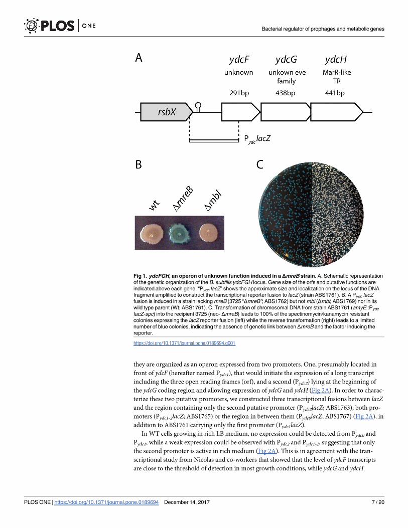
we created a transcriptional fusion between lacZ and the region upstream of ydcF (Pydc lacZ),

that extended from the upstream rsbX gene up to the middle of ydcF (Fig 1A), and introduced it

at the ectopic amyE locus. As expected, the fusion was not induced when placed in a wild-type

background (ABS1761) nor when combined with a deletion of mbl (ABS1769), but was strongly

induced when introduced into the ΔmreB strain 3725 (ABS1762)(Fig 1B), confirming our origi-

nal observation. However, when, in a reversed strategy, the chromosomal DNA of strain 3725

was transformed into ABS1761 containing the reporter and selected for the neomycin resis-

tance marker associated to the mreB deletion, the fusion was intriguingly not induced in the

vast majority of the neomycin resistant transformants (Fig 1C, right). We deduced from this

that the locus responsible for the induction of the reporter in ABS1762 was not genetically

linked to mreB, suggesting the presence of an extragenic mutation in strain 3725.

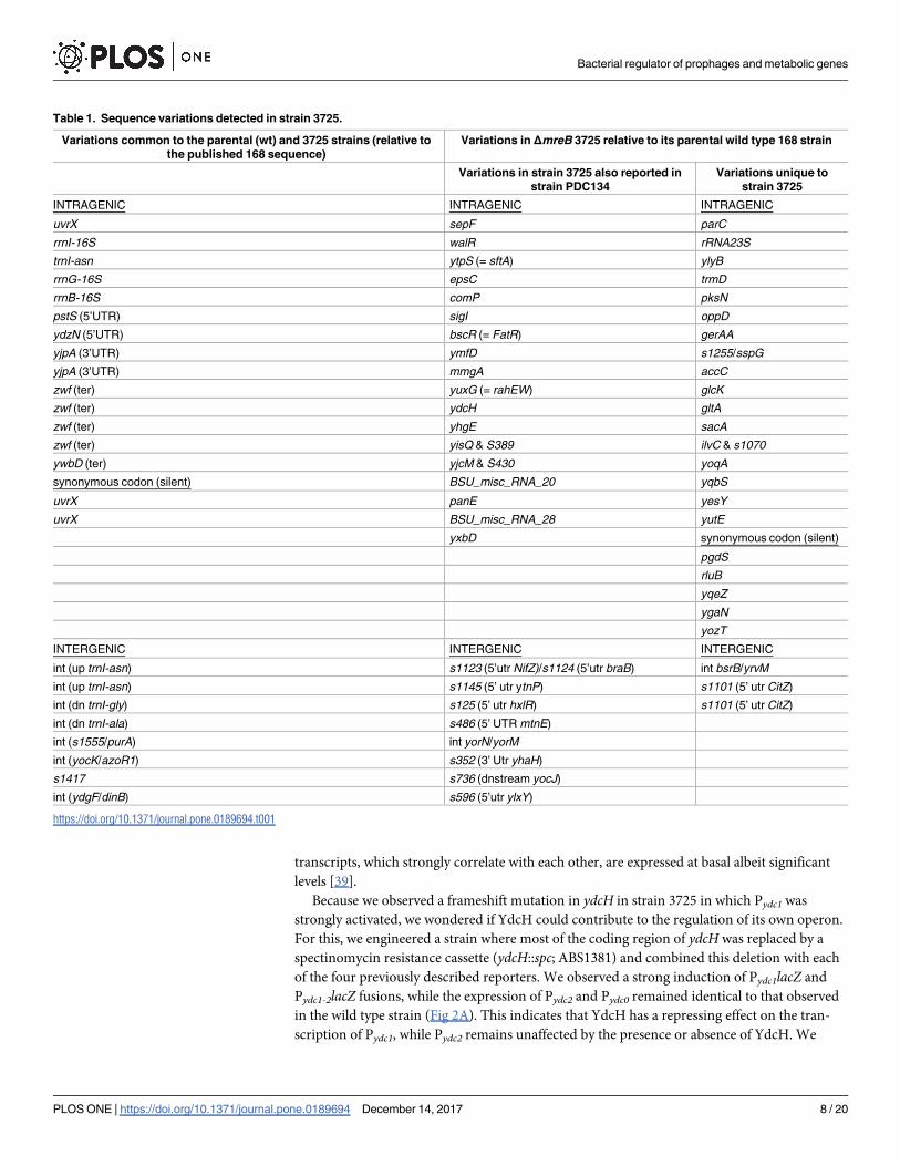
Using next generation sequencing, we completely sequenced the genome of this mutant,

along with that of the wild type 168 strain. We found 24 variations common to the 3725

mutant and its parental wild type strain 168 relative to the published sequence of B. subtilissubsp. subtilis str. 168 (GenBank AL009126.3)(Table 1 and S4 Table), indicating a polymor-

phism between wild types. Most mutations appeared to be silent or affecting untranslated

regions. However and unexpectedly, the sequencing revealed the overwhelming presence of 51

sequence variations in the mutant relative to its parental 168 wild type strain. Half of these

mutations were previously reported in a mutant of B. subtilis forming L-forms (PDC134) [38].

Among the uncovered mutations, we noticed a frameshift in ydcH leading to a premature stop

codon, cutting off more than half of the resulting protein. Since YdcH shares significant simi-

larities with the MarR family of transcriptional regulators, we hypothesized that this protein

could regulate the expression of the ydcF, ydcG and ydcH genes.

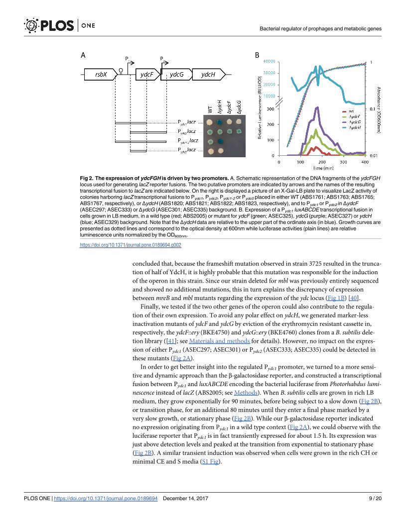
Expression of ydcFGH is driven by two promoters

The systematic mapping of transcription units of B. subtilis in a broad variety of conditions

[39] has shown that the ydcF, ydcG and ydcH genes are expressed as two transcripts, suggesting

Bacterial regulator of prophages and metabolic genes

PLOS ONE | https://doi.org/10.1371/journal.pone.0189694 December 14, 2017 6 / 20

they are organized as an operon expressed from two promoters. One, presumably located in

front of ydcF (hereafter named Pydc1), that would initiate the expression of a long transcript

including the three open reading frames (orf), and a second (Pydc2) lying at the beginning of

the ydcG coding region and allowing expression of ydcG and ydcH (Fig 2A). In order to charac-

terize these two putative promoters, we constructed three transcriptional fusions between lacZand the region containing only the second putative promoter (Pydc2lacZ; ABS1763), both pro-

moters (Pydc1-2lacZ; ABS1765) or the region in between them (Pydc0lacZ; ABS1767) (Fig 2A), in

addition to ABS1761 carrying only the first promoter (Pydc1lacZ).

In WT cells growing in rich LB medium, no expression could be detected from Pydc0 and

Pydc1, while a weak expression could be observed with Pydc2 and Pydc1-2, suggesting that only

the second promoter is active in rich medium (Fig 2A). This is in agreement with the tran-

scriptional study from Nicolas and co-workers that showed that the level of ydcF transcripts

are close to the threshold of detection in most growth conditions, while ydcG and ydcH

Fig 1. ydcFGH, an operon of unknown function induced in aΔmreB strain. A. Schematic representation

of the genetic organization of the B. subtilis ydcFGH locus. Gene size of the orfs and putative functions are

indicated above each gene. “Pydc lacZ” shows the approximate size and localization on the locus of the DNA

fragment amplified to construct the transcriptional reporter fusion to lacZ (strain ABS1761). B. A Pydc lacZ

fusion is induced in a strain lacking mreB (3725 “ΔmreB”; ABS1762) but not mbl (Δmbl; ABS1769) nor in its

wild type parent (Wt; ABS1761). C. Transformation of chromosomal DNA from strain ABS1761 (amyE::Pydc

lacZ-spc) into the recipient 3725 (neo- ΔmreB) leads to 100% of the spectinomycin/kanamycin resistant

colonies expressing the lacZ reporter fusion (left) while the reverse transformation (right) leads to a limited

number of blue colonies, indicating the absence of genetic link between ΔmreB and the factor inducing the

reporter.

https://doi.org/10.1371/journal.pone.0189694.g001

Bacterial regulator of prophages and metabolic genes

PLOS ONE | https://doi.org/10.1371/journal.pone.0189694 December 14, 2017 7 / 20

transcripts, which strongly correlate with each other, are expressed at basal albeit significant

levels [39].

Because we observed a frameshift mutation in ydcH in strain 3725 in which Pydc1 was

strongly activated, we wondered if YdcH could contribute to the regulation of its own operon.

For this, we engineered a strain where most of the coding region of ydcH was replaced by a

spectinomycin resistance cassette (ydcH::spc; ABS1381) and combined this deletion with each

of the four previously described reporters. We observed a strong induction of Pydc1lacZ and

Pydc1-2lacZ fusions, while the expression of Pydc2 and Pydc0 remained identical to that observed

in the wild type strain (Fig 2A). This indicates that YdcH has a repressing effect on the tran-

scription of Pydc1, while Pydc2 remains unaffected by the presence or absence of YdcH. We

Table 1. Sequence variations detected in strain 3725.

Variations common to the parental (wt) and 3725 strains (relative to

the published 168 sequence)

Variations in ΔmreB 3725 relative to its parental wild type 168 strain

Variations in strain 3725 also reported in

strain PDC134

Variations unique to

strain 3725

INTRAGENIC INTRAGENIC INTRAGENIC

uvrX sepF parC

rrnI-16S walR rRNA23S

trnI-asn ytpS (= sftA) ylyB

rrnG-16S epsC trmD

rrnB-16S comP pksN

pstS (5’UTR) sigI oppD

ydzN (5’UTR) bscR (= FatR) gerAA

yjpA (3’UTR) ymfD s1255/sspG

yjpA (3’UTR) mmgA accC

zwf (ter) yuxG (= rahEW) glcK

zwf (ter) ydcH gltA

zwf (ter) yhgE sacA

zwf (ter) yisQ & S389 ilvC & s1070

ywbD (ter) yjcM & S430 yoqA

synonymous codon (silent) BSU_misc_RNA_20 yqbS

uvrX panE yesY

uvrX BSU_misc_RNA_28 yutE

yxbD synonymous codon (silent)

pgdS

rluB

yqeZ

ygaN

yozT

INTERGENIC INTERGENIC INTERGENIC

int (up trnI-asn) s1123 (5’utr NifZ)/s1124 (5’utr braB) int bsrB/yrvM

int (up trnI-asn) s1145 (5’ utr ytnP) s1101 (5’ utr CitZ)

int (dn trnI-gly) s125 (5’ utr hxlR) s1101 (5’ utr CitZ)

int (dn trnI-ala) s486 (5’ UTR mtnE)

int (s1555/purA) int yorN/yorM

int (yocK/azoR1) s352 (3’ Utr yhaH)

s1417 s736 (dnstream yocJ)

int (ydgF/dinB) s596 (5’utr ylxY)

https://doi.org/10.1371/journal.pone.0189694.t001

Bacterial regulator of prophages and metabolic genes

PLOS ONE | https://doi.org/10.1371/journal.pone.0189694 December 14, 2017 8 / 20

concluded that, because the frameshift mutation observed in strain 3725 resulted in the trunca-

tion of half of YdcH, it is highly probable that this mutation was responsible for the induction

of the operon in this strain. Since our strain deleted for mbl was previously entirely sequenced

and showed no additional mutations, this in turn explains the discrepancy of expression

between mreB and mbl mutants regarding the expression of the ydc locus (Fig 1B) [40].

Finally, we tested if the two other genes of the operon could also contribute to the regula-

tion of their own expression. To avoid any polar effect on ydcH, we generated marker-less

inactivation mutants of ydcF and ydcG by eviction of the erythromycin resistant cassette in,

respectively, the ydcF::ery (BKE4750) and ydcG::ery (BKE4760) clones from a B. subtilis dele-

tion library ([41]; see Materials and methods for details). However, no impact on the expres-

sion of either Pydc1 (ASEC297; ASEC301) or Pydc2 (ASEC333; ASEC335) could be detected in

these mutants (Fig 2A).

In order to get better insight into the regulated Pydc1 promoter, we turned to a more sensi-

tive and dynamic approach than the β-galactosidase reporter, and constructed a transcriptional

fusion between Pydc1 and luxABCDE encoding the bacterial luciferase from Photorhabdus lumi-nescence instead of lacZ (ABS2005; see Methods). When B. subtilis cells are grown in rich LB

medium, they grow exponentially for 90 minutes, before being subject to a slow down (Fig 2B),

or transition phase, for an additional 80 minutes until they enter a final phase marked by a

very slow growth, or stationary phase (Fig 2B). While our β-galactosidase reporter indicated

no expression originating from Pydc1 in a wild type context (Fig 2A), we could observe with the

luciferase reporter that Pydc1 is in fact transiently expressed for about 1.5 h. Its expression was

just above detection levels and peaked at the transition from exponential to stationary phase

(Fig 2B). A similar transient induction was observed when cells were grown in the rich CH or

minimal CE and S media (S1 Fig).

Fig 2. The expression of ydcFGH is driven by two promoters. A. Schematic representation of the DNA fragments of the ydcFGH

locus used for generating lacZ reporter fusions. The two putative promoters are indicated by arrows and the names of the resulting

transcriptional fusion to lacZ are indicated below. On the right is displayed a picture of an X-Gal-LB plate to visualize LacZ activity of

colonies harboring lacZ transcriptional fusions to Pydc1, Pydc2, Pydc1-2 or Pydc0 placed in either WT (ABS1761; ABS1763; ABS1765;

ABS1767, respectively), or ΔydcH (ABS1820; ABS1821; ABS1822; ABS1823, respectively), and to Pydc1 or Pydc2 in ΔydcF

(ASEC297; ASEC333) or ΔydcG (ASEC301; ASEC335) background. B. Expression of a Pydc1 luxABCDE transcriptional fusion in

cells grown in LB medium, in a wild type (red; ABS2005) or mutant for ydcF (green; ASEC325), ydcG (purple; ASEC327) or ydcH

(blue; ASEC329) background. Note that the ΔydcH data are relative to the upper part of the ordinate axis (in blue). Growth curves are

presented as dotted lines and correspond to the optical density at 600nm while luciferase activities (plain lines) are relative

luminescence units normalized by the OD600nm.

https://doi.org/10.1371/journal.pone.0189694.g002

Bacterial regulator of prophages and metabolic genes

PLOS ONE | https://doi.org/10.1371/journal.pone.0189694 December 14, 2017 9 / 20

Thanks to the sensitivity of the luciferase reporter, we were able to observe, depending on

the medium, a 3 to 9 and 5 to 12 fold increase of expression in the absence of ydcF and ydcG,

respectively, compared to the wild type levels (Fig 2B and S1 Fig). Albeit significant, the

increase was two orders of magnitude lower that that observed in absence of ydcH (ASEC329;

Fig 2B and S1 Fig), matching our observations with the β-galactosidase reporter. This indicates

that repression was still largely maintained in the absence of YdcF or YdcG suggesting only a

minor role, if any, of these proteins on the repression of Pydc1. Taken together, our observa-

tions indicate that expression of the ydcFGH operon occurs during exponential growth, but is

maintained to very low levels in an YdcH dependent manner.

ydcH binds to the promoter of the ydcFGH operon

YdcH is predicted to belong to the MarR family of TR. These proteins are homodimers with a

characteristic winged-Helix-Turn-Helix (wHTH) domain conferring affinity for 16–20 base

pair (bp) inverted repeats [7]. Most MarR family members act as repressors, by binding close

to, or overlapping, the -35 or -10 sequences of their target promoters, hence preventing the

binding of the RNA polymerase through steric hindrance [6].

To test if the repressing effect of YdcH on Pydc1 expression that we observed could be due to

the direct binding of the protein to the promoter, we purified a recombinant form of the pro-

tein and tested its binding potential in an electrophoretic mobility shift assay (EMSA).

Although the promoter was not mapped, whole genome tailing arrays [39] had shown a tran-

scriptional upshift 165 bp upstream of the translational start site of the ydcF orf. Analysis of

this area revealed directly upstream of this upshift the presence of -35 and -10 sequences typi-

cal of σA-dependent promoters and, in between, a perfect inverted repeat (IR) sequence (TAA

TAAGnnnnCTTATTA) forming a palindrome (Fig 3A). We first incubated a 293 bp DNA

fragment extending from the end of the upstream rsbX gene to the beginning of the ydcF orf

with purified YdcH, in the presence of an excess of non-specific competitor DNA (Fig 3B). We

Fig 3. YdcH binds specifically to inverted repeats in the promoter region of ydcFGH. A. Sequence of the region upstream of the

ydcFGH operon. The two identified IR are indicated as green arrows. The transcriptional upshift previously identified is indicated as “up”,

putative -35, -10 and rbs sequences are underlined, and the ydcF orf is boxed. B. EMSAs (right panels) showing the specific binding of

PamR to DNA fragments corresponding to the wild type (wt) and mutated (IR1*) ydcF promoter, and schematic representation of the

corresponding area (left panel). IRs are drawn as facing triangles, plain for the wild types and hollowed for the mutated. The quantity of

YdcH (in pmol) incubated with 0.1 pmol of labeled target DNA is indicated above each lane.

https://doi.org/10.1371/journal.pone.0189694.g003

Bacterial regulator of prophages and metabolic genes

PLOS ONE | https://doi.org/10.1371/journal.pone.0189694 December 14, 2017 10 / 20

observed specific, concentration-dependent retardations, suggesting a direct and specific bind-

ing of the protein (Fig 3B; wt). Interestingly three species appeared indicating the formation of

two DNA-protein complexes.

The complete retardation was observed with a ratio of 4 moles of proteins per mole of

DNA, consistent with the binding of 2 dimers per DNA molecules. A rapid examination of the

promoter sequence revealed the presence of a second imperfect direct repeat (TAATAAGnnn

nCTTATcA) overlapping the probable transcriptional initiating base (Fig 3A; IR2), that could

account for the observed complexes. To test if the directed repeats correspond to the motif rec-

ognized by YdcH, we created a DNA fragment mutant for the most upstream IR, by swapping

between both strands of the seven bases forming the first half of the IR (ATTATTCnnnnCTTA

TTA). The resulting EMSA with the mutated DNA fragment showed that one of the protein-

DNA interactions was abolished, indicating that the IRs are the probable motifs recognized by

YdcH. We then scanned the entire B. subtilis genome for the two identified IR but these

sequences were not found anywhere else.

ydcFGH is not involved in resistance to a variety of stresses

We next wondered what could be the function of this operon. ydcF is predicted to encode a 97

amino-acid peptide with no similarity to known or uncharacterized proteins. The only notice-

able characteristic of YdcG is the presence of a domain of unknown function, EVE (formerly

DUF055), structurally related to the RNA-binding PUA domain, present in all three kingdoms

of life with a predominance in bacteria [42]. Thus, no clear function could be infered from

these two genes. However, our results are in full agreement with YdcH acting as a repressor.

MarR TRs are usually involved in the response to environmental changes, helping cells to

improve their survival, and are frequently linked to multiple resistance to antibiotic, salt and

aromatic molecules and to virulence [6, 43–45]. We therefore evaluated the effect of deleting

each of the ydcFGH genes on the ability of B. subtilis to resist to various stresses.

Using disc diffusion assays, we tested the resistance of the three deletion mutants to antibi-

otics of various classes, affecting cell wall synthesis (Vancomycin, Bacitracine, Methicillin),

membrane integrity (polymyxine), replication (ciprofloxacine, levofloxacine) transcription

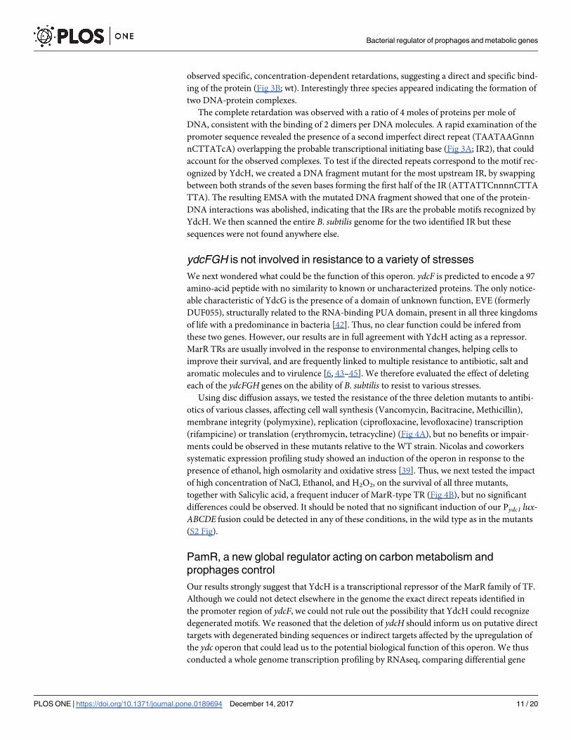
(rifampicine) or translation (erythromycin, tetracycline) (Fig 4A), but no benefits or impair-

ments could be observed in these mutants relative to the WT strain. Nicolas and coworkers

systematic expression profiling study showed an induction of the operon in response to the

presence of ethanol, high osmolarity and oxidative stress [39]. Thus, we next tested the impact

of high concentration of NaCl, Ethanol, and H2O2, on the survival of all three mutants,

together with Salicylic acid, a frequent inducer of MarR-type TR (Fig 4B), but no significant

differences could be observed. It should be noted that no significant induction of our Pydc1 lux-ABCDE fusion could be detected in any of these conditions, in the wild type as in the mutants

(S2 Fig).

PamR, a new global regulator acting on carbon metabolism and

prophages control

Our results strongly suggest that YdcH is a transcriptional repressor of the MarR family of TF.

Although we could not detect elsewhere in the genome the exact direct repeats identified in

the promoter region of ydcF, we could not rule out the possibility that YdcH could recognize

degenerated motifs. We reasoned that the deletion of ydcH should inform us on putative direct

targets with degenerated binding sequences or indirect targets affected by the upregulation of

the ydc operon that could lead us to the potential biological function of this operon. We thus

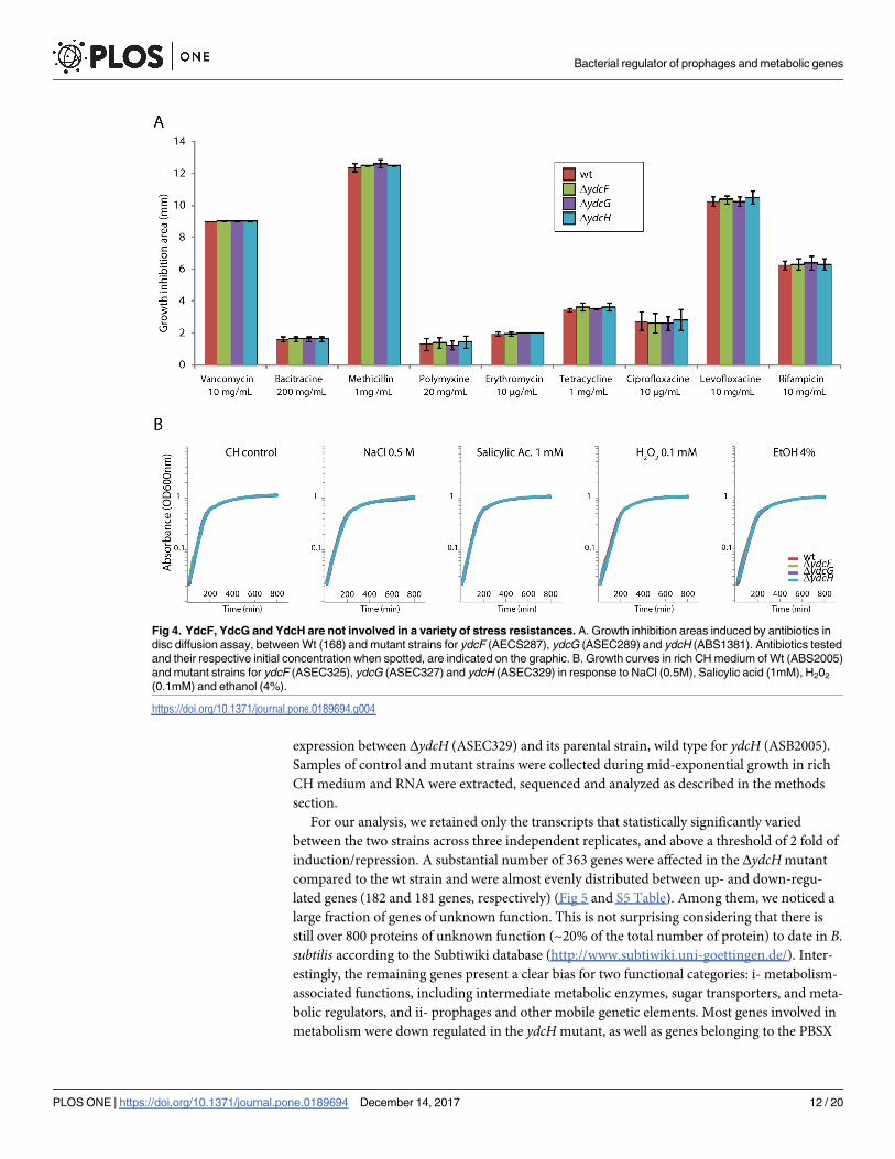
conducted a whole genome transcription profiling by RNAseq, comparing differential gene

Bacterial regulator of prophages and metabolic genes

PLOS ONE | https://doi.org/10.1371/journal.pone.0189694 December 14, 2017 11 / 20

expression between ΔydcH (ASEC329) and its parental strain, wild type for ydcH (ASB2005).

Samples of control and mutant strains were collected during mid-exponential growth in rich

CH medium and RNA were extracted, sequenced and analyzed as described in the methods

section.

For our analysis, we retained only the transcripts that statistically significantly varied

between the two strains across three independent replicates, and above a threshold of 2 fold of

induction/repression. A substantial number of 363 genes were affected in the ΔydcH mutant

compared to the wt strain and were almost evenly distributed between up- and down-regu-

lated genes (182 and 181 genes, respectively) (Fig 5 and S5 Table). Among them, we noticed a

large fraction of genes of unknown function. This is not surprising considering that there is

still over 800 proteins of unknown function (~20% of the total number of protein) to date in B.

subtilis according to the Subtiwiki database (http://www.subtiwiki.uni-goettingen.de/). Inter-

estingly, the remaining genes present a clear bias for two functional categories: i- metabolism-

associated functions, including intermediate metabolic enzymes, sugar transporters, and meta-

bolic regulators, and ii- prophages and other mobile genetic elements. Most genes involved in

metabolism were down regulated in the ydcH mutant, as well as genes belonging to the PBSX

Fig 4. YdcF, YdcG and YdcH are not involved in a variety of stress resistances. A. Growth inhibition areas induced by antibiotics in

disc diffusion assay, between Wt (168) and mutant strains for ydcF (AECS287), ydcG (ASEC289) and ydcH (ABS1381). Antibiotics tested

and their respective initial concentration when spotted, are indicated on the graphic. B. Growth curves in rich CH medium of Wt (ABS2005)

and mutant strains for ydcF (ASEC325), ydcG (ASEC327) and ydcH (ASEC329) in response to NaCl (0.5M), Salicylic acid (1mM), H202

(0.1mM) and ethanol (4%).

https://doi.org/10.1371/journal.pone.0189694.g004

Bacterial regulator of prophages and metabolic genes

PLOS ONE | https://doi.org/10.1371/journal.pone.0189694 December 14, 2017 12 / 20

prophage, while genes belonging to all others prophage or mobile genetic element (SPβ pro-

phage, skin element, Prophage1 and ICEBs1) were induced in absence of ydcH (Fig 5 and S5

Table). It is noticeable that 40 genes encoding TF or other regulators (including 16 known

transcription factors) (Table 2), acting either on metabolic pathways (LrpA, LrpB, ThrR,

TnrA, NtdR, PyrR), control of prophages (ImmR, ImmA, Xre, SknR, RapI) or during transi-

tion phase (AbrB, SinR) and stationary phase events such as sporulation (SigF, SpoVT, Sda,

YisI) or competence (ComK, Kre) were also affected. These regulators reflect well the diversity

of functional categories identified in our transcriptomic analysis by RNAseq (Fig 5) and may

also account for the large number of genes whose expression differ in our mutant. In

Fig 5. The YdcH regulon. Pie charts summarizing genome-wide transcriptional profiling by RNAseq

comparing gene expressions in a WT (ABS2005) and a ΔydcH strain (ASEC56). The 363 genes retained (left

chart) were reproducibly and statistically induced (182, right up) or repressed (181, right down) in the mutant

compared to the wt by at least a two-fold factor. Genes were sorted by functional categories (see S5 Table for

complete results), then regrouped into families of functions: Metabolism (carbon sources, amino acids, lipids,

nucleotides and other metabolic pathways; electron transport & ATP synthesis; transport of sugars and other

metabolites), stress response, information processing (DNA replication, segmentation, modification,

recombination and repair; RNA and protein synthesis, modification and degradation), cellular processes (cell

division; cell envelope synthesis, modification and degradation; ion homeostasis), lifestyles (motility &

chemotaxis; biofilms formation; competence; sporulation), prophages & mobile genetic elements, and

unknown. Numbers indicate the number of gene for each category.

https://doi.org/10.1371/journal.pone.0189694.g005

Bacterial regulator of prophages and metabolic genes

PLOS ONE | https://doi.org/10.1371/journal.pone.0189694 December 14, 2017 13 / 20

Table 2. Transcription factors and other regulators affected in the absence of PamR.

gene Effect3 Exp. Diff. TR & Mod 1 Functional category 2 Function

ypoP + 3,00 TF* U similar to transcriptional regulator (MarR family)

yopO + 4,12 TF* Prophages & mobile gen.

elemtssimilar to transcriptional regulator (Xre family)

yonR + 5,29 TF* Prophages & mobile gen.

elemtssimilar to transcriptional regulator (Xre family)

yobD + 3,69 TF* U similar to transcriptional regulator (Xre family)

ykvN + 2,64 TF* U unknown; MarR family transcription regulator

ydfL + 3,30 TF* U unknown; similar to multidrux efflux transporter regulator of MreR family

ydeB + 3,26 TF* U unknown; putative transcriptional regulator

yceK + 2,56 TF* U unknown; putative transcriptional regulator

ybzH + 3,84 TF* Prophages & mobile gen.

elemtsunknown; putative transcriptional regulator

xre + 3,83 TF Prophages & mobile gen.

elemtsregulation of PBSX prophage gene expression

tnrA + 2,45 TF Metabolism control of nitrogen assimilation (MerR family)

sknR + 3,04 TF Prophages & mobile gen.

elemtsrepression of yqaF operon of the skin element (Xre family)

sinR + 2,76 TF Lifestyle regulator of post-exponential-phase responses genes, and biofilm formation

ntdR + 3,73 TF Lifestyle activator of ntdABC-glcP operon (LacI family)

mntR + 2,77 TF Cellular processes regulation of manganese transport (DtxR family)

mgsR + 2,86 TF Stress response regulator of a subset of the SigB stress regulon; oxidative stress protection

lrpB + 3,51 TF Metabolism metabolism of glycine; Repression of glyA transcription and KinB-dependent

spo.

lrpA + 5,26 TF Metabolism metabolism of glycine; Repression of glyA transcription and KinB-dependent

spo.

immR + 3,86 TF Prophages & mobile gen.

elemtsControl of transfer of the mobile genetic element ICEBs1

comK + 2,74 TF Lifestyle master regulator for competence

abrB + 4,34 TF Lifestyle tansition state regulator

sigX + 3,07 TF Stress response resistance to cationic antimicrobial peptides; RNA polymerase ECF-type σfactor

pyrR + 3,56 R Metabolism transcriptional antiterminator of the pyr operon

kre + 3,31 R Lifestyle inhibitor of competence; ComK repressor

yydG + 4,50 R Stress response control of LiaR-LiaS activity; oxidoreductase

yisI + 4,16 R Lifestyle inhibition of phosphorelay; Spo0A-P phosphatase

sda + 2,68 R Lifestyle checkpoint coupling replication and sporulation; inhibitor of Spo0A

phosphorelay

rapI + 2,96 R Prophages & mobile gen.

elemtscontrol of ICEBs1, ImmR antagonist; response regulator aspartate

phosphatase

phrE + 3,48 R Lifestyle Regulator of RapE phosphatase

immA + 3,40 R Prophages & mobile gen.

elemtscontrol of ImmR activity; site-specific protease

degR + 3,58 R Metabolism control of DegU; Positive effector of DegU-phosphate stability

yhcF - 3,26 TF* U unknown; putative transcription factor (GntR family)

thrR - 2,75 TF Metabolism control of threonine biosynthesis

spoVT - 2,37 TF Lifestyle regulation of forespore gene expression

sigF - 2,56 TF Lifestyle RNA polymerase forespore-specific (early) sigma factor SigF

ycbM - 2,71 R* U predicted two-component sensor kinase

yhcY - 2,13 R U two component sensor kinase

yesM - 4,38 R U two-component sensor kinase

(Continued )

Bacterial regulator of prophages and metabolic genes

PLOS ONE | https://doi.org/10.1371/journal.pone.0189694 December 14, 2017 14 / 20

agreement, among the genes affected by ydcH absence we noticed that 34 fall under AbrB (a

transition state TR) regulation, a majority of them (25/34) variying as if AbrB was less active in

the ΔydcH strain (AbrB-induced genes are down and AbrB-repressed are up). We also noticed

that 42 known targets of CcpA, a transcriptional repressor of catabolic genes, are down regu-

lated as if CcpA was more active in the ΔydcH mutant. Since expression of ccpA did not seem

affected in absence of ydcH, it suggests that different levels of regulations could be affected in

this mutant.

Taken together, these results indicate that, albeit potentially indirect through activity of

other regulators, the absence of ydcH leads to a global reprogramming of the cell with a specific

prominence of metabolic pathways and prophage-related genes. We therefore propose to

rename the gene pamR for prophages and metabolism control regulator.

Discussion

Our study originally focused on the ydcFGH operon because of its potential relationship with

MreB, aiming at deciphering both its function and the link with this morphoprotein. However

our results indicate that induction of this operon was genetically unlinked with the morpho-

gene. Because mutants of mreB are notoriously sick and easily accumulate suppressor muta-

tions in the absence of high concentrations of Mg2+ [37, 46, 47], we cannot exclude that the

presence of the mutation affecting pamR (ydcH) in mreB 3725 strain is the result of a selective

pressure on this strain. Puzzlingly, we also noticed in the literature that a strain selected for L-

form proficiency (PDC134) (i.e. a strain supporting growth without cell wall) was reported to

bear 16 intragenic mutations (28 mutations in total), all of them present in the 3725 strain

[38]. The common origin of strain PDC134 and 3725 suggests that they share a common

ancestor presenting these mutations rather than having been selected independently. In addi-

tion, we could not observe any benefit of the deletion of pamR when inserted in a ΔmreBmutant, on the growth or shape of this mutant. Because the genome of strain 3725 contains 50

other sequence variations relative to its wild type parental strain, it is therefore possible that

one of the other mutations induced a change that in turn required the inactivation of pamR.

But what could be the function of the ydc operon? Our results have yet to demonstrate a

function for the first two genes of the operon. However, we showed that PamR (YdcH) con-

trols its own expression, specifically binds (probably as a dimer) its own promoter due to the

presence of two IRs motif (TAATAAGtgnnCTTATNA) overlapping the RNA polymerase

binding site, strongly suggesting that it is indeed a transcriptional regulator acting as a repres-

sor by preventing the recruitment of the RNA polymerase by steric hindrance. This is a fre-

quent feature of MarR TRs that often control their expression by binding to the promoter

controlling their own gene or operon [6–8]. Using genome wide transcription profiling, we

also showed that the absence of PamR correlates with the induction or repression of numerous

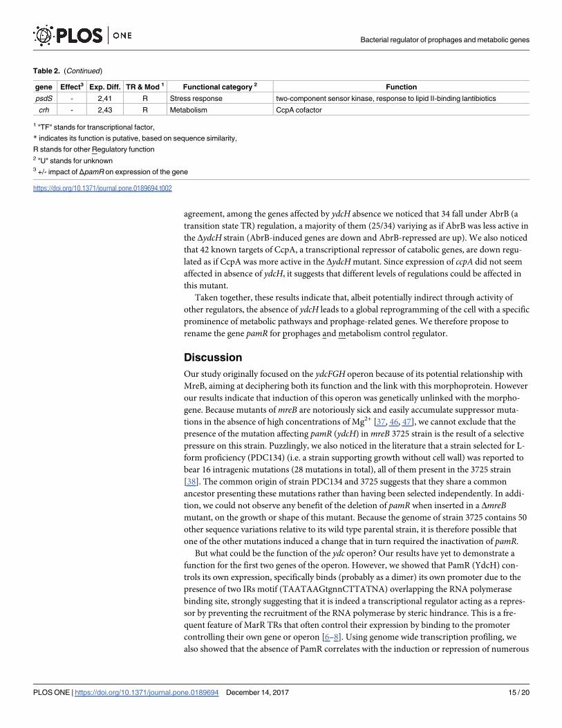
Table 2. (Continued)

gene Effect3 Exp. Diff. TR & Mod 1 Functional category 2 Function

psdS - 2,41 R Stress response two-component sensor kinase, response to lipid II-binding lantibiotics

crh - 2,43 R Metabolism CcpA cofactor

1 "TF" stands for transcriptional factor,

* indicates its function is putative, based on sequence similarity,

R stands for other Regulatory function2 "U" stands for unknown3 +/- impact of ΔpamR on expression of the gene

https://doi.org/10.1371/journal.pone.0189694.t002

Bacterial regulator of prophages and metabolic genes

PLOS ONE | https://doi.org/10.1371/journal.pone.0189694 December 14, 2017 15 / 20

genes, leading to a reprogramming of the B. subtilis genetic expression larger in its extent than

the general stress response (namely the SigB regulon [48, 49]), the SigH-dependent stationary

phase adaptation [14] and similar to stationary phase developmental processes as competence

or sporulation [50, 51]. TRs usually bind DNA with a strong affinity for a specific palindromic

sequence located in the promoter region of the controlled genes and inhibiting transcription

by steric hindrance or more rarely inducing transcription [7, 8]. While most MarR-type TRs

generally act as repressors and control small regulons, PamR uncommonly affects a very large

number of genes, some positively and some negatively in rather equal proportions. Although it

is at this stage possible that PamR is an atypical MarR-type TR, it is more likely that the vast

majority of affected genes, if not all but the ydcFGH operon, are downstream consequences of

expression changes affecting its “core” regulon, i.e. genes under direct PamR control. This is

re-enforced by the absence of conserved PamR binding site in the rest of the genome. A clue

advocating for this is the fact that many regulators or modulators of regulators are affected by

pamR deletion along with at least a fraction of their respective regulons (Xre, AbrB. . ..). Thus

by affecting the expression or/and activity of a limited number of key proteins, it would allow

a global reprogramming of the cell. What could be the purpose of such regulation? The pattern

of expression of the Pydc1 promoter (controlling the three genes of the ydcFGH operon) shows

induction during the transition between exponential and stationary phase. Although this may

be due to an additional regulatory process, it may nevertheless suggest a function in the transi-

tion between growth phases, a hypothesis reinforced by several observations. First, there is an

overlap between the PamR and AbrB regulons, AbrB being a well-known transition state regu-

lator [13]. In absence of PamR, we observed a release of the repression of a part of the AbrB

regulon suggesting a partial decrease in AbrB repression capacity. Thus PamR may contribute

to the previously reported sequential AbrB de-repression observed from mid-exponential

growth to stationary phase [52]. Next, we observed a change of expression of a large group (92)

of metabolic genes that could reflect a growth adaptation and a need to cope with new carbon

sources. We also observed the presence of numerous transition or stationary phase-involved

genes including antibiotic/toxin producing operons (albA, skf, yydG. . .) that suggest a scaveng-

ing strategy, and stress response genes that also advocate for adaptation to a complex life style

and/or stationary phase. Finally, the induction of genes from the lysogenic prophages SPβ and

the mobile elements ICEBs1 could also indicate a response to stress. DNA damages are the

most documented cause promoting their activation [20, 21]. It is however unlikely that the

SOS response could here be responsible for the induction of SPβ and ICEBs1 because only a

very limited number of LexA-controlled genes (the repressor controlling the SOS response)

are induced (and some are even down-regulated) in absence of PamR. In addition, the defec-

tive prophage PBSX, also known to be induced in response to DNA damage [53], is repressed

in this genetic context. Other stresses have been shown to promote their activation including

reactive oxygen species, heat, some antibiotics and toxic compounds [24] but the absence of

induction of the general stress response in absence of PamR advocate against this possibility.

Together this suggests that a more complex regulatory network could be responsible for the

differential expression of these prophage-encoded genes.

However, considering the very mild induction observed with our reporter (in the wt back-

ground) in every conditions tested so far compared to its levels in the strain deleted for pamR,

it is probable that in our conditions most of the pamR regulon will barely be affected. Uncover-

ing the conditions of optimal activation of the PamR regulon would help discovering the signal

sensed by the regulator. Typically, MarR-type TRs bind to one or several ligands upon which

the affinity to DNA decreases, allowing the release of repression–or activation- to their target

genes. From our results, the first two genes of the operon (ydcF and ydcG) are not -or

Bacterial regulator of prophages and metabolic genes

PLOS ONE | https://doi.org/10.1371/journal.pone.0189694 December 14, 2017 16 / 20

marginally- involved in Pydc1 regulation suggesting they are not involved in the production or

sensing of the yet to be determined signal.

Overall, based on the extent of the regulon and the observed influence on stationary-phase,

stress-response, metabolic or phage-related genes, our results suggest that PamR is switching

the genetic program of the bacteria in response to an unknown signal to help the cell cope with

some environmental changes or growth regime modification.

Supporting information

S1 Fig. Pydc1 is strongly induced in the absence of YdcH in rich and poor media. Expression

of a Pydc1 luxABCDE transcriptional fusion in cells grown in poor S and CE or rich CH media,

in a wild type (red; ABS2005) or mutant for ydcF (green; ASEC325), ydcG (purple; ASEC327)

or ydcH (blue; ASEC329) background. Left panels do not include ΔydcH (ASEC329) data that

would be off chart (notice the different axis ranges between right and left panels). Growth

curves are shown as dotted lines and correspond to the optical density at 600nm while lucifer-

ase activities (plain lines) are relative luminescence units normalized by the OD600nm.

(PDF)

S2 Fig. Pydc1 is not induced in response to various stresses. Maximum expression of a Pydc1

luxABCDE transcriptional fusion in cells exposed to NaCl (0.5 M), Salicylic acid (1 mM), H202

(0.1 mM) and ethanol (4%), in a wild type (ABS2005) or mutant for ydcF (ASEC325), ydcG(ASEC327) or ydcH (ASEC329) background.

(PDF)

S1 Table. Strains used in this study.

(PDF)

S2 Table. Plasmids used in this study.

(PDF)

S3 Table. Oligonucleotides used in this study.

(PDF)

S4 Table. Sequence variations detected in strain 3725 (detailed).

(PDF)

S5 Table. List of differentially expressed genes in a ΔpamR strain compared to its parental

wild type B. subtilis strain.

(PDF)

Acknowledgments

We thank L. Slamti for critical reading of the manuscript and P. Nicolas for constructive dis-

cussions and advices. We are grateful to the INRA MIGALE bioinformatics platform (http://

migale.jouy.inra.fr) for providing computational resources. This work has benefited from the

platforms and expertise of the High-Throughput Sequencing Platform of I2BC.

Author Contributions

Conceptualization: Alba De San Eustaquio-Campillo, Arnaud Chastanet.

Data curation: Cyprien Guerin.

Formal analysis: Cyprien Guerin.

Bacterial regulator of prophages and metabolic genes

PLOS ONE | https://doi.org/10.1371/journal.pone.0189694 December 14, 2017 17 / 20

Funding acquisition: Rut Carballido-Lopez.

Investigation: Alba De San Eustaquio-Campillo, Charlène Cornilleau, Arnaud Chastanet.

Methodology: Arnaud Chastanet.

Project administration: Arnaud Chastanet.

Supervision: Rut Carballido-Lopez, Arnaud Chastanet.

Validation: Arnaud Chastanet.

Writing – original draft: Alba De San Eustaquio-Campillo, Rut Carballido-Lopez, Arnaud

Chastanet.

References1. Aguilar C, Vlamakis H, Losick R, Kolter R. Thinking about Bacillus subtilis as a multicellular organism.

Current opinion in microbiology. 2007; 10(6):638–43. https://doi.org/10.1016/j.mib.2007.09.006 PMID:

17977783

2. Hamoen LW, Venema G, Kuipers OP. Controlling competence in Bacillus subtilis: shared use of regula-

tors. Microbiology (Reading, England). 2003; 149(Pt 1):9–17.

3. Hilbert DW, Piggot PJ. Compartmentalization of gene expression during Bacillus subtilis spore forma-

tion. Microbiology and molecular biology reviews. 2004; 68(2):234–62. https://doi.org/10.1128/MMBR.

68.2.234-262.2004 PMID: 15187183

4. Mielich-Suss B, Lopez D. Molecular mechanisms involved in Bacillus subtilis biofilm formation. Environ-

mental microbiology. 2015; 17(3):555–65. https://doi.org/10.1111/1462-2920.12527 PMID: 24909922

5. Piggot PJ, Hilbert DW. Sporulation of Bacillus subtilis. Current opinion in microbiology. 2004; 7(6):579–

86. https://doi.org/10.1016/j.mib.2004.10.001 PMID: 15556029

6. Grove A. MarR family transcription factors. Curr Biol. 2013; 23(4):R142–3. https://doi.org/10.1016/j.

cub.2013.01.013 PMID: 23428319

7. Perera IC, Grove A. Molecular mechanisms of ligand-mediated attenuation of DNA binding by MarR

family transcriptional regulators. Journal of molecular cell biology. 2010; 2(5):243–54. https://doi.org/10.

1093/jmcb/mjq021 PMID: 20716550

8. Wilkinson SP, Grove A. Ligand-responsive transcriptional regulation by members of the MarR family of

winged helix proteins. Current issues in molecular biology. 2006; 8(1):51–62. PMID: 16450885

9. Kumarevel T. The MarR Family of Transcriptional Regulators—A Structural Perspective. In: Pana M,

editor. Antibiotic resistant bacteria—A continuous challenge in the new millennium: InTech; 2012.

p. 403–18.

10. Shafikhani SH, Nunez E, Leighton T. Hpr (ScoC) and the phosphorelay couple cell cycle and sporula-

tion in Bacillus subtilis. FEMS microbiology letters. 2004; 231(1):99–110. https://doi.org/10.1016/

S0378-1097(03)00936-4 PMID: 14769473

11. Sonenshein AL. Control of key metabolic intersections in Bacillus subtilis. Nature reviews Microbiology.

2007; 5(12):917–27. https://doi.org/10.1038/nrmicro1772 PMID: 17982469

12. Strauch MA, Bobay BG, Cavanagh J, Yao F, Wilson A, Le Breton Y. Abh and AbrB control of Bacillus

subtilis antimicrobial gene expression. Journal of bacteriology. 2007; 189(21):7720–32. https://doi.org/

10.1128/JB.01081-07 PMID: 17720793

13. Strauch MA, Hoch JA. Transition-state regulators: sentinels of Bacillus subtilis post-exponential gene

expression. Molecular microbiology. 1993; 7(3):337–42. PMID: 8459762

14. Britton RA, Eichenberger P, Gonzalez-Pastor JE, Fawcett P, Monson R, Losick R, et al. Genome-wide

analysis of the stationary-phase sigma factor (Sigma-H) regulon of Bacillus subtilis. Journal of bacteriol-

ogy. 2002; 184(17):4881–90. https://doi.org/10.1128/JB.184.17.4881-4890.2002 PMID: 12169614

15. Chu F, Kearns DB, McLoon A, Chai Y, Kolter R, Losick R. A novel regulatory protein governing biofilm

formation in Bacillus subtilis. Molecular microbiology. 2008; 68(5):1117–27. https://doi.org/10.1111/j.

1365-2958.2008.06201.x PMID: 18430133

16. Phillips ZE, Strauch MA. Bacillus subtilis sporulation and stationary phase gene expression. Cell Mol

Life Sci. 2002; 59(3):392–402. PMID: 11964117

17. Chumsakul O, Takahashi H, Oshima T, Hishimoto T, Kanaya S, Ogasawara N, et al. Genome-wide

binding profiles of the Bacillus subtilis transition state regulator AbrB and its homolog Abh reveals their

Bacterial regulator of prophages and metabolic genes

PLOS ONE | https://doi.org/10.1371/journal.pone.0189694 December 14, 2017 18 / 20

interactive role in transcriptional regulation. Nucleic acids research. 2011; 39(2):414–28. https://doi.org/

10.1093/nar/gkq780 PMID: 20817675

18. Sonenshein AL. CodY, a global regulator of stationary phase and virulence in Gram-positive bacteria.

Current opinion in microbiology. 2005; 8(2):203–7. https://doi.org/10.1016/j.mib.2005.01.001 PMID:

15802253

19. Fujita Y. Carbon catabolite control of the metabolic network in Bacillus subtilis. Bioscience, biotechnol-

ogy, and biochemistry. 2009; 73(2):245–59. https://doi.org/10.1271/bbb.80479 PMID: 19202299

20. Auchtung JM, Lee CA, Garrison KL, Grossman AD. Identification and characterization of the immunity

repressor (ImmR) that controls the mobile genetic element ICEBs1 of Molecular microbiology. 2007; 64

(6):1515–28. https://doi.org/10.1111/j.1365-2958.2007.05748.x PMID: 17511812

21. Lazarevic V, Dusterhoft A, Soldo B, Hilbert H, Mauel C, Karamata D. Nucleotide sequence of the Bacil-

lus subtilis temperate bacteriophage SPbetac2. Microbiology (Reading, England). 1999; 145 (Pt

5):1055–67.

22. Imamovic L, Muniesa M. Characterizing RecA-independent induction of Shiga toxin2-encoding phages

by EDTA treatment. PloS one. 2012; 7(2):e32393. https://doi.org/10.1371/journal.pone.0032393 PMID:

22393404

23. Los JM, Los M, Wegrzyn G, Wegrzyn A. Differential efficiency of induction of various lambdoid pro-

phages responsible for production of Shiga toxins in response to different induction agents. Microbial

pathogenesis. 2009; 47(6):289–98. https://doi.org/10.1016/j.micpath.2009.09.006 PMID: 19761828

24. Nanda AM, Thormann K, Frunzke J. Impact of spontaneous prophage induction on the fitness of bacte-

rial populations and host-microbe interactions. Journal of bacteriology. 2015; 197(3):410–9. https://doi.

org/10.1128/JB.02230-14 PMID: 25404701

25. Martin-Verstraete I, Debarbouille M, Klier A, Rapoport G. Levanase operon of Bacillus subtilis includes

a fructose-specific phosphotransferase system regulating the expression of the operon. Journal of

molecular biology. 1990; 214(3):657–71. https://doi.org/10.1016/0022-2836(90)90284-S PMID:

2117666

26. Sterlini JM, Mandelstam J. Commitment to sporulation in Bacillus subtilis and its relationship to develop-

ment of actinomycin resistance. The Biochemical journal. 1969; 113(1):29–37. PMID: 4185146

27. Sharpe ME, Hauser PM, Sharpe RG, Errington J. Bacillus subtilis cell cycle as studied by fluorescence

microscopy: constancy of cell length at initiation of DNA replication and evidence for active nucleoid par-

titioning. Journal of bacteriology. 1998; 180(3):547–55. PMID: 9457856

28. Harwood CR, Cutting SM. Molecular Biological Methods for Bacillus. methods Mm, editor. New York:

J. Wileys and Sons; 1990.

29. Chen Y, Cao S, Chai Y, Clardy J, Kolter R, Guo JH, et al. A Bacillus subtilis sensor kinase involved in

triggering biofilm formation on the roots of tomato plants. Molecular microbiology. 2012; 85(3):418–30.

https://doi.org/10.1111/j.1365-2958.2012.08109.x PMID: 22716461

30. Wang X, Montero Llopis P, Rudner DZ. Bacillus subtilis chromosome organization oscillates between

two distinct patterns. Proceedings of the National Academy of Sciences of the United States of America.

2014; 111(35):12877–82. https://doi.org/10.1073/pnas.1407461111 PMID: 25071173

31. Milne I, Stephen G, Bayer M, Cock PJ, Pritchard L, Cardle L, et al. Using Tablet for visual exploration of

second-generation sequencing data. Briefings in bioinformatics. 2013; 14(2):193–202. https://doi.org/

10.1093/bib/bbs012 PMID: 22445902

32. Bolger AM, Lohse M, Usadel B. Trimmomatic: a flexible trimmer for Illumina sequence data. Bioinfor-

matics. 2014; 30(15):2114–20. https://doi.org/10.1093/bioinformatics/btu170 PMID: 24695404

33. Langmead B, Salzberg SL. Fast gapped-read alignment with Bowtie 2. Nature methods. 2012; 9

(4):357–9. https://doi.org/10.1038/nmeth.1923 PMID: 22388286

34. Anders S, Pyl PT, Huber W. HTSeq—a Python framework to work with high-throughput sequencing

data. Bioinformatics. 2015; 31(2):166–9. https://doi.org/10.1093/bioinformatics/btu638 PMID:

25260700

35. Strimmer K. fdrtool: a versatile R package for estimating local and tail area-based false discovery rates.

Bioinformatics. 2008; 24(12):1461–2. https://doi.org/10.1093/bioinformatics/btn209 PMID: 18441000

36. Robinson MD, McCarthy DJ, Smyth GK. edgeR: a Bioconductor package for differential expression

analysis of digital gene expression data. Bioinformatics. 2010; 26(1):139–40. https://doi.org/10.1093/

bioinformatics/btp616 PMID: 19910308

37. Formstone A, Errington J. A magnesium-dependent mreB null mutant: implications for the role of mreB

in Bacillus subtilis. Molecular microbiology. 2005; 55(6):1646–57. https://doi.org/10.1111/j.1365-2958.

2005.04506.x PMID: 15752190

Bacterial regulator of prophages and metabolic genes

PLOS ONE | https://doi.org/10.1371/journal.pone.0189694 December 14, 2017 19 / 20

38. Dominguez-Cuevas P, Mercier R, Leaver M, Kawai Y, Errington J. The rod to L-form transition of Bacil-

lus subtilis is limited by a requirement for the protoplast to escape from the cell wall sacculus. Molecular

microbiology. 2012; 83(1):52–66. https://doi.org/10.1111/j.1365-2958.2011.07920.x PMID: 22122227

39. Nicolas P, Mader U, Dervyn E, Rochat T, Leduc A, Pigeonneau N, et al. Condition-dependent transcrip-

tome reveals high-level regulatory architecture in Bacillus subtilis. Science (New York, NY. 2012; 335

(6072):1103–6.

40. Billaudeau C, Chastanet A, Yao Z, Cornilleau C, Mirouze N, Fromion V, et al. Contrasting mechanisms

of growth in two model rod-shaped bacteria. Nature communications. 2017; 8:15370. https://doi.org/10.

1038/ncomms15370 PMID: 28589952

41. Koo BM, Kritikos G, Farelli JD, Todor H, Tong K, Kimsey H, et al. Construction and Analysis of Two

Genome-Scale Deletion Libraries for Bacillus subtilis. Cell systems. 2017; 4(3):291–305 e7. https://doi.

org/10.1016/j.cels.2016.12.013 PMID: 28189581

42. Bertonati C, Punta M, Fischer M, Yachdav G, Forouhar F, Zhou W, et al. Structural genomics reveals

EVE as a new ASCH/PUA-related domain. Proteins. 2009; 75(3):760–73. https://doi.org/10.1002/prot.

22287 PMID: 19191354

43. Chang YM, Jeng WY, Ko TP, Yeh YJ, Chen CK, Wang AH. Structural study of TcaR and its complexes

with multiple antibiotics from Staphylococcus epidermidis. Proceedings of the National Academy of Sci-

ences of the United States of America. 2010; 107(19):8617–22. https://doi.org/10.1073/pnas.

0913302107 PMID: 20421503

44. Ellison DW, Miller VL. Regulation of virulence by members of the MarR/SlyA family. Current opinion in

microbiology. 2006; 9(2):153–9. https://doi.org/10.1016/j.mib.2006.02.003 PMID: 16529980

45. Vazquez-Torres A. Redox active thiol sensors of oxidative and nitrosative stress. Antioxidants & redox

signaling. 2012; 17(9):1201–14.

46. Abhayawardhane Y, Stewart GC. Bacillus subtilis possesses a second determinant with extensive

sequence similarity to the Escherichia coli mreB morphogene. Journal of bacteriology. 1995; 177(3):765–

73. PMID: 7836311

47. Schirner K, Errington J. The cell wall regulator {sigma}I specifically suppresses the lethal phenotype of

mbl mutants in Bacillus subtilis. Journal of bacteriology. 2009; 191(5):1404–13. https://doi.org/10.1128/

JB.01497-08 PMID: 19114499

48. Petersohn A, Brigulla M, Haas S, Hoheisel JD, Volker U, Hecker M. Global analysis of the general

stress response of Bacillus subtilis. Journal of bacteriology. 2001; 183(19):5617–31. https://doi.org/10.

1128/JB.183.19.5617-5631.2001 PMID: 11544224

49. Price CW, Fawcett P, Ceremonie H, Su N, Murphy CK, Youngman P. Genome-wide analysis of the

general stress response in Bacillus subtilis. Molecular microbiology. 2001; 41(4):757–74. PMID:

11532142

50. Berka RM, Hahn J, Albano M, Draskovic I, Persuh M, Cui X, et al. Microarray analysis of the Bacillus

subtilis K-state: genome-wide expression changes dependent on ComK. Molecular microbiology. 2002;

43(5):1331–45. PMID: 11918817

51. Molle V, Fujita M, Jensen ST, Eichenberger P, Gonzalez-Pastor JE, Liu JS, et al. The Spo0A regulon of

Bacillus subtilis. Molecular microbiology. 2003; 50(5):1683–701. PMID: 14651647

52. Chastanet A, Vitkup D, Yuan GC, Norman TM, Liu JS, Losick RM. Broadly heterogeneous activation of

the master regulator for sporulation in Bacillus subtilis. Proceedings of the National Academy of Sci-

ences of the United States of America. 2010; 107(18):8486–91. https://doi.org/10.1073/pnas.

1002499107 PMID: 20404177

53. McDonnell GE, Wood H, Devine KM, McConnell DJ. Genetic control of bacterial suicide: regulation of

the induction of PBSX in Bacillus subtilis. Journal of bacteriology. 1994; 176(18):5820–30. PMID:

8083174

Bacterial regulator of prophages and metabolic genes

PLOS ONE | https://doi.org/10.1371/journal.pone.0189694 December 14, 2017 20 / 20


Recommended