18
Pancreatic neuroendocrine tumors (PNETs) Dr Priyanka Vishwakarma 10 th April 2014
| Date post: | 12-Aug-2015 |
| Category: |
Health & Medicine |
| Upload: | dr-priyanka-vishwakarma |
| View: | 66 times |
| Download: | 1 times |

Pancreatic neuroendocrine tumors (PNETs)
Dr Priyanka Vishwakarma
10th April 2014

• 2% to 3% of all pancreatic tumors • 4th–6th decades of life• No gender predilection• The overall prognosis and long-term
survival for PNET patients are far better than for patients with ductal pancreatic cancer
• Furthermore, 19% of all pancreatic lesions incidentally detected by computed tomography (CT) are PNETs

By definition, PNETs express neuroendocrine markers—for example, synaptophysin , neuron-specific enolase (NSE) , and/or chromogranin A (CgA) —namely, proteins associated with the secretory apparatus of endocrine cells.
PETs often have been referred to as islet cell tumors; however, that term is no longer acceptable because of evidence that they do not arise from the islets of Langerhans, but rather from ductal pluripotent stem cells

Functional VS Non Functional
NF –PNET: hormones may be biologically inactive, of insufficient quantity to cause symptoms, or of a type that does not lead to a clinical syndrome (eg, pancreatic polypeptide)
Insulinoma is the most common syndromic ICT

Summary of PNET characteristics [15, 122].
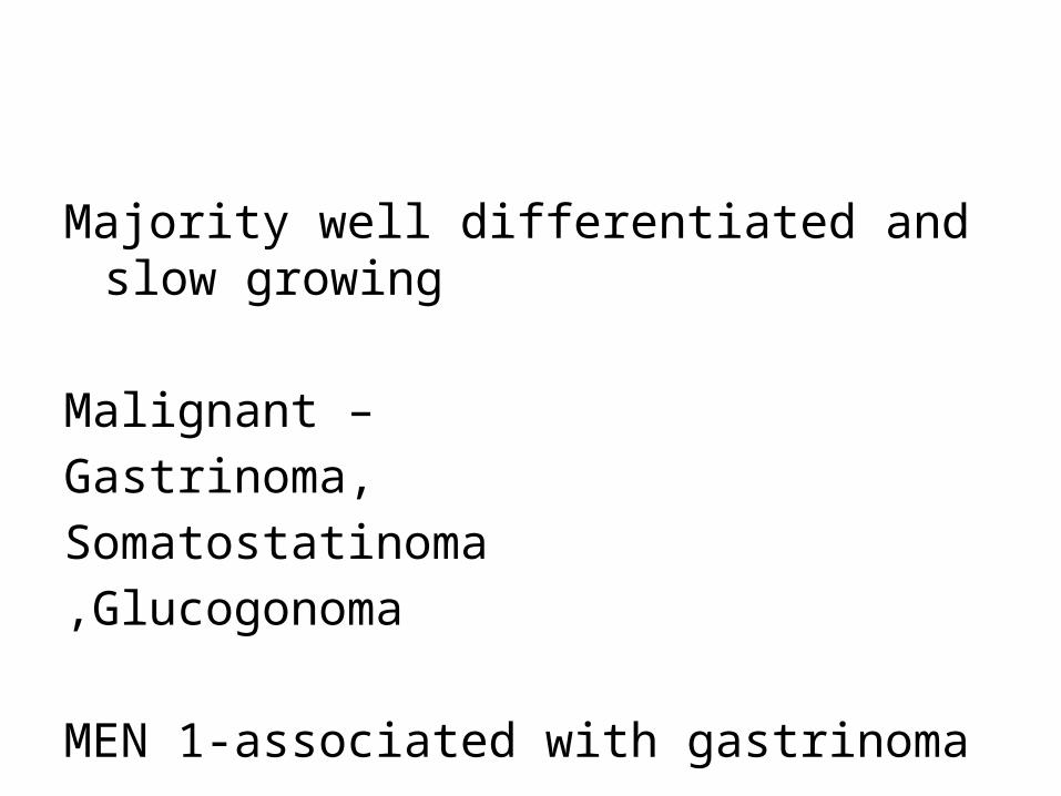
Majority well differentiated and slow growing
Malignant –
Gastrinoma,
Somatostatinoma
,Glucogonoma
MEN 1-associated with gastrinoma
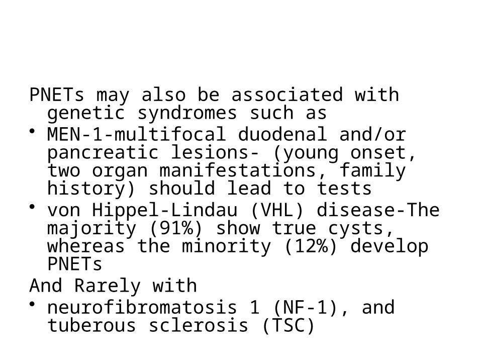
PNETs may also be associated with genetic syndromes such as
• MEN-1-multifocal duodenal and/or pancreatic lesions- (young onset, two organ manifestations, family history) should lead to tests
• von Hippel-Lindau (VHL) disease-The majority (91%) show true cysts, whereas the minority (12%) develop PNETs
And Rarely with• neurofibromatosis 1 (NF-1), and tuberous
sclerosis (TSC)

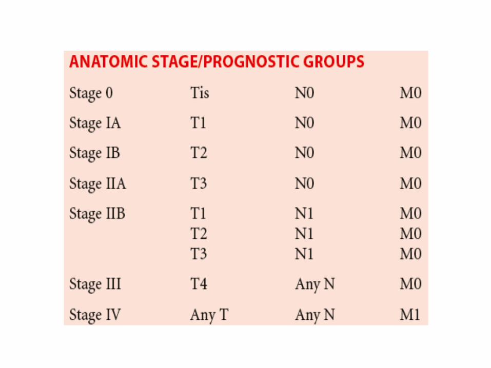
• The American Joint Committee on Cancer has published the 7th edition of the AJCC Cancer Staging Manual, which for the first time incorporates pancreatic neuroendocrine tumors in the same staging system as pancreatic exocrine tumors

• Primary Tumor (T)• TX Primary tumor cannot be assessed.• T0 No evidence of primary tumor.• Tis Carcinoma in situ.• T1 Tumor limited to the pancreas, ≤2 cm in greatest dimension.• T2 Tumor limited to the pancreas, >2 cm in greatest dimension.• T3 Tumor extends beyond the pancreas but without involvement of
the celiac axis or the superior mesenteric artery.• T4 Tumor involves the celiac axis or the superior mesenteric
artery (unresectable primary tumor).
• Regional Lymph Nodes (N) • NX Regional lymph nodes cannot be assessed.• N0 No regional lymph node metastasis.• N1 Regional lymph node metastasis.
• Distant Metastasis (M) • M0 No distant metastasis.• M1 Distant metastasis

Imaging
• PETs that are smaller than 0.5 cm are defined as microadenomas
• well circumscribed• displace, rather than invade, adjacent structures• Blush-Early enhancing lesion in in arterial phase• Small-homogenous:• Large-hetrogenous-necrotic
Functional PNETS-Small in size

NF PNETS• Large• Calcifications• venous tumor thrombus
Atypical findingsCan be cystic with peripheral enhancementCan be exophytic,Can enhance on venous phase
Metastasis• Liver Metastasis-Nodes-enhancing• Bone mets-sclerotic• Lung Mets
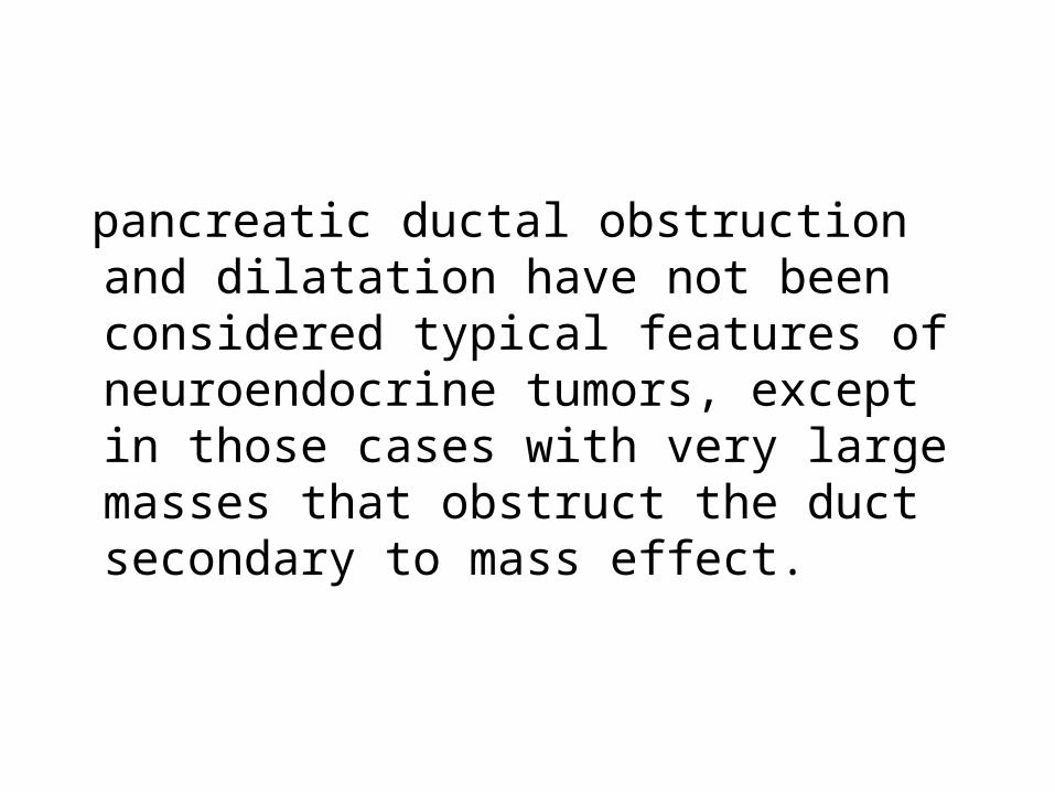
pancreatic ductal obstruction and dilatation have not been considered typical features of neuroendocrine tumors, except in those cases with very large masses that obstruct the duct secondary to mass effect.


Follow up on Imaging• RECIST 1.1 criterion for evaluating response to
treatment.(Response Evaluation Criteria in Solid Tumors)
• >1 cm• Not for necrotic,confluent lesions.not for
ascites,pleural fluid,bone mets,peritoneal or leptomeningeal disease.
• maximum of five total (and two per organ, maximum)
• short axis of >15 mm are considered measurable
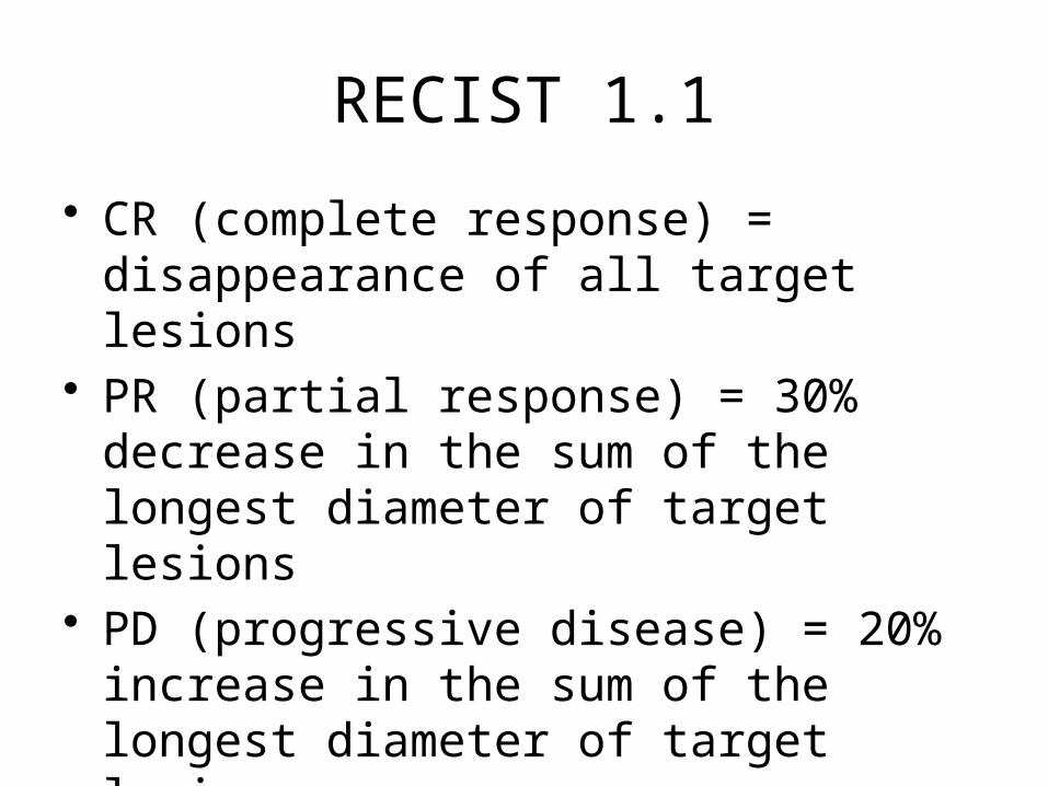
RECIST 1.1
• CR (complete response) = disappearance of all target lesions
• PR (partial response) = 30% decrease in the sum of the longest diameter of target lesions
• PD (progressive disease) = 20% increase in the sum of the longest diameter of target lesions
• SD (stable disease) = small changes that do not meet above criteria

• PR(WHO)====PR (Recist)• PD Rescist will require greater increase in
volume than WHO PD
• EORTC instrument
References1)http://www.ajronline.org/doi/full/10.2214/AJR.12.8627
2)Pancreatic Endocrine Tumors : Radiologic Clinicopathologic Correlation, RadioGraphics 2010 30:6, 1445-1464
2)The Oncologist May 2009 vol. 14no. 5 456-467
3)AJCC ,7Th edition
4) Multi–Detector Row CT of Pancreatic Islet Cell Tumors , RadioGraphics 2006


