Page 1
Paper of the month
Tasmanian Institute of Agriculture
www.utas.edu.au/tia
TIA is a joint venture between the University of
Tasmania and the Tasmanian Government
Interstrain Interactions between Bacteria Isolated from Vacuum-Packaged
Refrigerated Beef
Peipei Zhang,a József Baranyi,b and Mark Tamplina*
Tasmanian Institute of Agriculture, Food Safety Centre, University of Tasmania, Hobart,
Tasmania 7001, Australiaa; Institute of Food Research, Norwich Research Park, Colney, Norwich
NR47UA, United Kingdomb
Publication: Appl. Environ. Microbiol. (2015) 81:2753-2761.
Running title: Interaction among beef bacterial isolates
*Address correspondence to [email protected]
Abstract
The formation of bacterial spoilage communities in food is influenced by both extrinsic and intrinsic
environmental factors. While many reports describe how these factors affect bacterial growth, much
less is known about interactions among bacteria, which may influence community structure. This study
investigated interactions among representative species of bacteria isolated from vacuum-packaged (VP)
beef. Thirty-nine effectors and 20 target isolates were selected, representing 10 bacterial genera:
Carnobacterium, Pseudomonas, Hafnia, Serratia, Yersinia, Rahnella, Brochothrix, Bacillus, Leuconostoc
and Staphylococcus. The influence of live effectors on growth of target isolates was measured by spot-
lawn agar assay, and also in liquid culture medium broth using live targets and effector cell-free
supernatants. Inhibition on agar was quantified by diameter of inhibition zone, and in broth by
measuring detection time, growth rate, and maximum population density. A number of interactions
were observed, with 28.6% of isolates inhibiting and 4.2% promoting growth. The majority of
Pseudomonas isolates antagonised growth of approximately one-half of target isolates. Two Bacillus spp.
each inhibited 16 targets. Among lactic acid bacteria (LAB), Carnobacterium maltaromaticum inhibited a
wider range of isolates compared to other LAB. The majority of effector isolates enhancing target isolate
growth were Gram-negative, including Pseudomonas spp. and Enterobacteriaceae. These findings
markedly improve the understanding of potential interactions among spoilage bacteria, possibly leading
to more mechanistic descriptions of bacterial community formation in VP beef and other foods.
Page 2
2
Paper of the month
Tasmanian Institute of Agriculture
www.utas.edu.au/tia
TIA is a joint venture between the University of
Tasmania and the Tasmanian Government
Introduction
The shelf-life of meat is influenced, in part, by the composition and levels of bacteria within the spoilage
community (1). Independent laboratories have confirmed relatively high microbial diversity at the time
of meat packaging, and showing a progressive shift to lower community complexity towards the end of
shelf-life (2-4). For refrigerated vacuum-packaged (VP) beef, over time and under best-practice
conditions, lactic acid bacteria (LAB) tend to predominate and, to a lesser extent, Enterobacteriaceae (5).
Such change in bacterial community structure is based on intrinsic and extrinsic factors, including
temperature, atmosphere, pH, and organic acids, all of which may influence growth (5, 6). However, the
underlying forces of microbial interactions may also be important in shaping biodiversity of communities
(7-10); such studies have received relatively little attention in foods. Bacteria interact in any given
ecological niche through different mechanisms including quorum sensing, contact-dependent inhibition,
nutrient competition, and via production of defence compounds such as bacteriocins, antibiotics and
organic acids (10-14). There have been numerous reports exploring the effectiveness of protective
cultures and related antibacterial compounds at enhancing food safety and extending shelf-life (15-18),
however, few have investigated interactions among food bacteria, and of those which have, relatively
few species have been studied (19-22); far fewer have involved species from diverse communities (7, 23).
Nychas et al. (24) found quorum-sensing compounds extracted from meat increased the growth rate of
Serratia marcescens and Pseudomonas fluorescens. Also, Russo et al. (19) reported the growth of
Brochothrix thermosphacta, a meat spoilage bacteria, decreased in the presence of LAB. We postulate
testing a wide range of bacterial genera and species can provide a fuller understanding of potential
complex interactions.
The spot-lawn agar method (25) has been widely used to detect bacterial inhibitory activity, via
reporting an inhibition zone (8, 26, 27). However, this method does not supply specific information
about the effect of an effector on target growth, such as that achieved using broth-based assays. Also,
the latter assay more readily detects growth-promotion among isolates (24).
In this study, we applied both spot-lawn agar assay and broth assay, and investigated interactions
among a diverse group of bacteria isolated from VP beef produced at six Australian abattoirs. Network
maps illustrate the complexity of interactions, and the possible role of specific bacterial genera in
community structure. Such information might eventually be translated into models describing dynamic
changes in bacterial communities, and better inform processing and preservation strategies to enhance
meat quality and shelf-life.
Page 3
3
Paper of the month
Tasmanian Institute of Agriculture
www.utas.edu.au/tia
TIA is a joint venture between the University of
Tasmania and the Tasmanian Government
Materials and Methods
Bacterial isolates
The 180 bacterial isolates used in this study were previously obtained from VP beef primals produced at
six Australian abattoirs, stored at -0.5°C, and sampled at various time intervals for up to 30 weeks, as
described by Small et al. (28). Ten colonies, representing different morphologies, were obtained and
stored at -80°C. The isolates were identified by 16S rRNA gene sequences amplified using universal
primers 10F (5’-GAGTTTGATCCTGGCTCAG -3’) and 907R (5’-CCGTCAATTCCTTTGAGTTT-3’). The PCR
products were sent to Macrogen (Seoul, Korea) for sequencing. Sequences were compared with those in
Genbank using the BLAST function (http://blast.ncbi.nlm.nih.gov/Blast.cgi), and the closest matches of
each clone determined specific probable identities.
The 180 isolates were screened for inhibitory activity by using a spot-lawn method (25) at 25°C under
aerobic conditions. Thirty-nine of the isolates showing inhibition (effectors) were selected, representing
different species, abattoirs, storage times, and bacterial genera (Table 1). Twenty target (inhibited)
bacteria were selected using the same criterion as effector bacteria (i.e., different species, abattoirs,
storage times, and bacterial genera). Effector and target isolates comprised 10 genera, i.e.,
Carnobacterium, Pseudomonas, Brochothrix, Hafnia, Yersinia, Bacillus, Rahnella, Leuconostoc, Serratia
and Staphylococcus (Table 1 and 2). Six (Leuconostoc mesenteroides B30b, Staphylococcus epidermidis
F30c, Bacillus sp. strain A30g, Pseudomonas sp. D0g, Yersinia enterocolitica B8b and Rahnella aquatilis
B8f) were tested as both targets and effectors. The rationale for isolate selection was not based on the
species observed in a specific package of VP beef (24) but instead to have a panel of isolates
representing those species found in VP beef from different abattoirs. Isolates were stored at -80oC in
brain heart infusion broth (BHI; Amyl Media Ltd., Australia), supplemented with 20% (vol/vol) glycerol.
Inhibition activity measured on agar
The spot-lawn method described by Benkerroum et al. (25) was used to test for inhibitory activity of live
effectors on target isolates. Briefly, all isolates were transferred from -80°C, streaked on tryptone soya
agar (TSA; Oxoid Ltd., Australia), cultured for 24 h at 25°C, and then grown in BHI broth for 24 h at 25°C.
Cell density was adjusted to an optical density at 540 nm (OD540) 0.6-0.8 for effectors and 0.15-0.25 for
targets, a difference designed to enhance detection of growth inhibition or promotion. One hundred
microliters of each target was spread-plated on TSA, and then three replicate 10-μl aliquots of effectors
were spotted onto the target lawn. Inhibition was measured after 24 h of incubation at 25oC, when TSA
plates were photographed, and the diameter (D) of the inhibition zone was measured using the software
Page 4
4
Paper of the month
Tasmanian Institute of Agriculture
www.utas.edu.au/tia
TIA is a joint venture between the University of
Tasmania and the Tasmanian Government
program Image J (version 1.49 [http://rsb.info.nih.gov/ij/index.html]). The degree of inhibition was
classified at four levels: ++++, +++, ++, and +, corresponding to D ≥ 4 mm, 2 ≤ D < 4 mm, 0.5 < D < 2 mm
and 0 < D ≤ 0.5 mm, respectively (Fig. 1). This grouping considered variation in inhibition strength and
facilitated comparison. Inhibition patterns were also classified as having a well delineated or diffuse
edge.
Interaction activity measured by CFS assay
Overnight cultures (24 h, 25oC) of target isolates were adjusted to 104 cfu/ml. Effector isolates were
incubated for 48 h at 25oC until the stationary phase was reached. Cell-free supernatant (CFS) of each
effector isolates were made by centrifuging BHI cultures at 1,000 x g for 5 min, followed by filtration
through a 0.22 μm pore-sized filter (Whatman Ltd., Australia). Treatments consisted of mixing 100 µl of
the diluted target suspension with 100 µl of CFS in wells of a BioscreenC microwell plate (Growth Curve
Ab Ltd., Finland). Controls had the same volume of fresh BHI or phosphate-buffered saline (PBS; 1M, [pH
7.4]), instead of CFS. Duplicate wells were used for all treatments and controls. The BioscreenC
temperature was 25oC, and growth kinetics measured at 20-min intervals for 48 h. At the end of each
run, data were exported to an Excel spreadsheet. Detection time (DT; in hours) was calculated as the
time to reach an OD540 of 0.12 (background corrected data). The Baranyi model (29) was fitted to the
primary growth curves using DMFit (v3.0 [Combase; http://www.combase.cc/tools/]) to calculate
growth rate (GR; log10OD/h). Maximum population density (MPD; OD540) was calculated by averaging the
three highest OD readings. DT, GR and MPD were compared among treatments and controls, using the
Student t-Test in Excel. A P value below 0.05 was considered a significant interaction, i.e., as inhibition
comparing treatment and PBS or as promotion comparing treatment and BHI.
If P > 0.05, inhibition strength (IS) of CFS on individual target growth parameter was recorded as zero. If
P < 0.05, the IS was calculated by comparison of treatment and PBS control using the following formulas:
ISDT = |DTTreatment – DTControl| / DTControl (1)
ISGR = |GRTreatment – GRControl| / GRControl (2)
ISMPD = |MPDTreatment – MPDControl|/ MPDControl (3)
Page 5
5
Paper of the month
Tasmanian Institute of Agriculture
www.utas.edu.au/tia
TIA is a joint venture between the University of
Tasmania and the Tasmanian Government
The cumulative IS effect (ISTotal) on all three growth parameters was quantified using the formula:
ISTotal = (ISDT + ISGR + ISMPD) / 3 (4)
The promotion strength (PS) was calculated similar to IS, via comparison of test and BHI control.
IS was classified at four levels, ++++, +++, ++, and +, corresponding to IS = 1 (no detectable growth of the
target), 0.25 ≤ IS < 1, 0.15 ≤ IS < 0.25, and 0 < IS < 0.15, respectively (Fig. 2). In the relatively fewer
instances where CFS promoted growth, growth PS was classified at two levels, ++ and +, corresponding
to PS ≥ 0.1 and 0 < PS < 0.1, respectively.
Network maps of bacterial interactions.
Growth inhibition/promotion activity was described using a network diagram drawn with Cytoscape
(v3.1.1 [http://www.cytoscape.org/]) (Fig. 3). In maps, target and effector nodes were designated as
diamonds and circles, respectively. Isolates used as both inhibitors and targets were represented by
squares. Arrows (edges) connected interacting isolates. The strength of growth inhibition or promotion
was distinguished by line thickness.
In terms of node size, an arbitrary base number (BN) of 80 was first assigned. Then, a connection
number (CN) was calculated for each node according to the number of each interaction level as follows:
𝐶𝑁 = ∑ (𝑎𝑖
𝑏× 100 × 𝑖)4
𝑖=1 (5)
with i being the interaction level (1, +; 2, ++; 3, +++; and 4, ++++), ai the number of interactions at level i,
and b the number of effectors or targets for corresponding target or effector.
In the growth inhibition network map, the size of individual inhibiting nodes equalled the sum of BN and
CN. For target isolates, the diameter of the node was the difference between BN and CN; the smaller the
diamond, the greater the target was inhibited. In growth promotion network maps, the size of both
targets and effectors was set as the sum of BN and CN. For isolates being both a target and effector,
node size was calculated as target and effector, respectively, and then the final size displayed as the
average of these two values.
Page 6
6
Paper of the month
Tasmanian Institute of Agriculture
www.utas.edu.au/tia
TIA is a joint venture between the University of
Tasmania and the Tasmanian Government
Statistical analysis
The differences of distribution of growth-inhibiting and –promoting activity (IS and PS) among effectors
at isolate, species and genus levels were statistically analysed. An F-test was applied to examine overall
differences among different groups. If the F-test was significant (P < 0.05), a Student t test was used to
identify the significant pairwise differences. Differences between Gram-negative and -positive bacteria
were also examined in the same way. The dependent variable in analysis included IS from spot-lawn
assay (inhibition diameter, mm), and IS, PS, ISDT, ISGR, ISMPD, PSDT, PSGR, and PSMPD from CFS assay (%). The
arcsine transformation of square root of relative interaction strength was used to normalise the data
from CFS assay. A P value below 0.05 from Student t test was considered statistically significant. These
tests were performed using the GLM procedure in SAS (v 9.3; SAS, Inc., Rockville, MD).
Results
Total of 774 and 735 combinations of effector and target isolates were tested by spot-lawn and CFS
assay, respectively. The difference of 39 in total combinations between the two assays resulted from
Leuconostoc sp. F30e not sufficiently growing in BHI broth for CFS analysis.
Summary of interactions
Combined results of spot-lawn and CFS assays showed 31% of pairings produced an interaction, i.e., 28.6%
(221 pairings) inhibitions compared to 4.2% (31 pairings) promotions. A slightly larger number of
inhibitory reactions were detected by spot-lawn compared to CFS assay, i.e., 17.6% (136 pairings) versus
16.6% (122 pairings), respectively (Table 3).
Growth inhibition
Among the 774 effector-target pairings tested by spot-lawn assay, there were more weak (14.6%, + and
++) than strong inhibitions (3%, +++ and ++++) (Fig. 3 and Table 3). By CFS assay, 3.6% versus 13% of
interactions produced strong versus weak inhibition, respectively. Analysis of kinetic growth profiles of
target bacteria showed CFS primarily affected DT (Table 4), an effect particularly evident for
Carnobacterium (data not shown). On the whole, more inhibition events were associated with increased
DT (78.9% of inhibitions) than decreased GR (44.7%) and MPD (28.5%).
Growth promotion
Based on the nature of the two assays, growth promotion could only be detected by the CFS broth assay.
Among 31 pairings promoting growth, 9 were strong (++) and 22 were weak (+) (Table 3). Pseudomonas
spp. and Enterobacteriaceae were the most common growth-promoting effector isolates; less-common
Page 7
7
Paper of the month
Tasmanian Institute of Agriculture
www.utas.edu.au/tia
TIA is a joint venture between the University of
Tasmania and the Tasmanian Government
effectors included Bacillus sp. strains A30g and E0g, Yersinia frederiksenii A8h, and L. mesenteroides
B30b (Table 1 and Fig. 3C). The isolates stimulating the strongest growth promotion effects were Bacillus
sp. strains A30g and E0g, and Serratia sp. isolates C0b, C30b, E8c, E8i, and E30j. The targets most
strongly promoted were Pseudomonas sp. isolates D0g and D8g, B. thermosphacta A0b, C.
maltaromaticum D8c, Leuconostoc carnosum F30j, and L. mesenteroides (Fig. 3C).
Although most growth-promoting activity reduced DT and/or increased GR (data not shown), MPD was
enhanced in some interactions. For example, Bacillus subtilis E0g increased the MPD of Pseudomonas sp.
D8g by 0.45 OD540 units. Similarly, Serratia sp. E8c increased the MPD of Pseudomonas sp. D0g by 0.35
OD540 units (data not shown).
Effector species
Results of spot-lawn and CFS assays showed isolates inhibiting more than 10 targets predominantly
belonged to the genera Pseudomonas, Bacillus and Carnobacterium (Table 1; Fig. 3A and B). All six
Pseudomonas effector isolates, except B0i, inhibited at least nine targets, with Pseudomonas sp. D0b
inhibiting 18 targets (Table 1). Pseudomonas sp. B0i had a more limited spectrum, inhibiting only six
targets. Bacillus sp. A30g and E0g each inhibited 16 targets. Carnobacterium maltaromaticum inhibited 5
(C0a) to 10 (C8h) targets. Carnobacterium F8g, not identified by 16s rRNA sequencing at the species level,
inhibited seven targets, and Carnobacterium divergens three to eight targets. Staphylococcus
epidermidis, represented by one isolate (F30c), inhibited four targets. Live effector cells of the family
Enterobacteriaceae, including Hafnia alvei, Serratia spp., and R. aquatilis, produced lower levels of
inhibition against a small number of targets on spot lawns and against an even smaller group of targets
in the CFS assay (Fig. 3A and B). No inhibition by H. alvei E30e was observed in either assay.
Intraspecies inhibition was observed as well. For example, C. divergens D30f and C. maltaromaticum D8c
were inhibited by effector isolates of the same species in both spot-lawn and CFS assay (Fig. 3A and B).
Similarly, L. carnosum F30d and F30h inhibited L. carnosum F30j. Other interesting observations included
effectors inhibiting targets on agar, but promoting growth of the same target in broth (e.g.,
Pseudomonas sp. E0f as effector and C. divergens D30f as target) (Fig. 3).
Target species
Based on both assays, the most frequently inhibited species were C. divergens D30f, C. maltaromaticum
D8c, Pseudomonas sp. D0g, S. epidermidis F30c and B. thermosphacta A0b, with 51.3, 48.7, 47.4, 44.7,
and 43.6% of effectors inhibiting these isolates, respectively (Table 2). Interestingly, while being the
Page 8
8
Paper of the month
Tasmanian Institute of Agriculture
www.utas.edu.au/tia
TIA is a joint venture between the University of
Tasmania and the Tasmanian Government
most commonly inhibited species, growth of C. divergens D30f and C. maltaromaticum D8c was also
promoted by the largest number (25.6%) of effector isolates (Table 2).
Growth-promotion was target-dependent and restricted to a relatively small number of isolates, i.e.,
Carnobacterium sp. strains D30f and D8c, Pseudomonas sp. strains D8g and D0g, Bacillus sp. A30g, and B.
thermosphacta A0b (Table 2 and Fig. 3C). Among nine interactions showing strong growth promotion,
five targets were Pseudomonas spp. (Fig. 3C). Both Bacillus sp. strains A30g and E0g promoted the
growth of Pseudomonas sp. D8g, displaying PS of 0.15 and 0.32, respectively (data not shown). Serratia
sp. E8c promoted the growth of both Pseudomonas sp. D8g and D0g at PS of 0.37 and 0.12, respectively
(data not shown).
Interactions measured by spot-lawn versus CFS-broth assay.
Pseudomonas isolates inhibited more targets on agar (3 to 18 isolates) than in broth (1 to 4 isolates)
(Table 1and Fig. 3A and B). The influence of test method was especially evident for Pseudomonas sp.
D0b, which inhibited only one target in broth but inhibited 18 on agar. Pseudomonas isolates were often
associated with a diffuse inhibition zone (Fig. 3A). Specifically, diffuse zones were observed for thirteen,
nine and eight targets by Pseudomonas sp. strains D0b, F0b, and D0g, respectively.
Likewise, Bacillus sp. A30g inhibited 14 targets on agar versus seven in broth. Bacillus subtilis E0g,
however, inhibited the same number of targets by both assays. C. maltaromaticum effectors inhibited a
wider range of target strains/species in broth compared to agar (Fig. 3A and B). For example, C.
maltaromaticum C30h inhibited nine of 20 targets in broth, but only three on agar (Fig. 3A and B and
Table 1). Overall, by broth assay, Gram-positive bacteria inhibited more target bacteria and displayed
relatively stronger inhibition strength compared to Gram-negative bacteria (Fig. 3B). However, no
significant difference between these two groups was observed by agar assay (date not shown).
Discussion
In food, bacterial strains rarely exist in isolation (9) but as members of a microbial community
influencing food product quality and shelf-life. The structure of this community is not only affected by
intrinsic and extrinsic environmental factors but also possibly by interactions among specific bacteria (7-
9), influencing food quality and safety.
In the present study, we report numerous interactions, tested by both agar- and broth-based assays,
among a large and diverse group of bacteria isolated from commercial Australian VP beef (Fig. 3).
Page 9
9
Paper of the month
Tasmanian Institute of Agriculture
www.utas.edu.au/tia
TIA is a joint venture between the University of
Tasmania and the Tasmanian Government
Among the 39 effector and 20 target isolates tested, representing a total of 774 pair-wise tests, 28.6%
(221 pairings) showed inhibition and 4.2% (31 pairings) promotion of target growth.
These studies were conducted in bacteriological media, and under an aerobic atmosphere at 25°C.
Although it may be argued bacterial densities tested in these studies were high, such concentrations and
cell-cell proximities, may exist in food microenvironments, since bacteria are known to preferentially
bind and colonize to specific structures (30). While the interpretation of these studies is limited to these
specific conditions, they offer insight into potential inter-isolate interactions occurring before and
shortly after beef primals are vacuum-packaged. Additional studies are underway to quantify
interactions under conditions more relevant to long-term refrigerated storage of refrigerated VP beef.
LAB have been extensively studied as protective cultures for extending food shelf-life and enhancing
food safety. They inhibit growth of some spoilage and pathogenic bacteria, such as Carnobacterium spp.,
B. thermosphacta, Listeria spp., Salmonella spp., and Staphylococcus aureus, through the action of
bacteriocins, organic acids and/or other antibacterial substances (14, 18, 31). In the present study, C.
maltaromaticum isolates inhibited from five (C0a) to ten (D0h) target isolates (Table 1). In contrast,
other LAB species did not display as large an inhibition spectrum as C. maltaromaticum; for example,
most C. divergens inhibited no more than five targets, whereas L. carnosum inhibited two (Table 1).
Interestingly, C. maltaromaticum and C. divergens also showed strong intraspecies inhibition (Fig. 3A
and B), an observation consistent with the studies of Martin-Visscher et al. (31) and Worobo et al. (32).
As such, C. maltaromaticum, and to a lesser extent C. divergens, may have a strong influence on
bacterial community structure in VP beef.
The inhibition spectrum of most LAB measured by the agar spot-lawn assay was not as diverse as that by
CFS assay, for example, C. maltaromaticum D0h (Fig. 3), whereas in broth, extended DT and decreased
GR were more frequently observed than decreased MPD (data not shown). These differences may due
to inhibitory factors in CFS, such as disassociated lactic acid and bacteriocins, commonly produced by
Carnobacterium spp.(33).
When considering the combined results of spot-lawn and CFS assays, Pseudomonas spp., with the
exception of effector Pseudomonas sp. B0i, displayed high antagonistic behaviour, inhibiting, on average,
almost half of the targets (Fig. 3A and B and Table 1). Pseudomonas sp. D0b inhibited 18 of the 20
targets (Table 1). Similarly, Aguirre-von-Wobeser et al. (27), using the spot-lawn method, also found
Pseudomonas spp., isolated from an aquatic environment, were the most highly antagonistic strains.
Published reports show plant and clinical strains of Pseudomonas (e.g., Pseudomonas putida, P.
Page 10
10
Paper of the month
Tasmanian Institute of Agriculture
www.utas.edu.au/tia
TIA is a joint venture between the University of
Tasmania and the Tasmanian Government
fluorescens, and other Pseudomonas spp.) produce secondary antimicrobial metabolites, including
enzymes, volatiles (hydrogen cyanide), cyclic lipopeptides, and antibiotics (34-36). These have been
applied in plant pathology to control fungal pathogens and in clinical studies to inhibit pathogenic strains
(37-39).
However, antibacterial compounds might not explain all the inhibitory activities of Pseudomonas spp.,
since inhibition patterns of Pseudomonas spp. differed markedly between spot-lawn and CFS assays. For
example, Pseudomonas sp. D0b CFS only inhibited one target by CFS, but seventeen by spot-lawn. This
may indicate live effector cells, not just CFS, are required for target inhibition, as reported by Russell et
al. (40), who found Pseudomonas spp. killed bacteria by exporting functional molecules through the type
VI secretion system, a form of contact-mediated killing. It also may suggest physiological responses of
Pseudomonas spp. differ in solid versus liquid media.
It was also noted that growth of C. divergens D30f and C. maltaromaticum D8c was promoted by CFS
from most Pseudomonas isolates, although promotion strength was low. Thus, in the early stages of
vacuum-packaging of beef, when oxygen is present, the growth-promoting and/or -inhibiting effects of
Pseudomonas spp. on sensitive bacteria, such as Carnobacterium spp., may influence the levels and
composition of bacterial species during later stages of VP storage. Further studies are required to
elucidate the underlying interacting mechanism(s).
Both Bacillus sp. strains E0g and A30g influenced the growth of a wide spectrum of isolates, inhibiting 16
of 20 targets. Members of this genus are known to produce antimicrobial compounds (41). Baindara et
al. (42) characterized two antimicrobial peptides produced by a B. subtilis strain, which showed
antagonistic properties against Gram-positive bacteria, including S. aureus and Listeria monocytogenes.
Other Bacillus species have been reported to produce bacteriocins and biosurfactants (43, 44); the
bacteriocins inhibited the growth of a large range of Gram-positive and Gram-negative bacteria. Bacillus
subtilis E0g strongly inhibited most Gram-positive targets, including C. maltaromaticum D8c, B.
thermosphacta A0b, Bacillus sp. A30g, S. epidermidis F30c, L. carnosum F30j, and also some Gram-
negative species, such as Serratia spp. and Pseudomonas spp. (Fig. 3). Unlike B. subtilis E0g, Bacillus sp.
A30g only displayed a wide inhibition spectrum when tested by spot-lawn assay. This indicates inhibition
by Bacillus sp. A30g may be contact-dependent (12).
Enterobacteriaceae, such as H. alvei, Serratia spp., and R. aquatilis, produced a relatively lower level of
inhibition under the test conditions (Fig. 3A and B). Staphylococcus spp. were studied by Cogen et al. (45)
and were shown to possess antimicrobial activity against skin pathogens such as S. aureus via phenol-
Page 11
11
Paper of the month
Tasmanian Institute of Agriculture
www.utas.edu.au/tia
TIA is a joint venture between the University of
Tasmania and the Tasmanian Government
soluble modulins. Nevertheless, to our knowledge, S. aureus has not been well studied for antimicrobial
properties in food. The mechanism(s) of S. epidermidis F30c inhibition requires further study.
By broth assay, the growth of target isolates was promoted in 4.2% of the effector and target
combinations. Most effector isolates (84%) enhancing target growth were Gram-negative bacteria,
including Pseudomonas spp. and members of the Enterobacteriaceae, in addition to three other isolates
(L. mesenteroides B30b, Bacillus sp. A30g and B. subtilis E0g (Fig. 3C). Growth promotion also appeared
to be target-dependent, centering on a small range of targets, namely, Pseudomonas sp. D8g, B.
thermosphacta A0b, C. maltaromaticum D30f, C. divergens D8c and L. carnosum F30j. A review of the
literature shows promotion of bacterial growth by effector isolates has been less frequently reported
compared to inhibition. Possible reasons include the spot-lawn method, a test format not readily
detecting growth-promotion, being a primary method used in many previous studies (8, 26, 27), and
that primary interests of applied food microbiology are in extending shelf-life and food quality.
The growth of two Carnobacterium spp. isolates was enhanced by a large number of effector isolates,
including Serratia spp. and Pseudomonas spp. (Fig. 3C). As mentioned earlier, Carnobacterium spp. also
inhibited a large spectrum of targets. These combined observations, as well as Carnobacterium spp.
being a facultative anaerobe, may result in this genus being more dominant in meats stored under VP
conditions (46, 47).
In the present study, Leuconostoc sp. F30e failed to grow in BHI at 25oC, and thus influences on the
growth of this strain were not measured by CFS-broth assay. According to other studies, some
Leuconostoc species, such as Leuconostoc gelidum, are isolated form chill-stored foods and may not
readily grow at elevated temperature, including 25oC used here (48-50).
While our general focus was to measure growth inhibition and promotion, we observed different
inhibition zone morphologies on agar, possibly indicating different mechanisms of action. Undefined
(diffuse) inhibition zones have been observed in antibiotic resistance studies (51, 52), and interpreted as
low levels of bacterial resistance. We noted that Pseudomonas spp. often produced such a diffuse type
of inhibition zone.
We measured microbial interactions among bacteria isolated from Australian VP beef, which may, in
part, help explain the succession of bacterial communities. However, direct translation of these results
to actual bacterial community formation in beef environments must consider these studies used
bacteriological broth, relatively high densities of cells, and pair-wise comparison of isolates (7).
Page 12
12
Paper of the month
Tasmanian Institute of Agriculture
www.utas.edu.au/tia
TIA is a joint venture between the University of
Tasmania and the Tasmanian Government
Acknowledgements
We gratefully acknowledge funding by Meat and Livestock Australia. D. Ratkowsky is acknowledged for
assistance with statistical analyses. We thank T. Ross, C. Kocharunchitt, S. George, and M. Williams for
advice with the protocols. P. Z. acknowledges scholarship support provided by the Chinese Scholarship
Council and Zhejiang University.
Page 13
13
Paper of the month
Tasmanian Institute of Agriculture
www.utas.edu.au/tia
TIA is a joint venture between the University of
Tasmania and the Tasmanian Government
References
1. Nychas GJ, Skandamis PN, Tassou CC, Koutsoumanis KP. 2008. Meat spoilage during
distribution. Meat Sci. 78:77-89.
2. De Filippis F, La Storia A, Villani F, Ercolini D. 2013. Exploring the sources of bacterial
spoilers in beefsteaks by culture-independent high-throughput sequencing. PLoS ONE
8:e70222. doi:10.1371/journal.pone.0070222.
3. Powell SM, Tamplin ML. 2012. Microbial communities on Australian modified
atmosphere packaged Atlantic salmon. Food Microbiol. 30:226-232.
4. Sakala RM, Hayashidani H, Kato Y, Hirata T, Makino Y, Fukushima A, Yamada T,
Kaneuchi C, Ogawa M. 2002. Change in the composition of the microflora on vacuum-
packaged beef during chiller storage. Int. J. Food Microbiol. 74:87-99.
5. Doulgeraki AI, Ercolini D, Villani F, Nychas GJ. 2012. Spoilage microbiota associated to
the storage of raw meat in different conditions. Int. J. Food Microbiol. 157:130-141.
6. Nemergut DR, Schmidt SK, Fukami T, O'Neill SP, Bilinski TM, Stanish LF, Knelman JE,
Darcy JL, Lynch RC, Wickey P, Ferrenberg S. 2013. Patterns and processes of microbial
community assembly. Microbiol. Mol. Biol. Rev. 77:342-356.
7. Wolfe BE, Button JE, Santarelli M, Dutton RJ. 2014. Cheese rind communities provide
tractable systems for in situ and in vitro studies of microbial diversity. Cell 158:422-433.
8. Perez-Gutierrez RA, Lopez-Ramirez V, Islas A, Alcaraz LD, Hernandez-Gonzalez I,
Olivera BC, Santillan M, Eguiarte LE, Souza V, Travisano M, Olmedo-Alvarez G. 2013.
Antagonism influences assembly of a Bacillus guild in a local community and is depicted
as a food-chain network. ISME J 7:487-497.
9. Faust K, Raes J. 2012. Microbial interactions: from networks to models. Nat. Rev.
Microbiol. 10:538-550.
10. Blana VA, Nychas GJ. 2014. Presence of quorum sensing signal molecules in minced
beef stored under various temperature and packaging conditions. Int. J. Food Microbiol.
173:1-8.
11. Aoki SK, Pamma R, Hernday AD, Bickham JE, Braaten BA, Low DA. 2005. Contact-
dependent inhibition of growth in Escherichia coli. Science 309:1245-1248.
12. Dubey GP, Ben-Yehuda S. 2011. Intercellular nanotubes mediate bacterial
communication. Cell 144:590-600.
Page 14
14
Paper of the month
Tasmanian Institute of Agriculture
www.utas.edu.au/tia
TIA is a joint venture between the University of
Tasmania and the Tasmanian Government
13. Deriu E, Liu JZ, Pezeshki M, Edwards RA, Ochoa RJ, Contreras H, Libby SJ, Fang FC,
Raffatellu M. 2013. Probiotic bacteria reduce salmonella typhimurium intestinal
colonization by competing for iron. Cell Host Microbe 14:26-37.
14. Cotter PD, Ross RP, Hill C. 2013. Bacteriocins - a viable alternative to antibiotics? Nat.
Rev. Microbiol. 11:95-105.
15. Hastings JW, Stiles ME, von Holy A. 1994. Bacteriocins of leuconostocs isolated from
meat. Int. J. Food Microbiol. 24:75-81.
16. Budde BB, Hornbaek T, Jacobsen T, Barkholt V, Koch AG. 2003. Leuconostoc carnosum
4010 has the potential for use as a protective culture for vacuum-packed meats: culture
isolation, bacteriocin identification, and meat application experiments. Int. J. Food
Microbiol. 83:171-184.
17. Hequet A, Laffitte V, Simon L, De Sousa-Caetano D, Thomas C, Fremaux C, Berjeaud JM.
2007. Characterization of new bacteriocinogenic lactic acid bacteria isolated using a
medium designed to simulate inhibition of Listeria by Lactobacillus sakei 2512 on meat.
Int. J. Food Microbiol. 113:67-74.
18. Li M, Muthaiyan A, O'Bryan CA, Gustafson JE, Li Y, Crandall PG, Ricke SC. 2011. Use of
Natural Antimicrobials from a Food Safety Perspective for Control of Staphylococcus
aureus. Curr Pharm Biotechnol 12:1240-1254.
19. Russo F, Ercolini D, Mauriello G, Villani F. 2006. Behaviour of Brochothrix
thermosphacta in presence of other meat spoilage microbial groups. Food Microbiol.
23:797-802.
20. Vasilopoulos C, De Mey E, Dewulf L, Paelinck H, De Smedt A, Vandendriessche F, De
Vuyst L, Leroy F. 2010. Interactions between bacterial isolates from modified-
atmosphere-packaged artisan-type cooked ham in view of the development of a
bioprotective culture. Food Microbiol. 27:1086-1094.
21. Mellefont LA, McMeekin TA, Ross T. 2008. Effect of relative inoculum concentration on
Listeria monocytogenes growth in co-culture. Int. J. Food Microbiol. 121:157-168.
22. Dourou D, Ammor MS, Skandamis PN, Nychas GJ. 2011. Growth of Salmonella
enteritidis and Salmonella typhimurium in the presence of quorum sensing signalling
compounds produced by spoilage and pathogenic bacteria. Food Microbiol. 28:1011-
1018.
Page 15
15
Paper of the month
Tasmanian Institute of Agriculture
www.utas.edu.au/tia
TIA is a joint venture between the University of
Tasmania and the Tasmanian Government
23. Mounier J, Monnet C, Vallaeys T, Arditi R, Sarthou AS, Helias A, Irlinger F. 2008.
Microbial interactions within a cheese microbial community. Appl. Environ. Microbiol.
74:172-181.
24. Nychas GJ, Dourou D, Skandamis P, Koutsoumanis K, Baranyi J, Sofos J. 2009. Effect of
microbial cell-free meat extract on the growth of spoilage bacteria. J. Appl. Microbiol.
107:1819-1829.
25. Benkerroum N, Ghouati Y, Sandine WE, Tantaoui-Elaraki A. 1993. Methods to
demonstrate the bactericidal activity of bacteriocins. Lett. Appl. Microbiol. 17:78-81.
26. Lo Giudice A, Brilli M, Bruni V, De Domenico M, Fani R, Michaud L. 2007. Bacterium-
bacterium inhibitory interactions among psychrotrophic bacteria isolated from Antarctic
seawater (Terra Nova Bay, Ross Sea). FEMS Microbiol. Ecol. 60:383-396.
27. Aguirre-von-Wobeser E, Soberon-Chavez G, Eguiarte LE, Ponce-Soto GY, Vazquez-
Rosas-Landa M, Souza V. 2014. Two-role model of an interaction network of free-living
gamma-proteobacteria from an oligotrophic environment. Environ. Microbiol. 16:1366-
1377.
28. Small AH, Jenson I, Kiermeier A, Sumner J. 2012. Vacuum-packed beef primals with
extremely long shelf life have unusual microbiological counts. J. Food Prot. 75:1524-
1527.
29. Baranyi J, Roberts TA. 1994. A dynamic dpproach to predicting bacterial growth in food.
Int. J. Food Microbiol. 23:277-294.
30. Zulfakar SS, White JD, Ross T, Tamplin ML. 2012. Bacterial attachment to immobilized
extracellular matrix proteins in vitro. Int J Food Microbiol 157:210-217.
31. Martin-Visscher LA, van Belkum MJ, Garneau-Tsodikova S, Whittal RM, Zheng J,
McMullen LM, Vederas JC. 2008. Isolation and characterization of carnocyclin a, a novel
circular bacteriocin produced by Carnobacterium maltaromaticum UAL307. Appl.
Environ. Microbiol. 74:4756-4763.
32. Worobo RW, Van Belkum MJ, Sailer M, Roy KL, Vederas JC, Stiles ME. 1995. A signal
peptide secretion-dependent bacteriocin from Carnobacterium divergens. J. Bacteriol.
177:3143-3149.
33. Bali V, Panesar PS, Bera MB, Kennedy JF. 2014. Bacteriocins: Recent Trends and
Potential Applications. Crit Rev Food Sci Nutr. doi:10.1080/10408398.2012.729231.
34. Li W, Rokni-Zadeh H, De Vleeschouwer M, Ghequire MG, Sinnaeve D, Xie GL, Rozenski
J, Madder A, Martins JC, De Mot R. 2013. The antimicrobial compound xantholysin
Page 16
16
Paper of the month
Tasmanian Institute of Agriculture
www.utas.edu.au/tia
TIA is a joint venture between the University of
Tasmania and the Tasmanian Government
defines a new group of Pseudomonas cyclic lipopeptides. PLoS ONE 8:e62946.
doi:10.1371/journal.pone.0062946.
35. Kruijt M, Tran H, Raaijmakers JM. 2009. Functional, genetic and chemical
characterization of biosurfactants produced by plant growth-promoting Pseudomonas
putida 267. J. Appl. Microbiol. 107:546-556.
36. Kuiper I, Lagendijk EL, Pickford R, Derrick JP, Lamers GE, Thomas-Oates JE, Lugtenberg
BJ, Bloemberg GV. 2004. Characterization of two Pseudomonas putida lipopeptide
biosurfactants, putisolvin I and II, which inhibit biofilm formation and break down
existing biofilms. Mol. Microbiol. 51:97-113.
37. Afsharmanesh H, Ahmadzadeh M, Javan-Nikkhah M, Behboudi K. 2010.
Characterization of the antagonistic activity of a new indigenous strain of Pseudomonas
fluorescens isolated from onion rhizosphere. J. Plant Pathol. 92:187-194.
38. Cardozo VF, Oliveira AG, Nishio EK, Perugini MR, Andrade CG, Silveira WD, Duran N,
Andrade G, Kobayashi RK, Nakazato G. 2013. Antibacterial activity of extracellular
compounds produced by a Pseudomonas strain against methicillin-resistant
Staphylococcus aureus (MRSA) strains. Ann. Clin. Microbiol. Antimicrob. 12:12.
39. Trippe K, McPhail K, Armstrong D, Azevedo M, Banowetz G. 2013. Pseudomonas
fluorescens SBW25 produces furanomycin, a non-proteinogenic amino acid with
selective antimicrobial properties. BMC Microbiol. 13:111.
40. Russell AB, Hood RD, Bui NK, LeRoux M, Vollmer W, Mougous JD. 2011. Type VI
secretion delivers bacteriolytic effectors to target cells. Nature 475:343-347.
41. Teixeira ML, Dalla Rosa A, Brandelli A. 2013. Characterization of an antimicrobial
peptide produced by Bacillus subtilis subsp. spizezinii showing inhibitory activity towards
Haemophilus parasuis. Microbiology (Reading, Engl.) 159:980-988.
42. Baindara P, Mandal SM, Chawla N, Singh PK, Pinnaka AK, Korpole S. 2013.
Characterization of two antimicrobial peptides produced by a halotolerant Bacillus
subtilis strain SK.DU.4 isolated from a rhizosphere soil sample. AMB Express 3:2.
43. Singh PK, Chittpurna, Ashish, Sharma V, Patil PB, Korpole S. 2012. Identification,
purification and characterization of laterosporulin, a novel bacteriocin produced by
Brevibacillus sp. strain GI-9. PLoS ONE 7:e31498. doi:10.1371/journal.pone.0031498.
44. Velho RV, Basso AP, Segalin J, Costa-Medina LF, Brandelli A. 2013. The presence of
sboA and spaS genes and antimicrobial peptides subtilosin A and subtilin among Bacillus
strains of the Amazon basin. Genet. Mol. Biol. 36:101-104.
Page 17
17
Paper of the month
Tasmanian Institute of Agriculture
www.utas.edu.au/tia
TIA is a joint venture between the University of
Tasmania and the Tasmanian Government
45. Cogen AL, Yamasaki K, Sanchez KM, Dorschner RA, Lai Y, MacLeod DT, Torpey JW, Otto
M, Nizet V, Kim JE, Gallo RL. 2010. Selective antimicrobial action is provided by phenol-
soluble modulins derived from Staphylococcus epidermidis, a normal resident of the skin.
J. Invest. Dermatol. 130:192-200.
46. Kiermeier A, Tamplin M, May D, Holds G, Williams M, Dann A. 2013. Microbial growth,
communities and sensory characteristics of vacuum and modified atmosphere packaged
lamb shoulders. Food Microbiol. 36:305-315.
47. Casaburi A, Nasi A, Ferrocino I, Di Monaco R, Mauriello G, Villani F, Ercolini D. 2011.
Spoilage-related activity of Carnobacterium maltaromaticum strains in air-stored and
vacuum-packed meat. Appl. Environ. Microbiol. 77:7382-7393.
48. Kim BJ, Lee HJ, Park SY, Kim J, Han HU. 2000. Identification and characterization of
Leuconostoc gelidum, isolated from kimchi, a fermented cabbage product. J. Microbiol.
38:132-136.
49. Shaw BG, Harding CD. 1989. Leuconostoc gelidum sp. nov. and Leuconostoc carnosum
sp. nov. from chill-stored meats. Int. J. Syst. Bacteriol. 39:217-223.
50. Cai Y, Benno Y, Takeda A, Yoshida T, Itaya T, Nakase T. 1998. Characterization of
Leuconostoc species isolated from vacuum-packaged ham. J. Gen. Appl. Microbiol.
44:153-159.
51. Steward CD, Raney PM, Morrell AK, Williams PP, McDougal LK, Jevitt L, McGowan JE,
Jr., Tenover FC. 2005. Testing for induction of clindamycin resistance in erythromycin-
resistant isolates of Staphylococcus aureus. J. Clin. Microbiol. 43:1716-1721.
52. Deshpande LM, Fix AM, Pfaller MA, Jones RN, Group SASPP. 2002. Emerging elevated
mupirocin resistance rates among staphylococcal isolates in the SENTRY Antimicrobial
Surveillance Program (2000): correlations of results from disk diffusion, Etest and
reference dilution methods. Diagn. Microbiol. Infect. Dis. 42:283-290.
Page 18
18
Paper of the month
Tasmanian Institute of Agriculture
www.utas.edu.au/tia
TIA is a joint venture between the University of
Tasmania and the Tasmanian Government
Tables
TABLE 1 Growth inhibition and promotion activity for effector isolates, as tested by spot-lawn and CFS
assays
Effector Isolate code
Inhibition
Promotion (no.)
Agar CFS
Totala Targets Inhibited Targets Inhibited
Carnobacterium divergens
A0a 20 2 19 4 5 0
A0f 20 2 19 4 5 0
C8j 20 2 19 7 8 0
D30a 20 1 19 3 3 0
E0j 20 2 19 5 5 0
F8f 20 2 19 2 4 0
Carnobacterium maltaromaticum
B0f 20 3 19 7 8 0
C0a 20 0 19 5 5 0
C8h 20 0 19 9 9 0
C30h 20 3 19 9 9 0
D0h 20 4 19 9 10 0
Carnobacterium sp. F8g 20 0 19 7 7 0
Leuconostoc carnosum
F30d 20 0 19 2 2 0
F30h 20 0 19 2 2 0
Leuconostoc mesenteroides
B30b 19 0 18 3 3 1
Brochothrix thermosphacta
A8f 20 0 19 5 5 0
Staphylococcus epidermidis
F30c 19 7 18 4 7 0
Bacillus subtilis E0g 20 12 19 12 16 1
Bacillus sp. A30g 19 14 18 6 16 2
Pseudomonas fluorescens
B0i 20 3 19 4 6 2
C0c 20 8 19 3 9 1
Pseudomonas fragi F0b 20 12 19 2 13 2
Pseudomonas putida
D0b 20 18 19 1 18 2
Page 19
19
Paper of the month
Tasmanian Institute of Agriculture
www.utas.edu.au/tia
TIA is a joint venture between the University of
Tasmania and the Tasmanian Government
Pseudomonas sp. D0g 19 10 18 1 10 1
E0f 20 11 19 2 12 2
Hafnia alvei A8e 20 1 19 0 1 0
D0f 20 1 19 0 1 1
E30e 20 0 19 0 0 1
Yersinia enterocolitica
B8b 19 1 18 3 4 0
Yersinia frederiksenii
A8h 20 3 19 0 3 1
Yersinia sp. A8d 20 3 19 0 3 0
Rahnella aquatilis B8f 19 0 18 1 1 1
Serratia sp. C0b 20 1 19 0 1 3
C30b 20 3 19 0 3 2
E8i 20 2 19 0 2 1
E8c 20 3 19 0 3 4
E30g 20 1 19 0 1 2
E30h 20 0 19 0 0 0
E30j 20 1 19 0 1 1
a That is, the total number of unique inhibitions observed by spot-lawn and CFS assays.
Page 20
20
Paper of the month
Tasmanian Institute of Agriculture
www.utas.edu.au/tia
TIA is a joint venture between the University of
Tasmania and the Tasmanian Government
TABLE 2 Effectors inhibiting or promoting growth of target isolates
%a
Target Isolate code Inhibition Promotion
Carnobacterium divergens D30f 51.3 25.6
Carnobacterium maltaromaticum D8c 48.7 25.6
Hafnia alvei E30d 17.9 0
Brochothrix thermosphacta A0b 43.6 5.1
Yersinia enterocolitica B8b 21.1 0
Yersinia sp. D8b 25.6 0
Bacillus subtilis B30a 25.6 0
Bacillus sp. A30g 36.8 5.3
Serratia sp. B0h 5.1 0
Serratia sp. D0c 17.9 0
Serratia sp. D0d 23.1 0
Pseudomonas lundensis D8g 23.1 12.8
Pseudomonas fluorescens D8d 33.3 0
Pseudomonas sp. D0g 47.4 5.3
Staphylococcus saprophyticus E0c 38.5 0
Staphylococcus epidermidis F30c 44.7 0
Rahnella aquatilis B8f 13.2 0
Leuconostoc carnosum F30j 30.8 0
Leuconostoc mesenteroides B30b 15.8 0
Leuconostoc sp. F30e 7.7 0
a The percentages of target isolates where growth was inhibited or promoted are indicated.
Page 21
21
Paper of the month
Tasmanian Institute of Agriculture
www.utas.edu.au/tia
TIA is a joint venture between the University of
Tasmania and the Tasmanian Government
TABLE 3 Summary of growth inhibition and promotion activity
Interaction levela
Spot-lawn assay (total) CFS assayb
Total Inhibition Promotionc
No. % No. % No. %
++++ 6 0.8 19 2.6
+++ 17 2.2 7 1.0
++ 21 2.7 17 2.3 9 1.2
+ 92 11.9 79 10.7 22 3.0
Totald 136 17.6 122 16.6 31 4.2 a Spot-lawn assay: ++++, D ≥ 4 mm; +++, 2 mm ≤ D < 4 mm; ++, 0.5 mm < D < 2 mm; +, 0 < D ≤ 0.5 mm.
CFS broth assay and growth inhibition: ++++, no growth of the target (IS =1); +++, 0.25 ≤ IS < 1; ++, 0.15
≤ IS < 0.25; +, 0 < IS < 0.15. CFS assay and growth promotion: ++, PS ≥ 0.1; +, 0 < PS < 0.1. b Effector cell-free supernatant. c Growth promotion was classified at only two levels. d That is the total number or percentage of effector-target pairings displaying inhibition or promotion
among 774 and 735 effector-target parings studied by using spot-lawn and CFS assays, respectively.
Page 22
22
Paper of the month
Tasmanian Institute of Agriculture
www.utas.edu.au/tia
TIA is a joint venture between the University of
Tasmania and the Tasmanian Government
TABLE 4 Effects on growth parameters measured by CFS assay
Inhibition Promotion
Parametera %b No. % No.
DT 78.9 97 51.6 16
GR 44.7 55 32.3 10
MPD 28.5 35 29 9 a DT, detection time; GR, growth rate; MPD, maximum population density.
b The percentage was based on the number of interactions affecting a specific growth parameter,
divided the total number of interactions (inhibition, 122; promotion, 31).
List of Figures
FIG 1 Representative growth inhibition as determined by spot-lawn assay.
FIG 2 Representative growth inhibition and promotion by CFS-broth assay.
FIG 3 Interactions among effector and target isolates.
FIG 1 Representative growth inhibition as determined by spot-lawn assay.
Inhibition of target isolates was determined to be at four levels, ++++, +++, ++, and +, corresponding to D
≥ 4 mm, 2 mm ≤ D < 4 mm, 0.5 mm < D < 2 mm and 0 < D ≤ 0.5 mm, respectively.
Page 23
23
Paper of the month
Tasmanian Institute of Agriculture
www.utas.edu.au/tia
TIA is a joint venture between the University of
Tasmania and the Tasmanian Government
FIG 2 Representative growth inhibition and promotion by CFS-broth assay.
-0.1
0
0.1
0.2
0.3
0.4
0.5
0.6
0.0
4.3
8.7
13.0
17.3
21.7
26.0
30.3
34.7
39.0
43.3
OD
54
0n
m
++++ IS=1; no growth
-0.1
0
0.1
0.2
0.3
0.4
0.5
0.0
4.4
8.7
13.0
17.4
21.7
26.0
30.4
34.7
39.0
43.4
+++ 0.25 ≤ IS < 1
-0.1
0
0.1
0.2
0.3
0.4
0.5
0.6
0.0
4.7
9.4
14.0
18.7
23.4
28.0
32.7
37.4
42.0
46.7
++ 0.15 ≤ IS < 0.25
0
0.1
0.2
0.3
0.4
0.5
0.6
0.0
4.7
9.3
14.0
18.7
23.3
28.0
32.7
37.3
42.0
46.7
OD
54
0n
m
Time (h)
+ 0 < IS < 0.15
-0.2
0
0.2
0.4
0.6
0.8
1
1.2
1.4
0.0
4.7
9.3
14.0
18.7
23.3
28.0
32.7
37.3
42.0
46.7
Time (h)
++ PS ≥ 0.1
-0.1
0
0.1
0.2
0.3
0.4
0.5
0.6
0.0
4.3
8.7
13.0
17.3
21.7
26.0
30.3
34.7
39.0
43.3
Time (h)
+ 0 < PS < 0.1
Page 24
24
Paper of the month
Tasmanian Institute of Agriculture
www.utas.edu.au/tia
TIA is a joint venture between the University of
Tasmania and the Tasmanian Government
A
Page 25
25
Paper of the month
Tasmanian Institute of Agriculture
www.utas.edu.au/tia
TIA is a joint venture between the University of
Tasmania and the Tasmanian Government
B
Page 26
26
Paper of the month
Tasmanian Institute of Agriculture
www.utas.edu.au/tia
TIA is a joint venture between the University of
Tasmania and the Tasmanian Government
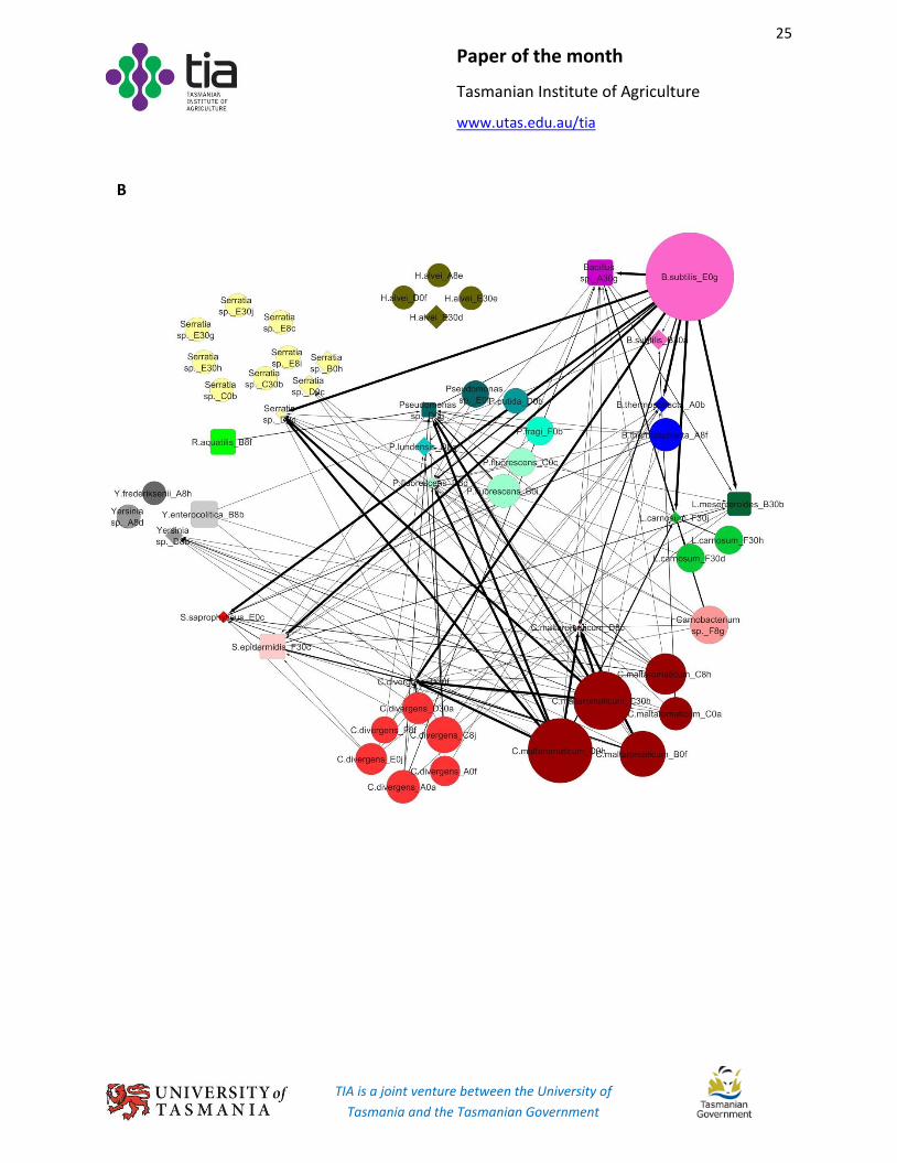
FIG 3 Interactions among effector and target isolates.
(A) Inhibition, spot-lawn assay; (B) Inhibition, CFS assay; (C), Promotion, CFS assay. Symnols:
, target; , effector; , isolate tested as both target and effector. a b = a inhibited (A and B) or
promoted (C) b. Thick to thin black (solid and dashed) arrows indicate “++++”, “+++”, “++”, and “+”
inhibition, respectively. Medium and thin green arrows indicate “++” and “+” growth promotion,
respectively. Dashed and solid black arrows indicate diffuse and clear inhibition zones, respectively, in
panel A. In panels A and B, the size of an effector and target node is, respectively, positively and
negatively correlated with the number and level of inhibitions. In panel C, the size of both an effector
and target node is positively correlated with the number and level of promotions.
C