Hong Kong J. Dermatol. Venereol. (2019) 27, 31-35
Case Report
Papillon-Lefevre syndrome: a rare case report and reviewof literature
D Vupperla, P Elaprolu, FT Taj
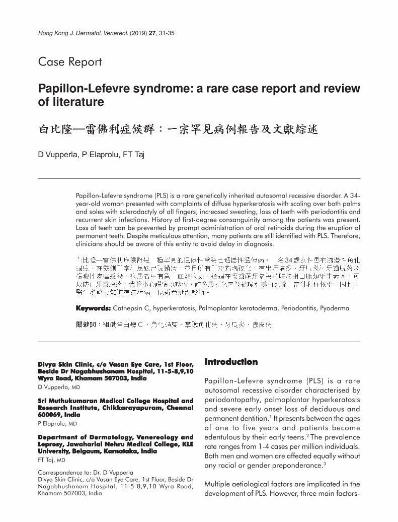
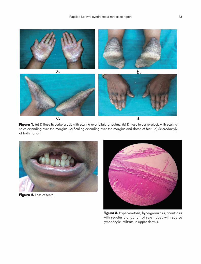
Papillon-Lefevre syndrome (PLS) is a rare genetically inherited autosomal recessive disorder. A 34-year-old woman presented with complaints of diffuse hyperkeratosis with scaling over both palmsand soles with sclerodactyly of all fingers, increased sweating, loss of teeth with periodontitis andrecurrent skin infections. History of first-degree consanguinity among the patients was present.Loss of teeth can be prevented by prompt administration of oral retinoids during the eruption ofpermanent teeth. Despite meticulous attention, many patients are still identified with PLS. Therefore,clinicians should be aware of this entity to avoid delay in diagnosis.
34
Keywords:Keywords:Keywords:Keywords:Keywords: Cathepsin C, hyperkeratosis, Palmoplantar keratoderma, Periodontitis, Pyoderma
C
Divya Skin Clinic, c/o VDivya Skin Clinic, c/o VDivya Skin Clinic, c/o VDivya Skin Clinic, c/o VDivya Skin Clinic, c/o Vasan Eye Care, 1st Floorasan Eye Care, 1st Floorasan Eye Care, 1st Floorasan Eye Care, 1st Floorasan Eye Care, 1st Floor,,,,,Beside Dr Nagabhushanam Hospital, 11-5-8,9,10Beside Dr Nagabhushanam Hospital, 11-5-8,9,10Beside Dr Nagabhushanam Hospital, 11-5-8,9,10Beside Dr Nagabhushanam Hospital, 11-5-8,9,10Beside Dr Nagabhushanam Hospital, 11-5-8,9,10Wyra RWyra RWyra RWyra RWyra Road, Khamam 507003, Indiaoad, Khamam 507003, Indiaoad, Khamam 507003, Indiaoad, Khamam 507003, Indiaoad, Khamam 507003, India
D Vupperla, MD
Sri Muthukumaran Medical College Hospital andSri Muthukumaran Medical College Hospital andSri Muthukumaran Medical College Hospital andSri Muthukumaran Medical College Hospital andSri Muthukumaran Medical College Hospital andRRRRResearch Institute, Chikkesearch Institute, Chikkesearch Institute, Chikkesearch Institute, Chikkesearch Institute, Chikkarayapuram, Chennaiarayapuram, Chennaiarayapuram, Chennaiarayapuram, Chennaiarayapuram, Chennai600069, India600069, India600069, India600069, India600069, India
P Elaprolu, MD
Department of DermatologyDepartment of DermatologyDepartment of DermatologyDepartment of DermatologyDepartment of Dermatology, V, V, V, V, Venereology andenereology andenereology andenereology andenereology andLeprosy, Jawaharlal Nehru Medical College, KLELeprosy, Jawaharlal Nehru Medical College, KLELeprosy, Jawaharlal Nehru Medical College, KLELeprosy, Jawaharlal Nehru Medical College, KLELeprosy, Jawaharlal Nehru Medical College, KLEUniversityUniversityUniversityUniversityUniversity, Belgaum, Karnatak, Belgaum, Karnatak, Belgaum, Karnatak, Belgaum, Karnatak, Belgaum, Karnataka, Indiaa, Indiaa, Indiaa, Indiaa, India
FT Taj, MD
Correspondence to: Dr. D VupperlaDivya Skin Clinic, c/o Vasan Eye Care, 1st Floor, Beside DrNagabhushanam Hospital, 11-5-8,9,10 Wyra Road,Khamam 507003, India
IntroductionIntroductionIntroductionIntroductionIntroduction
Papillon-Lefevre syndrome (PLS) is a rare
autosomal recessive disorder characterised by
periodontopathy, palmoplantar hyperkeratosis
and severe early onset loss of deciduous and
permanent dentition.1 It presents between the ages
of one to five years and patients become
edentulous by their early teens.2 The prevalence
rate ranges from 1-4 cases per million individuals.
Both men and women are affected equally without
any racial or gender preponderance.3
Multiple aetiological factors are implicated in the
development of PLS. However, three main factors-
D Vupperla et al32
immunological, microbial and genetic factors are
responsible for initiation and progression of PLS.
Firstly, cellular immune defects with reduced
phagocyt ic and chemotact ic funct ion of
neutrophils, as well as other granulocytes are
found in most of the patients. Secondly,
pathogenic microorganisms such as Actinobacillus
actinomycetemcomitans, Capnocytophaga
g ing i va l i s , Fu sobac t e r i um nuc l ea t um ,
Peptostreptococcus micros, spirochetes and
Porphyromonas gingivalis, act as the causative
agents for periodontal problems in PLS.4 Another
recently identified genetic defect in PLS patients is
diminution in cathepsin C activity. The mutation
in coding sequence of cathepsin C located on
chromosome 11q14-q21 indicates PLS. The
cathepsin C gene, also known as dipeptidyl-
peptidase I, has endopeptidase activity. It is mostly
expressed in the palms, keratinised oral gingiva,
knees, and soles which are the most commonly
affected areas in PLS. In addition, this gene is also
expressed in many immune cells such as
polymorphonuclear leukocytes, macrophages,
and their precursors.3 Despite advances in
characterising the syndrome, the pathogenic
mechanisms involved in periodontal lesions still
remain obscure. Various treatments including
advice on oral hygiene, scaling, root planning,
non-surgical treatment along with the use of
systemic antibiotics, periodontal surgery have
been tried but without success.5 We hereby report
one case of PLS with the clinical characteristic
features and a brief literature review.
Case presentationCase presentationCase presentationCase presentationCase presentation
A 34-year-old woman presented with complaints
of skin thickening and fine scaling on the soles
and palms with a foul smell, itching, and increased
sweating since the age of two years. There was
total loss of dentition and recurrent skin infections
by the age of 10 years. History revealed similar
complaints in her elder brother. History of first-
degree consanguinity among the patients was
present although both parents were healthy.
On clinical examination, diffuse hyperkeratosis
with scaling was observed on both palms and soles
extending over the margins and dorsa of the hands
and feet (Figures 1 a-c). The presence of
sclerodactyly of fingers of bilateral upper limb was
also observed (Figure 1d). Oral cavity examination
revealed loss of upper incisors and canines
( lef t s ide) wi th periodonti t is (Figure 2).
Histopathological examination of epidermis
showed hyperkeratosis, hypergranulosis, and
acanthosis with regular elongation of rete ridges.
Examination of the upper dermis showed a sparse
lymphocytic infiltrate (Figure 3). These findings
were consistent with palmoplantar keratoderma.
Hence, the diagnosis of the PLS was made based
on history, clinical, and histopathological features.
The patient was treated with oral isotretinoin
20 mg daily for one month together with
17% salicylic acid ointment. Systemic antibiotics
were also given as advised by the dental
department, along with dental advice. A significant
improvement in hyperkeratosis and hyperhidrosis
was observed after one month. Nevertheless,
due to financial constraints, the patient stopped
the treatment regimen and defaulted further
follow-up.
DiscussionDiscussionDiscussionDiscussionDiscussion
Papillon-Lefevre syndrome is generally considered
as a manifestation of homozygosity of autosomal
recess ive genes. A 2.8-cm in terval on
chromosome 11q14 is mapped as a major gene
locus for PLS. The mutational inheritance of
cathepsin C gene is identified in homozygotes of
PLS.6 Cathepsin C is one of the components
required for T-cell activation. Recurrent pyogenic
infection occurs due to neutrophil phagocytosis
and impaired reactivity to T-cell and B-cell
mitogens.7
Papillon-Lefevre syndrome generally starts in
childhood with cutaneous lesions appearing with
oral lesions. Cutaneous lesions, characterised by
Papillon-Lefevre syndrome: a rare case report 33
Figure 1.Figure 1.Figure 1.Figure 1.Figure 1. (a) Diffuse hyperkeratosis with scaling over bilateral palms. (b) Diffuse hyperkeratosis with scaling
soles extending over the margins. (c) Scaling extending over the margins and dorsa of feet. (d) Sclerodactyly
of both hands.
Figure 2.Figure 2.Figure 2.Figure 2.Figure 2. Loss of teeth.
Figure 3.Figure 3.Figure 3.Figure 3.Figure 3. Hyperkeratosis, hypergranulosis, acanthosis
with regular elongation of rete ridges with sparse
lymphocytic infiltrate in upper dermis.
D Vupperla et al34
erythematous hyperkeratotic plaques, involve the
entire surface of the palms and soles and
sometimes extend onto the skin above the Achilles
tendon and external malleoli (transgradiens).
Hyperhidrosis of the palms and soles causes a
foul smell. The psoriasiform plaques may also
extend on to the elbows and knees and the
condition may be associated with painful fissures,
usually worsening during winter. The nails may
also show transverse ridging and fissuring. Severe
periodontitis and premature loss of primary and
permanent dentition is also seen in PLS patients.
All permanent teeth are usually lost between the
ages of 14 to 16 years. The probable reasons of
shedding of teeth are loss of alveolar bone,
gingival infect ion, periodontal l igament
destruction, and abscess formation.3 As a result
of resorption of underlying alveolar bone,
radiography shows a "floating in air" appearance
of the tooth. The patient in this case also showed
similar clinical and radiological characteristics. In
addition to the oral and dermatological findings,
there have been reports of decreased monocyte,
lymphocyte or neutrophil function. In addition,
pyogenic liver abscess, which is associated with
impairment of immune system, has also been
observed in a few cases.8
The differential diagnosis includes three rare allelic
variants of PLS: acrodynia, cyclic neutropenia,
hypophosphatasia. However, this was not
implemented in our study. The clinical features of
muscle pain, insomnia, tachycardia, presence of
erythrocyanosis, psychological disturbances and
premature teeth eruption along with dystrophic
enamel differentiates PLS from acrodynia. The
absence of palmoplantar hyperkeratosis
differentiates cyclic neutropenia from PLS.
The c l in ica l features that d i f ferent ia te
hypophosphatasia from this disease include
knock-knee, hypoplastic teeth, enlarged wrists,
and phosphoethanolaminuria. Palmoplantar
hyperkeratosis is not present in patients with cyclic
neutropenia. Other conditions including Greither
syndrome, keratosis punctate and Howel-Evans
syndrome also can be included in differential
diagnosis. Although palmoplantar hyperkeratosis
may be p re sen t i n t he se s ynd romes ,
periodontopathy is not observed in these cases.9
Molecular genetic testing can be used to detect
mutations in the Cathepsin C gene. This diagnostic
service is available at specialised research
laboratories. Genetic testing was not performed
in this study due to low financial status of the
parents. However, history, cl inical, and
histopathological features strongly supported the
diagnosis of PLS. Furthermore, a consanguineous
lineage has been defined for this syndrome.10
Similarly, in this case first-degree consanguinity
was observed among the patients. Phenotypically,
both the parents were healthy and no family
history of this syndrome suggests an autosomal
recessive genetic syndrome.10
Patients with PLS should be managed with a
multidisciplinary approach. Emollients, salicylic
acid, and urea are involved in the treatment of
skin manifestations. Oral retinoids including
acitretin, isotretinoin and etretinate are mainly
used to treat keratoderma and periodontitis.4,5 If
the treatment is initiated before the onset of
eruption of permanent teeth, normal dentition is
expected for patients on treatment with retinoids.
Treatment is more efficacious if it is continued
during the development of permanent teeth.
Shedding of the teeth can be delayed with advice
on oral hygiene. A proper course of antibiotics
should also be prescribed to preserve teeth, to
control active periodontitis, to treat bacteraemia
and pyrogenic liver abscess. Early extraction of
infected teeth has also been recommended to
avoid bone loss and allows protection of a solid
base for implantation of artificial dentures.3
Furthermore, genetic counselling should also be
given to the parents as well as caregivers of
affected persons regarding the chances of passing
the condition to their offspring.
Papillon-Lefevre syndrome: a rare case report 35
ConclusionConclusionConclusionConclusionConclusion
Pap i l lon-Le fev re syndrome requ i res a
multidisciplinary approach with dynamic
participation of a dermatologist and dental
surgeon. The current case report illustrates that
continuous administration of isotretinoin along
with salicylic acid and systemic antibiotics can
improve the condition of the patient. Permanent
loss of teeth can be prevented by prompt
administration of oral retinoids during the eruption
of permanent teeth. Despite meticulous attention,
several patients are still identified with PLS.
Therefore, prompt diagnosis and intervention is
mandatory. A periodontist may be the first person
to see and treat patients afflicted with unusual PLS,
and therefore, greater awareness of this syndrome
will be helpful in identifying more cases for further
study. Hence, clinicians should be familiar with
this condition to avoid delay in diagnosis.
RRRRReferenceseferenceseferenceseferenceseferences
1. Reddy KV, Maloth KN, Anusha NV, Thummala VS,Thakur M. Papillon-Lefèvre syndrome: A case series withreview of literature. J Oral Med, Oral Surg, Oral PatholOral Radiol 2016;2:248-51.
2. Hart TC, Shapira L. Papillon-Lefèvre syndrome.Periodontol 2000 1994;6:88-100.
3. Pavankumar K. Papillon-Lefèvre syndrome: A casereport. Saudi Dent J 2010;22:95-8.
4. Kaur B. Papillon lefevre syndrome: A case report withreview. Dentistry 2013;3:156.
5. Ramadan ARM, Bashir AH. Papillon-Lefèvre syndrome-a case report and a multidisciplinary approach in thetreatment of 18 years old Sudanese female. Am JDermatol Venereol 2017;6:11-5.
6. Mahajan VK, Thakur NS, Sharma NL. Papillon-Lefèvresyndrome. Ind Pediatr 2003;40:1197-200.
7. Shah J, Goel S. Papillon-Lefèvre syndrome: Two casereports. Ind J Dent Res 2007;18:210.
8. Raghoji S, Ravishankar S. Papillon-Lefèvre syndrome:A case report with review of literature. Ind J MednodentAllied Sci 2014;2.
9. Sreeramulu B, Shyam ND, Ajay P, Suman P. Papillon-Lefèvre syndrome: Clinical presentation andmanagement options. Clin Cosmet Invest Dent 2015;7:75-81.
10. Khan FY, Jan SM, Mushtaq M. Papillon-Lefèvresyndrome: Case report and review of the literature.J Ind Soc Periodontol 2012;16:261-5.