Paracrine inhibition of GM-CSF signaling by human cytomegalovirus inmonocytes differentiating to dendritic cellsJerome Carlier,1-3 Helene Martin,1-3 Bernard Mariame,1-3 Benjamin Rauwel,1-3 Catherine Mengelle,4 Hugo Weclawiak,1-3
Alain Coaquette,5 Charline Vauchy,6 Pierre Rohrlich,6 Nassim Kamar,1-3,7 Lionel Rostaing,1-3,7 Georges Herbein,5 andChristian Davrinche1-3
1Inserm U1043, Toulouse, France; 2Centre National de la Recherche Scientifique U5282, Toulouse, France; 3Universite de Toulouse, UPS, Centre dePhysiopathologie de Toulouse Purpan, Toulouse, France; 4Centre Hospitalo-University (CHU) Toulouse, Hopital Purpan, Service de Virologie, Toulouse, France;5Departement de Virologie, IFR133, EA3186, Universite de Franche Comte, CHU Besancon, Besancon, France; 6Service d’Hematologie Clinique, CHUBesancon, Inserm U-645/IFR 133, Besancon, France; and 7CHU Toulouse, Hopital Rangueil, Service de Nephrologie, Toulouse, France
A primary HCMV infection or virus reacti-vation may cause severe disease in hostswith a deficient immune system. The vi-rus can disturb both innate and adaptiveimmunity by targeting dendritic cell (DC)functions. Monocytes, the precursors ofDCs in vivo (MoDCs), are the primarytargets of HCMV; they can also harborlatent virus. The DCs generated from in-fected monocytes (CMV-MoDCs) have analtered phenotype and functional defects.We have shown that CMV-MoDCs do not
secrete IL-12 in response to lipopolysac-charide stimulation, cannot ingest deadcells, induce TH1 differentiation, or theproliferation of naive allogeneic CD4�
T cells. We found that the GM-CSF signal-ing in an entire population of CMV-MoDCswas impaired, although only half of thecells were productively infected, and thatIL-6 secretion and suppressors of cyto-kine signaling 3 induction contributed tothis bystander effect. We also showedthat MoDCs derived ex vivo from mono-
cytes of viremic patients had the samealtered phenotype as CMV-MoDCs, includ-ing decreased STAT5 phosphorylation,indicating defective GM-CSF signaling.We have thus described a new mecha-nism of HCMV-induced immunosupres-sion, indicated how infection may disturbboth GM-CSF–dependent physiologicprocesses and proposed GM-CSF–basedtherapeutic approaches. (Blood. 2011;118(26):6783-6792)
Introduction
Human CMV (HCMV) is a �-herpesvirus that infects most of thehuman population during their lifetime. HCMV opportunisticallyexploits its host immune defects to replicate and spread, so causingsevere disease. Transplant recipients, fetuses, and newborns are allat risk of disease or death because of primary infections or virusreactivation. HCMV also encodes a variety of proteins that disruptTCD4� and TCD8� antiviral responses; the latter are critical forcontrolling the spread of virus and preventing associated diseases(for a review see Crough and Khanna1). Dendritic cells (DCs) arethe sites of primary infection and are essential for capturing thevirus Ags needed to mould an appropriate CD8� T-cell repertoireagainst the virus. Thus, DCs are main targets of virus subversion.DCs infected with HCMV have defects in all stages of Agpresentation, migration to secondary lymphoid organs, and T-cellactivation (reviewed in Varani et al2). We have developed aworking hypothesis that these virus-induced changes can beovercome by uninfected DC cross-presenting Ags to CD8� T cells.3
Monocytes are primary targets of HCMV, can harbor latent virus,and are a source of inflammatory DCs in vivo.4-6 We thereforeassumed that DC dysfunction was because of abnormalities arisingduring the differentiation of their monocyte precursors. DCsderived from infected monocytes in vitro and in patients whoreceived a transplant experiencing an active infection7,8 all haveabnormal phenotypes and function that may contribute to immuno-suppression. However, the molecular and cellular mechanisms
underlying these defects are poorly documented. We have obtainedevidence that the infection of monocytes with HCMV results indefective GM-CSF signaling, with inhibition of STAT5 phosphory-lation that leads to dysfunctional derived DCs. These DCs had lostcritical features such as the ability to potentiate TH1 differentia-tion and proliferation because of secreted factors such as IL-6that is essential for suppressors of cytokine signaling 3 (SOCS3)synthesis. We have therefore provided new insights into the wayHCMV infections cause immunosuppressive damage and theensuing side effects.
Methods
Generation and culture of monocyte-derived DCs
DCs were generated from monocytes, and cell cultures were phenotyped asdescribed in supplemental Methods (available on the Blood Web site; seethe Supplemental Materials link at the top of the online article).
Viruses
The endothelial cell–adapted strain HCMVTB40/E GFP was provided byM. Messerle (Hanover, Germany) and HCMVVHLE was provided by C.Sinzger (Tubingen, Germany). HCMV BACTB40/E GFP (green fluorescentprotein) was designed and generated by E. Borst (Hanover, Germany) from
Submitted February 17, 2011; accepted October 11, 2011. Prepublished onlineas Blood First Edition paper, October 26, 2011; DOI 10.1182/blood-2011-02-337956.
The online version of this article contains a data supplement.
The publication costs of this article were defrayed in part by page chargepayment. Therefore, and solely to indicate this fact, this article is herebymarked ‘‘advertisement’’ in accordance with 18 USC section 1734.
a HCMV BACTB40/E genome9 in which the US1 to US7 genomic fragmentwas replaced by a GFP cassette under the control of mouse MIEP.
Viruses were amplified on MRC5 fibroblasts and collected by centrifu-gation at 100 000g for 35 minutes. Virus titers (PFU/mL) were determinedon MRC5 cells. Viruses were inactivated by UV irradiation for 20 minutesat 0.17 Amp with the use of a Spectroline device.
Monocyte infection and treatment
Monocytes in differentiation medium were either mock-infected (medium)or infected with live or UV-inactivated virus at a MOI of 5. Alternatively,culture medium was supplemented with human recombinant IL-6 (25-50 ng/mL; Ozyme), anti–IL-6 or anti–IL-6R blocking Abs, or IgG isotype control(15 �g/mL; Gen-Probe Diaclone), and phosphonoacetic acid (PAA; 250 �g/mL; Sigma-Aldrich). CMV-MoDC defines cells produced by the differentia-tion of infected monocytes and IL-6-MoDC defines cell supplemented withIL-6. In experiments with conditioned medium, the culture medium was amixture of fresh differentiation medium and the medium to be tested (1/1;vol/vol). When applicable, culture media were cleared of infectious virus bycentrifugation (100 000g for 35 minutes).
Flow cytometric analysis
Cells were treated, stained, and analyzed by flow cytometry on a FACSCali-bur cytometer (BD Biosciences; supplemental Methods).
Western blot analysis
Electrophoresis of cell lysates and protein analysis by immunoblotting wasperformed with standard procedures (supplemental Methods).
RT-PCR analysis
PCRs were performed on reverse-transcribed total RNA as detailed insupplemental Methods.
Phagocytosis assays
MRC5 cells were stained with PKH26 (Sigma-Aldrich) and cultured withTNF-� (1 �g/mL) and cycloheximide (25 �g/mL; Sigma-Aldrich) incomplete DMEM medium for 24 hours to obtain apoptotic bodies. Apopto-tic MRC5 cells were cocultured with DCs (1 DC for 2 MRC5 cells). Cellswere harvested and stained with an FITC-conjugated anti–HLA-DR Ab,and double-positive cells were quantified on a FACSCalibur device. Thepercentage of PKH26�/HLA-DR� MoDCs was considered as 100% ofphagocytic activity. Alternatively, the Vybrant phagocytosis assay kit(Escherichia coli bioparticles; Invitrogen) was used.
Cytokine quantification
Secreted inflammatory cytokines were detected with a dedicated Ab arrayfrom Raybiotech. TNF-�, IL-6, and IL-12p70 were measured with acytometric bead array assay (CBA; BD Biosciences) and the soluble formof GM-CSF receptor (sGMR�) by ELISA (Gen-Probe Diaclone).
Small interfering RNA transfection
Purified monocytes were treated with Lipofectamine RNAiMax (Invitro-gen) and 50nM RNAi was directed against SOCS1 (3�-UCGAAGAGGCA-GUCGAAGCUCUCGC-5�) or SOCS3 (3�-AGUAGAUGUAAUAGGCU-CUUCUGGG-5�) mRNA (Invitrogen) or control RNAi (Invitrogen).Corresponding protein expressions were checked by Western blot analysiswith the use of anti-SOCS3 or anti-SOCS1 Abs (Abcam).
Isolation of naive CD4� T cells and polarization experiments
Naive CD4�CD45RA� T cells were isolated from PBMCs with the use of amagnetic bead separation system (naive CD4� T-cell isolation kit; MiltenyiBiotec). Allogeneic stimulators were infected with HCMV or were treatedwith anti–IL-6 or isotypic Abs during differentiation and then with ultrapurelipopolysaccharide (LPS; Invivogen) for 24 hours, washed with culture
medium, and cocultured for 5 days in 96-well plates with T cells (95%-98%purity) at a ratio of 1:10 (1 � 104 allogeneic stimulators for 1 � 105
T cells/well in 200 �L). IL-2 (50 U/mL; PeproTech) was then added to eachwell, and culture was continued for a further 3 days. For TH2 polarizationexperiments, IL-4 (5 ng/mL; PeproTech) and anti–IFN-� Ab (10 �g/mL;BD Biosciences) were added to cocultures. Intracellular cytokines wereassayed on day 8 (see supplemental Methods). In CFSE experiments, naiveT cells were stained with CFSE (Invitrogen), cocultured for 5 days at a ratioof 1 stimulator to 10 T cells as above, and T-cell proliferation was analyzed.
Patients
The project was approved by the ethics committees of the RangueilHospital (Toulouse) and Besancon Hospital (Besancon), and all patientswho provided samples gave their written informed consent in accordancewith the Declaration of Helsinki. PBMCs were isolated from patients’ bloodsamples with the use of a Ficoll density gradient, and CD14� cells weresorted as described in supplemental Methods: “Generation and culture ofmonocytes derived DCs.” Cells were differentiated to MoDCs for 5 days,and their CD1a and pSTAT5 contents were determined. Secreted IL-6 inblood plasma and MoDC supernatants were assayed with CBA. HCMVDNA in patients’ blood was quantified by real-time PCR.10
Statistical analysis
Values are expressed as the means � SDs of � 3 independent experiments.Data were analyzed for significance with the Mann-Whitney U test orStudent t test, and a P value � .05 was considered significant. All data wereanalyzed with Prism Version 5 (GraphPad) and FlowJo Version 7.6.5(TreeStar) software.
Results
Phenotype of DCs differentiated from HCMV-infectedmonocytes
Adherent monocytes obtained from the peripheral blood of healthydonors were either mock-infected or infected with HCMV (VHLE;MOI of 5). The resulting MoDCs and CMV-MoDCs were obtainedby differentiation in a culture medium containing GM-CSF andIL-13 for 5 days. IL-4 treatment always generated the samephenotype as IL-13 (not shown). The MoDCs had the phenotype ofimmature DCs bearing CD1a (95%) and CD1c (98%), CD80 (52%)and CD86 (53%), but no CD14 (0%) or CD83 (3%). Only 10% ofinfected MoDCs expressed CD1a, and only 19% had CD1c,whereas most of them had the maturation marker CD83 (83%) andhad increased CD80 (85%) and CD86 (97%; Figure 1A). Identicalresults were obtained when monocytes were positively selectedfrom PBMCs with the use of CD14 magnetic beads (data notshown). Alternatively, the HCMV inoculum was UV-inactivatedbefore adding it to monocytes to determine whether the phenotypemodifications required live virus. UVCMV-MoDCs had the regularMoDC phenotype (not shown), indicating that de novo synthesis ofvirus proteins was necessary for production of the modifiedphenotype. Surprisingly, monocytes are poorly permissive toHCMV, but most CMV-MoDCs were modified. We thereforemeasured the percentage of infected cells with an HCMV BACconstruct encoding a GFP. Approximately 50% of CMV-MoDCscontained GFP-labeled fluorescence, whereas 82% of them werepositive for CD83 and 6% were positive for CD1a compared with11% (CD83) and 90% (CD1a) of MoDCs (Figure 1B). These datasuggest that infected cells inhibited their uninfected counterpartsby secreting soluble factors. We therefore cultured monocytes for5 days in standard medium (GM-CSF, IL-13) supplemented with
6784 CARLIER et al BLOOD, 22 DECEMBER 2011 � VOLUME 118, NUMBER 26
For personal use only.on April 4, 2019. by guest www.bloodjournal.orgFrom
supernatants recovered from 5-day cultures of MoDCs, CMV-MoDCs, and UVCMV-MoDC. Most of the cells generated frommonocytes that had been exposed to medium conditioned byCMV-MoDCs expressed no CD1a (11% of CD1a� cells), whereasthose exposed to supernatants from UVCMV-MoDCs were un-changed (Figure 1C). Thus, monocytes infected with live HCMVsecrete soluble factors that alter their differentiation into standardimmature DCs.
Paracrine inhibition of STAT5 phosphorylation in CMV-MoDCs
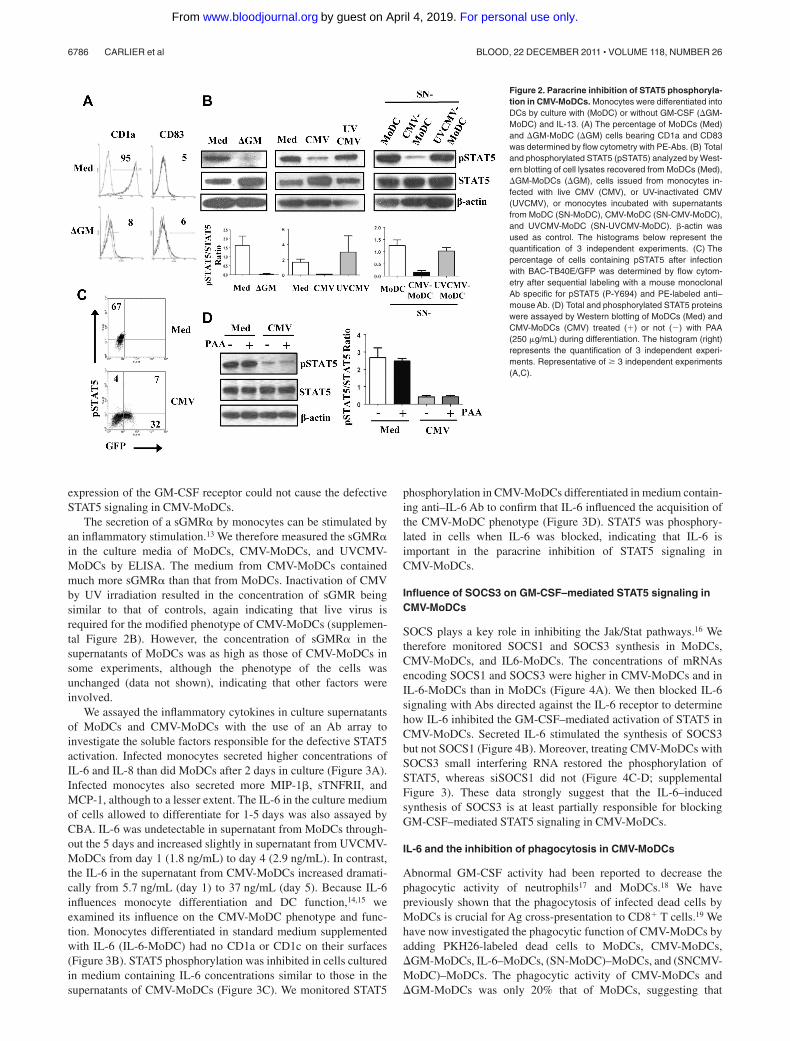
We checked that monocytes that differentiated in the absence ofGM-CSF gave rise to DCs bearing no CD1a, as previouslyreported11 (Figure 2A). We then investigated whether CMV-MoDCphenotype could be because of, at least in part, a defect in GM-CSFsignaling. Because GM-CSF triggers STAT5 activation,12 whichrequires the phosphorylation of tyrosine at position 694, wemeasured the phosphorylated STAT5 (pSTAT5) in CMV-MoDCsby Western blot analysis. The pSTAT5/total STAT5 ratio in DCsgenerated in the absence of GM-CSF (GM-MoDCs) was lowerthan that of standard MoDCs (Figure 2B). This was also true for
CMV-MoDCs and monocytes differentiated in the presence ofCMV-MoDC supernatants. The altered phenotype of CMV-MoDCs could therefore be because of a blockage of GM-CSFsignaling, perhaps by soluble factors secreted by infected mono-cytes during their differentiation. STAT5 phosphorylation was notinhibited in cells treated with UVCMV, confirming that blockingSTAT5 activation requires live CMV. We substantiated the impair-ment of STAT5 activation in CMV-MoDCs by flow cytometry todetect pSTAT5 in cells infected with BAC-GFP (Figure 2C). Thisagain pointed to HCMV impairing monocyte differentiation via aparacrine effect. Inhibiting virus replication with PAA, whichinhibits late viral protein synthesis (supplemental Figure 1), did notreverse the inhibition of STAT5 phosphorylation by HCMV,suggesting that immediate early (IE) or early (E) virus products areinvolved (Figure 2D).
Paracrine effect of IL-6 on differentiating monocytes
Cytometric analysis showed that there was more GM-CSF recep-tors on the cell surface of CMV-MoDCs than on MoDCs at day 2 ofdifferentiation (supplemental Figure 2A). Hence, impaired surface
Figure 1. Abnormal phenotype of DCs derived fromHCMV-infected monocytes results from a paracrineeffect. Monocytes (Mo) were differentiated into DCsafter mock-infection (MoDC) or infection with HCMV-VHLE (CMV-MoDC) by culture for 5 days with GM-CSFand IL-13. (A) Surface markers (CD1a, CD1c, CD14,CD83, CD80, and CD86) were analyzed by flow cytom-etry, and the percentages of positive cells was deter-mined as indicated. (B) Monocytes were infected withBAC-TB40E/GFP and checked for GFP fluorescenceand by flow cytometry after labeling with anti-CD1a andanti-CD83 Abs. (C) Monocytes were differentiated byculture for 5 days with GM-CSF and IL-13 standardmedium supplemented with supernatants recovered fromMoDC (SN-MoDC), MoDC infected with live CMV(SN-CMV-MoDC) or UV-inactivated CMV (SN-UVCMV-MoDC). Cells were labeled with PE-anti-CD1a Abs, andthe percentage of CD1a� cells was determined asindicated. Representative of � 3 independent experi-ments.
HCMV IMPAIRS GM-CSF SIGNALING AND DC FUNCTIONS 6785BLOOD, 22 DECEMBER 2011 � VOLUME 118, NUMBER 26
For personal use only.on April 4, 2019. by guest www.bloodjournal.orgFrom
expression of the GM-CSF receptor could not cause the defectiveSTAT5 signaling in CMV-MoDCs.
The secretion of a sGMR� by monocytes can be stimulated byan inflammatory stimulation.13 We therefore measured the sGMR�in the culture media of MoDCs, CMV-MoDCs, and UVCMV-MoDCs by ELISA. The medium from CMV-MoDCs containedmuch more sGMR� than that from MoDCs. Inactivation of CMVby UV irradiation resulted in the concentration of sGMR beingsimilar to that of controls, again indicating that live virus isrequired for the modified phenotype of CMV-MoDCs (supplemen-tal Figure 2B). However, the concentration of sGMR� in thesupernatants of MoDCs was as high as those of CMV-MoDCs insome experiments, although the phenotype of the cells wasunchanged (data not shown), indicating that other factors wereinvolved.
We assayed the inflammatory cytokines in culture supernatantsof MoDCs and CMV-MoDCs with the use of an Ab array toinvestigate the soluble factors responsible for the defective STAT5activation. Infected monocytes secreted higher concentrations ofIL-6 and IL-8 than did MoDCs after 2 days in culture (Figure 3A).Infected monocytes also secreted more MIP-1�, sTNFRII, andMCP-1, although to a lesser extent. The IL-6 in the culture mediumof cells allowed to differentiate for 1-5 days was also assayed byCBA. IL-6 was undetectable in supernatant from MoDCs through-out the 5 days and increased slightly in supernatant from UVCMV-MoDCs from day 1 (1.8 ng/mL) to day 4 (2.9 ng/mL). In contrast,the IL-6 in the supernatant from CMV-MoDCs increased dramati-cally from 5.7 ng/mL (day 1) to 37 ng/mL (day 5). Because IL-6influences monocyte differentiation and DC function,14,15 weexamined its influence on the CMV-MoDC phenotype and func-tion. Monocytes differentiated in standard medium supplementedwith IL-6 (IL-6-MoDC) had no CD1a or CD1c on their surfaces(Figure 3B). STAT5 phosphorylation was inhibited in cells culturedin medium containing IL-6 concentrations similar to those in thesupernatants of CMV-MoDCs (Figure 3C). We monitored STAT5
phosphorylation in CMV-MoDCs differentiated in medium contain-ing anti–IL-6 Ab to confirm that IL-6 influenced the acquisition ofthe CMV-MoDC phenotype (Figure 3D). STAT5 was phosphory-lated in cells when IL-6 was blocked, indicating that IL-6 isimportant in the paracrine inhibition of STAT5 signaling inCMV-MoDCs.
Influence of SOCS3 on GM-CSF–mediated STAT5 signaling inCMV-MoDCs
SOCS plays a key role in inhibiting the Jak/Stat pathways.16 Wetherefore monitored SOCS1 and SOCS3 synthesis in MoDCs,CMV-MoDCs, and IL6-MoDCs. The concentrations of mRNAsencoding SOCS1 and SOCS3 were higher in CMV-MoDCs and inIL-6-MoDCs than in MoDCs (Figure 4A). We then blocked IL-6signaling with Abs directed against the IL-6 receptor to determinehow IL-6 inhibited the GM-CSF–mediated activation of STAT5 inCMV-MoDCs. Secreted IL-6 stimulated the synthesis of SOCS3but not SOCS1 (Figure 4B). Moreover, treating CMV-MoDCs withSOCS3 small interfering RNA restored the phosphorylation ofSTAT5, whereas siSOCS1 did not (Figure 4C-D; supplementalFigure 3). These data strongly suggest that the IL-6–inducedsynthesis of SOCS3 is at least partially responsible for blockingGM-CSF–mediated STAT5 signaling in CMV-MoDCs.
IL-6 and the inhibition of phagocytosis in CMV-MoDCs
Abnormal GM-CSF activity had been reported to decrease thephagocytic activity of neutrophils17 and MoDCs.18 We havepreviously shown that the phagocytosis of infected dead cells byMoDCs is crucial for Ag cross-presentation to CD8� T cells.19 Wehave now investigated the phagocytic function of CMV-MoDCs byadding PKH26-labeled dead cells to MoDCs, CMV-MoDCs,GM-MoDCs, IL-6–MoDCs, (SN-MoDC)–MoDCs, and (SNCMV-MoDC)–MoDCs. The phagocytic activity of CMV-MoDCs andGM-MoDCs was only 20% that of MoDCs, suggesting that
Figure 2. Paracrine inhibition of STAT5 phosphoryla-tion in CMV-MoDCs. Monocytes were differentiated intoDCs by culture with (MoDC) or without GM-CSF (GM-MoDC) and IL-13. (A) The percentage of MoDCs (Med)and GM-MoDC (GM) cells bearing CD1a and CD83was determined by flow cytometry with PE-Abs. (B) Totaland phosphorylated STAT5 (pSTAT5) analyzed by West-ern blotting of cell lysates recovered from MoDCs (Med),GM-MoDCs (GM), cells issued from monocytes in-fected with live CMV (CMV), or UV-inactivated CMV(UVCMV), or monocytes incubated with supernatantsfrom MoDC (SN-MoDC), CMV-MoDC (SN-CMV-MoDC),and UVCMV-MoDC (SN-UVCMV-MoDC). �-actin wasused as control. The histograms below represent thequantification of 3 independent experiments. (C) Thepercentage of cells containing pSTAT5 after infectionwith BAC-TB40E/GFP was determined by flow cytom-etry after sequential labeling with a mouse monoclonalAb specific for pSTAT5 (P-Y694) and PE-labeled anti–mouse Ab. (D) Total and phosphorylated STAT5 proteinswere assayed by Western blotting of MoDCs (Med) andCMV-MoDCs (CMV) treated (�) or not () with PAA(250 �g/mL) during differentiation. The histogram (right)represents the quantification of 3 independent experi-ments. Representative of � 3 independent experiments(A,C).
6786 CARLIER et al BLOOD, 22 DECEMBER 2011 � VOLUME 118, NUMBER 26
For personal use only.on April 4, 2019. by guest www.bloodjournal.orgFrom
GM-CSF signaling also plays a role in this process (Figure 5A).The CMV-induced IL-6–mediated inhibition of STAT5 accountedat least in part for this defect in CMV-MoDCs because thephagocytic capacity of IL-6–MoDCs was � 50% that of MoDCs.However, although adding conditioned medium from CMV-MoDCs to MoDCs mimicked the effect of IL-6 (50% reduction;Figure 5B), neutralization of IL-6 with anti–IL-6 Abs did notrestore phagocytosis. Thus, IL-6 may not be the only secretedfactor involved in this effect. Assays of phagocytosis which usedE coli bioparticles gave similar results (data not shown). Thus, thedefective phagocytosis of dead cells by CMV-MoDCs probablyinvolves both autocrine and paracrine inhibition of STAT5 signal-ing, some of which involves IL-6.
IL-6 secretion and blockage of allogeneic T-cell proliferationand TH1 polarization by CMV-MoDCs
DCs and their environment help determine the programming ofT cells toward TH1 or TH2 subsets. We have examined how thepolarization of naive CD4� T cells is influenced by CMV-MoDCs.One of the mechanisms by which DCs drive TH1 differentiationinvolves IL-12p70. It is generally agreed that the secretion ofIFN-� by differentiated T cells correlates with the amount of IL-12secreted by stimulated MoDCs2 in mixed lymphocyte reactions(MLRs),20 whereas IL-4 production promotes TH2 differentiation.We measured the amount of IL-12p70 secreted by MoDCs,CMV-MoDCs, and IL-6–MoDCs that had been stimulated withLPS, a known activator of IL-12 secretion. CMV-MoDCs weremuch less responsive (67 pg/mL) than MoDCs (380 pg/mL),whereas IL-6–MoDCs produced almost no detectable IL-12p70(Figure 6A). No DC population secreted any IL-12p70 withoutLPS stimulation. The production of IL-12p70 by stimulatedMoDCs was inhibited by adding CMV-MoDC supernatants. How-ever, neutralization of IL-6 did not restore IL-12p70 production,suggesting that IL-6 is not the only factor involved (Figure 6B). We
used MLR experiments to study TH polarization by DCs. Unacti-vated and LPS-activated MoDCs, CMV-MoDCs, and IL-6-MoDCswere washed and cocultured for 8 days with allogeneic naiveT cells. The TH1 and TH2 responses were assessed by flowcytometry by measuring the IL-4 and IFN-� produced by T cells.MoDCs promoted the production of IFN-� in 19.6% and LPS-stimulated MoDCs in 33.0% of T cells (Figure 6C), correlating theability of MoDCs to potentiate the TH1 response with IL-12p70secretion. In contrast, LPS-stimulated CMV-MoDCs induced IFN-�secretion in very few T cells (6.56%). Few T cells secreted IFN-�when incubated with LPS-unstimulated CMV-MoDCs, suggestingthat CMV-MoDCs are intrinsically unable to prime a TH1 response(data not shown). IL-6–MoDCs treated with LPS also showed areduced capacity to prime TH1 (Figure 6C), suggesting that IL-6alters the ability of MoDCs to polarize T cells toward TH1. T cellsstimulated with MoDCs that had been treated with CMV-MoDCconditioned medium (SN-CMV-MoDCs) and LPS formed fewerIFN-�–producing T cells (18%) than did those stimulated withsupernatants from MoDCs (28.0%). The rate of TH1 formation wasrestored (33.9%) by blocking the IL-6 in these supernatants (Figure6D). The IL-6 secreted by CMV-MoDCs during differentiationtherefore prevents them from stimulating T cells to form TH1. Noneof the DC populations induced detectable IL-4– (Figure 6) orIL-17– (data not shown) secreting cells after stimulation with LPS.This indicates that CMV-MoDCs do not stimulate naive T cells toform either TH2 or TH17, although they were able to differentiatetoward TH2 when stimulated with MoDCs treated with IL-4 andanti–IFN-� Abs (supplemental Figure 4). Besides impaired TH1commitment, incubation of naive T cells with LPS-stimulatedCMV-MoDCs or MoDCs conditioned with SN-CMV-MoDCsblocked their proliferation (Figure 6E). Thus, the infection ofmonocytes with CMV causes profound changes in the ability ofCMV-MoDCs to promote TH1 proliferation and activation, partlybecause of secreted IL-6.
Figure 3. Increased IL-6 secretion by CMV-MoDCsand inhibition of STAT5 phosphorylation by IL-6.Monocytes were differentiated into DCs by culture for5 days with GM-CSF and IL-13 after mock-infection(Med), infection with live (CMV-MoDC), or UV-inactivatedCMV (UVCMV). (A) MoDCs (Med) and CMV-MoDCs(CMV) were differentiated by culture for 5 days; theirinflammatory cytokines were assayed with a dedicatedAb array (Raybiotech), and IL-6 was quantified by CBA.(B) Monocytes were also differentiated in the presenceof recombinant IL-6. The percentages of cells expressingCD1a, CD1c, and CD83 were determined by flow cytom-etry. (C-D) Total and phosphorylated STAT5 (pSTAT5)were analyzed by Western blotting of cell lysates ofIL-6–MoDCs (IL-6) and CMV-MoDCs (CMV) or CMV-MoDCs treated with anti–IL-6 neutralizing Abs (�IL-6;15 �g/mL). The histograms below their respective West-ern blots represent the quantification of 3 independentexperiments. Error bars indicate the SD. Representativeof � 3 independent experiments.
HCMV IMPAIRS GM-CSF SIGNALING AND DC FUNCTIONS 6787BLOOD, 22 DECEMBER 2011 � VOLUME 118, NUMBER 26
For personal use only.on April 4, 2019. by guest www.bloodjournal.orgFrom
Correlation of the phenotype of cells from patients with HCMVviremia and CMV-MoDCs
We investigated the relevance of these observations in vivo withthe use of monocytes from patients who received a transplantexperiencing CMV viremia (Table 1). The monocytes were differ-entiated into DCs (MoDCs). The MoDCs from viremic patientsformed cell clusters (Figure 7A), a feature indicating cell activationas observed in cultured CMV-MoDCs. The medium recovered after5 days of differentiation contained no detectable viral genome,suggesting the absence of virus replication. Significantly fewerMoDCs from CMV-positive patients bore CD1a than those fromCMV-negative patients (Figure 7B). IL-6 was quantified in thegrowth medium and blood plasma, and the phosphorylation ofSTAT5 in MoDCs was analyzed. Patients 7 and 8 had very highvirus loads (480 000 and 340 000 copies/mL, respectively); thesewere correlated with large amounts of IL-6 secreted by theirMoDCs and in their plasma (Figure 7C-D) and few CD1a� cells
(Figure 7A). Furthermore, the MoDCs derived from all the viremicpatients lost their ability to phosphorylate STAT5 on differentiation(Figure 7E). These data reinforce the in vitro findings of the role ofIL-6 and the subsequently defective GM-CSF signaling in DCsderived from infected monocytes.
Discussion
Infection by HCMV is known to disturb both innate and adaptiveimmune responses. DCs are main virus targets to prevent thestimulation of antiviral T cells. However, this is always incompletebecause the virus immune evasion functions are circumvented bythe host.3 Because monocytes are the precursors of DCs in vivo andHCMV may infect them to further establish latency, we examinedthe effects of infection on monocyte differentiation and DCdysfunction.
Monocytes were differentiated into DCs by the coordinatedaction of GM-CSF and IL-13, which models the phenotype andfunction of inflammatory and migratory DCs in vivo.21 MoDCsinfected by HCMV lost their standard CD1a� immature phenotypeand acquired the maturation marker CD83, probably because of thebystander effect of TNF-�, a known inducer of DC maturation,secreted by infected cells in the early phase of differentiation.Discrepancies with published data7 about both cell maturation andthe requirement for live virus could be partly because of the use byothers of the laboratory strain AD169 that has a 19-kbp genomedeletion not found in clinical strains. CD1a and CD1c bind andpresent lipid Ags to T cells,22 in addition to being DC phenotypemarkers. Their expressions are subnormal in CMV-MoDCs and ininfected DCs,23 but their role in controlling HCMV infectionremains to be studied. We have found evidence for a powerfulparacrine mechanism that leads to modification of the phenotype ofthe whole population of CMV-MoDCs. Even though only a fractionof the cells contained the IE/E virus proteins, this viral proteinexpression seems to be critical for initiating this process. Weconsidered the possibility that there was a failure of GM-CSFsignaling in CMV-MoDCs because GM-CSF is needed to generateinflammatory DCs from monocytes in vivo21 and because there arefunctional and phenotypic defects (CD1a) in MoDCs generated inthe absence of GM-CSF.11 We showed that STAT5 activation was
Figure 5. IL-6 is not crucial for inhibition of phagocytosis in CMV-MoDCs.Monocytes differentiated for 5 days as described below were cocultured withPKH-26–labeled apoptotic fibroblasts for 24 hours. Phagocytic activity was deter-mined as described in “Phagocytosis assays.” (A) Monocytes were differentiated instandard medium (Med), after infection (CMV), with added IL-6 (50 ng/mL), or withoutGM-CSF (GM). (B) Monocytes were differentiated in the presence of supernatantfrom MoDCs (SN-MoDC) or CMV-MoDCs (SN-CMV-MoDC) supplemented withmedium (NT), IL-6 neutralizing Ab (�IL-6; 15 �g/mL), or isotype control Ab (iso;15 �g/mL). Results are expressed as the percentage of phagocytic activity relative toMoDCs (100%). Error bars indicate the SD. Representative of � 3 independentexperiments.
Figure 4. Increased SOCS3 and inhibition of STAT5 phosphorylation in CMV-MoDCs. (A-B) Mock-infected (Med), HCMV-infected (CMV), or IL-6–treated (50 ng/mL) monocytes were differentiated by culture with GM-CSF and IL-13 for 5 days.Alternatively, medium (NT), or neutralizing Ab against IL-6 receptor (�IL-6R; 15 �g/mL) or isotype Ab (iso; 15 �g/mL) was added to infected cells during differentiation.SOCS1, SOCS3, and �-actin mRNAs were analyzed by RT-PCR. (C-D) Total andphosphorylated STAT5 protein (pSTAT5) were assessed by Western blotting ofmock-infected (Med) and HCMV-infected (CMV) cells. Cells were transfected withSOCS1, SOCS3, and control small interfering RNA (siRNA; Ctrl) as indicated. �-actinwas used as control. The histograms below RT-PCR (A-B) and Western blots (C-D)represent the quantification of 3 independent experiments.
6788 CARLIER et al BLOOD, 22 DECEMBER 2011 � VOLUME 118, NUMBER 26
For personal use only.on April 4, 2019. by guest www.bloodjournal.orgFrom
inhibited in the whole population of CMV-MoDCs, which mayblock GM-CSF signaling, and that this was because of solublefactors secreted by a fraction of infected monocytes.
Our first hypothesis was that the soluble GM-CSF receptorsGMR� blocked the binding of GM-CSF to its receptor. Thesignificantly increased sGMR� concentration in some preparationsof CMV-MoDCs pointed to its implication in impairing STAT5signaling. However, individual variations in the concentrations ofconstitutive sGMR� in monocytes and our inability to neutralizesGMR� in culture supernatants have made it impossible to decide
on the effect of sGMR� at this stage. One of the inflammatorycytokines induced by HCMV, IL-6, is suspected to be a key factorin the failure of organ and BM transplantations.24 IL-6 has alsobeen identified in the HCMV secretome of endothelial cells as amediator of angiogenesis and cell survival25 and in tumor cells as aparacrine factor in tumor progression and vascularization throughactivation of the STAT3 cascade.26 We find that DCs derived fromthe differentiation of monocytes supplemented with IL-6 bore noCD1a or CD1c. Moreover, neutralizing IL-6 during the differentia-tion of infected monocytes restored phosphorylation of STAT5,suggesting that IL-6 blocks GM-CSF receptor signaling. Previousdata have shown that IE1 increases the activity of the IL-6promoter,27 which may explain why UV-inactivated virus barelyincreased IL-6 secretion and had no effect on STAT5 phosphoryla-tion. Thus, the secretion of IL-6 by UVCMV is initiated by theactivation of NF-�B but cannot be maintained because of blockadeof IE1 transcription after UV irradiation.
The inhibition of STAT5 phosphorylation in DCs generatedfrom monocytes involves SOCS3 and SOCS1,18 and SOCS1 canabolish GM-CSF signal transduction in DC precursors28 but not inconditions of viral infection. Furthermore, induction of SOCS3 byIL-6 is crucial for the regulation of the inflammatory response,whereas the IL-6–mediated induction of SOCS3 could inhibitinsulin receptor signaling.16,29 We have now obtained evidence thatIL-6 secreted by CMV-MoDCs is crucial for the induction ofSOCS3, which in turn leads to the inhibition of STAT5 phosphory-lation that may reflect blockage of the GM-CSF receptor signalingby SOCS3 but not SOCS1.
The influence of STAT5 blockage by SOCS on the phagocyticactivity of MoDCs was deduced18 from the inability of cells tointernalize dextran. Defects in the phagocytosis of dextran andyeast particles by CMV-MoDCs have been reported,7 but they havelittle physiologic relevance. The sequestration of tumor and viralAgs through phagocytosis of dead cells by DCs is a key step inT-cell activation.30,31 We examined the capacity of CMV-MoDCs toingest apoptotic and necrotic bodies prepared from infectedfibroblasts because this phagocytosis is a critical step in thecross-presentation of HCMV Ags to CD8� T cells.19 The inhibitionof this process involved soluble factors secreted by CMV-MoDCs,but IL-6 was not the only factor involved. It has been reported thatIL-6 can cause monocytes to differentiate to macrophages ratherthan DCs,15 but no information was provided about their phago-cytic capacities. IL-6 can also reactivate latent HCMV in a modelof primary monocytes infected in vitro,32 which makes our findingsalso relevant to the differentiation of latently infected monocytes.Giving GM-CSF to patients with carcinoma improved their clear-ance of apoptotic cells,33 suggesting a link between the activationof STAT5 and the expression of genes promoting phagocytosis.Because PU.1, a transcription factor induced by pSTAT5, drivessuch genes,34 we looked for PU.1 mRNA and protein in CMV-MoDCs, but their expressions were similar to those in MoDCs(data not shown). CD36 and �v�5 integrin could be involved in theuptake of apoptotic cells by DCs,31 but we observed no alteration inCD36 in DCs (data not shown). The �v�5 integrin remains to beexplored. Because inhibition of phagocytosis could provide thevirus with a new way of escaping the host immune response, itmight be worth exploring the capacity of CMV-MoDCs to cross-present viral Ag to CD8� T cells.
We performed MLRs with CMV-MoDCs as stimulators becauseIL-6 influences T-cell activation and the commitment of naiveT cells to form TH1 or TH2. Cells were treated with LPS, whichinduces APCs to secrete IL-12 and is usually a prerequisite for
Figure 6. Blockage of allogenic T-cell proliferation and TH1 polarization byCMV-MoDCs is partially because of IL-6 secretion. (A,C) Monocytes were eithermock-infected (Med) or infected with CMV (CMV) or supplemented with IL-6(50 ng/mL), differentiated in the presence of GM-CSF and IL-13 for 5 days andtreated (or not) with ultrapure LPS (200 ng/mL) for 24 hours. (B,D) Supernatant fromMoDCs (SN-MoDC) or CMV-MoDCs (SN-CMV-MoDC) were added to monocytesbefore differentiation in the presence (�IL-6; 15 �g/mL) or not (NT) of anti–IL-6neutralizing Abs or isotypic Ab as control (iso; 15 �g/mL). (A-B) IL-12 was quantifiedby CBA on day 6 of culture. (C-D) DCs derived from monocytes as described abovewere cocultured with naive allogenic T cells for 8 days after adding IL-2 (50 U/mL) onday 5. Cells were fixed, permeabilized, double-labeled with anti–IL-4 and anti–IFN-�Abs, and analyzed by flow cytometry. (E) Monocytes were either mock-infected(Med), or infected with CMV (CMV), or treated with supernatant (SN) as indicatedbefore differentiation. Naive allogenic T cells were stained with CFSE and coculturedfor 5 days with monocyte-derived DCs as indicated. The proliferation of T cells wasanalyzed by flow cytometry. Error bars represent SD. Representative of � 3 indepen-dent experiments. P values were obtained with Student t test.
HCMV IMPAIRS GM-CSF SIGNALING AND DC FUNCTIONS 6789BLOOD, 22 DECEMBER 2011 � VOLUME 118, NUMBER 26
For personal use only.on April 4, 2019. by guest www.bloodjournal.orgFrom
naive cells to produce IFN-�. Our data showing that HCMVinhibited IL-12p70 secretion by LPS-activated CMV-MoDCs ex-tend previous reports that used infected DCs from adults35 andneonates.36 The defect in IL-12p70 secretion by CMV-MoDCs wasnot because of IL-6, even though SOCS1, a negative regulator ofboth IL-12 synthesis37,38 and TH1 promotion,39 was induced.Nevertheless, the secretion of IFN-� by T cells was restored when
the IL-6 in supernatants of CMV-MoDCs was neutralized beforeadding it to T cells, suggesting the dissociation of IL-12p70secretion from TH1 commitment as reported in experiments thatimplicated Notch signaling.40 We have demonstrated the capacityof HCMV to impair the commitment of T cells to form TH1, by amechanism that may disturb the overall host response. Proteinarray studies have reported that CMV-MoDCs secreted more
Table 1. Demographic, immunosuppressive regimen, and virologic data from organ recipients
Patients Sex Age, yTransplanted
organTime from
transplantation, moImmunossupressive
regimenCMV serologic statusbefore transplantation
CMV viremia,copies/mL
CMV�
1 M 58 Heart 156 CsA/MPA/CS R� ND
2 M 45 Kidney 168 Tac/MPA R� ND
3 M 63 Liver 1 Tac/MPA/CS R� ND
4 F 63 Kidney 6 Tac/MPA/CS R� ND
5 M 67 Kidney 26 Tac/MPA/CS R ND
CMV�
6 M 50 Kidney 240 MPA/CS R� 36 000
7 M 56 Kidney 8 Tac/MPA/CS R 480 000
8 M 49 Kidney 2 Jak3i/MPA/CS R� 341 000
9 M 20 Kidney 12 Tac/MPA/CS R� 67 000
CsA indicates cyclosporin A; MPA, mycophenolic acid; CS, corticosteroid; R, recipient; ND, not detectable; Tac, tacrolimus; and JAK3i, JAK3 inhibitor.
Figure 7. Reduced CD1a and pSTAT5 in MoDCs from viremic patientswho had received a transplant. Monocytes were isolated from the bloodof patients who had received a transplant with the use of Ficoll gradientsfollowed by sorting with anti-CD14 magnetic separation beads. Cells weredifferentiated into MoDCs in culture medium containing GM-CSF andIL-13 for 5 days. (A) Light microscopy of monocytes after 3 days in culture(�200; Zeiss, Telaval 31). Images were acquired with a Nikon Eclipse TE2000_v; 20�/0.35 objective; culture medium 37°C; camera: NikonDX71200C; Adobe Photo-shop 9.02. (B) The percentage of MoDCsexpressing CD1a was determined by flow cytometry. (C-D) Amounts ofIL-6 secreted by MoDCs (C) and in plasma of patients who received atransplant (D) were determined by CBA. (E) Total and phosphorylatedSTAT5 (pSTAT5) was assessed by Western blotting of MoDC extracts with�-actin as control. The histogram below the Western blots representsquantification of proteins from all the patients. Lines represent medianvalues. Mann-Whitney U tests were used to determine statisticalsignificance.
6790 CARLIER et al BLOOD, 22 DECEMBER 2011 � VOLUME 118, NUMBER 26
For personal use only.on April 4, 2019. by guest www.bloodjournal.orgFrom
MIP-1� and IL-6 than did MoDCs. The chemokine MIP-1�reduces the polarization of T cells to TH1 when added to LPS-stimulated MoDCs in MLRs.20 Thus, HCMV may interfere withthe polarization of naive T cells via MIP-1� and the resulting ERKactivation, but this needs further investigation. The negativeregulation of GM-CSF receptor signaling by a Src-like adaptor41
and the disruption of DC metabolism because of SOCS3 blockingM2 pyruvate kinase42 have yet to be investigated. PPAR� activa-tion may be involved in the alterations in DC phenotype andfunction43; this is being investigated because we have shown thatHCMV activates PPAR�.44
The relevance of our in vitro observations is supported by dataobtained from naturally infected MoDCs derived from viremic patientswhose high virus loads were correlated with the absence of CD1a,confirming previous observations.8 The blockage of STAT5 phosphory-lation in MoDCs and the high IL-6 contents of both MoDC supernatantsand patients’blood plasma further suggest abnormal GM-CSF signalingbecause of HCMV. Because MoDCs from patients produced no virussuggests that the ability of monocytes infected in vivo to differentiateinto standard DCs was modified. HCMV may enhance the host’ssusceptibility to secondary bacterial infections, because GM-CSF isessential for neutrophil function and proliferation. These findingsprovide new insights into how HCMV infection impairs the overallinnate and adaptive responses, and these should be taken into accountwhen using immunomodulatory therapies that are based on GM-CSF totreat infectious diseases in solid-organ transplant recipients,45 as inprophylaxis against sepsis in preterm neonates46 and in fighting cancer.47
Acknowledgments
The authors thank all the patients who donated blood, S. Chavanas,E. Champagne, and K. Leghmari for critically reading the manu-script, and F. L’faqihi and V. Duplan for technical assistance in flowcytometry. The English text was edited by Dr Owen Parkes.
This work was supported by institutional grants from Inserm.J.C. is supported by a Fondation pour la Recherche Medicalefellowship.
Authorship
Contribution: J.C. designed and performed experiments, analyzeddata, and wrote the manuscript; H.M. designed experiments; B.M.prepared HCMV-BAC; B.R. analyzed data; C.M. performed viro-logic diagnosis; H.W. collected patients and analyzed data; A.C.,C.V., P.R., N.K., L.R., and G.H. collected patients; C.D. designedexperiments, analyzed data, and wrote the manuscript; and all theauthors approved the final manuscript.
Conflict-of-interest disclosure: The authors declare no compet-ing financial interests.
1. Crough T, Khanna R. Immunobiology of humancytomegalovirus: from bench to bedside. Clin Mi-crobiol Rev. 2009;22(1):76-98.
2. Varani S, Frascaroli G, Landini MP, Soderberg-Naucler C. Human cytomegalovirus targets differ-ent subsets of antigen-presenting cells withpathological consequences for host immunity:implications for immunosuppression, chronic in-flammation and autoimmunity. Rev Med Virol.2009;19(3):131-145.
3. Martin H, Mandron M, Davrinche C. Interplay be-tween human cytomegalovirus and dendritic cellsin T cell activation. Med Microbiol Immunol. 2008;197(2):179-184.
4. Reeves MB, Sinclair JH. Analysis of latent viralgene expression in natural and experimental la-tency models of human cytomegalovirus and itscorrelation with histone modifications at a latentpromoter. J Gen Virol. 2010;91(Pt 3):599-604.
5. Geissmann F, Manz MG, Jung S, Sieweke MH,Merad M, Ley K. Development of monocytes,macrophages, and dendritic cells. Science. 2010;327(5966):656-661.
6. Cheong C, Matos I, Choi JH, et al. Microbialstimulation fully differentiates monocytes toDC-SIGN/CD209(�) dendritic cells for immuneT cell areas. Cell. 2010;143(3):416-429.
7. Gredmark S, Soderberg-Naucler C. Human cyto-megalovirus inhibits differentiation of monocytesinto dendritic cells with the consequence of de-pressed immunological functions. J Virol. 2003;77(20):10943-10956.
8. Varani S, Frascaroli G, Gibellini D, et al. Impaireddendritic cell immunophenotype and function inheart transplant patients undergoing active cyto-megalovirus infection. Transplantation. 2005;79(2):219-227.
9. Sinzger C, Hahn G, Digel M, et al. Cloning andsequencing of a highly productive, endotheliotro-pic virus strain derived from human cytomegalovi-rus TB40/E. J Gen Virol. 2008;89(Pt 2):359-368.
10. Mengelle C, Mansuy JM, Da Silva I, Davrinche C,
Izopet J. Comparison of 2 highly automatednucleic acid extraction systems for quantitation ofhuman cytomegalovirus in whole blood. DiagnMicrobiol Infect Dis. 2011;69(2):161-166.
11. Roy KC, Bandyopadhyay G, Rakshit S, Ray M,Bandyopadhyay S. IL-4 alone without the involve-ment of GM-CSF transforms human peripheralblood monocytes to a CD1a(dim), CD83(�) my-eloid dendritic cell subset. J Cell Sci. 2004;117(Pt16):3435-3445.
12. Lehtonen A, Matikainen S, Miettinen M, Julkunen I.Granulocyte-macrophage colony-stimulating fac-tor (GM-CSF)-induced STAT5 activation and tar-get-gene expression during human monocyte/macrophage differentiation. J Leukoc Biol. 2002;71(3):511-519.
13. Prevost JM, Pelley JL, Zhu W, et al. Granulocyte-macrophage colony-stimulating factor (GM-CSF)and inflammatory stimuli up-regulate secretion ofthe soluble GM-CSF receptor in human mono-cytes: evidence for ectodomain shedding of thecell surface GM-CSF receptor alpha subunit.J Immunol. 2002;169(10):5679-5688.
14. Mitani H, Katayama N, Araki H, et al. Activity ofinterleukin 6 in the differentiation of monocytes tomacrophages and dendritic cells. Br J Haematol.2000;109(2):288-295.
15. Chomarat P, Banchereau J, Davoust J, Palucka AK.IL-6 switches the differentiation of monocytesfrom dendritic cells to macrophages. Nat Immu-nol. 2000;1(6):510-514.
16. Yoshimura A, Naka T, Kubo M. SOCS proteins,cytokine signaling and immune regulation. NatRev Immunol. 2007;7(6):454-465.
17. Uchida K, Beck DC, Yamamoto T, et al. GM-CSFautoantibodies and neutrophil dysfunction in pul-monary alveolar proteinosis. N Engl J Med. 2007;356(6):567-579.
18. Stevenson NJ, Addley MR, Ryan EJ, et al. CCL11blocks IL-4 and GM-CSF signaling in hematopoi-etic cells and hinders dendritic cell differentiationvia suppressor of cytokine signaling expression.J Leukoc Biol. 2009;85(2):289-297.
19. Mandron M, Martin H, Bonjean B, Lule J, Tartour E,Davrinche C. Dendritic cell-induced apoptosis ofhuman cytomegalovirus-infected fibroblasts pro-motes cross-presentation of pp65 to CD8�T cells. J Gen Virol. 2008;89(Pt 1):78-86.
20. Perrin-Cocon L, Agaugue S, Diaz O, et al. Th1disabled function in response to TLR4 stimulationof monocyte-derived DC from patients chroni-cally-infected by hepatitis C virus. PLoS One.2008;3(5):e2260.
22. Salio M, Silk JD, Cerundolo V. Recent advancesin processing and presentation of CD1 boundlipid antigens. Curr Opin Immunol. 2010;22(1):81-88.
23. Raftery MJ, Hitzler M, Winau F, et al. Inhibition ofCD1 antigen presentation by human cytomegalo-virus. J Virol. 2008;82(9):4308-4319.
24. Humbert M, Delattre RM, Fattal S, et al. In situproduction of interleukin-6 within human lung allo-grafts displaying rejection or cytomegaloviruspneumonia. Transplantation. 1993;56(3):623-627.
25. Botto S, Streblow DN, Defilippis V, et al. IL-6 inhuman cytomegalovirus secretome promotes an-giogenesis and survival of endothelial cellsthrough the stimulation of survivin. Blood. 2011;117(1):352-361.
26. Slinger E, Maussang D, Schreiber A, et al.HCMV-encoded chemokine receptor US28 medi-ates proliferative signaling through the IL-6-STAT3 axis. Sci Signal. 2010;3(133):ra58.
29. Emanuelli B, Peraldi P, Filloux C, Sawka-Verhelle D,Hilton D, Van Obberghen E. SOCS-3 is an insu-lin-induced negative regulator of insulin signaling.J Biol Chem. 2000;275(21):15985-15991.
30. Arrode G, Boccaccio C, Lule J, et al. Incominghuman cytomegalovirus pp65 (UL83) containedin apoptotic infected fibroblasts is cross-pre-sented to CD8(�) T cells by dendritic cells. J Vi-rol. 2000;74(21):10018-10024.
31. Albert ML, Pearce SF, Francisco LM, et al. Imma-ture dendritic cells phagocytose apoptotic cellsvia alphavbeta5 and CD36, and cross-presentantigens to cytotoxic T lymphocytes. J Exp Med.1998;188(7):1359-1368.
32. Hargett D, Shenk TE. Experimental human cyto-megalovirus latency in CD14� monocytes. ProcNatl Acad Sci U S A. 2010;107(46):20039-20044.
33. Galati G, Rovere P, Citterio G, et al. In vivo ad-ministration of GM-CSF promotes the clearanceof apoptotic cells: effects on monocytes and poly-morphonuclear leukocytes. J Leukoc Biol. 2000;67(2):174-182.
34. Berclaz PY, Shibata Y, Whitsett JA, Trapnell BC.GM-CSF, via PU. 1, regulates alveolar macro-phage Fcgamma R-mediated phagocytosis andthe IL-18/IFN-gamma-mediated molecular con-nection between innate and adaptive immunity inthe lung. Blood. 2002;100(12):4193-4200.
35. Moutaftsi M, Mehl AM, Borysiewicz LK, Tabi Z.Human cytomegalovirus inhibits maturation and
impairs function of monocyte-derived dendriticcells. Blood. 2002;99(8):2913-2921.
36. Renneson J, Dutta B, Goriely S, et al. IL-12 andtype I IFN response of neonatal myeloid DC tohuman CMV infection. Eur J Immunol. 2009;39(10):2789-2799.
42. Zhang Z, Liu Q, Che Y, et al. Antigen presentation
by dendritic cells in tumors is disrupted by alteredmetabolism that involves pyruvate kinase M2 andits interaction with SOCS3. Cancer Res. 2010;70(1):89-98.
43. Gogolak P, Rethi B, Szatmari I, et al. Differentia-tion of CD1a- and CD1a� monocyte-derived den-dritic cells is biased by lipid environment andPPARgamma. Blood. 2007;109(2):643-652.
44. Rauwel B, Mariame B, Martin H, et al. Activationof peroxisome proliferator-activated receptorgamma by human cytomegalovirus for de novoreplication impairs migration and invasiveness ofcytotrophoblasts from early placentas. J Virol.2010;84(6):2946-2954.
45. Page AV, Liles WC. Granulocyte colony-stimulatingfactor, granulocyte-macrophage colony-stimulatingfactor, and other immunomodulatory therapies forthe treatment of infectious diseases in solid organtransplant recipients. Curr Opin Organ Transplant.2008;13(6):575-580.
46. Carr R, Brocklehurst P, Dore CJ, Modi N.Granulocyte-macrophage colony stimulating fac-tor administered as prophylaxis for reduction ofsepsis in extremely preterm, small for gestationalage neonates (the PROGRAMS trial): a single-blind, multicentre, randomised controlled trial.Lancet. 2009;373(9659):226-233.
47. Burke JM. GM-CSF-armed, replication-competentviruses for cancer. Cytokine Growth Factor Rev.2010;21(2-3):149-151.
6792 CARLIER et al BLOOD, 22 DECEMBER 2011 � VOLUME 118, NUMBER 26
For personal use only.on April 4, 2019. by guest www.bloodjournal.orgFrom
online October 26, 2011 originally publisheddoi:10.1182/blood-2011-02-337956
2011 118: 6783-6792
Georges Herbein and Christian DavrincheWeclawiak, Alain Coaquette, Charline Vauchy, Pierre Rohrlich, Nassim Kamar, Lionel Rostaing, Jérome Carlier, Hélène Martin, Bernard Mariamé, Benjamin Rauwel, Catherine Mengelle, Hugo monocytes differentiating to dendritic cellsParacrine inhibition of GM-CSF signaling by human cytomegalovirus in
http://www.bloodjournal.org/content/118/26/6783.full.htmlUpdated information and services can be found at:
(5662 articles)Immunobiology and Immunotherapy Articles on similar topics can be found in the following Blood collections
http://www.bloodjournal.org/site/misc/rights.xhtml#repub_requestsInformation about reproducing this article in parts or in its entirety may be found online at:
http://www.bloodjournal.org/site/misc/rights.xhtml#reprintsInformation about ordering reprints may be found online at:
http://www.bloodjournal.org/site/subscriptions/index.xhtmlInformation about subscriptions and ASH membership may be found online at:
Copyright 2011 by The American Society of Hematology; all rights reserved.of Hematology, 2021 L St, NW, Suite 900, Washington DC 20036.Blood (print ISSN 0006-4971, online ISSN 1528-0020), is published weekly by the American Society
For personal use only.on April 4, 2019. by guest www.bloodjournal.orgFrom


![Paracrine Growth Stimulation of Androgen-responsive Shionogi … · (CANCER RESEARCH 50, 4979-4983. August 15. 1990] Paracrine Growth Stimulation of Androgen-responsive Shionogi Carcinoma](https://static.documents.pub/doc/80x56/5e73928322d4f1405956059e/paracrine-growth-stimulation-of-androgen-responsive-shionogi-cancer-research-50.jpg)


