Parallel mRNA and MicroRNA Profiling of HEV71-Infected Human Neuroblastoma Cells Reveal the Up-Regulation of miR-1246 in Association with DLG3 Repression Li-Juan Xu 1,2. , Tao Jiang 1. , Wei Zhao 3. , Jian-Feng Han 1 , Juan Liu 1 , Yong-Qiang Deng 1 , Shun-Ya Zhu 1 , Yue-Xiang Li 1,4 , Qing-Gong Nian 1 , Yu Zhang 1 , Xiao-Yan Wu 1 , E-De Qin 1 , Cheng-Feng Qin 1,4 * 1 Department of Virology, State Key Laboratory of Pathogen and Biosecurity, Beijing Institute of Microbiology and Epidemiology, Beijing, China, 2 PLA 404 Hospital, Weihai, Shandong, China, 3 School of Public Health and Tropical Medicine, Southern Medical University, Guangzhou, Guangdong, China, 4 Graduate School, Anhui Medical University, Hefei, Anhui, China Abstract Human enterovirus 71 (HEV71) has emerged as the leading cause of viral encephalitis in children in most Asian countries. The roles of host miRNAs in the neurological pathogenesis of HEV71 infection remain unknown. In the present study, comprehensive miRNA expression profiling in HEV71-infected human neuroblastoma SH-SY5Y cells was performed using the Affymetrix Gene Chip microarray assay and was validated using real-time RT-PCR. Among the 69 differentially expressed miRNAs, miR-1246 was specifically induced by HEV71 infection in human neuroblastoma cells, but inhibition of miR-1246 failed to affect HEV71 replication. Parallel mRNA and microRNA profiling based on the 35 K Human Genome Array identified 182 differentially regulated genes. Target prediction of miR-1246 and network modeling revealed 14 potential target genes involved in cell death and cell signaling. Finally, a combined analysis of the results from mRNA profiling and miR-1246 target predication led to the identification of disc-large homolog 3 (DLG3), which is associated with neurological disorders, for further validation. Sequence alignment and luciferase reporter assay showed that miR-1246 directly bound with the 39-UTR of DLG3 gene. Down-regulation of miR-1246 induced significant changes in DLG3 expression levels in HEV71-infected SHSY5Y cells. Together, these results suggested that miR-1246 might play a role in neurological pathogenesis of HEV71 by regulating DLG3 gene in infected cells. These findings provide new information on the miRNA and mRNA profiles of HEV71- infected neuroblastoma cells. The biological significance of miR-1246 and DLG3 during the course of HEV71 infection deserves further investigation. Citation: Xu L-J, Jiang T, Zhao W, Han J-F, Liu J, et al. (2014) Parallel mRNA and MicroRNA Profiling of HEV71-Infected Human Neuroblastoma Cells Reveal the Up- Regulation of miR-1246 in Association with DLG3 Repression. PLoS ONE 9(4): e95272. doi:10.1371/journal.pone.0095272 Editor: Dong-Yan Jin, University of Hong Kong, Hong Kong Received September 10, 2013; Accepted March 26, 2014; Published April 16, 2014 Copyright: ß 2014 Xu et al. This is an open-access article distributed under the terms of the Creative Commons Attribution License, which permits unrestricted use, distribution, and reproduction in any medium, provided the original author and source are credited. Funding: This work was supported by the Beijing Natural Science Foundation (No. 7122129 and No. 7112108) and the National Natural Science Foundation of China (No. 81000721 and No. 31270195). CFQ was supported by the Beijing Nova Program of Science and Technology (No. 2010B041). The funders had no role in study design, data collection and analysis, decision to publish, or preparation of the manuscript. Competing Interests: The authors have declared that no competing interests exist. * E-mail: [email protected]. These authors contributed equally to this work. Introduction Human enterovirus71 (HEV71) is a single-stranded, positive- sense RNA virus belonging to the genus Enterovirus, family Picornaviridae[1,2]. HEV71 has been identified as one of the leading pathogensof hand-foot-and-mouthdisease (HFMD) and is associated with encephalitis, meningitis, and neurological compli- cations, even death, in infants and young children. Since the 1998 outbreak in Taiwan, HEV71 has become a newly emerging, life- threatening pathogen in children, specifically in the Asia-Pacific region [3]. Millions of cases, including hundreds of deaths, are confirmed annually in mainland China. Thus far, no vaccine or effective antiviral therapy is commercially available. The neuropathogenesis of HEV71 infection remains largely elusive. Previous studies have provided rigorous radiological or histopathological evidence regarding the induction of paralysis via the infection and destruction of the anterior horn motor neurons of the spinal cord [4,5]. Some cases of HEV71-mediated paralysis may induce acute flaccid paralysis by several mechanisms, leading to the virus-mediated destruction of anterior horn motor neurons [6,7]. HEV71 radiculomyelitis is most frequently observed in the medulla oblongata, reticular formation, pons, and midbrain structures by magnetic resonance imaging and post-mortem studies [8,9]. Other studies in cynomolgus macaques demonstrat- ed that the development of paralytic disease following HEV71 infection was associated with inflammatory infiltrates in the spinal cord and medulla oblongata [10,11]. Importantly, HEV71 viral antigen has been found in neurons following immunocytochemical staining [12,13]. miRNAs are a class of non-coding, single-stranded RNAs 18–25 nucleotides in length that are found in the genomes of all multicellular organisms and some viruses[14]. Functionally, miRNAs prevent the translation of mRNAs or result in the degradation of mRNAs by binding to complementary sequences in the mRNA[15]; in this way, miRNAs work to regulate the cell cycle, cell differentiation, proliferation, development, apoptosis, PLOS ONE | www.plosone.org 1 April 2014 | Volume 9 | Issue 4 | e95272
Transcript
Parallel mRNA and MicroRNA Profiling of HEV71-InfectedHuman Neuroblastoma Cells Reveal the Up-Regulationof miR-1246 in Association with DLG3 RepressionLi-Juan Xu1,2., Tao Jiang1., Wei Zhao3., Jian-Feng Han1, Juan Liu1, Yong-Qiang Deng1, Shun-Ya Zhu1,
1Department of Virology, State Key Laboratory of Pathogen and Biosecurity, Beijing Institute of Microbiology and Epidemiology, Beijing, China, 2 PLA 404 Hospital,
Weihai, Shandong, China, 3 School of Public Health and Tropical Medicine, Southern Medical University, Guangzhou, Guangdong, China, 4Graduate School, Anhui
Medical University, Hefei, Anhui, China
Abstract
Human enterovirus 71 (HEV71) has emerged as the leading cause of viral encephalitis in children in most Asian countries.The roles of host miRNAs in the neurological pathogenesis of HEV71 infection remain unknown. In the present study,comprehensive miRNA expression profiling in HEV71-infected human neuroblastoma SH-SY5Y cells was performed usingthe Affymetrix Gene Chip microarray assay and was validated using real-time RT-PCR. Among the 69 differentially expressedmiRNAs, miR-1246 was specifically induced by HEV71 infection in human neuroblastoma cells, but inhibition of miR-1246failed to affect HEV71 replication. Parallel mRNA and microRNA profiling based on the 35 K Human Genome Array identified182 differentially regulated genes. Target prediction of miR-1246 and network modeling revealed 14 potential target genesinvolved in cell death and cell signaling. Finally, a combined analysis of the results from mRNA profiling and miR-1246 targetpredication led to the identification of disc-large homolog 3 (DLG3), which is associated with neurological disorders, forfurther validation. Sequence alignment and luciferase reporter assay showed that miR-1246 directly bound with the 39-UTRof DLG3 gene. Down-regulation of miR-1246 induced significant changes in DLG3 expression levels in HEV71-infectedSHSY5Y cells. Together, these results suggested that miR-1246 might play a role in neurological pathogenesis of HEV71 byregulating DLG3 gene in infected cells. These findings provide new information on the miRNA and mRNA profiles of HEV71-infected neuroblastoma cells. The biological significance of miR-1246 and DLG3 during the course of HEV71 infectiondeserves further investigation.
Citation: Xu L-J, Jiang T, Zhao W, Han J-F, Liu J, et al. (2014) Parallel mRNA and MicroRNA Profiling of HEV71-Infected Human Neuroblastoma Cells Reveal the Up-Regulation of miR-1246 in Association with DLG3 Repression. PLoS ONE 9(4): e95272. doi:10.1371/journal.pone.0095272
Editor: Dong-Yan Jin, University of Hong Kong, Hong Kong
Received September 10, 2013; Accepted March 26, 2014; Published April 16, 2014
Copyright: � 2014 Xu et al. This is an open-access article distributed under the terms of the Creative Commons Attribution License, which permits unrestricteduse, distribution, and reproduction in any medium, provided the original author and source are credited.
Funding: This work was supported by the Beijing Natural Science Foundation (No. 7122129 and No. 7112108) and the National Natural Science Foundation ofChina (No. 81000721 and No. 31270195). CFQ was supported by the Beijing Nova Program of Science and Technology (No. 2010B041). The funders had no role instudy design, data collection and analysis, decision to publish, or preparation of the manuscript.
Competing Interests: The authors have declared that no competing interests exist.
with HEV71 was carried out using the 35 K Human Genome
Array (Operon), which comprised ,70 bp oligonucleotide probes
for 35035 genes from the human genome Oligodatabase
(human_V4.0) (CapitalBio). Firstly, SH-SY5Y cells were transient-
ly transfected with the miR-1246 inhibitor or the negative control
using the HiPerFect Transfection Reagent (QIAGEN) according
to the manufacturer’s instructions. At 12 hpi, the cells were lysed
with TRIzol (Invitrogen) and frozen for mRNA profiling analysis
according to the manufacturer’s protocol. All data were submitted
to the GEO microarray database according to LuxScan 3.0
standards (CapitalBio). All files were transformed and normalized
using Loess normalization techniques. The degree of fold-change
(relative fluorescence intensity) was analyzed for all of the
differentially regulated genes. The significant genes list was
determined for hierarchical clustering.
Computational Analysis Validating the miR-1246 Targetsand mRNAsPotential targets of miR-1246 were predicted using miRanda
and TargetScan6.0. The mRNA target pairs that were up- or
down-regulated .1.5-fold or ,21.5-fold (P,0.05) were then
selected for further analyses using the CapitalBio Molecule
Annotation System V3.0 (CMAS3.0) (http://bioinfo.capitalbio.
com/mas3/). Predicted interactions represent both direct and
indirect associations that are derived from various sources,
including GenBank, EMBL, SwissProt, Gene Ontology, KEGG,
and BioCarta. The cut-off values for the inclusion in these analyses
included a differential gene expression, with p-value ,0.05, and a
fold change .1.5 or ,21.5 (based on SAM) [36].
Selected co-occurrence genes were further analyzed by qRT-
PCR; GAPDH was used as an internal control. Real-time RT-
PCR primers were designed using the Beacon Designer software
(Table S5). The qRT-PCR reaction was performed according to
the manufacturer’s protocol. Each assay was performed in
triplicate.
Inhibition of miR-1246 in HEV71-infected CellsSH-SY5Y cells were infected with HEV71 followed by
transfection with varying doses of miR-1246 inhibitor. The cells
were then harvested, and qRT-PCR was performed to determine
the relative level of DLG3 transcription.
Construction of miR-1246 Expression PlasmidTwo complementary oligonucleotides were designed and
synthesized based on the cDNA sequence of the Homo sapiens
miR-1246 precursor including restriction enzyme sites as well as
protecting bases. The two annealed complementary oligonucleo-
tides were then ligated into the pSilencer 2.1-U6 hygro expression
vector. The recombinant plasmid, pS-miR-1246, was confirmed
by restriction enzyme digestion and DNA sequencing.
Luciferase Reporter AssayTo create a luciferase reporter construct, the 39-UTR segments
of DLG3 that contained the putative binding sites for miR-1246
were generated by PCR using the following primers: 59-
tcattctagatcatcatgtgactgtgcc-39, and 59-ccatggccggcctacgttgcaccgtt-
caga-39. The purified PCR products were cloned into the pGL3-C
(Promega, Madison, WI) vector downstream of the luciferase gene
to generate the pGL3-DLG3 construct. Site-directed mutagenesis
of the predicted miR-1246 target sites in the 39-UTR of DLG3
was generated with the following primers: 59-ctctgtacctaattg-
cacctgtgctagcgcttgggaaa-39 and 59-aggtgcaattaggtacagagccattgtttt-
39. The construct was confirmed by sequencing and named pGL3-
DLG3-mut. For reporter assay, HepG2 cells cultured in 24-well
plates were co-transfected with pS-miR-1246 and pGL3-DLG3
using Lipofectamine 2000 (Life Technologies). The plasmids
pGL3-C and pGL3-DLG3-mut were set as control. The pRL-
CMV plasmid (Promega) expressing Renilla luciferase was co-
transfected to normalize the transfection efficiency. Then, cells
were harvested 48 h after transfection, and Firefly and Renilla
luciferase activity levels were measured using the Dual-luciferase
Reporter Assay (Promega, Madison, WI) according to the
manufacturer’s instruction. Each assay was performed in triplicate.
Statistical AnalysisThe data were analyzed using Microsoft Excel 2007 and Graph-
Pad Prism v5.0. For real-time PCR assays, relative quantitation
value of each miRNA was calculated by using the equation
22DDCT and underwent log2-transformation to show the relatively
expression levels of each target miRNAs. Statistical significance
was determined using the Student’s t test. In all figures, values are
expressed as mean 6 standard deviation (SD), a P value ,0.05
was considered to be statistically significant.
Microarray Data SubmissionThe raw microarray data were submitted to Gene Expression
Omnibus database, and is available under the following accession
numbers GSE45816 and GSE45817.
Results
miRNA Profiling in HEV71-infected SH-SY5Y CellsThe expression profiling of human-specific miRNAs was
performed in HEV71-infected SH-SY5Y cells using the Affymetrix
GeneChip microarray assay. SH-SY5Y cells were infected with
HEV71 at an MOI of 1, and the infected cells were harvested after
6 and 12 hpi. The hierarchical clustering heatmap shows all
differentially regulated miRNAs in the four independent samples
according to the criteria of a fold change$1.5 or#21.5 (P,0.05)
at 6 (C1 and E1) and 12 (C2 and E2) hpi (Fig. 1). Further analysisshowed that 69 miRNAs were differentially expressed during
HEV71 infection compared with those in the uninfected cells
(Table S1). Among the 69 identified miRNAs, miR-1246
exhibited significant 7- and 9-fold increases at 6and 12 hpi,
respectively, compared with those in the uninfected cells.
To validate the microarray results, six miRNAs were selected
for independent qRT-PCR assays. As shown in Fig. 2, the
expression levels of miR-125a significantly decreased, while miR-
320b and miR-1246 significantly increased at 6 and 12 hpi; No
significant change was observed for miR-1268 at either time
points. These results were in agreement with the microarray
analysis. These results strongly indicated that miR-1246 was
significantly up-regulated in HEV71-infected SH-SY5Y cells.
miR-1246 is Specifically Induced by Enterovirus Infectionin SH-SY5Y CellsPreviously, the expression of miR-1246 was identified in human
embryonic stem cells [37]. To further characterize the expression
pattern of miR-1246 in response to virus infection, SH-SY5Y cells
were infected with various viruses, and the levels of miR-1246
were analyzed at 6 and 12 hpi. As shown in Fig. 3A, PV-3 and
CV-B5 significantly induced the up-regulation of miR-1246 in
SH-SY5Y cells, as did HEV71. However, no significant difference
in miR-1246 levels was observed in the JEV-infected cells
(Fig. 3A). Another two enterovirus-permissive cell lines, RD cells
HEV71 Down-Regulates DLG3 by miR-1246
PLOS ONE | www.plosone.org 3 April 2014 | Volume 9 | Issue 4 | e95272
Figure 1. Heatmap and hierarchical clustering of miRNA. The heatmap represents the results of the two-way hierarchical clustering of miRNAand samples. Each row represents a miRNA, and each column represents a sample. C_1 and C_2 represent the non-infected cells at 6 and 12 hpi,respectively, and E_1 and E_2 represent the infected cells at 6 and 12 hpi, respectively. Up-regulated miRNAs are shown in red, and down-regulatedmiRNAs are shown in green.doi:10.1371/journal.pone.0095272.g001
HEV71 Down-Regulates DLG3 by miR-1246
PLOS ONE | www.plosone.org 4 April 2014 | Volume 9 | Issue 4 | e95272
and Vero cells, were infected with HEV71, followed by miR-1246
assay at 6 and 12 hpi. The results showed that up-regulation of
miR-1246 was only observed in HEV71-infected SH-SY5Y cells;
no significant change was observed in RD cells, and only a slight
change was observed in Vero cells at 12 hpi (Fig. 3B). Together,these results indicated that the up-regulation of miR-1246 is a
specific response to HEV71 infection in human neuroblastoma
cells.
miR-1246 does not Affect HEV71 ReplicationmiRNAs can exert regulatory effects on both the host and the
pathogen. To further investigate the potential effects of miR-1246
on viral RNA replication, SH-SY5Y cells were infected with
HEV71 at an MOI of 1, following transfection with the miR-1246
inhibitor or the negative control. The miR-1246 inhibitor is a
chemically synthetic oligonucleotide with a complete complemen-
tary sequence to endogenous miR-1246. At 6 and 12 hpi, viral
titers in culture supernatants were then determined using qRT-
PCR. As shown in Fig. S1, no significant difference was observed
at 6 and 12 hpi with or without the miR-1246 inhibitor, indicating
the inhibition of miR-1246 did not significantly affect HEV71
replication.
Gene Expression Profiling and Target PredicationmiRNAs regulate cellular gene expression at the post-transcrip-
increased or decreased during HEV71 infection, as shown in
Table S2 and Table S3. Among the 35035 transcripts tested
(from human genome Oligo database), 182 genes were identified
to be differentially regulated in the infected cells compared to the
uninfected controls. Of the 182 differentially regulated genes, 97
genes increased in abundance with a fold change .1.5 (P,0.005),
while 85 mRNAs decreased in abundance with a fold change,2
1.5 (P,0.005).
The potential target genes of miR-1246 were predicted using
TargetScan6.0 online, and a total of 178 conserved target genes
were shown in Table S4. To further analyze the potential
correlation between the up-/down-regulated mRNAs and the
predicted targets, a total of 14 co-occurrence genes were identified
(Table S5). All these genes were then modeled into ontological
networks using MAS3.0, and potential interaction is shown in
Fig. 4. Among the 14 co-occurrence genes, four of them, VPS53,
PSD3, C6orf35 and Cxorf36, were identified as orphan genes
without interaction with any genes from the inclusive database.
Furthermore, all the co-occurrence genes were validated using
qRT-PCR. As shown in Fig. 5, a total of eight genes, including
ADRB1, CREB5, KIAA0240, SLC112A2, DSG3, NFIB,
DNAJC3, and VPS53, were significantly induced by HEV71
infection, which was consistent with the mRNA microarray data.
Finally, the DLG3 gene was selected for the following assays.
miR-1246 Target the 39-UTR of DLG3To clarify the roles of host miR-1246 in HEV71 infection,
miRanda and TargetScan databases were initially used to screen
for potential targets of miR-1246 within HEV71 genome. The
results showed that no miR-1246 complementary sequences were
found in the HEV71 genome RNA transcripts. The potential
miR-1246-binding sites within the 39-UTR of DLG3 were further
predicted using TargetScan 6.0, and one specific miR-1246
binding site was accessed (Fig. 6A). Further, to test whether miR-
1246 directly target the 39-UTR of DLG3, dual luciferase reporter
assay was performed in pS-miR-1246-transfected cells. As shown
in Fig. 7, the expression of DLG3 was significantly inhibited by
Figure 2. Validation of the microarray data using qRT-PCR.Selected miRNAs in HEV71-infected SH-SY5Y cells were validated usingqRT-PCR. The gray bar and the black bar represent 6 and 12 hpi,respectively. The fold change was calculated based on endogenouscontrol normalization using the equation 22DDCt. ***P,0.0001; valuesrepresent the mean6SD.doi:10.1371/journal.pone.0095272.g002
Figure 3. miR-1246 is specially induced in enterovirus-infectedSH-SY5Y cells. a. SH-SY5Y cells were infected with HEV71, PV-3, CV-B5, and JEV, respectively, and the expression of miR-1246 was detectedby qRT-PCR. b. SH-SY5Y cells, RD cells, and Vero cells were infected withHEV71 and subjected to qRT-PCR analysis for miR-1246. All data arenormalized against a mock-infected sample. ***P,0.0001; valuesrepresent the mean6SD.doi:10.1371/journal.pone.0095272.g003
HEV71 Down-Regulates DLG3 by miR-1246
PLOS ONE | www.plosone.org 5 April 2014 | Volume 9 | Issue 4 | e95272
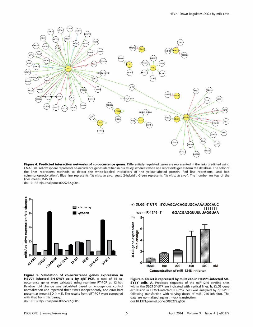
Figure 4. Predicted interaction networks of co-occurrence genes. Differentially regulated genes are represented in the links predicted usingCMAS 3.0. Yellow sphere represents co-occurrence genes identified in our study, whereas white one represents genes form the database. The color ofthe lines represents methods to detect the white-labeled interactors of the yellow-labeled protein. Red line represents ‘‘anti baitcoimmunoprecipitation’’. Blue line represents ‘‘in vitro; in vivo; yeast 2-hybrid’’. Green represents ‘‘in vitro; in vivo’’. The number on top of thelines means MAS ID.doi:10.1371/journal.pone.0095272.g004
Figure 5. Validation of co-occurrence genes expression inHEV71-infected SH-SY5Y cells by qRT-PCR. A total of 14 co-occurrence genes were validated using real-time RT-PCR at 12 hpi.Relative fold change was calculated based on endogenous controlnormalization and repeated three times independently, and error barspresent as mean6SD (n = 3). The results from qRT-PCR were comparedwith that from microarray.doi:10.1371/journal.pone.0095272.g005
Figure 6. DLG3 is repressed by miR1246 in HEV71-infected SH-SY5Y cells. A. Predicted sequence of the miR-1246 binding siteswithin the DLG3 39-UTR are indicated with vertical lines. b. DLG3 geneexpression in HEV71-infected SH-SY5Y cells was analyzed by qRT-PCRfollowing transfection with varying doses of miR-1246 inhibitor. Thedata are normalized against mock transfection.doi:10.1371/journal.pone.0095272.g006
HEV71 Down-Regulates DLG3 by miR-1246
PLOS ONE | www.plosone.org 6 April 2014 | Volume 9 | Issue 4 | e95272
miR-1246, while pGL3-DLG3-mut that destroyed the binding
sites and vector control were not affected by miR1246 (p,0.001).
Finally, to clarify the relationship between miR-1246 and DLG3
regulation in response to HEV71 infection, varying doses of miR-
1246 inhibitor were transfected to inhibit the induction of miR-
1246. The results showed that the suppression of DLG3 caused by
HEV71 infection was significantly eliminated by the transfection
of miR-1246 inhibitor in dose-dependent manner (Fig. 6B).
Together, all these results clearly demonstrated that the up-
regulation of miR-1246 in response to HEV71 infection inhibited
the expression of DLG3.
Discussion
Human SH-SY5Y cells are the third successive subclone of the
SK-N-SH (human neuroblastoma cell) cells [38–40], and these
cells have been used to study HEV71 neural cell tropism [41]. In
this study, the first miRNA profiling of SH-SY5Y cells infected
with HEV71 was performed. Sixty-nine miRNAs were differen-
tially regulated, suggesting that virus infection does alter the
miRNA expression profile in SH-SY5Y cells. On the one hand,
the HEV71-mediated miRNA expression was assessed in the SH-
SY5Y cell line, as well as in different cell types. Interestingly, one
of the 69 miRNAs, miR-1246, was expressed at significantly
higher levels in SH-SY5Y cells than in RD or Vero cells. These
results differ from a previous study that showed that miR-141
significantly increased in RD cells infected with enteroviruses [28].
It is not surprising because miRNAs play a major part in the
tissue-specific regulation of gene expression patterns [42,43], and
several miRNAs have been shown to modulate the tissue tropism
of a number of viruses from different families [44–47]. In addition,
the special role ofmiR-1246 has been studied, especially on cancer
and cystic fibrosis [48–51]. Thus, how miR-1246 interacts with
HEV71 is of highly interests. Recent studies have reported that
miRNAs play important roles in the host cellular response to viral
infections [18,21,52], which can be attributed to both antiviral
defenses and viral factors altering the cellular environment.
Moreover, cellular or viral miRNAs have been shown to be
involved in reciprocal interaction between the host cells and virus
[25,53]; for example, miR-122 treatment has been shown to
enhance hepatitis C virus (HCV) replication by targeting the 59-
UTR [54]. However, our results showed that inhibition of miR-
1246 did not affect HEV71 production at 6 and 12 hpi (Fig. S1).These results ruled out the possibility that up-regulation of miR-
1246 may facilitate or suppress viral replication.
Alterations of the miRNA profile underlie global cell and tissue
transcriptional changes during developmental processes and
senescence, as well as during neoplastic transformation [55,56].
Likewise, it has been shown that miRNAs are involved in the
pathogenesis of the neurodegenerative disorders in multiple
diseases, including AD and Parkinson’s disease [57,58]. According
to our results, HEV71 infection can disturb the expression of host
miRNAs. It is well known that miRNAs can regulate post-
transcriptional processes by binding to the 39-UTR of the target
transcript. miR-1246 has been shown to target the 39-UTR
sequence of the DYRK1A mRNA, resulting in a reduction in
DYRK1A levels [59]; however, in our study, sequence analysis
showed that this miRNA does not target the 39-UTR or 59-UTR
of HEV71, which is in agreement with virus growth experiments
(Fig. S1).
Precise identification of miRNA targets is necessary for the
functional characterization of individual miRNAs and for a better
understanding of complex human diseases. Gene expression
profiling has been used to improve the target prediction for the
identification of functional targets [60,61]. A miRNA-mRNA
regulatory module consists of a set of miRNAs and a set of their
targets. One miRNA can potentially regulate multiple mRNAs,
and the opposite is also possible [62]. Our results demonstrated
that the 39-UTR of DLG3 gene were a potential target of miR-
1246. DLG3 is the first XLMR gene and is linked directly to
NMDA receptor-mediated signaling and synaptic plasticity
[63,64]. Compared with the classical transcription factors, miR-
1246 directly targeted the 39-UTR of DLG3 mRNA (Fig. 7), andits expression was inversely correlated with DLG3 expression in
HEV71-infected SH-SY5Y cells (Fig. 6). In this study, we
combined results from gene chips, mRNA assay, computational
predictions, and dual luciferase assay to confirm miR-1246 directly
targets DLG3 in HEV71-infected neuroblastoma cells.
The predicted interaction networks of genes in Fig. 4 show
significant over representation of known mental disorder suscep-
tibility genes [65,66], supporting the increased power of the
network-based approach in identifying disease-relevant transcrip-
tional changes. DLG3 is a membrane-associated guanylate kinase
protein family (MAGUK) that is localized to the postsynaptic
density of excitatory synapses [67]. Synapse-associated proteins
are thought to have important functional roles within neuronal
cells [68,69]. Moreover, investigators reported that DLG3 was part
of a trafficking complex toward the synapse and was regulated by
its PPxY motifs, which bind to the domain of Nedd4 and Nedd4-2
E3 ubiquitin ligases in neurons [70,71]. DLG3 has been
demonstrated to control apical epithelial polarity and tight
junction formation and to contribute to neural induction in mouse
development [72]. Mutation of DLG3 leads to synaptic dysfunction
and learning and memory impairments [69,73].
Dysregulation of synaptic plasticity has been involved in a
variety of psychiatric disorders, neurological disorders, and in age-
related cognitive impairment. Previous studies have reported that
neurodevelopment and cognitive function may be affected by viral
encephalitis or by bacterial meningitis [74–76]. Recently, Chang et
al. showed that HEV71 infection with central nervous system
(CNS) involvement and cardiopulmonary failure may be associ-
Figure 7. The 39-UTR of DLG3 was the target of miR-1246.Reporter construct pLG3-DLG3-mut that destroyed the putative bindingsites (underlined) is shown in comparison with wild type pLG3-DLG3construct. The expression of pLG3-DLG3 was significantly decreased bymiR-1246 in comparison with vector control and pLG3-DLG3-mut(***p,0.001). Each transfection was performed in triplicate.doi:10.1371/journal.pone.0095272.g007
HEV71 Down-Regulates DLG3 by miR-1246
PLOS ONE | www.plosone.org 7 April 2014 | Volume 9 | Issue 4 | e95272
ated with neurologic sequelae, delayed neurodevelopment, and
reduced cognitive functioning [77]. Considering the presence of
HEV71 viral antigens in neurons [12,13], direct HEV71 invasion
and subsequent neuron damage may contribute to the main cause
of neurologic sequelae. Our results set up connection between the
neurologic sequelae caused by HEV71 infection and host miR-
1246 regulation in association with DLG3 gene.
In summary, this is the first report on whole-genome joint
mRNA and miRNA profile analysis from EV71-infected SH-
SY5Y cells. Among the 69 differentially expressed miRNAs, miR-
1246 was expressed at significantly higher levels. The HEV71-
mediated up-regulation of miR-1246 reduced the levels of DLG3
protein in SH-SY5Y cells. Together, these results indicate that
miR-1246 play a potential role in the neurological process and cell
death pathways by regulating DLG3 upon HEV71 infection in
human neuroblastoma cells.
Supporting Information
Figure S1 HEV71 virus replication was uncorrelated to miR-
1246 in SH-SY5Y cells.
(DOCX)
Table S1 Differentially expressed miRNAs in SH-SY5Y cells
infected and non-infected with HEV71 virus by microarray assay.
(DOCX)
Table S2 Up-regulated genes in SH-SY5Y cells infected with
HEV71 after transfection miR-1246 inhibitor by mRNA micro-
array assay.
(DOCX)
Table S3 Down-regulated genes in SH-SY5Y cells infected with
HEV71 after transfection miR-1246 inhibitor by mRNA micro-
array assay.
(DOCX)
Table S4 The predicted miRNA targets of miR-1246 by
Targetscan Human 6.0.
(DOCX)
Table S5 Co-occurrence genes and detection primers used for
qRT-PCR analysis.
(DOCX)
Author Contributions
Conceived and designed the experiments: CFQ LJX TJ. Performed the
29. Zheng Z, Ke X, Wang M, He S, Li Q, et al. (2013) Human microRNA hsa-
miR-296-5p suppresses Enterovirus 71 replication by targeting the viral genome.
J Virol.
30. Wen BP, Dai HJ, Yang YH, Zhuang Y, Sheng R (2013) MicroRNA-23b inhibits
enterovirus 71 replication through downregulation of EV71 VPl protein.
Intervirology 56: 195–200.
31. Xu LJ, Jiang T, Zhang FJ, Han JF, Liu J, et al. (2013) Global transcriptomic
analysis of human neuroblastoma cells in response to enterovirus type 71
infection. PLoS One 8: e65948.
32. Cao RY, Han JF, Jiang T, Tian X, Yu M, et al. (2011) In vitro and in vivo
characterization of a new enterovirus type 71-specific human intravenous
immunoglobulin manufactured from selected plasma donors. J Clin Virol 51:
246–249.
33. Han JF, Jiang T, Fan XL, Yang LM, Yu M, et al. (2012) Recombination of
human coxsackievirus B5 in hand, foot, and mouth disease patients, China.
Emerg Infect Dis 18: 351–353.
HEV71 Down-Regulates DLG3 by miR-1246
PLOS ONE | www.plosone.org 8 April 2014 | Volume 9 | Issue 4 | e95272
34. Ye Q, Li XF, Zhao H, Li SH, Deng YQ, et al. (2012) A single nucleotide
mutation in NS2A of Japanese encephalitis-live vaccine virus (SA14-14-2) ablatesNS1’ formation and contributes to attenuation. J Gen Virol 93: 1959–1964.
35. Han JF, Cao RY, Deng YQ, Tian X, Jiang T, et al. (2011) Antibody dependent
enhancement infection of enterovirus 71 in vitro and in vivo. Virol J 8: 106.36. Tusher VG, Tibshirani R, Chu G (2001) Significance analysis of microarrays
applied to the ionizing radiation response. Proc Natl Acad Sci U S A 98: 5116–5121.
37. Morin RD, O’Connor MD, Griffith M, Kuchenbauer F, Delaney A, et al. (2008)
Application of massively parallel sequencing to microRNA profiling anddiscovery in human embryonic stem cells. Genome Res 18: 610–621.
38. Ross RA, Spengler BA, Biedler JL (1983) Coordinate morphological andbiochemical interconversion of human neuroblastoma cells. J Natl Cancer Inst
71: 741–747.39. Huang SC, Chang CL, Wang PS, Tsai Y, Liu HS (2009) Enterovirus 71-induced
autophagy detected in vitro and in vivo promotes viral replication. J Med Virol
81: 1241–1252.40. Tung WH, Hsieh HL, Yang CM (2010) Enterovirus 71 induces COX-2
expression via MAPKs, NF-kappaB, and AP-1 in SK-N-SH cells: Role ofPGE(2) in viral replication. Cell Signal 22: 234–246.
41. Cordey S, Petty TJ, Schibler M, Martinez Y, Gerlach D, et al. (2012)
Identification of site-specific adaptations conferring increased neural cell tropismduring human enterovirus 71 infection. PLoS Pathog 8: e1002826.
43. Lagos-Quintana M, Rauhut R, Yalcin A, Meyer J, Lendeckel W, et al. (2002)Identification of tissue-specific microRNAs from mouse. Curr Biol 12: 735–739.
44. Barnes D, Kunitomi M, Vignuzzi M, Saksela K, Andino R (2008) Harnessing
endogenous miRNAs to control virus tissue tropism as a strategy for developingattenuated virus vaccines. Cell Host Microbe 4: 239–248.
45. Kelly EJ, Hadac EM, Greiner S, Russell SJ (2008) Engineering microRNAresponsiveness to decrease virus pathogenicity. Nat Med 14: 1278–1283.
46. Kelly EJ, Nace R, Barber GN, Russell SJ (2010) Attenuation of vesicular
stomatitis virus encephalitis through microRNA targeting. J Virol 84: 1550–1562.
47. Lauring AS, Jones JO, Andino R (2010) Rationalizing the development of liveattenuated virus vaccines. Nat Biotechnol 28: 573–579.
48. Cui FM, Li JX, Chen Q, Du HB, Zhang SY, et al. (2013) Radon-inducedalterations in micro-RNA expression profiles in transformed BEAS2B cells.
J Toxicol Environ Health A 76: 107–119.
49. Jones CI, Zabolotskaya MV, King AJ, Stewart HJ, Horne GA, et al. (2012)Identification of circulating microRNAs as diagnostic biomarkers for use in
multiple myeloma. Br J Cancer 107: 1987–1996.50. Gillen AE, Gosalia N, Leir SH, Harris A (2011) MicroRNA regulation of
expression of the cystic fibrosis transmembrane conductance regulator gene.
Biochem J 438: 25–32.51. Pigati L, Yaddanapudi SC, Iyengar R, Kim DJ, Hearn SA, et al. (2010) Selective
release of microRNA species from normal and malignant mammary epithelialcells. PLoS One 5: e13515.
58. Nunez-Iglesias J, Liu CC, Morgan TE, Finch CE, Zhou XJ (2010) Joint genome-wide profiling of miRNA and mRNA expression in Alzheimer’s disease cortex
reveals altered miRNA regulation. PLoS One 5: e8898.
59. Zhang Y, Liao JM, Zeng SX, Lu H (2011) p53 downregulates Down syndrome-
associated DYRK1A through miR-1246. EMBO Rep 12: 811–817.
60. Giraldez AJ, Mishima Y, Rihel J, Grocock RJ, Van Dongen S, et al. (2006)Zebrafish MiR-430 promotes deadenylation and clearance of maternal mRNAs.
Science 312: 75–79.
61. Wang X, El Naqa IM (2008) Prediction of both conserved and nonconserved
microRNA targets in animals. Bioinformatics 24: 325–332.
62. Shalgi R, Lieber D, Oren M, Pilpel Y (2007) Global and local architecture of themammalian microRNA-transcription factor regulatory network. PLoS Comput
Biol 3: e131.
63. Kuwahara H, Araki N, Makino K, Masuko N, Honda S, et al. (1999) A novel
NE-dlg/SAP102-associated protein, p51-nedasin, related to the amidohydrolasesuperfamily, interferes with the association between NE-dlg/SAP102 and N-
64. Lickert H, Van Campenhout CA (2012) Evolution of the Discs large gene family
provides new insights into the establishment of apical epithelial polarity and theetiology of mental retardation. Commun Integr Biol 5: 287–290.
65. Zhang B, Horvath S (2005) A general framework for weighted gene co-
expression network analysis. Stat Appl Genet Mol Biol 4: Article17.
66. Lang UE, Puls I, Muller DJ, Strutz-Seebohm N, Gallinat J (2007) Molecular
mechanisms of schizophrenia. Cell Physiol Biochem 20: 687–702.
67. Nishikawa T (2011) Analysis of free D-serine in mammals and its biologicalrelevance. J Chromatogr B Analyt Technol Biomed Life Sci 879: 3169–3183.
68. Katsanis N (2004) The oligogenic properties of Bardet-Biedl syndrome. Hum
Mol Genet 13 Spec No1: R65–71.
69. Katoh M (2006) Cross-talk of WNT and FGF signaling pathways at GSK3beta
to regulate beta-catenin and SNAIL signaling cascades. Cancer Biol Ther 5:1059–1064.
70. Sans N, Petralia RS, Wang YX, Blahos J, 2nd, Hell JW, et al. (2000) A
developmental change in NMDA receptor-associated proteins at hippocampal
synapses. J Neurosci 20: 1260–1271.
71. Fujita A, Kurachi Y (2000) SAP family proteins. Biochem Biophys Res Commun269: 1–6.
72. Tarpey P, Parnau J, Blow M, Woffendin H, Bignell G, et al. (2004) Mutations in
the DLG3 gene cause nonsyndromic X-linked mental retardation. Am J Hum
Genet 75: 318–324.
73. Huibregtse JM, Scheffner M, Beaudenon S, Howley PM (1995) A family ofproteins structurally and functionally related to the E6-AP ubiquitin-protein
ligase. Proc Natl Acad Sci U S A 92: 2563–2567.
74. Sans N, Prybylowski K, Petralia RS, Chang K, Wang YX, et al. (2003) NMDA
receptor trafficking through an interaction between PDZ proteins and theexocyst complex. Nat Cell Biol 5: 520–530.
75. Van Campenhout CA, Eitelhuber A, Gloeckner CJ, Giallonardo P, Gegg M, et
al. (2011) Dlg3 trafficking and apical tight junction formation is regulated bynedd4 and nedd4–2 e3 ubiquitin ligases. Dev Cell 21: 479–491.
76. Cuthbert PC, Stanford LE, Coba MP, Ainge JA, Fink AE, et al. (2007) Synapse-associated protein 102/dlgh3 couples the NMDA receptor to specific plasticity
pathways and learning strategies. J Neurosci 27: 2673–2682.
77. Baraff LJ, Lee SI, Schriger DL (1993) Outcomes of bacterial meningitis inchildren: a meta-analysis. Pediatr Infect Dis J 12: 389–394.
HEV71 Down-Regulates DLG3 by miR-1246
PLOS ONE | www.plosone.org 9 April 2014 | Volume 9 | Issue 4 | e95272


![Cloning and Functional Characterization of a Novel ... · sodium pyruvate, 100 U/ml penicillin, 0.1 mg/ml streptomycin, 0.2 mg/ml gentamycin]. The oocytes were defolliculated enzymatically](https://static.documents.pub/doc/80x56/5fb1ef49560727203112a519/cloning-and-functional-characterization-of-a-novel-sodium-pyruvate-100-uml.jpg)


