Parathyroid hormone receptor signaling induces boneresorption in the adult skeleton by directly regulatingthe RANKL gene in osteocytes
Abdullah N. Ben-awadh,1, Jesus Delgado-Calle,1,3, Xiaolin Tu,1,Kali Kuhlenschmidt,1, Matthew R. Allen,1, Lilian I. Plotkin,1,3, Teresita Bellido*1,2,3
1Department of Anatomy and Cell Biology, 2Department of Medicine, Division of Endocrinology, INUniversity School of Medicine, Indianapolis, IN, USA, 3Roudebush Veterans Administration MedicalCenter, Indianapolis, IN, USA
PTH upregulates RANKL in cells of the osteoblastic lineage, but the precise differentiation stage ofthe PTH target cell responsible for RANKL-mediated stimulation of bone resorption remains un-defined. We report that constitutive activation of PTH receptor signaling only in osteocytes intransgenic mice (DMP1-caPTHR1) was sufficient to increase RANKL expression and bone resorption.Resorption in DMP1-caPTHR1 mice crossed with mice lacking the distal control region regulated byPTH in the RANKL gene (DCR-/-) was similar to DMP1-caPTHR1 mice at 1 month of age, but pro-gressively declined to reach values undistinguishable from wild type (WT) mice at 5 months of age.Moreover, DMP1-caPTHR1 mice exhibited low tissue material density and increased serum alkalinephosphatase activity at 5 month of age, and these indices of high remodeling were partially andtotally corrected in compound DMP1-caPTHR1;DCR-/- male mice, and less affected in female mice.RANKL expression in bones from DMP1-caPTHR1 mice was elevated at both 1 and 5 months of age;whereas it was high, similar to DMP1-caPTHR1 mice at 1 month, but low, similar to WT levels at 5months in compound mice. Moreover, PTH increased RANKL and decreased Sost and OPG expres-sion in ex vivo bone organ cultures established from WT mice, but only regulated Sost and OPGexpression in cultures from DCR-/- mice. PTH also increased RANKL expression in osteocyte-con-taining primary cultures of calvarial cells, in isolated murine osteocytes, and in WT but not in DCR-/-
osteocyte-enriched bones. Thus, PTH upregulates RANKL expression in osteocytes in vitro, ex vivoand in vivo, and resorption induced by PTH receptor signaling in the adult skeleton requires directregulation of the RANKL gene in osteocytes.
Accumulating evidence demonstrates that some of ac-tions of PTH on the skeleton are mediated by direct
effects of the hormone on osteocytes, the most abundantand highly communicated cells in bone (1). PTH down-regulates the expression of the Sost gene, which encodesthe potent inhibitor of bone formation sclerostin, ex-pressed by osteocytes in bone (2, 3); and also increases theexpression of FGF23, a hormone expressed in osteocytes(and osteoblasts) that regulates phosphate reabsorption inthe kidney, contributing to mineral homeostasis (1, 4, 5).
Earlier studies have shown that the major skeletal ef-fects of PTH are recapitulated in transgenic mice express-
ing a constitutively active PTH receptor in osteocytes,named DMP1-caPTHR1 mice (1, 4, 6, 7). Bones fromthese transgenic mice exhibit decreased Sost/sclerostin ex-pression and elevated Wnt signaling, increased osteoblastsand bone formation, and a remarkable increase in bonemineral density (BMD) in both the axial and appendicularskeletons (6, 7). DMP1-caPTHR1 mice also exhibit highbone resorption, as evidenced by elevated resorptionmarkers in the blood and urine, high number of osteoclastsin bone, and elevated cortical porosity. Bones fromDMP1-caPTHR1 mice display an increased RANKL/OPG ratio and elevated M-CSF expression. The elevated
The Endocrine Society. Downloaded from press.endocrine.org by [${individualUser.displayName}] on 10 July 2014. at 01:46 For personal use only. No other uses without permission. . All rights reserved.
bone formation and increased bone mass in DMP1-caP-THR1 mice are abolished by overexpressing Sost in os-teocytes, demonstrating that the increase in osteoblasts isdue to reduced sclerostin expression unleashing Wnt sig-naling (6, 7). On the other hand, increased osteoclast num-bers and resorption are not eliminated by Sost overexpres-sion. Taken together, these findings demonstrate that PTHreceptor signaling in osteocytes regulates bone formationand resorption by distinct mechanisms.
The pro-osteoclastogenic actions of PTH are long rec-ognized to be associated with up-regulation of RANKLand down-regulation of OPG. However, the precise dif-ferentiation stage of the PTH target cell that supports os-teoclastogenesis remains undefined. Osteocytes expresshigh levels of RANKL and OPG compared to other os-teoblastic cells (8–10); and also express M-CSF (11, 12).In addition, deletion of the RANKL gene from osteocytesleads to osteopetrosis in mice (9, 10). These pieces of ev-idence raise the possibility that elevated resorption drivenby PTH receptor activation in osteocytes could result fromdirect regulation of osteoclastogenic genes in these cells.By taking advantage of the genetic model of osteocyte-driven bone resorption induced by PTH receptor signaling(DMP1-caPTHR1 mice), we show in this study that re-moval of the distal control region (DCR) of the RANKLgene regulated by PTH gradually corrects the increasedresorption exhibited by DMP1-caPTHR1 mice, andblunts the high RANKL levels in bone. In addition, PTHupregulated RANKL expression in authentic osteocytes,in primary cultures of bone cells containing osteocytes,and in bone organ cultures established from WT mice;however, it fail to do so in cultures established from DCR-/-
mice. These data show that PTH receptor signaling in os-teocytes enhances resorption by regulating the expressionof the RANKL gene directly in osteocytes.
Materials and Methods
Generation of experimental miceExperimental animals were generated by a two-step breeding
strategy using transgenic mice expressing a constitutively activePTHR1 in osteocytes under the control of the 8kb fragment ofthe promoter of the dentin matrix protein 1 gene (DMP1-caP-THR1) (6) and knock in mice lacking the distant transcriptionalenhancer region in the RANKL gene (DCR-/-) (13). Heterozy-gous DMP1-caPTHR1 mice were crossed with DCR-/� and theresulting DMP1-caPTHR1;DCR-/� were subsequently crossedwith DCR-/� mice. Littermates of the four generated genotypeswere used for the experiments: wild type (WT), DMP1-caP-THR1, DCR-/-, and compound DMP1-caPTHR1;DCR-/- mice.All mice were born at the expected Mendelian frequency, werefertile, and exhibited normal size and weight. Cohorts of maleand female mice composed of 9–16 mice per genotype and gen-
der were used. Mice expressing green fluorescent protein (GFP)in osteocytes (DMP1-GFP) were provided by Dr. David W.Rowe and were maintained as homozygous (14). Mice were feda regular diet (Harlan/Teklad, Indianapolis, IN, USA), had adlibitum water, and were maintained on a 12 hour light/dark cycle(7). All animal protocols were approved by the Institutional An-imal Care and Use Committee at Indiana University School ofMedicine.
Quantification of bone turnover markersBlood was collected monthly by facial vein bleeding into hep-
arinized tubes and plasma was separated from blood cells bycentrifugation at 1500 g for 10 minutes. C-telopeptide fragmentsof type I collagen (CTX) were measured using an enzyme-linkedimmunosorbent assay (ELISA) (RatlapsTM EIA, Immunodiag-nostic Systems, Fountain Hills, AZ, USA). Alkaline Phosphatasewas measured using the AMP Buffer (Randox #AP 3802)method on a Randox Daytona analyzer (Randox LaboratoriesLimited, Crumlin, Country Antrim, United Kingdom) (7).
Bone mineral density (BMD) and microcomputedtomography (micro-CT) measurements
Total, femoral and spine BMD was determined by dual en-ergy x-ray absorptiometry (DXA) using a PIXImus II densitom-eter (G.E. Medical Systems, Lunar Division, Madison, WI), aspreviously described (6). Mice were anesthetized via inhalationof 2.5% isoflurane (IsoFlo; Abbott laboratories, North Chicago,IL) mixed with O2 (1.5 L/min). For micro-CT analysis, boneswere dissected, cleaned of soft tissue, stored in 70% ethanol, andscanned at 6 micron resolution (Skyscan 1172, SkyScan, Kon-tich, Belgium), as previously described (6, 7).
Bone formation rate (BFR) and TRAPase stainingMice were injected with calcein and alizarin red to allow for
dynamic histomorphometric measurements, as previously pub-lished (7). Femora were fixed in neutral buffer formalin and cutat the midshaft. The distal half of the femur was embedded inmethyl metacrylate and unstained sections were analyzed usingthe OsteoMeasure High Resolution Digital Video System (Os-teoMetrics, Inc.) attached to an Olympus BX51TRF microscope(Olympus America Inc.) Only measurements on the endocorticalsurface were done since by 5 month of age, the action of PTHreceptor signaling on the periosteal surface had subsided and thefluorochrome labels on cancellous bone are very intricate pre-cluding accurate measurement of BFR. The proximal half of thefemur was decalcified, sections stained for TRAPase and coun-terstained with toluidine blue to visualize osteoclasts, as previ-ously described (15). Sections were viewed on a Leitz DMRXEmicroscope (Leica Mikroskopie und System GmbH). Imageswere captured using a SPOT digital camera (Diagnostic Instru-ments, Inc.).
Ex vivo bone organ culture and in vitro cell cultureCalvarial bones, femur and tibia were harvested from
5-month old DCR-/- mice and littermate WT controls, main-tained in alpha MEM containing 10% FBS for 24 hours. Forosteocyte-enriched bone preparations, tibiae were harvestedfrom 5-month old mice, bone marrow cells were flushed out andbones were subjected to serial digestions with collagenase andEDTA, as previously published (16). Sost expression was 1.5-
2 Osteocytic RANKL and PTH receptor signaling Endocrinology
The Endocrine Society. Downloaded from press.endocrine.org by [${individualUser.displayName}] on 10 July 2014. at 01:46 For personal use only. No other uses without permission. . All rights reserved.
and 2.4-fold higher in digested WT and DCR-/- bones, respec-tively, demonstrating osteocyte enrichment. Bones were treatedwith PTH (100 nM) or vehicle for 4 hours and mRNA was thenisolated. Calvarial cells obtained from 1 month-old C57BL/6mice were cultured to reach confluence and then media withascorbic acid (50 �g/ml) was added for 5–6 days, changing halfof the medium every 2–3 days, as previously described (2). Cellswere then treated with PTH (100 nM), dibutyryl cAMP (DBA; 1mM) or vehicle for 4 hours. Osteoblasts and osteocytes wereisolated from calvarial bones of 9–11 day-old DMP1-GFP micein which osteocytes are labeled with GFP, as previously pub-lished (6, 14). Calvarial bones were subjected to 9 sequential20-minute digestions with a mixture of Trypsin/EDTA/collage-nase P (17). The first digestion was discarded and cells from allother digestions were pulled and immediately subjected to flu-orescence activated cell sorting (FACS), as previously reported(6, 18). GFP positive (osteocytes) and GFP negative (osteoblasts)cells were cultured separately on plates coated with collagen I asdescribed earlier (19, 20) for 48 hours, and treated with PTH(100 nM) or vehicle for 4 hours.
Gene expression analysis by quantitative PCR(qPCR)
Calvariae from 1 or 5 month old mice were cleaned and frozenimmediately in liquid nitrogen, and stored at -80C until RNAisolation. Cutured bones or cells were rinsed with with PBS andRNA immediately isolated. Total RNA was purified using Trizolreagent (Invitrogen, Foster City, CA, USA) according to the man-ufacturer’s instructions. Gene expression was analyzed by qPCRas previously described using primer probe sets from AppliedBiosystems (Foster City, CA, USA) or from Roche Applied Sci-ence (Indianapolis, IN, USA) (6, 7). Relative mRNA expressionlevels were normalized to the house-keeping genes ribosomalprotein S2 (ChoB) or glyceraldehyde-3-phosphate dehydroge-nase (GAPDH) using the �Ct method.
Statistical analysisData were analyzed using SigmaStat (SPSS Science, Chicago,
IL). Differences between group means were evaluated using two-way ANOVA, followed by pair wise multiple comparisons usingStudent-Newman-Keuls method. All values are reported as themean � standard deviations (SD).
Results
Deletion of the DCR gradually corrects highresorption and remodeling driven by PTH receptorsignaling in osteocytes
To investigate whether direct regulation of the RANKLgene by PTH in osteocytes is required to stimulate boneresorption, we examined resorption in DMP1-caPTHR1mice crossed with knock-in mice lacking the distal controlregion (DCR) of the RANKL gene regulated by PTH. Lon-gitudinal analysis of circulating CTX in male mice showedthe expected high resorption in growing mice of all geno-types that progressively decreased to plateau at 3–5 monthof age (Figure 1A). Resorption was significantly higher
(approximately 100%) in DMP1-caPTHR1 mice andnonsignificantly lower (between 15 and 30%) in DCR-/-
mice, vs WT littermates across all ages. CTX in compoundDMP1-caPTHR1;DCR-/- mice was similar to DMP1-caP-THR1 mice at 1 and 2 months of age, but deletion of theDCR gradually corrected the increased resorption exhib-ited by DMP1-caPTHR1 mice. By 3 months of age, CTXin compound mice was significantly lower compared toDMP1-caPTHR1 mice (and 50% higher than WT), and by5 months, it was undistinguishable from WT mice. Al-though accurate measurements of osteoclast numbers forDMP1-caPTHR1 mice at this age are not possible,TRAPase staining of crosssections at the femoral midshaftshow that the abundant TRAPase staining exhibited byDMP1-caPTHR1 bones is decreased in compound micelacking the DCR (Figure 1B).
Similar results were found in the longitudinal analysisof CTX for female mice. Resorption was high at the firsttwo months for all genotypes (Figure 1C). As found formale mice, resorption in DMP1-caPTHR1 female micewas about 70%–100% higher compared to WT litter-mates and it showed a nonsignificant decrease (7%–13%)in DCR-/- mice compared to WT at all ages. DMP1-caP-THR1; DCR-/- female mice had similar CTX levels toDMP1-caPTHR1 mice for the first two months. But at 3months DMP1-caPTHR1; DCR-/- female mice showed asignificant decrease in CTX compared to DMP1-caP-THR1 mice. CTX remained significantly lower in com-pound mice at 4 and 5 months of age; but, in contrast tomale mice, it never reached WT levels. Thus, in femalemice, the absence of DCR reduced but did not completelynormalize the high resorption induced by the DMP1-caP-THR1 transgene.
DCR deletion did not affect the high BMD exhibited bymale or female DMP1-caPTHR1 mice. Thus, longitudinalBMD analysis revealed no significant changes in total,femoral, or spinal BMD in compound DMP1-caPTHR1;DCR-/- compared to DMP1-caPTHR1 littermates male orfemale mice (Figure 2A and B). However, micro-CT anal-ysis of femoral bone revealed decreased bone volume incompound male mice compared to DMP1-caPTHR1 lit-termates (Table 1 and Figure 3A). Both bone volume in thedistal femur (BV/TV) and in the femoral midshaft (BA/TA)was lower in DMP-caPTHR1 mice with deleted DCR.Dynamic histomorphometric analysis showed that the de-creased bone volume in the compound mice was accom-panied by reduced bone formation rate (BFR) on the en-docortical surface of the femur compared to single DMP1-caPTHR1 transgenic mice (Table 1 and Figure 3B). Thiseffect was due to combined reduction in the numbers ofosteoblasts covering bone surfaces (MS/BS) and in the ac-tivity of osteoblasts teams (MAR). Moreover, although
doi: 10.1210/en.2014-1046 endo.endojournals.org 3
The Endocrine Society. Downloaded from press.endocrine.org by [${individualUser.displayName}] on 10 July 2014. at 01:46 For personal use only. No other uses without permission. . All rights reserved.
intracortical bone formation could not be quantified alsodue to the convoluted fluorochrome incorporation, dele-tion of the DCR markedly decreased the amount and in-tensity of fluorescent labels (Figure 3B).
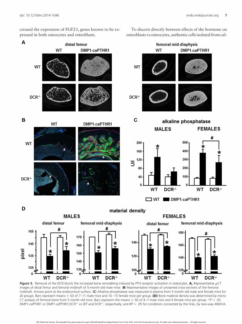
Consistent with these effects on CTX and BFR, circu-lating levels of the osteoblast marker alkaline phosphatasewere elevated in 5 month old DMP1-caPTHR1 male miceand were reduced to WT levels in compound DMP1-caP-THR1;DCR-/- male mice (Figure 3C). A reduction in al-
kaline phosphatase was also found in female mice, al-though, similar to CTX levels, alkaline phosphatase levelsin compound female mice did not reach WT values (Figure3C). In addition, DMP1-caPTHR1 bones exhibited lowermaterial density, an index of tissue mineralization thattypically is reduced in conditions of high bone remodeling,and this effect was partially corrected in DMP1-caPTHR1male mice lacking the DCR (Figure 3D). Consistent withthe findings for CTX and alkaline phosphatase, the de-
Figure 1. Deletion of the DCR progressively corrects the high resorption induced by PTH receptor signaling in osteocytes in male and female mice.(A) Longitudinal analysis of CTX for the male cohort from 1 to 5 months. Symbols represent the means � SD of 7–16 mice per group. #P � .05DMP1-caPTHR1 vs DMP1-caPTHR1;DCR-/-.CTX levels at 1 month and 5 month. Bars represent means � SD of 7–16 mice per group. *P � .05DMP1-caPTHR1 or DMP1-caPTHR1;DCR-/- vs WT and DCR-/-, respectively, by two-way ANOVA. (B) Representative images of cross-sections of thefemoral midshaft of 5-month-old males stained for TRAPase/toluidine blue. Bars indicate 50 �m. (C) Longitudinal analysis of CTX for the femalecohort from 1 to 5 months. Symbols represent the means � SD of 7–16 mice per group. # P � .05 DMP1-caPTHR1 vs DMP1-caPTHR1;DCR-/-. CTXlevels at 1 month and 5 month. Bars represent means � SD of 7–16 mice per group. * P � .05 DMP1-caPTHR1 or DMP1-caPTHR1;DCR-/- vs WTand DCR-/-, respectively; and # P � .05 for conditions connected by the lines, by two-way ANOVA.
4 Osteocytic RANKL and PTH receptor signaling Endocrinology
The Endocrine Society. Downloaded from press.endocrine.org by [${individualUser.displayName}] on 10 July 2014. at 01:46 For personal use only. No other uses without permission. . All rights reserved.
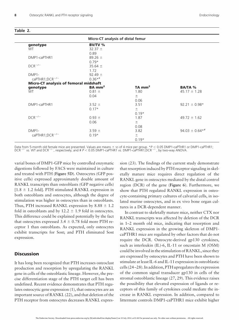
crease in bone material density exhibited by DMP1-caP-THR1 mice was also prevented to a lesser extent in femalethan in male mice. Further, micro-CT analysis demon-strate that deletion of the DCR in DMP1-caPTHR1 femalemice does not reduce bone volume in either the distal fe-mur or the femoral midshaft (Table 2), confirming that theeffect of the DCR deletion is weaker in females. Indeed,bone volume at both sites is increased in female compoundmice, suggesting that a mild reduction in resorption in-duced by the DCR deletion is not sufficient to inhibit boneformation coupled to resorption and bone is preserved.
PTH receptor signaling upregulates RANKLexpression in osteocytes in vivo, ex vivo and invitro
The increased resorption exhibited by DMP1-caP-THR1 male mice was accompanied by high RANKLmRNA in bone at 1 and 5 months of age (Figure 4A). Incompound DMP1-caPTHR1;DCR-/- mice, RANKL ex-pression closely matched the described effects on boneresorption. Thus, RANKL expression levels were similarto DMP1-caPTHR1 mice at 1 month of age and reducedto WT levels at 5 months. Similar pattern of expressionwas observed for M-CSF (Figure 4B), a survival factor forosteoclast progenitors and mature osteoclasts previously
Figure 2. Removal of the DCR does not affect the increase in BMD exhibited by DMP1-caPTHR1 mice. Longitudinal analysis of total, spinal, andfemoral BMD in both males (A) and females (B) at 1 month intervals up to 5 months of age. Symbols are means � standard deviations. N � 7–19mice per group. No significant differences were detected among phenotypes.
doi: 10.1210/en.2014-1046 endo.endojournals.org 5
The Endocrine Society. Downloaded from press.endocrine.org by [${individualUser.displayName}] on 10 July 2014. at 01:46 For personal use only. No other uses without permission. . All rights reserved.
shown to be upregulated by RANKL in cultured RAW264.7 macrophage cells (21). In addition, the increasedexpression in bone of TRAPase and cathepsin K, genesexpressed in osteoclasts, exhibited by DMP1-caPTHR1mice was decreased to WT levels in compound DMP1-caPTHR1;DCR-/- male mice (Figures 4C). Similar regula-tion of RANKL, M-CSF, TRAPase, and cathepsin K wasfound in bones from 5 month old female mice of the fourgenotypes (Figure 4D). These results demonstrate thatPTH receptor signaling in osteocytes regulates RANKL, aswell as M-CSF and osteoclastic genes, through the DCR invivo.
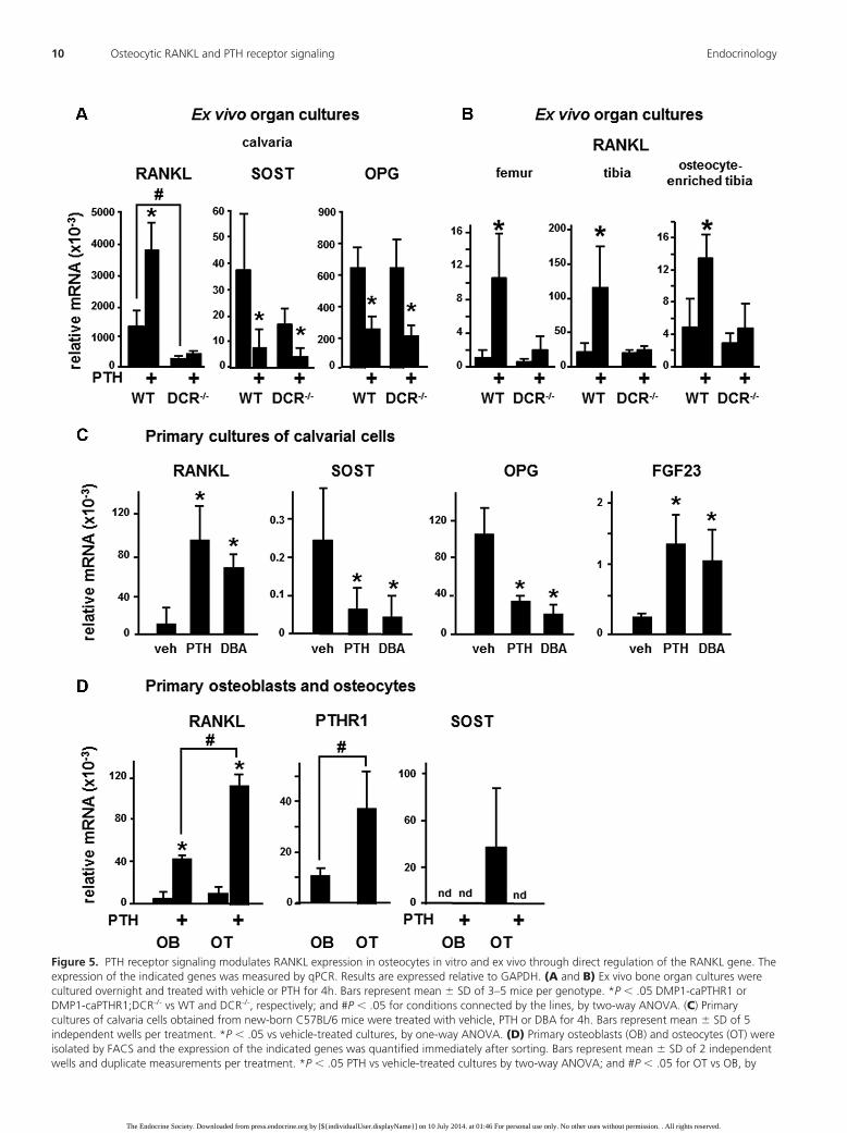
Deletion of the DCR also blunted the increase inRANKL expression induced by PTH ex vivo (Figure 5Aand B). Thus, PTH increased RANKL and decreased Sostand OPG expression in calvaria organ cultures establishedfrom WT mice. In contrast, the upregulation of theRANKL gene was eliminated in bones from DCR-/- mice,whereas Sost and OPG expression was regulated by PTH
the same extent in DCR-/- and WT bones. Similar regula-tion of RANKL expression by PTH in WT but not inDCR-/- mice was observed in organ cultures of femur ortibia (Figure 5B). Further, PTH increased RANKL expres-sion in organ cultures established from tibial bones en-riched in osteocytes from WT but not from DCR-/- mice,demonstrating that PTH upregulates RANKL expressionin osteocytes ex vivo, through the DCR.
PTH also increased RANKL expression in differenti-ating primary cultures of mouse calvarial cells (Figure 5C).Similar increase was induced by addition of the stable an-alog of cAMP dibutyryl cAMP (DBA). We previouslydemonstrated that these cultures contain osteocytes, asevidenced by a time-dependent increase in Sost transcriptsand the presence of sclerostin positive cells (2). Consistentwith these previous findings, Sost mRNA was readily de-tectable after 6 days of culture, and addition of PTH orDBA for 4 hours markedly decreased Sost expression.PTH and DBA also decreased expression of OPG and in-
Table 1.
Micro-CT analysis of distal femur
genotype BV/TV %WT 27.37 �
3.05DMP1-caPTHR1 58.37 �
10.37*DCR�/� 31.01 �
0.48DMP1-
caPTHR1;DCR�/�46.07 �10.86*#
Micro-CT analysis of femoral midshaftgenotype BA mm2 TA mm2 BA/TA %WT 0.85 �
0.061.96 �0.22
43.34 � 3.68
DMP1-caPTHR1 2.52 �0.28*
3.03 �0.15*
83.29 � 6.69*
DCR�/� 0.91 �0.06
2.04 �0.22
44.88 � 1.98
DMP1-caPTHR1;DCR�/�
2.55 �0.63*
3.38 �0.39*
75.04 � 12.08*#
Dynamic histomorphometric analysis of endocortical bone at the femoral midshaftgenotype Ec. MAR
�m/dayEc. MS/BS%
Ec. BFR �m3/�m2/day
WT 0.75 �0.28
57.89�12.52
0.44 � 0.22
DMP1-caPTHR1 1.03 �0.25*
53.62�10.77
0.57 � 0.25
DCR�/� 0.68 �0.26
48.34 � 7.56 0.33 � 0.16
DMP1-caPTHR1;DCR�/�
0.52 �0.21#
37.95�7.75*#
0.23 � 0.10#
Data from 5-month old male mice are presented. Values are means � SD of 4–10 mice per group. *P � 0.05 DMP1-caPTHR1 or DMP1-caPTHR1;DCR�/� vs. WT and DCR�/�, respectively; and # P � 0.05 DMP1-caPTHR1 vs. DMP1-caPTHR1;DCR�/�, by two-way ANOVA.
6 Osteocytic RANKL and PTH receptor signaling Endocrinology
The Endocrine Society. Downloaded from press.endocrine.org by [${individualUser.displayName}] on 10 July 2014. at 01:46 For personal use only. No other uses without permission. . All rights reserved.
creased the expression of FGF23, genes known to be ex-pressed in both osteocytes and osteoblasts.
To discern directly between effects of the hormone onosteoblasts vs osteocytes, authentic cells isolated from cal-
Figure 3. Removal of the DCR blunts the increased bone remodeling induced by PTH receptor activation in osteocytes. A, Representative �CTimages of distal femur and femoral midshaft of 5-month-old male mice. (B) Representative images of unstained cross-sections of the femoralmidshaft. Arrows point at the endocortical surface. (C) Alkaline phosphatase was measured in plasma from 5 month old male and female mice forall groups. Bars represent means � SD of 7–11 male mice and 10–15 female mice per group. (D) Bone material density was determined by micro-CT analysis of femoral bone from 5 month old mice. Bars represent the means � SD of 4–7 male mice and 4 female mice per group. *P � .05DMP1-caPTHR1 or DMP1-caPTHR1;DCR-/- vs WT and DCR-/-, respectively; and #P � .05 for conditions connected by the lines, by two-way ANOVA.
doi: 10.1210/en.2014-1046 endo.endojournals.org 7
The Endocrine Society. Downloaded from press.endocrine.org by [${individualUser.displayName}] on 10 July 2014. at 01:46 For personal use only. No other uses without permission. . All rights reserved.
varial bones of DMP1-GFP mice by controlled enzymaticdigestions followed by FACS were maintained in cultureand treated with PTH (Figure 5D). Osteocytes (GFP pos-itive cells) expressed approximately double amount ofRANKL transcripts than osteoblasts (GFP negative cells)[1.8 � 1.2 fold]. PTH stimulated RANKL expression inboth osteoblasts and osteocytes, although the degree ofstimulation was higher in osteocytes than in osteoblasts.Thus, PTH increased RANKL expression by 8.88 � 1.2fold in osteoblasts and by 12.2 � 1.9 fold in osteocytes.This difference could be explained potentially by the factthat osteocytes expressed 3.4 � 0.78 fold more PTH re-ceptor 1 than osteoblasts. As expected, only osteocytesexhibit transcripts for Sost; and PTH eliminated Sostexpression.
Discussion
It has long been recognized that PTH increases osteoclastproduction and resorption by upregulating the RANKLgene in cells of the osteoblastic lineage. However, the pre-cise differentiation stage of the PTH target cell has beenundefined. Recent evidence demonstrates that PTH regu-lates osteocytic gene expression (1), that osteocytes are animportant source of RANKL (22), and that deletion of thePTH receptor from osteocytes decreases RANKL expres-
sion (23). The findings of the current study demonstratethat resorption induced by PTH receptor signaling in skel-etally mature mice requires direct regulation of theRANKL gene in osteocytes mediated by the distal controlregion (DCR) of the gene (Figure 6). Furthermore, weshow that PTH regulated RANKL expression in osteo-cyte-containing primary cultures of calvarial cells, in iso-lated murine osteocytes, and in ex vivo bone organ cul-tures in a DCR-dependent manner.
In contrast to skeletally mature mice, neither CTX norRANKL transcripts was affected by deletion of the DCRin 1–2 month old mice, indicating that resorption andRANKL expression in the growing skeleton of DMP1-caPTHR1 mice are regulated by other factors that do notrequire the DCR. Osteocyte-derived gp130 cytokines,such as interleukin (IL)-6, IL-11 or oncostain M (OSM)could be involved in the stimulation of RANKL, since theyare expressed by osteocytes and PTH have been shown tostimulate at least IL-6 and IL-11 expression in osteoblasticcells (24–28). In addition, PTH upregulates the expressionof the common signal transducer gp130 in cells of thestromal osteoblastic lineage (27, 29). This evidence raisesthe possibility that elevated expression of ligands or re-ceptors of this family of cytokines could mediate the in-crease in RANKL expression. In addition, compared tolittermate controls DMP1-caPTHR1 mice exhibit higher
Table 2.
Micro-CT analysis of distal femur
genotype BV/TV %WT 32.37 �
0.89DMP1-caPTHR1 89.26 �
0.79*DCR�/� 35.64 �
1.72DMP1-
caPTHR1;DCR�/�92.49 �0.36*#
Micro-CT analysis of femoral midshaftgenotype BA mm2 TA mm2 BA/TA %WT 0.81 �
0.041.80�0.06
45.17 � 1.28
DMP1-caPTHR1 3.52 �0.17*
3.51�0.19*
92.21 � 0.98*
DCR�/� 0.93 �0.06
1.87�0.08
49.72 � 1.62
DMP1-caPTHR1;DCR�/�
3.59 �0.19*
3.82�0.19*
94.03 � 0.64*#
Data from 5-month old female mice are presented. Values are means � SD of 4 mice per group. *P � 0.05 DMP1-caPTHR1 or DMP1-caPTHR1;DCR�/� vs. WT and DCR�/�, respectively; and # P � 0.05 DMP1-caPTHR1 vs. DMP1-caPTHR1;DCR�/�, by two-way ANOVA.
8 Osteocytic RANKL and PTH receptor signaling Endocrinology
The Endocrine Society. Downloaded from press.endocrine.org by [${individualUser.displayName}] on 10 July 2014. at 01:46 For personal use only. No other uses without permission. . All rights reserved.
circulating levels of 1,25-dihydroxy-Vitamin D3 (4, 6),another recognized stimulator of RANKL genetranscription.
Most of the circulating products of collagen degrada-tion (CTX) in growing mice (like our 1 month old) areexpected to derive from osteoclast activity in cortical
Figure 4. Deletion of the DCR corrects the elevated expression of RANKL and M-CSF in adult, but not in young DMP1-caPTHR1 mice. (A-C) qPCRanalysis of mRNA transcripts in calvaria from 1 and 5 month old male mice. Bars represent means � SD of 3–5 mice per group. (D) geneexpression in calvaria from 5 month old female mice. Bars represent means � SD of 5 mice per group. Results are expressed relative to CHO B.*P � .05 DMP1-caPTHR1 or DMP1-caPTHR1;DCR-/- vs WT and DCR-/-, respectively, by two-way ANOVA.
doi: 10.1210/en.2014-1046 endo.endojournals.org 9
The Endocrine Society. Downloaded from press.endocrine.org by [${individualUser.displayName}] on 10 July 2014. at 01:46 For personal use only. No other uses without permission. . All rights reserved.
Figure 5. PTH receptor signaling modulates RANKL expression in osteocytes in vitro and ex vivo through direct regulation of the RANKL gene. Theexpression of the indicated genes was measured by qPCR. Results are expressed relative to GAPDH. (A and B) Ex vivo bone organ cultures werecultured overnight and treated with vehicle or PTH for 4h. Bars represent mean � SD of 3–5 mice per genotype. *P � .05 DMP1-caPTHR1 orDMP1-caPTHR1;DCR-/- vs WT and DCR-/-, respectively; and #P � .05 for conditions connected by the lines, by two-way ANOVA. (C) Primarycultures of calvaria cells obtained from new-born C57BL/6 mice were treated with vehicle, PTH or DBA for 4h. Bars represent mean � SD of 5independent wells per treatment. *P � .05 vs vehicle-treated cultures, by one-way ANOVA. (D) Primary osteoblasts (OB) and osteocytes (OT) wereisolated by FACS and the expression of the indicated genes was quantified immediately after sorting. Bars represent mean � SD of 2 independentwells and duplicate measurements per treatment. *P � .05 PTH vs vehicle-treated cultures by two-way ANOVA; and #P � .05 for OT vs OB, by
10 Osteocytic RANKL and PTH receptor signaling Endocrinology
The Endocrine Society. Downloaded from press.endocrine.org by [${individualUser.displayName}] on 10 July 2014. at 01:46 For personal use only. No other uses without permission. . All rights reserved.
bone, the so-called resorptive modeling (30). In contrast,CTX in adult mice (like our 5 month old) probably rep-resent osteoclast activity involved in bone remodeling, towhich cancellous bone is a major contributor. The basalphenotype of DCR-/- mice is manifested in adult animals,which exhibit reduced bone remodeling and a modest in-crease in bone mass confined to the cancellous bone com-partment (31). Together with this earlier evidence, ourfindings showing an age-dependent effect of the DCR de-letion on DMP1-caPTHR1 mice suggest that PTH recep-tor signaling in osteocytes drives resorptive modeling inthe growing skeleton mainly through DCR-independentstimulators of RANKL expression, such as IL-6 type cy-tokines; whereas it stimulates resorption in cancellousbone in the adult skeleton via DCR-dependent mecha-nisms. Future studies are required to identify the DCR-independent mechanisms and bone compartments in-volved in resorption-driven by PTH receptor signaling inthe growing skeleton.
Remarkably, in contrast with our findings showing thatRANKL is equally stimulated in 1-month old DMP1-caP-THR1 mice regardless of whether DCR is deleted or not,earlier studies showed that endogenous elevation of PTHinduced by calcium deficiency does not increase RANKLexpression in growing mice lacking the DCR (32). Theapparent discrepancy could be explained by the differentways of activating PTH receptor signaling. In the paper byO’Brien and colleagues endogenous PTH is elevated sys-temically, thus affecting all PTH responsive cells; whereasin the current study the PTH receptor is activated only inosteocytes. Taken together, these findings suggest thatPTH actions in cells other than osteocytes are responsiblefor RANKL increase with systemic elevation of PTH ingrowing mice. In contrast, similar to our study, RANKLexpression induced by endogenous PTH elevation was notincreased in adult mice lacking the DCR, suggesting thatosteocytes are the major contributors to RANKL regula-tion by PTH in the mature skeleton.
We have previously shown that DMP1-caPTHR1 miceexhibit a consistent decrease in material density of about10% compared to WT littermates (6, 33). This index mea-sured by micro-CT is indicative of tissue mineralizationand age of the mineral. Reduced values of bone materialdensity reveal accumulation of undermineralized bone,which could result from increased bone remodeling ratethat does not give enough time for the new bone to fullymineralize, or from abnormal mineralization. We haveshown that further stimulation of resorption in the DMP1-
caPTHR1 mice decreased material density even more.Conversely, treatment of DMP1-caPTHR1 mice with bi-sphosphonates, which strongly inhibits resorption, in-creased material density by 7.4% towards values found inWT mice (33). In the current study, we found that deletionof the DCR, which only blocks PTH-induced RANKLmediated resorption, also increases material density inDMP1-caPTHR1 mice but to a lesser extent, by 3.7%.Taken together, these pieces of evidence suggest that in theDMP1-caPTHR1 mouse model, bone material density isregulated by changes in resorption, and that deletion of theDCR decreases the rate of bone remodeling leading tobone with higher levels of mineralization. Nevertheless,decreased material density could result also from defectivemineralization, potentially caused by decreased 1,25-vi-tamin D3 or phosphate, or to increased FGF23. However,1,25-vitamin D3 is actually increased and phosphate is notaltered in DMP-caPTHR1 mice, as reported earlier (4, 6).Although young DMP-caPTHR1 mice do exhibit in-creased FGF23 that could have an impact on bone min-eralization (4), FGF23 was practically undetectable inbones from the 5 month-old mice used in this study, of anygenotype and gender. Therefore, we cannot attribute thepartial rescue of bone material density observed in com-pound DMP1-caPTHR1;DCR-/- mice to changes inFGF23 expression.
RANKL and M-CSF are two major factors that con-tribute to osteoclast differentiation and maturation. In thepresent study, we found that M-CSF mRNA levels in boneexhibited similar pattern of expression to RANKL tran-scripts. Deletion of the DCR eliminates the ability of thePTH receptor/cAMP pathway to increase RANKL expres-sion. Thus, the fact that at 5 month of age the expressionof both RANKL and M-CSF was reduced in DMP1-caP-THR1; DCR-/- mice compared to single DMP1-caPTHR1transgenic mice demonstrates that M-CSF regulation issecondary to RANKL regulation. To our knowledge, thisevidence is the first in vivo demonstration of M-CSF reg-ulation by RANKL and is consistent with an earlier in vitrostudy showing that RANKL augmented M-CSF produc-tion in preosteoclastic cells (21). The regulation of M-CSFby RANKL appears counterintuitive, since M-CSF in-creases the expression of the RANKL receptor RANK inosteoclast precursors (34), suggesting that M-CSF isneeded for RANKL signaling. However, deletion of theDCR only reverses the PTHR1-induced M-CSF expres-sion demonstrating that basal levels of M-CSF are suffi-cient to induce RANK expression and to allow the initial
Legend to Figure 5 Continued. . .two-way ANOVA (for RANKL) or t test (for PTHR1).
The Endocrine Society. Downloaded from press.endocrine.org by [${individualUser.displayName}] on 10 July 2014. at 01:46 For personal use only. No other uses without permission. . All rights reserved.
action of PTH-stimulated osteocytic RANKL. RANKL/RANK signaling in turn could induce higher M-CSF ex-pression inapositive feedback loop, stimulatingosteoclas-togenesis. Further, M-CSF is expressed not only inosteoclast precursors but also in osteocytes (11, 12) sug-gesting that RANKL (either membrane-bound or solubleform) could upregulate M-CSF gene expression in oste-oclast precursors or in osteocytes. Future studies are re-quired to determine the cellular source of M-CSF in os-teocyte-driven resorption induced by PTH receptoractivation.
Despite the marked effects on resorption driven by PTHreceptor signaling in osteocytes, deletion of the DCR didnot affect BMD. DMP1-caPTHR1 mice exhibit high bonemass with elevated bone resorption and formation (6, 7,33). Therefore, inhibition of resorption could lead to evenhigher BMD. However, we found that bone volume isreduced and alkaline phosphatase levels in the circulationare decreased in compound DMP1-caPTHR1;DCR-/-
male mice compared to single transgenic DMP1-caP-
THR1 mice. Moreover, bone formation on the endocor-tical surface of the femoral midshaft is also reduced bydeletion of the DCR. Thus, the lack of an increase in BMDin the face of reduced resorption is explained by concom-itant reduced bone formation. These findings are consis-tent with our earlier evidence demonstrating that boneformation on the endocortical surface of DMP1-caP-THR1 mice results from a combination of Wnt-dependentincreased osteoblast number and resorption-driven osteo-blast activity (33).
In contrast to our findings with male mice in which theDCR deletion abolishes completely resorption at 5 monthof age, DCR deletion has a weaker effect on resorption infemale DMP-caPTHR1 mice. This could be explained bythe higher bone remodeling rate exhibited by female com-pared to male mice (2-fold higher CTX and 3-fold higheralkaline phosphatase). Nevertheless, deletion of the DCRreduces effectively the expression of RANKL, M-CSF, andosteoclast markers in females, suggesting that givenenough time, compound female mice would exhibit a com-
plete reversal of resorption towardsWT levels.
It is important to distinguish ourstudy from that of Leder and col-leagues in which daily injections ofPTH (1–34) combined with an anti-RANKL antibody increased BMDmore than PTH alone in humans(35). In our study, activation of PTHreceptor signaling is confined to os-teocytes and, consequently, the ex-pression of RANKL is increased (ornot in the absence of DCR) solely inosteocytes. Thus, deleting the DCRdoes not remove the effect of PTH onRANKL expressed in other bonecells or the effects of the hormone onother genes such as Sost and OPG.Further, PTH receptor signaling inthe DMP1-caPTHR1 mice is acti-vated in a continuous manner, not inan intermittent mode. These differ-ences might explain the failure ofDCR deletion to further increaseBMD in DMP1-caPTHR1 mice. Anexperiment more comparable to thestudy by Leder would be administra-tion of bisphosphonates to DMP1-caPTHR1 mice, which did increaseBMD over the anabolic effect of PTHreceptor activation in osteocytes aswe published earlier (33).
Figure 6. Osteocytic PTH receptor-driven resorption is controlled by the distal control region(DCR) of the RANKL gene in the mature skeleton. PTHR1 signaling through cAMP acts directly onthe DCR to control RANKL expression and resorption in the mature murine skeleton. Stimulationof RANKL expression and resorption in the growing skeleton might result from increased gp130cytokine signaling through STAT3 response elements or from increased 1,25(OH)2D3 mediated bythe vitamin D3 receptor (VDR), or both.
12 Osteocytic RANKL and PTH receptor signaling Endocrinology
The Endocrine Society. Downloaded from press.endocrine.org by [${individualUser.displayName}] on 10 July 2014. at 01:46 For personal use only. No other uses without permission. . All rights reserved.
In conclusion, resorption induced by PTH receptor sig-naling in osteocytes requires direct regulation of theRANKL gene in osteocytes. Thus, whereas DCR-indepen-dent mechanisms operate in the growing skeleton, DCR-dependent, cAMP-activated mechanisms mediate resorp-tion induced by PTH receptor signaling in the adultskeleton.
Acknowledgments
The authors thank Dr. Keith Condon and Ms. Naomie Olivos fortechnical assistance and Dr. Munro Peacock for measurement ofalkaline phosphatase. This research was supported by the Na-tional Institutes of Health (R01DK076007 and American Re-covery and Reinvestment Act supplement S10-RR023710 to TB)and the Veterans Administration (Merit Review I01BX002104to TB).
Address all correspondence and requests for reprints to:Teresita Bellido, PhD, Department of Anatomy and Cell Biology,and Department of Internal Medicine, Endocrinology; IndianaUniversity School of Medicine; 635 Barnhill Drive, MS5045A;Indianapolis, IN 46202; Phone 317–274-7410; Fax 317–278-2040; [email protected]
Conflict of interest: The authors declare that no conflict ofinterest exists
This work was supported by the National Institutes of Health(R01 DK076007 and S10-RR023710 to TB) and the VeteransAdministration (Merit Review I01BX002104 to TB).
References
1. Bellido T, Saini.V, Divieti Pajevic P. Effects of PTH on osteocytefunction. Bone. 2013;54:250–257.
2. Bellido T, Ali AA, Gubrij I, Plotkin LI, Fu Q, O’Brien CA, Mano-lagas SC, Jilka RL. Chronic elevation of PTH in mice reduces ex-pression of sclerostin by osteocytes: a novel mechanism for hor-monal control of osteoblastogenesis. Endocrinology. 2005;146:4577–4583.
3. Keller H, Kneissel M. SOST is a target gene for PTH in bone. Bone.2005;37:148–158.
4. Rhee Y, Bivi N, Farrow EG, Lezcano V, Plotkin LI, White KE, Bel-lido T. Parathyroid hormone receptor signaling in osteocytes in-creases the expression of fibroblast growth factor-23 in vitro and invivo. Bone. 2011;49:636–643.
5. Lavi-Moshayoff V, Wasserman G, Meir T, Silver J, Naveh-Many T.PTH increases FGF23 gene expression and mediates the high FGF23levels of experimental kidney failure: a bone parathyroid feedbackloop. Am J Physiol Renal Physiol. 2010;299:F882–F889.
6. O’Brien CA, Plotkin LI, Galli C, Goellner J, Gortazar AR, Allen MR,Robling AG, Bouxsein M, Schipani E, Turner CH, Jilka RL, Wein-stein RS, Manolagas SC, Bellido T. Control of bone mass and re-modeling by PTH receptor signaling in osteocytes. PLoS ONE.2008;3:e2942.
7. Rhee Y, Allen MR, Condon K, Lezcano V, Ronda AC, Galli C,Olivos N, Passeri G, O’Brien CA, Bivi N, Plotkin LI, Bellido T. PTHreceptor signaling in osteocytes governs periosteal bone formation
and intra-cortical remodeling. J Bone Miner Res. 2011;26:1035–1046.
8. Kramer I, Halleux C, Keller H, Pegurri M, Gooi JH, Weber PB, FengJQ, Bonewald LF, Kneissel M. Osteocyte Wnt/beta-catenin signal-ing is required for normal bone homeostasis. Mol Cell Biol. 2010;30:3071–3085.
9. Xiong J, Onal M, Jilka RL, Weinstein RS, Manolagas SC, O’BrienCA. Matrix-embedded cells control osteoclast formation. Nat Med.2011;17:1235–1241.
10. Nakashima T, Hayashi M, Fukunaga T, Kurata K, OH-hora M,Feng JQ, Bonewald LF, Kodama T, Wutz A, Wagner EF, PenningerJM, Takayanagi H. Evidence for osteocyte regulation of bone ho-meostasis through RANKL expression. Nat Med. 2011;17:1231–1234.
11. Zhao S, Zhang YK, Harris S, Ahuja SS, Bonewald LF. MLO-Y4osteocyte-like cells support osteoclast formation and activation.J Bone Miner Res. 2002;17:2068–2079.
12. Harris SE, MacDougall M, Horn D, Woodruff K, Zimmer SN, RebelVI, Fajardo R, Feng JQ, Heinrich-Gluhak J, Harris MA, AbboudWS. Meox2Cre-mediated disruption of CSF-1 leads to osteopetrosisand osteocyte defects. Bone. 2012;50:42–53.
13. Fu Q, Manolagas SC, O’Brien CA. Parathyroid hormone controlsreceptor activator of NFkB ligand gene expression via a distant tran-scriptional enhancer. Mol Cell Biol. 2006;26:6453–6468.
14. Kalajzic I, Braut A, Guo D, Jiang X, Kronenberg MS, Mina M,Harris MA, Harris SE, Rowe DW. Dentin matrix protein 1 expres-sion during osteoblastic differentiation, generation of an osteocyteGFP-transgene. Bone. 2004;35:74–82.
15. Erlebacher A, Derynck R. Increased expression of TGF-beta 2 inosteoblasts results in an osteoporosis-like phenotype. J Cell Biol.1996;132:195–210.
16. Stern AR, Stern MM, Van Dyke ME, Jahn K, Prideaux M, BonewaldLF. Isolation and culture of primary osteocytes from the long bonesof skeletally mature and aged mice. Biotechniques. 2012;52:361–373.
17. Paic F, Igwe JC, Nori R, Kronenberg MS, Franceschetti T, Har-rington P, Kuo L, Shin DG, Rowe DW, Harris SE, Kalajzic I. Iden-tification of differentially expressed genes between osteoblasts andosteocytes. Bone. 2009;45:682–692.
18. Bivi N, Condon KW, Allen MR, Farlow N, Passeri G, Brun L, RheeY, Bellido T, Plotkin LI. Cell autonomous requirement of connexin43 for osteocyte survival: consequences for endocortical resorptionand periosteal bone formation. J Bone Min Res. 2012;27:374–389.
19. Plotkin LI, Weinstein RS, Parfitt AM, Roberson PK, Manolagas SC,Bellido T. Prevention of osteocyte and osteoblast apoptosis by bi-sphosphonates and calcitonin. J Clin Invest. 1999;104:1363–1374.
20. Kato Y, Windle JJ, Koop BA, Mundy GR, Bonewald LF. Establish-ment of an osteocyte-like cell line, MLO-Y4. J Bone Miner Res.1997;12:2014–2023.
21. Islam S, Hassan F, Tumurkhuu G, Dagvadorj J, Koide N, Naiki Y,Yoshida T, Yokochi T. Receptor activator of nuclear factor-kappaB ligand induces osteoclast formation in RAW 264.7 macrophagecells via augmented production of macrophage-colony-stimulatingfactor. Microbiol Immunol. 2008;52:585–590.
22. O’Brien CA, Nakashima T, Takayanagi H. Osteocyte control ofosteoclastogenesis. Bone. 2013;54:258–263.
23. Saini V, Marengi DA, Barry KJ, Fulzele KS, Heiden E, Liu X, DedicC, Maeda A, Lotinun S, Baron R, Pajevic PD. Parathyroid hormone(PTH)/PTH-related peptide type 1 receptor (PPR) signaling in os-teocytes regulates anabolic and catabolic skeletal responses to PTH.J Biol Chem. 2013;288:20122–20134.
24. Sakagami Y, Girasole G, Yu X-P, Boswell HS, Manolagas SC. Stim-ulation of interleukin-6 production by either calcitonin gene-relatedpeptide or parathyroid hormone in two phenotypically distinct bonemarrow-derived murine stromal cell lines. J Bone Min Res. 1993;8:811–816.
25. Feyen JHM, Elford P, Dipadova RE, Trechsel U. Interleukin-6 is
The Endocrine Society. Downloaded from press.endocrine.org by [${individualUser.displayName}] on 10 July 2014. at 01:46 For personal use only. No other uses without permission. . All rights reserved.
produced by bone and modulated by parathyroid hormone. J BoneMin Res. 1989;4:633–638.
26. Lin S-C, Yamate T, Taguchi Y, Borba VZC, Girasole G, O’BrienCA, Bellido T, Abe E, Manolagas SC. Regulation of the gp80 andgp130 subunits of the IL-6 receptor by sex steroids in the murinebone marrow. J Clin Invest. 1997;100:1980–1990.
27. Romas E, Udagawa N, Zhou H, Tamura T, Saito M, Taga T, HiltonDJ, Suda T, Ng KW, Martin TJ. The role of gp130 -mediated signalsin osteoclast development: Regulation of Interleukin 11 productionby osteoblasts and distribution of its receptor in bone marrow cul-tures. J Exp Med. 1996;183:2581–2591.
28. O’Brien CA. Control of RANKL gene expression. Bone. 2010;46:911–919.
29. Shin HI, Divieti P, Sims NA, Kobayashi T, Miao D, Karaplis AC,Baron R, Bringhurst R, Kronenberg HM. Gp130-mediated signal-ing is necessary for normal osteoblastic function in vivo and in vitro.Endocrinology. 2004;145:1376–1385.
30. Allen MR, Burr DB 2014 Bone modeling and remodeling. In: BurrD, Allen M, eds. Basic and Applied Bone Biology. First ed. Elsevier;75–90.
31. Galli C, Zella LA, Fretz JA, Fu Q, Pike JW, Weinstein RS, ManolagasSC, O’Brien CA. Targeted deletion of a distant transcriptional en-hancer of the RANKL gene reduces bone remodeling and increasesbone mass. Endocrinology. 2007;149:146–153.
32. Onal M, Galli C, Fu Q, Xiong J, Weinstein RS, Manolagas SC,O’Brien CA. The RANKL distal control region is required for theincrease in RANKL expression, but not the bone loss, associatedwith hyperparathyroidism or lactation in adult mice. Mol Endocri-nol. 2012;26:341–348.
33. Rhee Y, Lee EY, Lezcano V, Ronda AC, Condon KW, Allen MR,Plotkin LI, Bellido T. Resorption controls bone anabolism driven byPTH receptor signaling in osteocytes. J Biol Chem. 2013;288:29809–29820.
34. Bellido T, Plotkin LI, Bruzzaniti A. 2014 Bone cells. In: Burr D, AllenM, eds. Basic and Applied Bone Biology. First ed. Elsevier; 27–45.
35. Tsai JN, Uihlein AV, Lee H, Kumbhani R, Siwila-Sackman E, Mc-Kay EA, Burnett-Bowie SA, Neer RM, Leder BZ. Teriparatide anddenosumab, alone or combined, in women with postmenopausalosteoporosis: the DATA study randomised trial. Lancet. 2013;382:50–56.
14 Osteocytic RANKL and PTH receptor signaling Endocrinology
The Endocrine Society. Downloaded from press.endocrine.org by [${individualUser.displayName}] on 10 July 2014. at 01:46 For personal use only. No other uses without permission. . All rights reserved.