Scuola Internazionale Superiore di Studi Avanzati Trieste PARIETAL LOBE CONTRIBUTION TO SPATIAL PROCESSING: Evidence from brain tumour patients CANDIDATE Tania Buiatti SUPERVISOR Professor Tim Shallice Thesis submitted for the degree of Philosophiae Doctor in Cognitive Neuroscience at International School for Advanced Studies, Trieste, Italy SISSA - Via Bonomea 265 – 34136 TRIESTE,
Transcript
1
Scuola Internazionale Superiore di Studi Avanzati
Trieste
PARIETAL LOBE CONTRIBUTION TO SPATIAL PROCESSING:
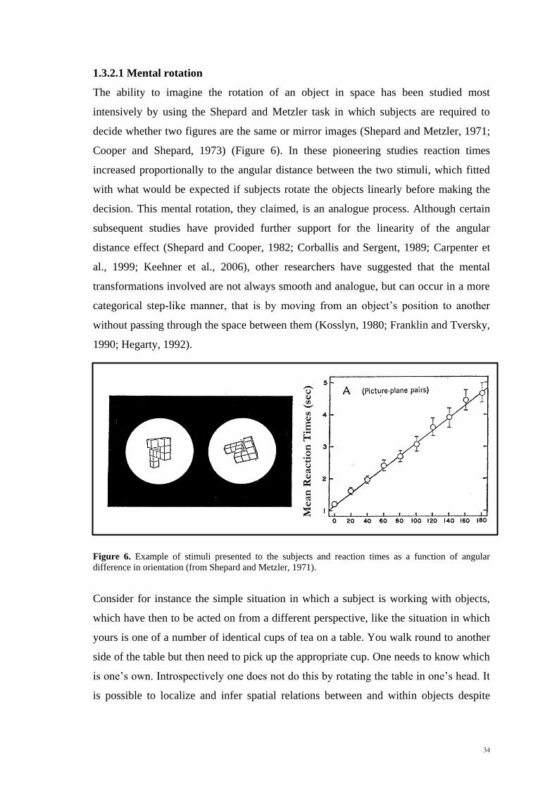
Evidence from brain tumour patients
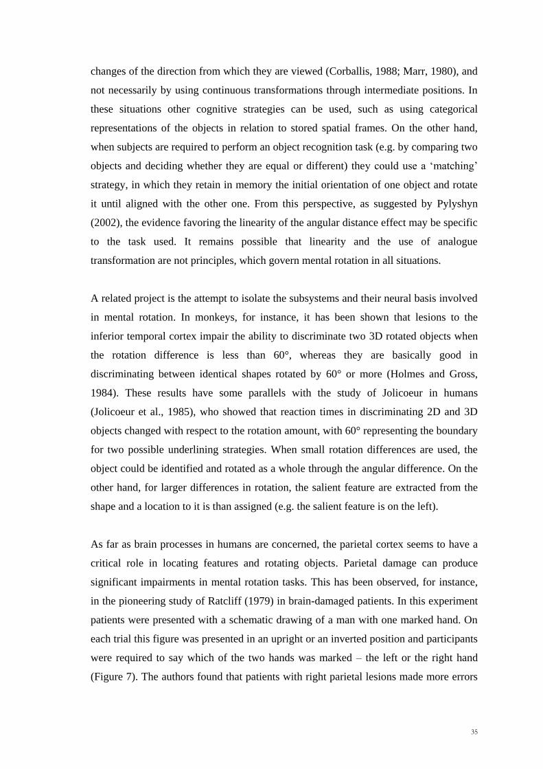
CANDIDATE Tania Buiatti
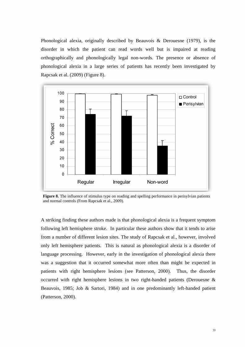
SUPERVISOR Professor Tim Shallice
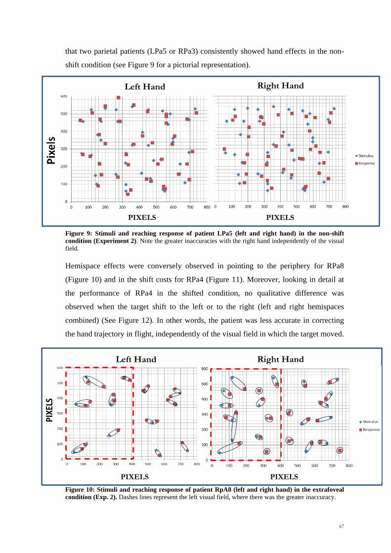
Thesis submitted for the degree of Philosophiae Doctor in Cognitive Neuroscience at
International School for Advanced Studies, Trieste, Italy
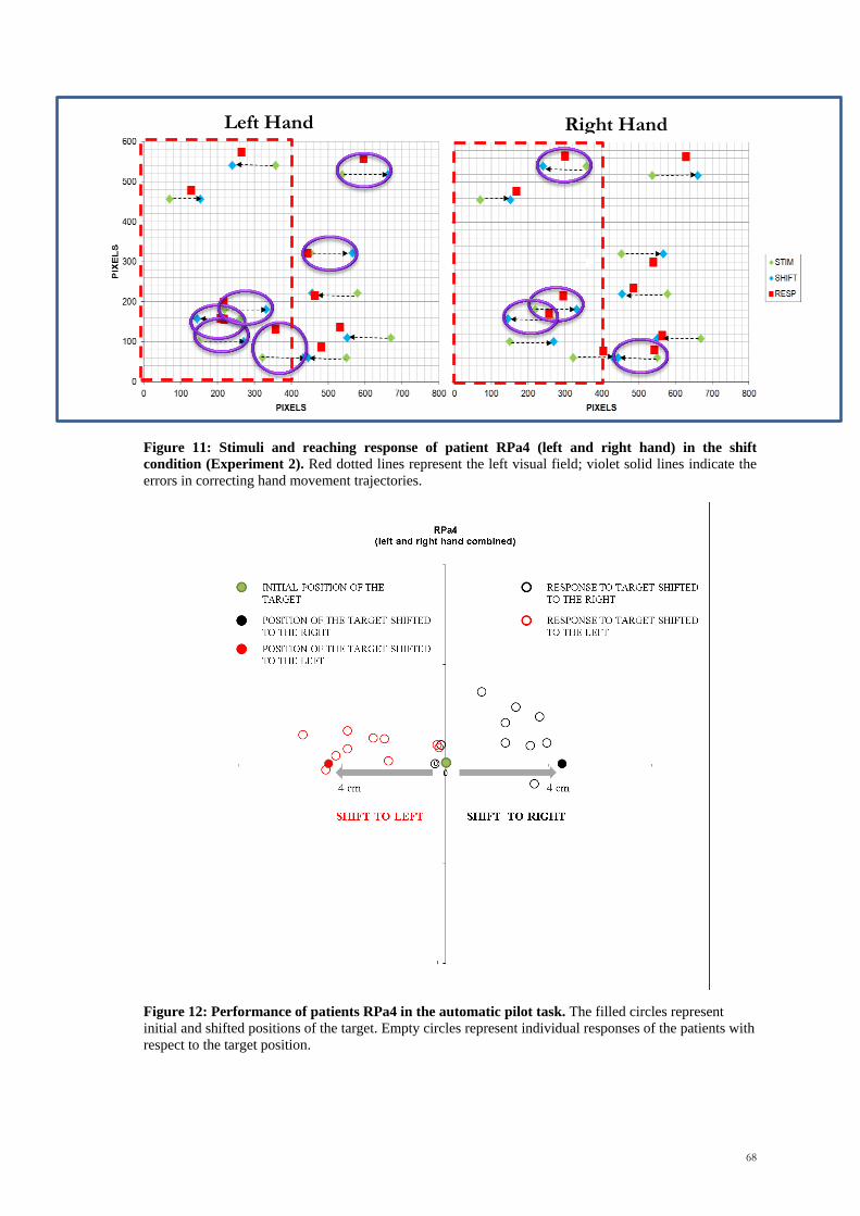
SISSA - Via Bonomea 265 – 34136 TRIESTE,
2
The research presented in this thesis was carried out at the ‗Scuola Internazionale Superiore di Studi
Avanzati – SISSA, Cognitive Neuroscience Sector, Trieste, Italy, in collaboration with the Neurosurgery
Department of the ‗Santa Maria della Misericordis‘ hospital, Udine, Italy.
All rights reserved. No part of this publication may be reproduced or transmitted in any form or by any
means, without the permission from the author
3
“Among other things, you'll find that you're not the first person who was ever confused and frightened and even sickened by human behavior. You're by no means alone on that score, you'll be excited and stimulated to know. Many, many men have been just as troubled morally and spiritually as you are right now. Happily, some of them kept records of their troubles. You'll learn from them - if you want to. Just as someday, if you have something to offer, someone will learn something from you. It's a beautiful reciprocal arrangement. And it isn't education. It's history. It's poetry.”
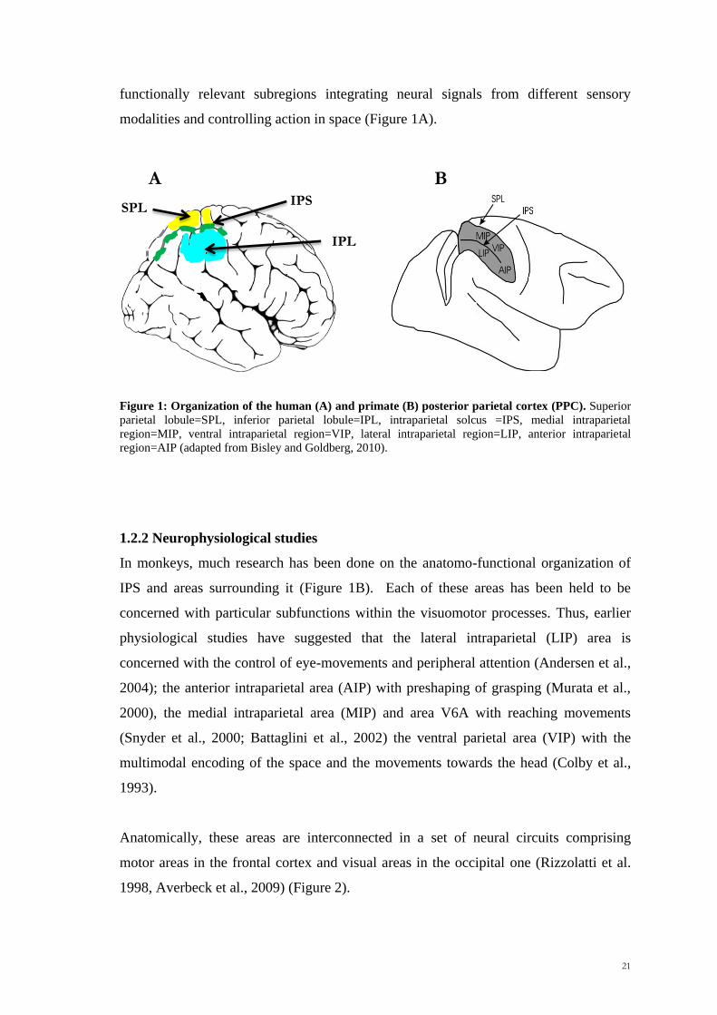
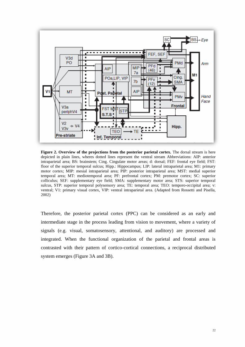
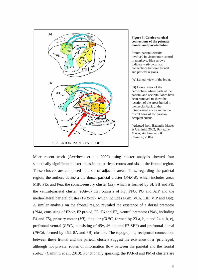
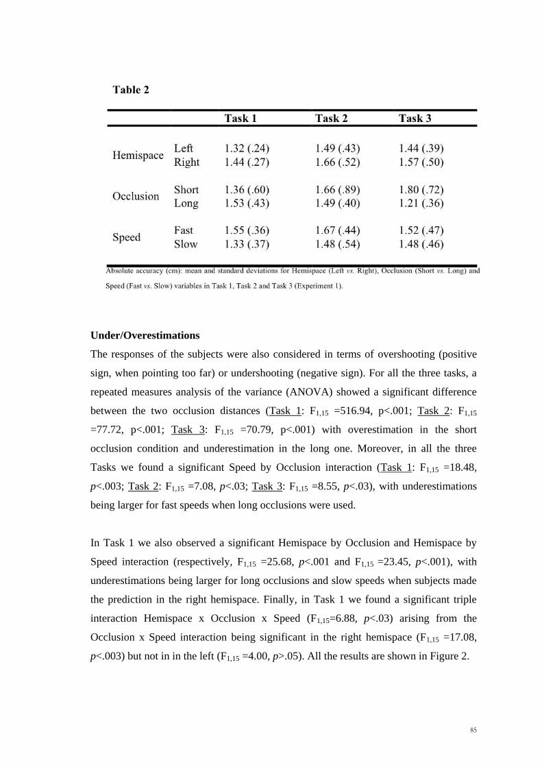
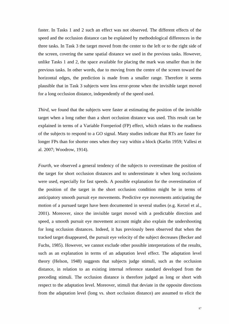
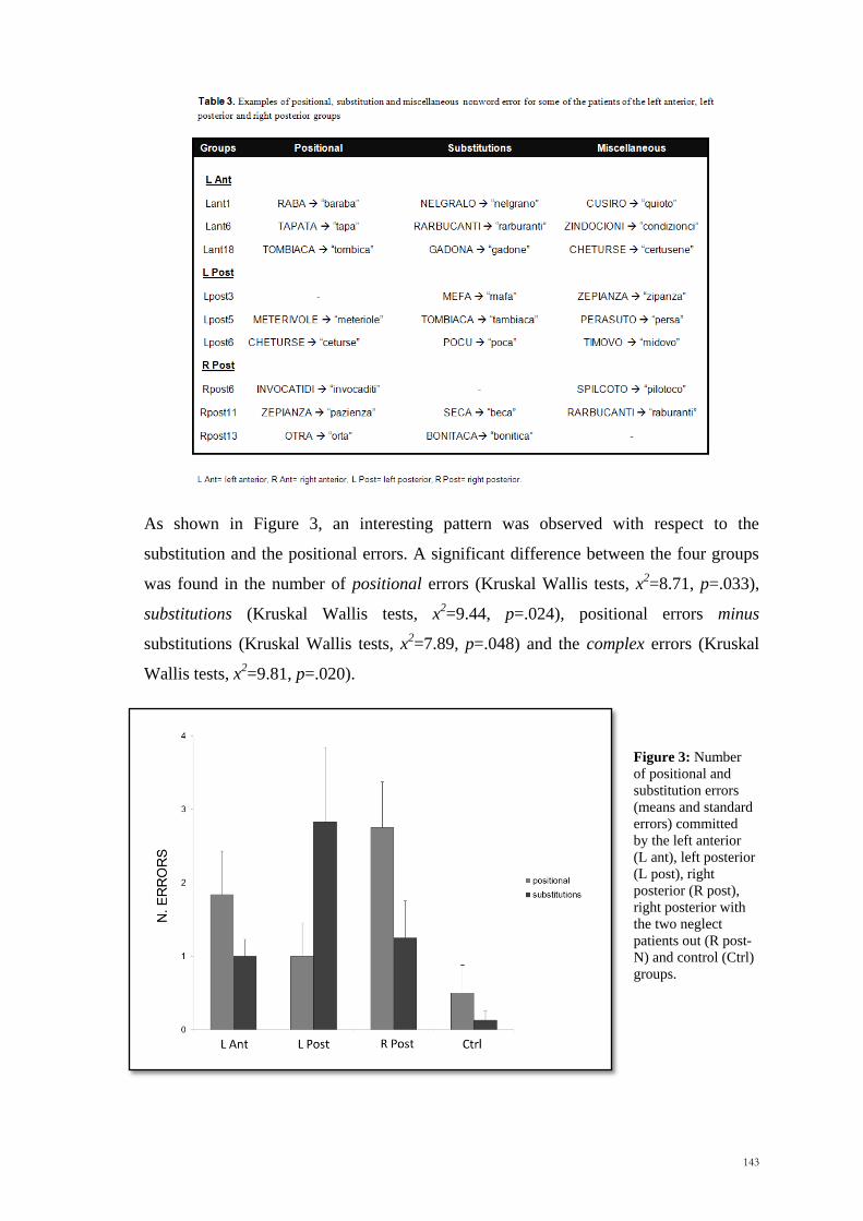
being larger for fast speeds when long occlusions were used.
In Task 1 we also observed a significant Hemispace by Occlusion and Hemispace by
Speed interaction (respectively, F1,15 =25.68, p<.001 and F1,15 =23.45, p<.001), with
underestimations being larger for long occlusions and slow speeds when subjects made
the prediction in the right hemispace. Finally, in Task 1 we found a significant triple
interaction Hemispace x Occlusion x Speed (F1,15=6.88, p<.03) arising from the
Occlusion x Speed interaction being significant in the right hemispace (F1,15 =17.08,
p<.003) but not in in the left (F1,15 =4.00, p>.05). All the results are shown in Figure 2.
86
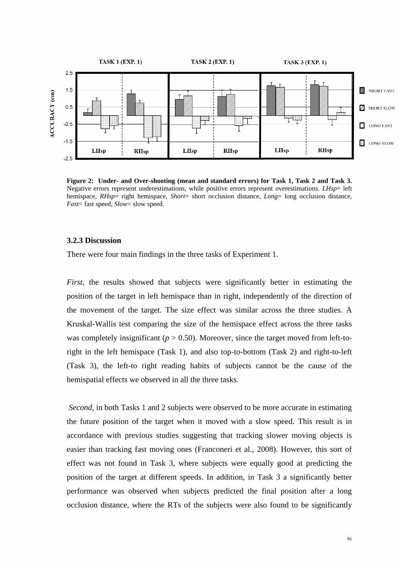
Figure 2: Under- and Over-shooting (mean and standard errors) for Task 1, Task 2 and Task 3. Negative errors represent underestimations, while positive errors represent overestimations. LHsp= left
hemispace, RHsp= right hemispace, Short= short occlusion distance, Long= long occlusion distance,
Fast= fast speed, Slow= slow speed.
3.2.3 Discussion
There were four main findings in the three tasks of Experiment 1.
First, the results showed that subjects were significantly better in estimating the
position of the target in left hemispace than in right, independently of the direction of
the movement of the target. The size effect was similar across the three studies. A
Kruskal-Wallis test comparing the size of the hemispace effect across the three tasks
was completely insignificant (p > 0.50). Moreover, since the target moved from left-to-
right in the left hemispace (Task 1), and also top-to-bottom (Task 2) and right-to-left
(Task 3), the left-to right reading habits of subjects cannot be the cause of the
hemispatial effects we observed in all the three tasks.
Second, in both Tasks 1 and 2 subjects were observed to be more accurate in estimating
the future position of the target when it moved with a slow speed. This result is in
accordance with previous studies suggesting that tracking slower moving objects is
easier than tracking fast moving ones (Franconeri et al., 2008). However, this sort of
effect was not found in Task 3, where subjects were equally good at predicting the
position of the target at different speeds. In addition, in Task 3 a significantly better
performance was observed when subjects predicted the final position after a long
occlusion distance, where the RTs of the subjects were also found to be significantly
87
faster. In Tasks 1 and 2 such an effect was not observed. The different effects of the
speed and the occlusion distance can be explained by methodological differences in the
three tasks. In Task 3 the target moved from the center to the left or to the right side of
the screen, covering the same spatial distance we used in the previous tasks. However,
unlike Tasks 1 and 2, the space available for placing the mark was smaller than in the
previous tasks. In other words, due to moving from the centre of the screen toward the
horizontal edges, the prediction is made from a smaller range. Therefore it seems
plausible that in Task 3 subjects were less error-prone when the invisible target moved
for a long occlusion distance, independently of the speed used.
Third, we found that the subjects were faster at estimating the position of the invisible
target when a long rather than a short occlusion distance was used. This result can be
explained in terms of a Variable Foreperiod (FP) effect, which relates to the readiness
of the subjects to respond to a GO signal. Many studies indicate that RTs are faster for
longer FPs than for shorter ones when they vary within a block (Karlin 1959; Vallesi et
al. 2007; Woodrow, 1914).
Fourth, we observed a general tendency of the subjects to overestimate the position of
the target for short occlusion distances and to underestimate it when long occlusions
were used, especially for fast speeds. A possible explanation for the overestimation of
the position of the target in the short occlusion condition might be in terms of
anticipatory smooth pursuit eye movements. Predictive eye movements anticipating the
motion of a pursued target have been documented in several studies (e.g. Kerzel et al.,
2001). Moreover, since the invisible target moved with a predictable direction and
speed, a smooth pursuit eye movement account might also explain the undershooting
for long occlusion distances. Indeed, it has previously been observed that when the
tracked target disappeared, the pursuit eye velocity of the subject decreases (Becker and
Fuchs, 1985). However, we cannot exclude other possible interpretations of the results,
such as an explanation in terms of an adaptation level effect. The adaptation level
theory (Helson, 1948) suggests that subjects judge stimuli, such as the occlusion
distance, in relation to an existing internal reference standard developed from the
preceding stimuli. The occlusion distance is therefore judged as long or short with
respect to the adaptation level. Moreover, stimuli that deviate in the opposite directions
from the adaptation level (long vs. short occlusion distance) are assumed to elicit the
88
opposite type of responses, one leading to underestimation and the other to an
overshoot. Responses are skewed by the context in which the stimuli are presented. In a
positively skewed context (where short occlusion distances occurred more often),
subjects can place the mark ahead of the actual position of the target. Conversely, in a
negative skewed context (long occlusions) they can place the mark behind (Parducci
and Wedell, 1986).
From the results of the three tasks, two other important questions arise:
1. Could all these effects depend upon the hand used to place the mark?
2. Do the laterality effects truly reflect a right hemisphere superiority in integrating
spatial and temporal information or are they related only to spatial processing?
We investigated these questions in Experiment 2 (paragraph 3.3) and Experiment 3
(paragraph 3.4) respectively. In Experiment 2 we asked subjects to perform the same as
Task 1 of Experiment 1, but using the left (non-dominant) hand. In Experiment 3 we
developed a pure spatial task in which subjects were required to remember the last
spatial position of a moving target after it disappeared for a short (1 s) or a long (3 sec)
temporal interval.
3.3 Experiment 2
3.3.1 Methods
Participants
Sixteen healthy right-handed subjects participated in Experiment 2 (9 males and 7
females, aged between 17 and 31 years). All had normal or corrected-to-normal vision,
no past neurological or psychiatric history and used no medication.
Apparatus, Stimuli and Design
Apparatus, stimuli, design and data analysis procedures were the same as Task 1 of
Experiment 1, with the exception of the hand used to place the mark. In this experiment
the subjects were required to use the left (non-dominant) hand.
89
3.3.2 Results
RTs
A repeated measures analysis of the variance (ANOVA) showed a main effect for the
Occlusion condition (F1,15=8.43, p<.03) with subjects being faster with long occlusion
distances than with short (means were respectively 382.22 msec and 413.78 msec). No
other significant main effects and interactions were observed.
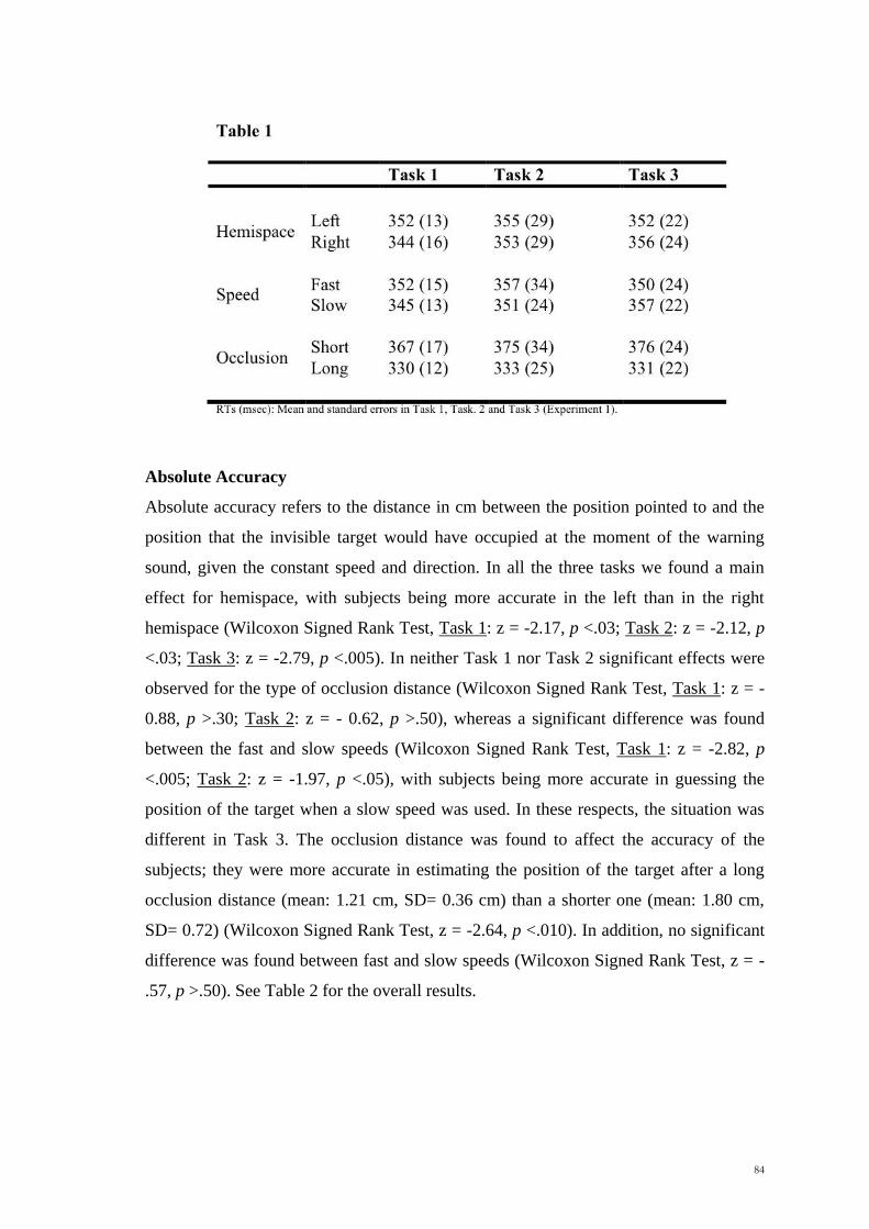
Absolute Accuracy
A main effect of Hemispace (Wilcoxon Signed Rank Test, z = -2.02, p <.05) showed
that subjects were more accurate in guessing the position of the moving target in the left
hemispace (mean: 1.72 cm, SD=0.31) than the right (mean: 1.88 cm, SD=0.52). No
significant differences were observed between the short and the long occlusion distance
conditions.
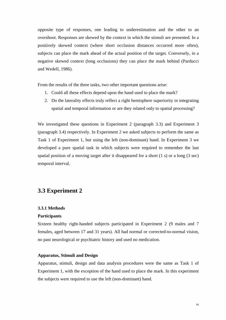
Under/Overestimations
A repeated measures analysis of variance (ANOVA) gave a significant difference
between the two occlusion distances (F1,15=75.97, p<.001), with overestimations for the
short interval and underestimations for the long occlusion condition. A significant
interaction was observed between Hemispace and Occlusion (F1,15=80.32, p<.001), with
larger overestimations for short distances in the right hemispace. Moreover, we also
found an Occlusion by Speed interaction (F1,15=4.61, p<.05), with underestimations
being larger for long distances and fast speeds. The results are summarized in Figure 3.
Figure 3: Under- and Over-shooting (mean and standard errors) for Exp 2 and Exp 3. Negative
errors represent underestimations, while positive errors represent overestimations. LHsp= left hemispace,
RHsp= right hemispace, Short= short occlusion distance, Long= long occlusion distance, Fast= fast
speed, Slow= slow speed.
90
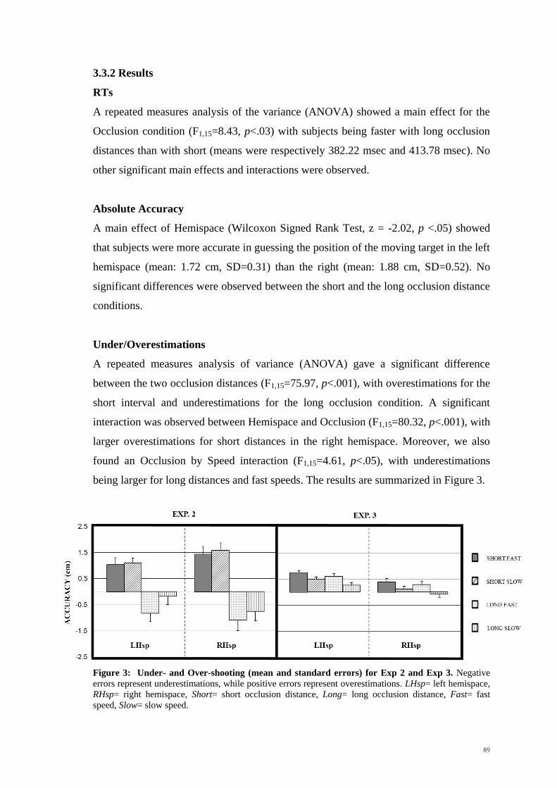
3.3.3 Discussion
In line with the findings from the previous tasks we observed that subjects were more
accurate in guessing the position of an invisible moving target when the prediction had
to be made in the left rather than the right hemispace. Moreover, when we looked at the
qualitative nature of such errors (under- and over-estimations) we again observed a
main effect for Occlusion and a significant Occlusion by Speed interaction. The
subjects overestimated the position of the target for short occlusions and underestimated
for long occlusion distance, with the undershooting being larger for fast speeds. Since
the subject used the left and not the right (dominant) hand in the present study, we can
conclude that all these effects cannot be just be explained by the hand used to reach the
target.
3.4 Experiment 3
3.4.1 Methods
Participants
Sixteen healthy right-handed subjects participated in Experiment 3 (11 males and 5
females, aged between 21 and 32 years). All had normal or corrected-to-normal vision,
no past neurological or psychiatric history and used no medication.
Apparatus, Stimuli and Design
In Experiment 3 we used the same apparatus, stimuli and data analysis procedure used
in Task 1 of Experiment 1. The target appeared at one side of the screen and moved
along the x-axis with a fast (4.4 cm/s) or a slow (1.8 cm/s) speed. After an unpredictable
spatial interval the target disappeared for a short (1 sec) or a long (3 sec) retention
interval. Then, a 1000 Hz tone warned the subjects to point as quickly and accurately as
possible to where they thought the target had disappeared. As in the previous tasks, two
separate block conditions were used in an ABAB design counterbalanced over subjects:
in one condition the circle moved from left to right in the left hemispace and in the
other condition the circle moved from right to left in the right hemispace.
91
3.4.2 Results
RTs
No significant differences in response times were observed between the right and the
left hemispace (Wilcoxon Signed Rank Test, z = -1.19, p>.10) and between the fast and
slow speeds (Wilcoxon Signed Rank Test, z = -.931, p>.30). A significant effect was
found between the two retention intervals (Wilcoxon Signed Rank Test, z = -3.26, p<
.003), with subjects being faster with long intervals than with short (means were
respectively 399.11 and 468.64 msec).
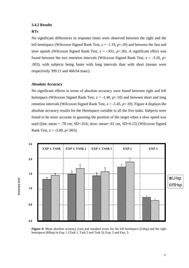
Absolute Accuracy
No significant effects in terms of absolute accuracy were found between right and left
hemispace (Wilcoxon Signed Rank Test, z = -1.40, p>.10) and between short and long
retention intervals (Wilcoxon Signed Rank Test, z = -1.45, p>.10). Figure 4 displays the
absolute accuracy results for the Hemispace variable in all the five tasks. Subjects were
found to be more accurate in guessing the position of the target when a slow speed was
used (fast: mean = .78 cm; SD=.016; slow: mean=.61 cm, SD=0.15) (Wilcoxon Signed
Rank Test, z = -3.00, p<.003).
Figure 4: Mean absolute accuracy (cm) and standard errors for the left hemispace (LHsp) and the right
hemispace (RHsp) in Exp. 1 (Task 1, Task 2 and Task 3), Exp. 2 and Exp. 3.
EXP 1: TASK 1
EXP 1: TASK 2 EXP 1: TASK 3 EXP 2 EXP 3
92
Underestimation/Overestimations
As did for the previous tasks, also in Experiment 3 we looked at the existence of
possible directional errors (Results shown in Figure 3). In other words we checked
whether the response of the subject was directed towards the centre of the screen
(pointing forward with respect to the direction of the target) or away from the centre
(pointing backward). A repeated measures analyses of the variance (ANOVA) showed a
main effect for Hemispace, suggesting that the overestimations were larger in the left
than the right hemispace (F1,15=7.08, p<.03), namely when the dot moved from the left
to the right. Main effects were observed also for retention intervals (F1,15=21.04, p
<.001) and Speed (F1,15=48.62, p <.001). Subjects have been found to show larger
overshooting for short retention intervals and fast speeds.
3.4.3 Discussion
The main result of Experiment 3 was the lack of a laterality effect for absolute
accuracy. In other words, the subjects showed no significant differences in accuracy
between the left and the right hemispace, when required to perform a purely spatial task
such as remembering the last spatial position of a target. This ‗negative‘ result is in
contrast with any hypothesis that the results of Experiment 1 and 2 could be explained
just in terms of spatial processing per se and not by the need to produce spatio-temporal
integration. Interestingly, for long occlusions intervals the RTs to respond were over
100 msec slower in Experiment 3 than in the three conditions of Experiment 1. Thus,
the lack of better performance in the left hemispace could therefore also represent a
possible dissociation between responses mediated by the dorsal stream (Experiment 1)
and ones based partially on conscious judgements (possibly Experiment 3). In other
words, the right hemispare might have a critical role especially when an immediate
action response in a spatial task is required. Moreover, in contrast with the previous
tasks, we observed a general tendency to overestimate the position of the target with
errors being larger in the left hemispace, for short retention intervals and fast speeds.
The results can be explained in terms of a Representational Momentum Effect, a
memory distortion which has been first reported by Freyd and Finke (1984). When
observers are first presented with a stationary or moving target that vanishes without
warning and then asked to judge where the target has disappeared, observers have been
found to be more likely to indicate a point which is ahead of the actual vanishing point
in terms of the direction of the moving dot (for a review, Hubbard, 2005). This effect is
93
influenced by many factors such as the velocity, the direction of the target and the
temporal interval between the responses of the subjects and when the target vanished.
For instance, Hubbard and Bharucha (1988) reported that faster speeds led to larger
forward displacements, while Halpern and Kelly (1993) observed that the effect was
larger for targets presented in the left visual field. These results are consistent with the
data we reported.
3.5 General discussion
In the current study five tasks were run to investigate the probable lateralized spatial
effects on accuracy in estimating the position of an invisible moving target. The first
four main tasks differed from each other with respect to the direction of the moving
target within the display (Experiment 1: Task 1, Task 2 and Task 3) and to the hand
used (Experiment 2). Moreover, in the additional task (Experiment 3), subjects were
required to remember the final spatial position of a moving target after a temporal
interval of 1 or 3 seconds.
In Experiment 1 an analysis of the accuracy of the subjects revealed a significant main
effect of the hemispace variable, indicating that participants were more accurate when
they predicted the position of an invisible moving target in the left hemispace rather
than the right. This suggested that a right hemisphere superiority could exist for
spatiotemporal integration.
Our findings seem to be in contrast with those of previous fMRI studies in which
collision and trajectory judgments caused an increase in the neural activity of the left
parietal cortex (Assmus et al., 2005; Coull et al. 2008). However, it shoul be noted that
in both the latter experiments, healthy volunteers were required to judge whether the
stimuli would have collided or not by selecting and pressing the corresponding response
button. Therefore, one can claim that the left parietal activation is not directly related to
on-line spatio-temporal integration, but to conscious action selection and action
preparation. Interestingly, previous brain imaging studies showed that when the
selection of the movement is crucial for the task (e.g. as in a choice reaction time task),
an activation of the left parietal cortex could be observed, independently of the hand
94
used (Kawashima et al., 1993; Schluter et al., 2001). Conversely, this left hemisphere
dominance was not observed in a simple reaction time task, where brain activation was
contralateral to the hand used. Notwithstanding, our findings do not preclude a role for
the left parietal cortex in tasks other than the one used, the simple prediction of the
spatial position an invisible target would take over in time. In other words, predicting if
a collision would occur could require additional cognitive processes, not spatiotemporal
integration alone.
Conversely, the results fit well with the ATOM theory (Walsh, 2003), according to
which the right parietal cortex could have a role in the integration of spatial and
temporal information. Also findings with neglect patients support this view. Indeed,
patients with neglect following a right hemisphere lesion frequently show impairments
in temporal representation as well as spatial (Basso et al., 1996; Danckert et al., 2007;
Calabria et al., 2011). If the interpretation of the laterality effects as related to a right
hemisphere superiority for spatio-temporal integration is correct, we should have
observed a different pattern in patients with brain damage involving the right parietal
cortex. We will investigate this issue in the next Chapter (Chapter 4).
Finally, in Experiment 2 and Experiment 3 we demonstrated that the left hemispace
advantage for spatio-temporal integration could not be explained by a hand effect or by
a spatial processing per se. Indeed, the effect was still present when patients used the
left (non-dominant) hand and it was not present anymore when subjects were required
to remember the last spatial position of the moving target.
95
Chapter 4
Hemispatial effects on spatio-temporal integration: evidence from brain tumour
patients
96
97
4.1 Introduction
In Chapter 3 evidence has been provided that the right hemisphere plays an important
role in integrating spatial and temporal information. In particular, we observed that
subjects are generally more accurate in estimating the position of an invisible moving
target when the predicted position is in the left hemispace rather than the right. In
accordance with the theory of Walsh (ATOM; 2003) (see paragraph 1.3.1.2, Chapter 1
and paragraph 3.6, Chapter 3), the results were explained in terms of a right hemisphere
superiority for the process responsible for spatio-temporal integration.
In the study presented in this chapter, we have investigated this issue more directly in a
population of unilateral brain tumour patients with lesion occurring in the anterior or in
the posterior (parietal or parietal-occipital) regions of the brain. The specific aim is to
examine whether the laterality effects observed in healthy subjects will no be longer to
be present in patients with damage to the right posterior cortex.
Methodologically, a slight change to the procedure used in Task 1 of Experiment 1
(paragraph 3.1.2, Chapter 3) was made, due to the smaller size of the monitor that was
available for patient testing. In the left-to-right condition, patients track the visible
moving target in the left hemispace but need to predict the position of the no longer
visible target when it would be in the right hemispace. We refer to this as a right
hemispace (RHsp) effect. In a complementary fashion, in the right-to-left condition,
patients track the visible moving object in the right hemispace and respond when it
would be in the left hemispace (LHsp, left hemispace effect).
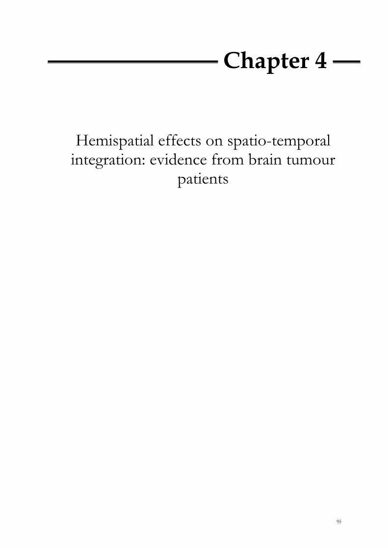
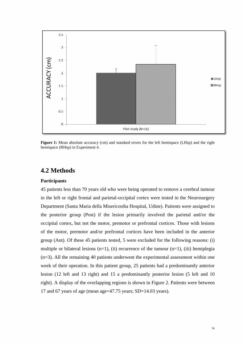
A pilot study with 16 healthy controls confirmed the validity of the new adapted task in
generating similar hemispatial effects to those observed in studies reported in Chapter 3
(see Figure 1). As before, subjects were more accurate in making the judgement about
the position of the invisible moving target in the left hemispace rather than the right
(Wilcoxon Signed Rank Test, z = -2.64, p<.01), with no significant differences in terms
of speed (Wilcoxon Signed Rank Test, z = -.36, p>.5).
98
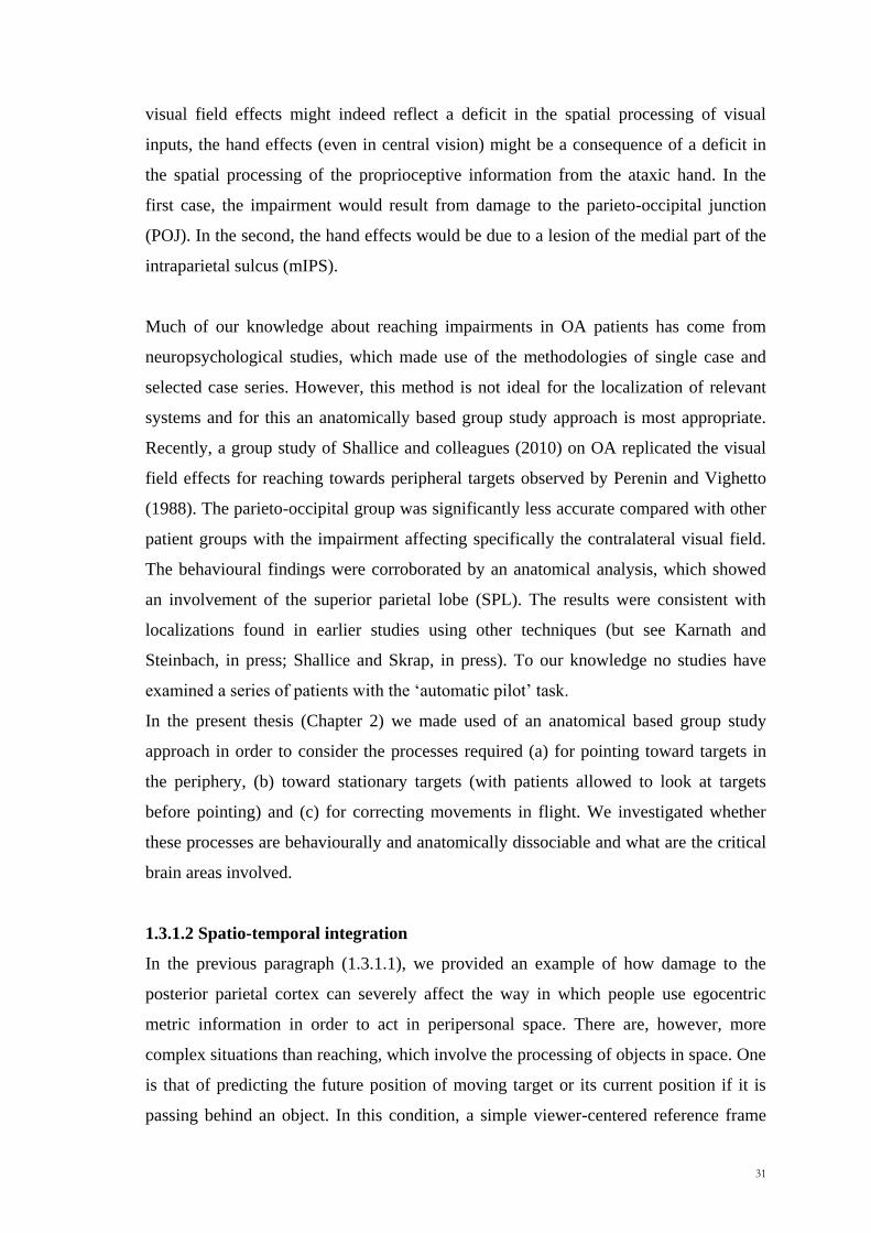
Figure 1: Mean absolute accuracy (cm) and standard errors for the left hemispace (LHsp) and the right
hemispace (RHsp) in Experiment 4.
4.2 Methods
Participants
45 patients less than 70 years old who were being operated to remove a cerebral tumour
in the left or right frontal and parietal-occipital cortex were tested in the Neurosurgery
Department (Santa Maria della Misericordia Hospital, Udine). Patients were assigned to
the posterior group (Post) if the lesion primarily involved the parietal and/or the
occipital cortex, but not the motor, premotor or prefrontal cortices. Those with lesions
of the motor, premotor and/or prefrontal cortices have been included in the anterior
group (Ant). Of these 45 patients tested, 5 were excluded for the following reasons: (i)
multiple or bilateral lesions (n=1), (ii) recurrence of the tumour (n=1), (iii) hemiplegia
(n=3). All the remaining 40 patients underwent the experimental assessment within one
week of their operation. In this patient group, 25 patients had a predominantly anterior
lesion (12 left and 13 right) and 15 a predominantly posterior lesion (5 left and 10
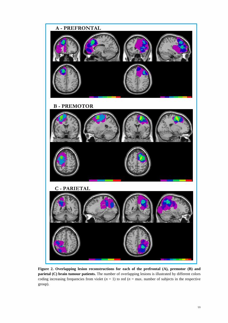
right). A display of the overlapping regions is shown in Figure 2. Patients were between
17 and 67 years of age (mean age=47.75 years; SD=14.03 years).
99
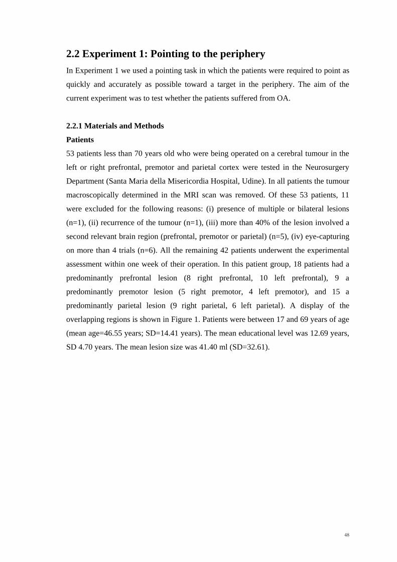
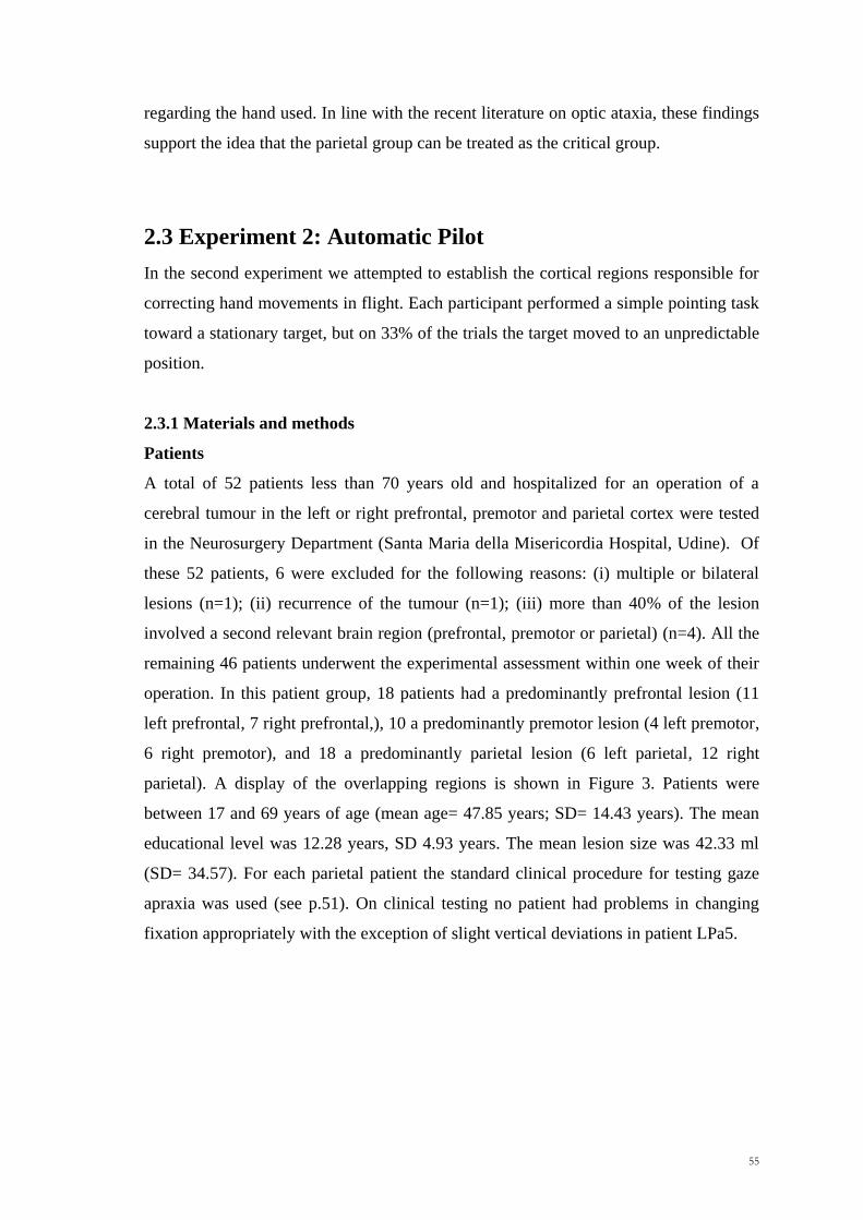
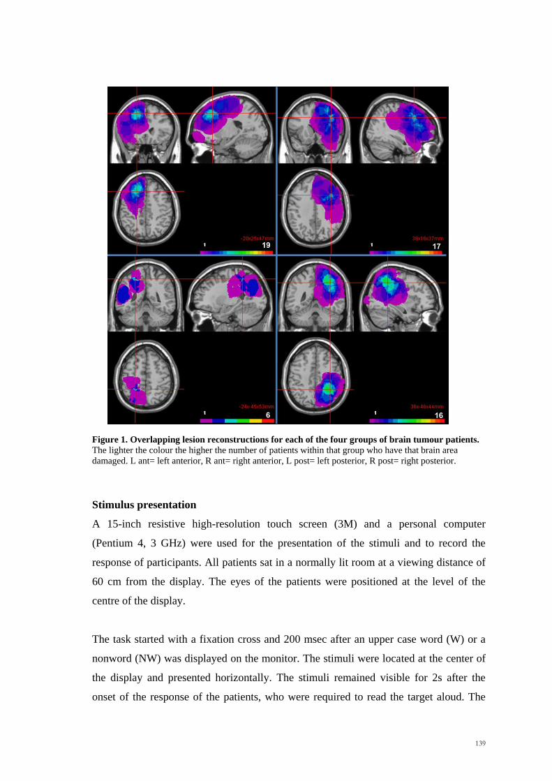
Figure 2. Overlapping lesion reconstructions for each of the prefrontal (A), premotor (B) and
parietal (C) brain tumour patients. The number of overlapping lesions is illustrated by different colors
coding increasing frequencies from violet (n = 1) to red (n = max. number of subjects in the respective
group).
A - PREFRONTAL
B - PREMOTOR
C - PARIETAL
100
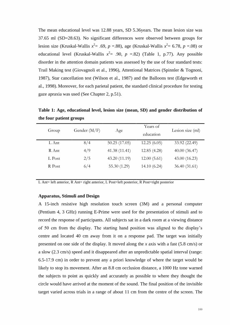
The mean educational level was 12.88 years, SD 5.36years. The mean lesion size was
37.65 ml (SD=28.63). No significant differences were observed between groups for
lesion size (Kruskal-Wallis x2= .69, p =.88), age (Kruskal-Wallis x
2= 6.78, p =.08) or
educational level (Kruskal-Wallis x2= .90, p =.82) (Table 1, p.77). Any possible
disorder in the attention domain patients was assessed by the use of four standard tests:
Trail Making test (Giovagnoli et al., 1996), Attentional Matrices (Spinnler & Tognoni,
1987), Star cancellation test (Wilson et al., 1987) and the Balloons test (Edgeworth et
al., 1998). Moreover, for each parietal patient, the standard clinical procedure for testing
gaze apraxia was used (See Chapter 2, p.51).
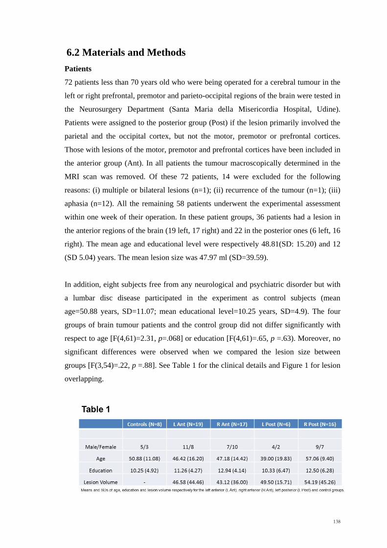
Table 1: Age, educational level, lesion size (mean, SD) and gender distribution of
the four patient groups
Group Gender (M/F) Age Years of
education Lesion size (ml)
L Ant 8/4 50.25 (17.05) 12.25 (6.05) 33.92 (22.49)
R Ant 4/9 41.38 (11.41) 12.85 (4.28) 40.00 (36.47)
L Post 2/5 43.20 (11.19) 12.00 (5.61) 43.00 (16.23)
R Post 6/4 55.30 (1.29) 14.10 (6.24) 36.40 (31.61)
L Ant= left anterior, R Ant= right anterior, L Post=left posterior, R Post=right posterior
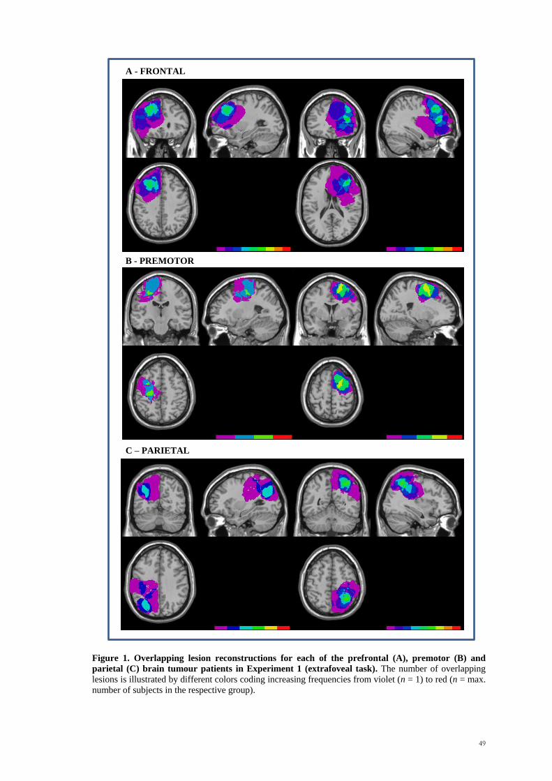
Apparatus, Stimuli and Design
A 15-inch resistive high resolution touch screen (3M) and a personal computer
(Pentium 4, 3 GHz) running E-Prime were used for the presentation of stimuli and to
record the response of participants. All subjects sat in a dark room at a viewing distance
of 50 cm from the display. The starting hand position was aligned to the display‘s
centre and located 40 cm away from it on a response pad. The target was initially
presented on one side of the display. It moved along the x axis with a fast (5.8 cm/s) or
a slow (2.3 cm/s) speed and it disappeared after an unpredictable spatial interval (range:
6.5-17.9 cm) in order to prevent any a priori knowledge of where the target would be
likely to stop its movement. After an 8.8 cm occlusion distance, a 1000 Hz tone warned
the subjects to point as quickly and accurately as possible to where they thought the
circle would have arrived at the moment of the sound. The final position of the invisible
target varied across trials in a range of about 11 cm from the centre of the screen. The
101
pointing response on the touchscreen allows us to record the display coordinates.
Participants were not allowed to trace the target with the finger on the screen, but they
were free to follow the target with the eyes. Once they touched the screen, they were
required to place the hand back on the starting position. No response was collected if
the subjects leave the response pad before the tone. Patients were required to perform
four blocks of trials counterbalanced for the moving direction of the dot (ABBA
design). In one block the dot moved from left-to-right in the left hemispace, in the other
one it moved from right-to-left in the right hemispace. As mentioned before
(Introduction, paragraph 4.1, Chapter 4), for the first type of stimuli patients predict the
position of the invisible target when it is in the right hemispace, whereas in the second
type they responded to where it should be in the left hemispace. Moreover, in contrast
with the previous experiments (Experiment 1 and 2, Chapter 3), only a long occlusion
distance was used. Within each block, the slow and fast speeds were presented in a
pseudo-randomized order. Each session began with a short practice (4 trials for the dot
moving left-to-right and 4 trials for the dot moving right-to-left). No eye-movements
instructions were given to the patients.
Data analysis
Behavioural data
For each participant we calculated the mean Reaction Times (RTs), Movement Times
(MTs) and the accuracy. RTs were measured from the onset of the target to the release
of the response pad. MTs were measured from the release of the contact switch to the
moment at which patients touched the touch-screen. Accuracy was calculated as the
absolute distance in millimetres between the real position of the invisible target and the
point of contact on the touch screen. Trials were discarded if the RTs and Accuracy
were four SDs below or above the grand mean of each participant or if the touch screen
failed to record the response of the patient. For all patients this accounted for less than
5% of trials.
For all the measures, we used the same statistical procedure based on that adapted by
Stuss et al. (2005), which involved two levels of analysis:
(i) We first selected and divided patients into four groups according to the side and the
predominant location of the brain tumour (left anterior, L Ant, right anterior, R Ant,
left posterior, L Post and right posterior, R Post) and we first compared the
102
performance among these groups. Three different measures were examined: (i) the
overall effects on RTs, MTs and absolute accuracy; (ii) the difference in terms of
accuracy, reaction times and movement times between the left minus the right
hemispace (LHsp-RHsp, hemispatial effect) and (iii) between fast minus slow speed
(Fast-Slow, speed effect). The results were corrected for multiple comparisons (p <
.017).
(ii) If a significant overall effect was observed at this level, we contrasted the
performance of each group of patients with those of the other groups combined (e.g.
R Post vs. L Ant, R Ant and L Post combined). In this way we were able to be more
specific about the location of any impairment with respect to our patient population.
The raw data were first checked for normality using the Kolmogorov-Smirnov test and
for homogeneity of variance by applying the Levene test. As the data were not normally
distributed, non-parametric tests were used. The results were considered significant if
the p value was <.05; all the significance tests were two-tailed.
Anatomical data
The location and the extension of the tumour were carried out using a digital format
contrast-enhanced t1-weighted MRI scans obtained 1-7 days before operation using a
1.5T machine. The preoperative MRI scans were selected, as they are the scans
generally used by the neurosurgeon during the operation with the Neuronavigator as the
best indicator of macroscopic tumour extent. This allowed us to avoid any possible
confusion in draw lesions due to the replacement of neural brain tissue that occurs after
surgical removal. MRicro reconstructional software was used to extrapolate a 3D
representation of the lesion from digital MR scans (Rorden and Brett, 2000). The scans
and ROIs were normalized to the Montreal Neurological Institute template by using
SPM05b with 12 affine transformations and 7 x 9 x 7 basis functions.
4.3 Results
4.3.1 RTs and MTs
The Kruskal-Wallis test showed no significant effect of the overall RTs and MTs
among the four groups of patients (RTs: x2=2.18, p =.54; MTs: x
2=2.57, p =.46;
Kruskal-Wallis test), as well as hemispatial (LHsp-RHsp, RTs: x2=1.80, p =.61; MTs:
103
x2=4.46, p =.22; Kruskal-Wallis test) and speed effects (Fast-Slow, RTs: x
2=2.26, p
=.52; MTs: x2=5.83, p =.12; Kruskal-Wallis test).
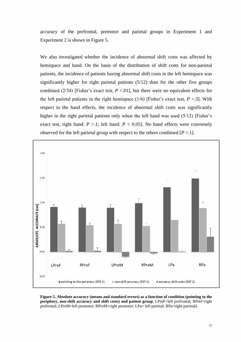
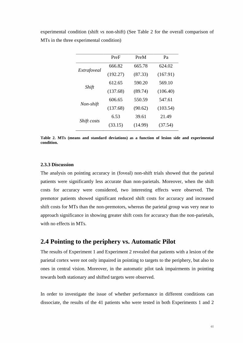
Absolute Accuracy
Absolute accuracy refers to the distance in cm between the position pointed by the
patient and the real position of the invisible target at the moment of the sound, given the
axis and the constant speed. A non-parametric analysis between the four groups of
patients revealed that they did not differ significantly in terms of overall accuracy (left
and right hemispace combined) (Kruskal-Wallis, x2=1.67, p =.64).
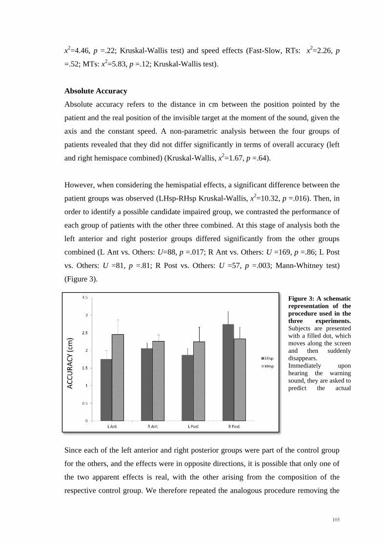
However, when considering the hemispatial effects, a significant difference between the
patient groups was observed (LHsp-RHsp Kruskal-Wallis, x2=10.32, p =.016). Then, in
order to identify a possible candidate impaired group, we contrasted the performance of
each group of patients with the other three combined. At this stage of analysis both the
left anterior and right posterior groups differed significantly from the other groups
combined (L Ant vs. Others: U=88, p =.017; R Ant vs. Others: U =169, p =.86; L Post
vs. Others: U =81, p =.81; R Post vs. Others: U =57, p =.003; Mann-Whitney test)
(Figure 3).
Since each of the left anterior and right posterior groups were part of the control group
for the others, and the effects were in opposite directions, it is possible that only one of
the two apparent effects is real, with the other arising from the composition of the
respective control group. We therefore repeated the analogous procedure removing the
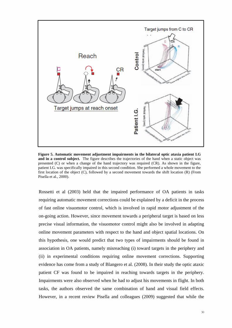
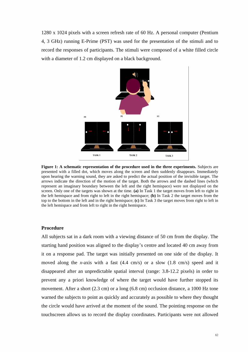
Figure 3: A schematic
representation of the
procedure used in the
three experiments.
Subjects are presented
with a filled dot, which
moves along the screen
and then suddenly
disappears.
Immediately upon
hearing the warning
sound, they are asked to
predict the actual
position of the invisible
104
left anterior and the right posterior groups from the respective control group. On this
second round, only the right posterior group differed statistically from the other two
groups combined (R Post vs. R Ant+L Post, U=46, p =.008; Mann-Whitney test). The
difference observed between the left anterior and the other two groups was not
significant (L Post vs. R Ant+L Post, U=58, p =.17; Mann-Whitney test). This means
that only the right posterior effect is clearly genuine. Moreover, the right posterior
group was significantly worse than the other groups combined in the left hemispace, but
it was not in the right one (LHsp: U= 85, p =.043; RHsp: U=150, p =1).
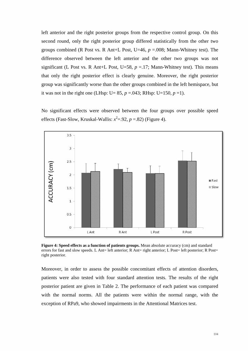
No significant effects were observed between the four groups over possible speed
effects (Fast-Slow, Kruskal-Wallis: x2=.92, p =.82) (Figure 4).
Figure 4: Speed effects as a function of patients groups. Mean absolute accuracy (cm) and standard
errors for fast and slow speeds. L Ant= left anterior; R Ant= right anterior; L Post= left posterior; R Post=
right posterior.
Moreover, in order to assess the possible concomitant effects of attention disorders,
patients were also tested with four standard attention tests. The results of the right
posterior patient are given in Table 2. The performance of each patient was compared
with the normal norms. All the patients were within the normal range, with the
exception of RPa9, who showed impairments in the Attentional Matrices test.
105
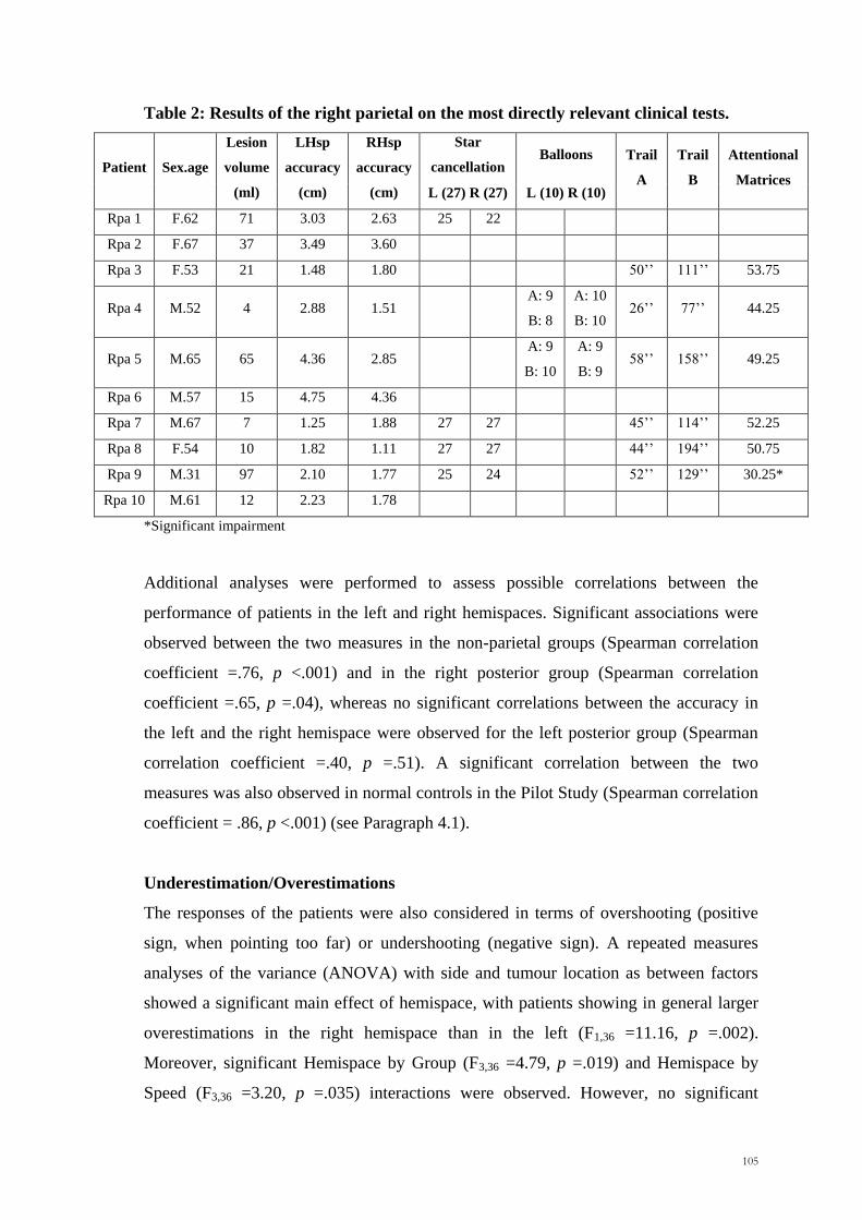
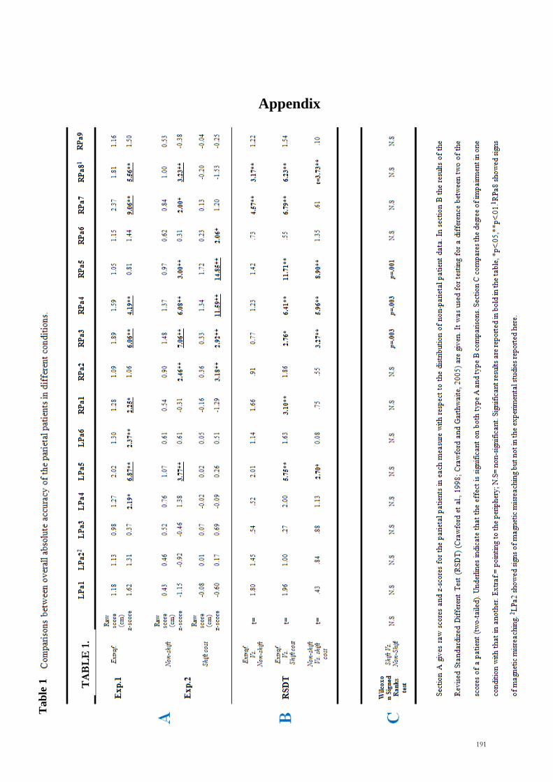
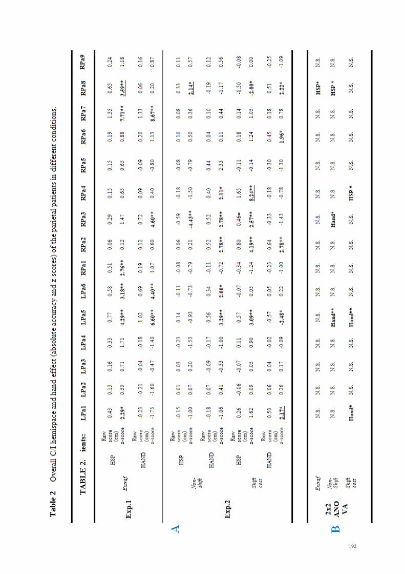
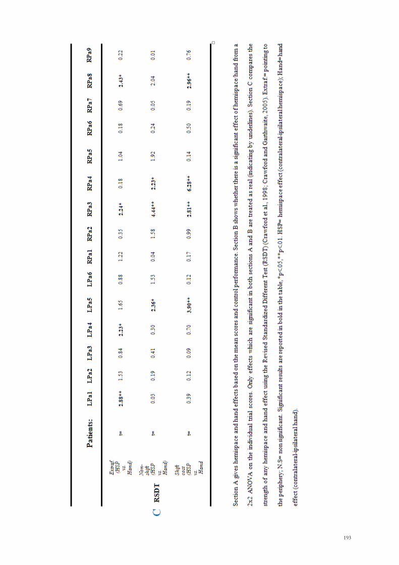
Table 2: Results of the right parietal on the most directly relevant clinical tests.
Patient Sex.age
Lesion
volume
(ml)
LHsp
accuracy
(cm)
RHsp
accuracy
(cm)
Star
cancellation Balloons Trail
A
Trail
B
Attentional
Matrices L (27) R (27) L (10) R (10)
Rpa 1 F.62 71 3.03 2.63 25 22
Rpa 2 F.67 37 3.49 3.60
Rpa 3 F.53 21 1.48 1.80 50‘‘ 111‘‘ 53.75
Rpa 4 M.52 4 2.88 1.51 A: 9
B: 8
A: 10
B: 10 26‘‘ 77‘‘ 44.25
Rpa 5 M.65 65 4.36 2.85 A: 9
B: 10
A: 9
B: 9 58‘‘ 158‘‘ 49.25
Rpa 6 M.57 15 4.75 4.36
Rpa 7 M.67 7 1.25 1.88 27 27 45‘‘ 114‘‘ 52.25
Rpa 8 F.54 10 1.82 1.11 27 27 44‘‘ 194‘‘ 50.75
Rpa 9 M.31 97 2.10 1.77 25 24 52‘‘ 129‘‘ 30.25*
Rpa 10 M.61 12 2.23 1.78
*Significant impairment
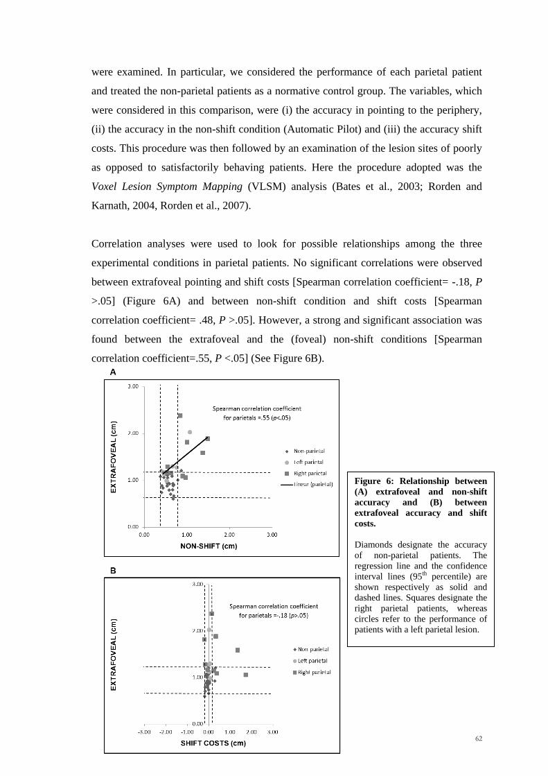
Additional analyses were performed to assess possible correlations between the
performance of patients in the left and right hemispaces. Significant associations were
observed between the two measures in the non-parietal groups (Spearman correlation
coefficient =.76, p <.001) and in the right posterior group (Spearman correlation
coefficient =.65, p =.04), whereas no significant correlations between the accuracy in
the left and the right hemispace were observed for the left posterior group (Spearman
correlation coefficient =.40, p =.51). A significant correlation between the two
measures was also observed in normal controls in the Pilot Study (Spearman correlation
coefficient = .86, p <.001) (see Paragraph 4.1).
Underestimation/Overestimations
The responses of the patients were also considered in terms of overshooting (positive
sign, when pointing too far) or undershooting (negative sign). A repeated measures
analyses of the variance (ANOVA) with side and tumour location as between factors
showed a significant main effect of hemispace, with patients showing in general larger
overestimations in the right hemispace than in the left (F1,36 =11.16, p =.002).
Moreover, significant Hemispace by Group (F3,36 =4.79, p =.019) and Hemispace by
Speed (F3,36 =3.20, p =.035) interactions were observed. However, no significant
106
interaction were observed between hemispace effects and location of the brain tumour
(Hemispace*Group, F2,36 =1.93, p =.16).
4.4 Discussion
This study aimed to assess whether damage to the right posterior cortex can disrupt the
hemispatial effects we reported in normal subjects (Chapter 3). From a behavioural
point of view, there was a significant difference between groups with respect to the
hemispatial effect. The present work confirmed the results of Chapter 3 by
demonstrating that patients with a lesion involving the left anterior, right anterior and
left posterior cortex behave in a similar fashion to normal subjects with respect to the
hemispatial effects. However, the right posterior group showed a different pattern. They
were worse then the other patients in the left hemispace, but not in the right.
One way in which one might try to interpret the different pattern of results for the right
posterior group compared with the others is in terms of a right hemisphere superiority
for spatio-temporal processing (Walsh, 2003; Olivieri et al., 2009). According to this
hypothesis, damage to the right posterior cortex would impair the ability to integrate
spatial and temporal information, independently of whether the prediction has to be
made for the left or for the right hemispace. However, on a closer inspection, there are
suggestions that the effect observed in the right posterior group might not be simply
interpreted in this way. Thus, clear evidence for impairments in both hemispaces was
obtained, which is what one expects if the right posterior cortex is the only structure
involved in integrating spatial and temporal information.
Why might this specific pattern of results occur? It is possible that two factors operate
in spatio-temporal integration. One, in accordance with the theory of Walsh (2003), is
that the right posterior cortex has greater resources than the left one for integrating
spatial and temporal information. The second is that systems in each hemisphere
operate more effectively for stimuli in the contralateral than in its ipsilateral
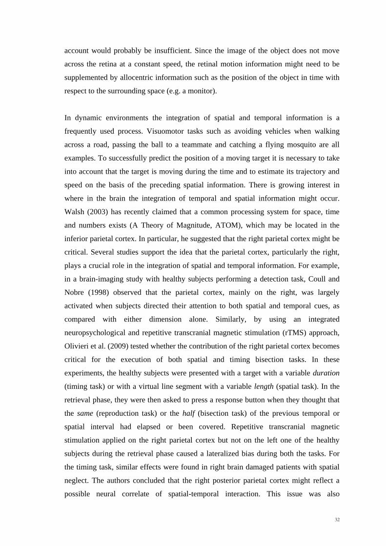
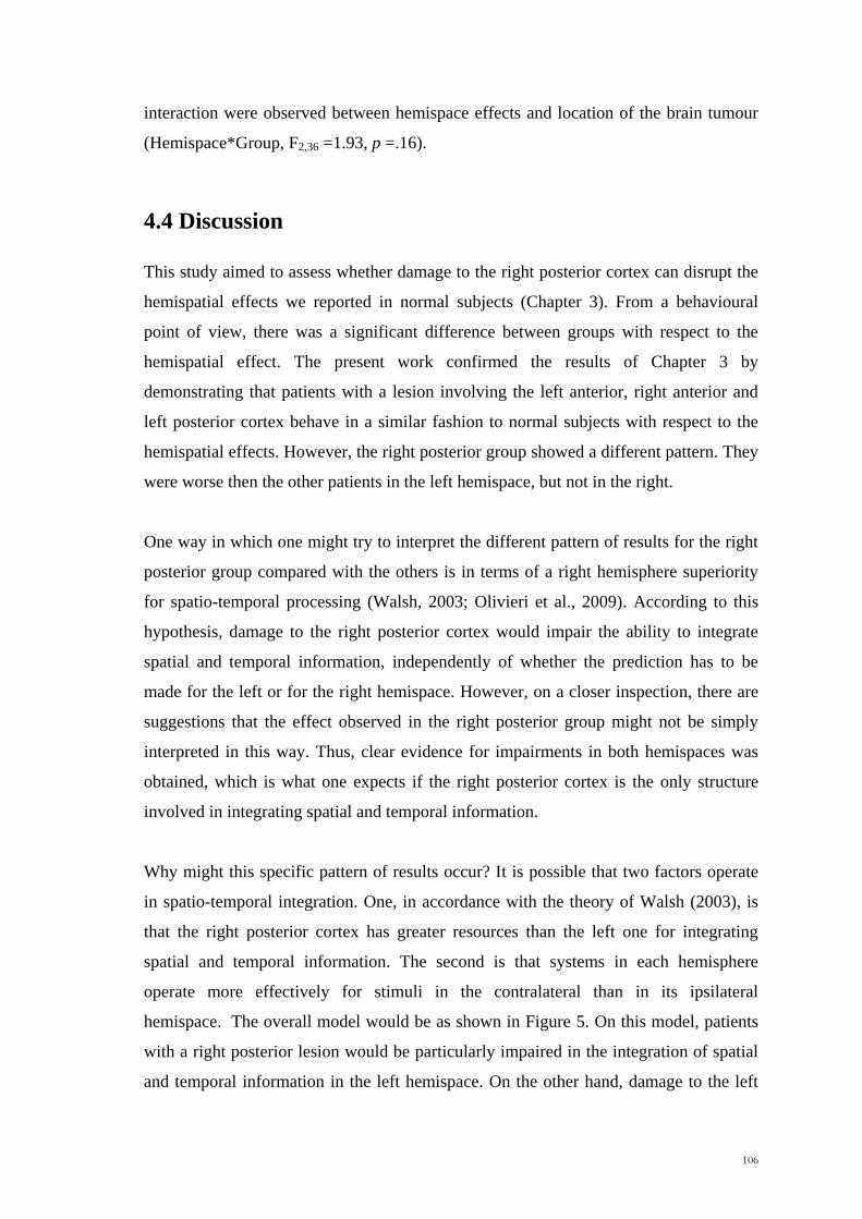
hemispace. The overall model would be as shown in Figure 5. On this model, patients
with a right posterior lesion would be particularly impaired in the integration of spatial
and temporal information in the left hemispace. On the other hand, damage to the left
107
posterior cortex would have less effect on the performance of patients, since the right
posterior cortex would combine spatial and temporal information with respect to targets
in both the left and right hemispace.
This hypothesis is supported by the correlation analysis we performed. The strong
positive correlation between the accuracy in the left hemispace and the right hemispace
in both healthy (pilot study) and non-parietal patients supports the idea that spatial and
temporal information are combined in a single module (placed in the right posterior
cortex, in unimpaired subjects). Damage to the critical right posterior module would
lessen the correlation as the left posterior system would be partially responsable for
right hemispace effects, as there were suggestions of this effect in the right posterior
group (Spearman correlation coefficient = .65, p =.04). The correlation of the right
posterior group is in fact weaker than the non-parietals (Spearman correlation
coefficient =.76) and the normal controls (Spearman correlation coefficient =.86).
However, in the model proposed, significant correlations between the accuracy in left
and right hemispace should also be observed in the left posterior group. We failed to
find this positive association. Since the number of patients with left posterior lesion that
could be tested in the present study was very small (N=5), the model we propose clearly
needs to be tested by future studies.
108
One alternative hypothesis to explain the lower performance of the right posterior group
in the left hemispace is in terms of neglect. In the present study, clear signs of left
neglect were not observed in any of the six right posterior patients who were tested (see
Table 2, p.82). Moreover, no significant differences in RTs and MTs were observed
between the four groups of patients and more critically, no hemispatial effects in these
measures were found. Therefore, a possible role of neglect in the spatio-temporal
integration impairments remains only a remote possibility.
Of course, in this study we only investigated a single task, the most basic spatio-
temporal one. Other investigations would be needed to assess whether the spatio-
temporal system like the trajectory setting and reaching ones involved in optic ataxia
are influenced by the hand used. Conceivably, one could obtain effects analogous to
those we obtained with respect to hemispace. In addition, the possibility needs to be
considered that slowing up of RTs occurring in the spatial condition could lead to the
involvement of the ventral route.
109
Chapter 5
Two qualitatively different impairments in making rotation operations
Buiatti T, Mussoni A, Toraldo A, Skrap M, & Shallice T (2011). Two qualitatively different impairments
in making rotation operations. Cortex, 47(2): 166-179.
110
111
5.1 Introduction
In the previous chapters, processes underlying reaching and spatio-temporal integration
were investigated in detail. In this chapter we will focus on a more cognitive process,
such as mental rotation. The aims of the study reported in the current chapter are
twofold. A first purpose is to investigate whether a unilateral brain tumour occurring in
different part of the brain, such as the prefrontal, premotor and parietal cortex lead to
impairments in performing mental rotation operations. A second aim is to further
investigate the categorical-metric account (Kosslyn et al., 1989) by means of
neuropsychological tools (see paragraph 1.3.2.1, Chapter 1). For this purpose, we used
the experimental paradigm based on the previous work by Bricolo et al. (2000), which
required patients to remember the position of a dot inside an upright or a tilted frame of
reference and to reproduce it inside a subsequent identical upright reference frame after
the frame was re-oriented vertically.
In this work, we used different methods of analysis. The first traditional methodology
used was an anatomically based group study approach. In the initial comparisons,
following the procedure of Stuss et al. (2005), the relative performance of patients with
tumours in six different regions of cortex was contrasted. This analysis allowed us to
investigate the contaminating effects of variables such as lesion size and age. This
procedure was then followed by an examination of the lesion sites of poorly as opposed
to satisfactorily behaving patients. Here the procedure adopted was the Voxel Lesion
Symptom Mapping (VLSM) analysis (Bates et al., 2003; Rorden and Karnath, 2004,
Rorden et al., 2007). Finally, in order to validate the main findings of the group analysis
and to exclude any possibility that the pattern of responding observed was achieved by
chance, we also contrasted our empirical findings with a Monte-Carlo simulation study.
5.2 Methods
Patients
A total of 95 patients with a single circumscribed brain tumour confined in the left or
right prefrontal, premotor and parietal cortex were selected and tested in the
Neurosurgery Department (Santa Maria della Misericordia Hospital, Udine) within a
time period of about three years. Of these 95 patients, 40 were excluded by means of
112
the following criteria: (i) multiple or bilateral lesions; (ii) recurrence of the tumour; (iii)
hemianopia, severe neglect or right hand motor impairment, (iv) diagnosed stroke, head
injury or other neurological and psychiatric diseases. We performed the experimental
test on the remaining 55 patients (Tab. 1). All the 55 patients underwent the
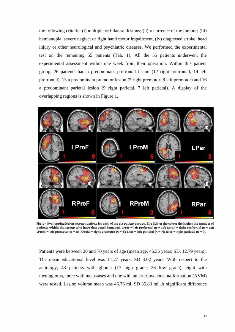
experimental assessment within one week from their operation. Within this patient
group, 26 patients had a predominant prefrontal lesion (12 right prefrontal, 14 left
prefrontal), 13 a predominant premotor lesion (5 right premotor, 8 left premotor) and 16
a predominant parietal lesion (9 right parietal, 7 left parietal). A display of the
overlapping regions is shown in Figure 1.
Patients were between 20 and 70 years of age (mean age, 45.35 years; SD, 12.79 years).
The mean educational level was 11.27 years, SD 4.02 years. With respect to the
aetiology, 43 patients with glioma (17 high grade; 26 low grade), eight with
meningioma, three with metastases and one with an arteriovenous malformation (AVM)
were tested. Lesion volume mean was 46.76 ml, SD 35.83 ml. A significant difference
1 1 1
1 1 1
113
between groups was found for lesion size [Kruskal-Wallis 2 = 11.88, p = .04].
Premotor patients tended to have smaller lesions than parietal and prefrontal patients.
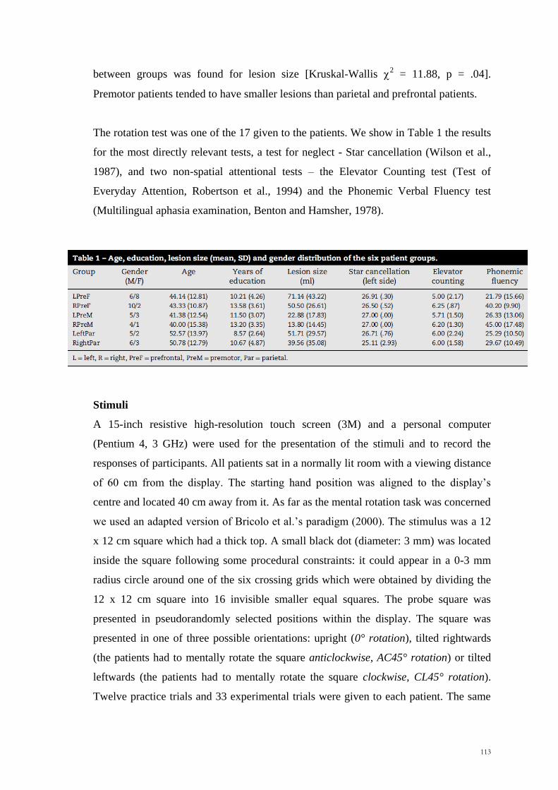
The rotation test was one of the 17 given to the patients. We show in Table 1 the results
for the most directly relevant tests, a test for neglect - Star cancellation (Wilson et al.,
1987), and two non-spatial attentional tests – the Elevator Counting test (Test of
Everyday Attention, Robertson et al., 1994) and the Phonemic Verbal Fluency test
(Multilingual aphasia examination, Benton and Hamsher, 1978).
Stimuli
A 15-inch resistive high-resolution touch screen (3M) and a personal computer
(Pentium 4, 3 GHz) were used for the presentation of the stimuli and to record the
responses of participants. All patients sat in a normally lit room with a viewing distance
of 60 cm from the display. The starting hand position was aligned to the display‘s
centre and located 40 cm away from it. As far as the mental rotation task was concerned
we used an adapted version of Bricolo et al.‘s paradigm (2000). The stimulus was a 12
x 12 cm square which had a thick top. A small black dot (diameter: 3 mm) was located
inside the square following some procedural constraints: it could appear in a 0-3 mm
radius circle around one of the six crossing grids which were obtained by dividing the
12 x 12 cm square into 16 invisible smaller equal squares. The probe square was
presented in pseudorandomly selected positions within the display. The square was
presented in one of three possible orientations: upright (0° rotation), tilted rightwards
(the patients had to mentally rotate the square anticlockwise, AC45° rotation) or tilted
leftwards (the patients had to mentally rotate the square clockwise, CL45° rotation).
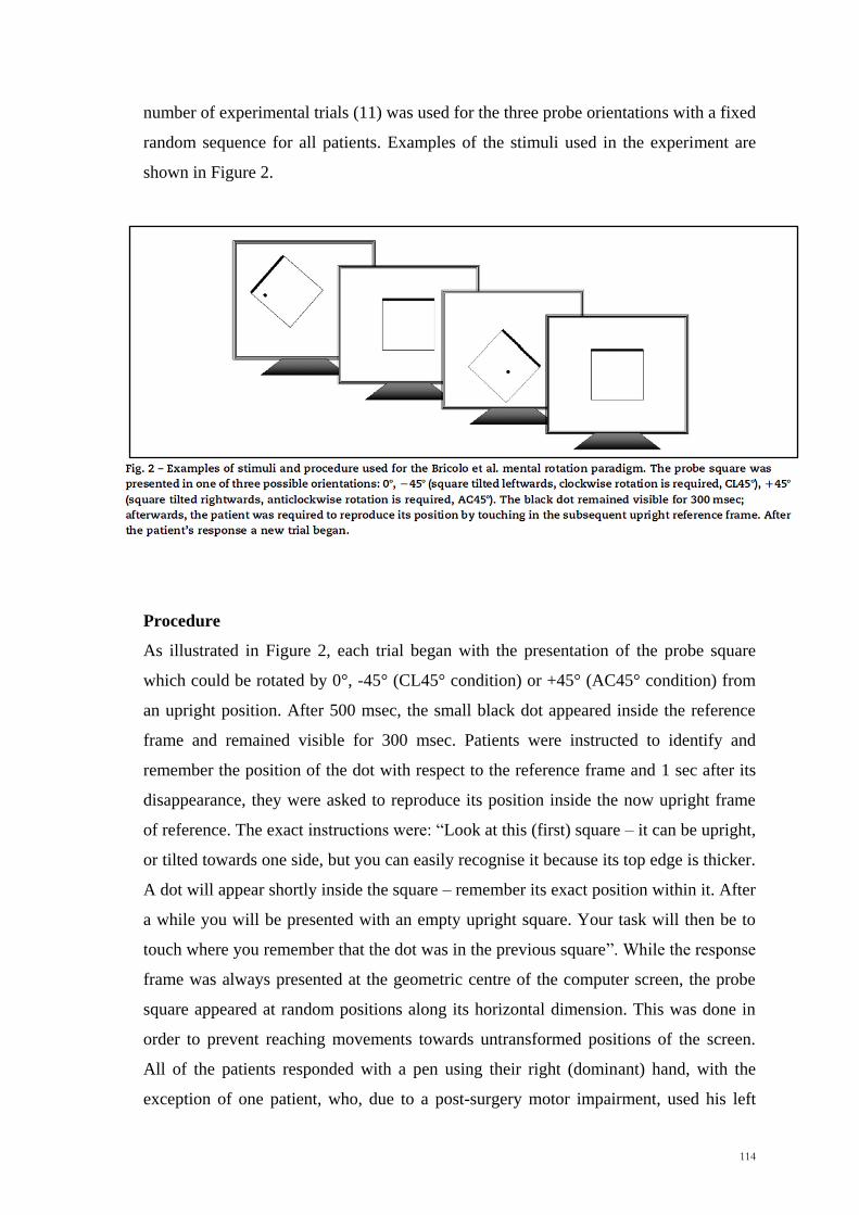
Twelve practice trials and 33 experimental trials were given to each patient. The same
114
number of experimental trials (11) was used for the three probe orientations with a fixed
random sequence for all patients. Examples of the stimuli used in the experiment are
shown in Figure 2.
Procedure
As illustrated in Figure 2, each trial began with the presentation of the probe square
which could be rotated by 0°, -45° (CL45° condition) or +45° (AC45° condition) from
an upright position. After 500 msec, the small black dot appeared inside the reference
frame and remained visible for 300 msec. Patients were instructed to identify and
remember the position of the dot with respect to the reference frame and 1 sec after its
disappearance, they were asked to reproduce its position inside the now upright frame
of reference. The exact instructions were: ―Look at this (first) square – it can be upright,
or tilted towards one side, but you can easily recognise it because its top edge is thicker.
A dot will appear shortly inside the square – remember its exact position within it. After
a while you will be presented with an empty upright square. Your task will then be to
touch where you remember that the dot was in the previous square‖. While the response
frame was always presented at the geometric centre of the computer screen, the probe
square appeared at random positions along its horizontal dimension. This was done in
order to prevent reaching movements towards untransformed positions of the screen.
All of the patients responded with a pen using their right (dominant) hand, with the
exception of one patient, who, due to a post-surgery motor impairment, used his left
115
(non-dominant). The upright response frame remained visible until the participants
responded. When they pointed to the touch screen, the stimulus disappeared and the
experimenter started the next trial by pressing the spacebar. Patients were given four
practice trials for each orientation condition. Each session lasts about ten minutes.
Data analyses
Behavioural data: For the data analyses we employed an anatomically based group
study approach that was based on the Stuss et al.‘s 2005 procedure. The methodology
used to infer brain-behaviour relations involved three levels of analysis:
(i) We selected and divided patients into six groups according to the side and the
predominant location of the brain tumour (right prefrontal, RPreF; left prefrontal,
LPreF; right premotor, RPreM; left premotor, LPreM; right parietal, RPar; left
parietal, LPar) and we first compared the performance among these groups.
(ii) If a significant overall effect was obtained, we compared the performance of each
group of patients with those of the other groups combined (e.g. RPar vs. RPreF,
LPreF, RPreM, LPreM, and LPar combined). In this way we were able to be more
specific about the location of any impairment with respect to our patient
population.
(iii) If we found a significant effect at this level, we performed more detailed analyses.
We applied the following procedure of error classification to the data set of each
individual patient (Toraldo and Shallice, in preparation):
1. Errors. An error was assigned when the patient reached out to a point more than
1.5 cm away from the correct position. The 1.5 cm criterion corresponds to the
25% of the width of one of the four quadrants into which the 12 x 12 square was
divided for the qualitative analyses (see below).
2. Classification of errors in spatial categories. The reference frame was
considered as a square divided into four quadrants (top-left, top-right, bottom-
left, bottom-right) and we determined whether the target‘s position and the
wrong response of the patients were in the same or in a different quadrant. In
this way, each response was broadly classified as ―Correct Quadrant‖ (CQ,
response in the correct quadrant but more than 1.5 cm away from target
position) or as ―Quadrant error‖ (Q, response in an incorrect quadrant). A
116
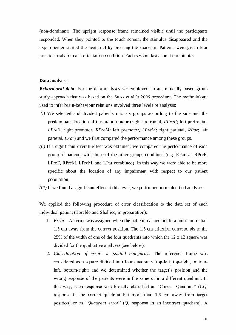
subsequent analysis was carried out on the direction of Quadrant errors. Thus,
we evaluated whether the Q errors were in the same (Q+) or in the opposite
direction (Q-) with respect to the required rotation (Figure 3).
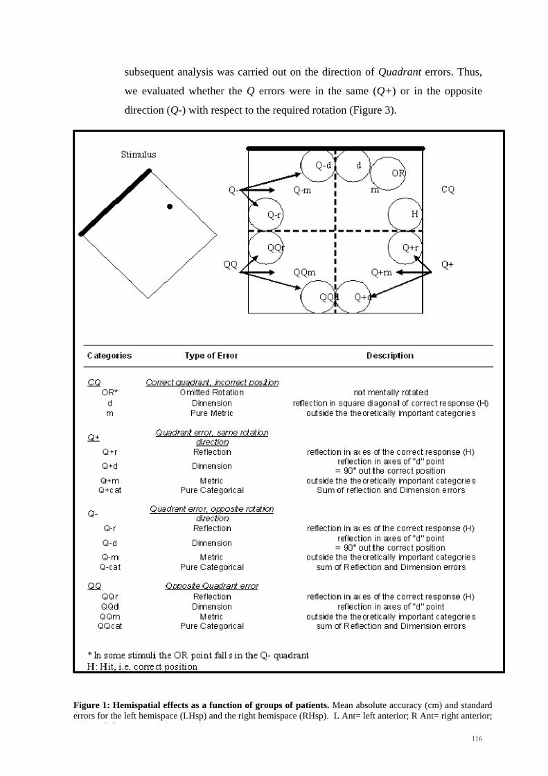
Figure 1: Hemispatial effects as a function of groups of patients. Mean absolute accuracy (cm) and standard
errors for the left hemispace (LHsp) and the right hemispace (RHsp). L Ant= left anterior; R Ant= right anterior;
L Post= left posterior; R Post= right posterior.
117
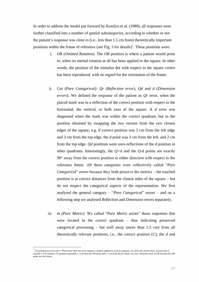
In order to address the model put forward by Kosslyn et al. (1989), all responses were
further classified into a number of spatial subcategories, according to whether or not
the patient‘s response was close to (i.e., less than 1.5 cm from) theoretically important
positions within the frame of reference (see Fig. 3 for details)1. These positions were:
i. OR (Omitted Rotation). The OR position is where a patient would point
to, when no mental rotation at all has been applied to the square. In other
words, the position of the stimulus dot with respect to the square centre
has been reproduced, with no regard for the orientation of the frame.
ii. Cat (Pure Categorical): Qr (Reflection error), Qd and d (Dimension
errors). We defined the response of the patient as Qr error, when the
placed mark was in a reflection of the correct position with respect to the
horizontal, the vertical, or both axes of the square. A d error was
diagnosed when the mark was within the correct quadrant, but in the
position obtained by swapping the two vectors from the two closest
edges of the square; e.g. if correct position was 2 cm from the left edge
and 3 cm from the top edge, the d point was 3 cm from the left, and 2 cm
from the top edge. Qd positions were axes-reflections of the d position in
other quadrants. Interestingly, the Q+d and the Q-d points are exactly
90° away from the correct position in either direction with respect to the
reference frame. All these categories were collectively called ―Pure
Categorical‖ errors because they both preserve the metrics – the touched
position is at correct distances from the closest sides of the square – but
do not respect the categorical aspects of the representation. We first
analysed the general category – ―Pure Categorical‖ errors – and on a
following step we analysed Reflection and Dimension errors separately.
iii. m (Pure Metric). We called ―Pure Metric errors‖ those responses that
were located in the correct quadrant – thus indicating preserved
categorical processing – but well away (more than 1.5 cm) from all
theoretically relevant positions, i.e., the correct position (C), the d and
1 Disambiguation procedure. When more than one error category could be applied to a given response, we chose the closest theo- retical point to
classify it. For instance, if a patient responded 1.1 cm from the OR point and 1.3 cm from the Q-r point, we clas- sified the error as OR because the OR
point was the closer.
118
the OR points. This indicates selective damage to the metric component
of the processing.
iv. Qm (Quadrant and Metric error). We called ―Qm‖ errors those
responses located in an incorrect quadrant and outside all of the
theoretically important areas (d and r).
Kosslyn et al.‘s (1989) distinction between categorical and metric processing is best
characterized, in this error classification procedure, by classes (ii) and (iii) above, i.e.
―Pure Categorical‖ and ―Pure Metric‖ errors.
The raw data were first checked for normality using the Kolmogorov-Smirnov test and
for homogeneity of variance by the Levene test. As the data were not normally
distributed, non-parametric tests were used. The results were considered significant if
the p value was < .05. All the significant tests were two- tailed unless otherwise
specified.
Anatomical data: The pre-operative location of the tumour was carried out using a
digital format contrast-enhanced t1-weighted MRI scan obtained 1-2 days before
operation, using a 1.5T machine and a GRE-3D T1-weighted scan (TI 600 msec, TR
1400 msec, TE 31 msec, TH 1 mm, DF 1 mm); this image was selected as it is the scan
generally used by the neurosurgeon during the operation with the Neuronavigator as the
best indicator of macroscopic tumour extent. MRicro reconstructional software was
used to extrapolate a 3D representation of the lesion from digital MR scans (Rorden and
Brett, 2000). The boundary of a lesion was drawn as a region of interest (ROI) on each
sagittal slide in collaboration with the neurosurgeon and a neuroradiologist, who did not
know the behavioural results, so as to limit the lesion‘s boundary to the brain tissue
removed during the surgical approach. The scans and ROIs were normalized using
SPM05b with 12 affine transformations and 7 x 9 x 7 basis functions. Each patient‘s
lesion was referred to an anatomical template image AAL (Automated Anatomical
Labelling) (Tzourio-Mazoyer et al., 2002), a macroscopic anatomical parcellation of
MNI volume (Collins et al., 1998). Afterwards, the Voxel-based Lesion-Symptom
Mapping analyses were run. The procedure allows one to use the statistical relation
between behavioral data and the specific voxels affected by the lesion without grouping
119
patients for lesion location or relying on behavioral cut-offs (Bates et al., 2003; Rorden
and Karnath, 2004). The Non-Parametric Mapping software (NPM) (Holmes et al.,
1996) was used to run the Brunnel-Munzel test (Brunner and Munzel 2000) and
compute a statistical map for continuous variable results (Rorden et al. 2007). The
results are shown using Bonferonni corrected significance values, requiring a minimum
of three patients affected for a voxel for it to be included.
5.3 Results
The Background variables were analysed first. No significant differences were found
among the six groups for educational level [F(5,48) = 1.6, p = .18] and age [F(5,48) =
0.77, p = .57]. We also studied the effects of the variables on performance in our
experimental task. Age did not significantly influence error rate in the two rotation
conditions combined [F(1,46) = 1.13, p = .29, with error rate being normalized by a log-
transformation]. A significant effect of educational level on error rate was, however,
found [F(1,46) = 4.77, p = .03].
Overall error analysis
An exploratory analysis was performed by comparing the overall number of errors in
the rotation conditions (CL45° and AC45° combined) and in the non-rotation condition
(0°). The average number of errors was greater for the CL45°/AC45° conditions
combined than for the 0° condition. This result was significant for almost all patient
groups [LPreF: z = -2.94, p = .002, RPreF: z = -2.49, p = .007, LPreM: z = -1.81, p =
.036, RPreM: z = -1.00, p = .159, LPar: z = -1.63, p = .051, RPar: z = -2.67, p = .004;
one-tailed Wilcoxon Signed Rank tests]. No significant differences were observed by
comparing the number of error responses in the CL45° and the AC45° rotation
conditions within each patient group (for all groups: p >.05, Wilcoxon Signed Rank
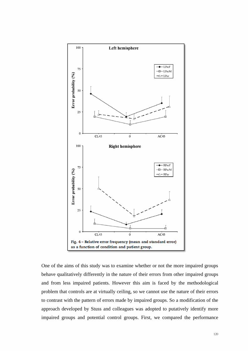
test) (Figure 4).
A one-way non-parametric ANOVA across all six groups on the number of errors
occurring in the rotation conditions (CL45°/AC45°) showed that the groups differed
significantly [Kruskal-Wallis; 2(5) = 13.18, p = .02].
120
One of the aims of this study was to examine whether or not the more impaired groups
behave qualitatively differently in the nature of their errors from other impaired groups
and from less impaired patients. However this aim is faced by the methodological
problem that controls are at virtually ceiling, so we cannot use the nature of their errors
to contrast with the pattern of errors made by impaired groups. So a modification of the
approach developed by Stuss and colleagues was adopted to putatively identify more
impaired groups and potential control groups. First, we compared the performance
121
among all the six groups to determine whether there was a significant difference
between them. Given at what was found, we then contrasted the performance of each
group of patients with the other five groups combined. At this stage the left prefrontal
group differed significantly from the other five (CL45°/AC45° errors combined, Mann-
Whitney U=178, p = .035), but the right parietal did not. In order to investigate whether
there were differences among the five groups, we removed the left prefrontal group and
repeated the analogous procedure. On this second round only the right parietal group
performed statistically worse than the other groups combined (RP: U=77, p = .03;
Mann-Whitney). Repeating the procedure a third time did not lead to any new
significant effects (p > .35). We will therefore putatively take the right parietal and left
prefrontal groups as impaired groups and treat the other groups combined as a control
group.
In order to determine whether the effects observed were related to differences in lesion
size, we correlated the patients performance in the CL45°/AC45° conditions combined
with lesion size. Overall the correlation of the total number of errors with lesion size
was completely insignificant [F(1,46) = 0.83, p = .37]. Spearman correlation
coefficients for the six subgroups ranged from -.87 to .42 for the six groups, in all cases
being far from significance (p >.3).
Direction of errors
The statistical analyses revealed that both the right parietal and the left prefrontal
groups made a significantly greater number of Quadrant errors (Q) compared to the
other four groups combined [RPar vs. Others: Mann-Whitney U=75, p = .03; LPreF vs.
Others: Mann-Whitney U=130, p = .02]. For the negative Quadrant errors (Q-) –
moving in the direction opposite to that of the rotation required – the right parietal
patients made a significantly greater number than the other groups combined [RPar vs.
Others: Mann-Whitney U=62, p = .01]. However, for the positive Quadrant errors (Q+)
– moving too far in the same direction as that of the required rotation, the left prefrontal
patients made a larger number than the other groups combined [LPreF vs. Others:
Mann-Whitney U=153.5, p = .04] (Table 2).
When a direct comparison of the number of Q- and Q+ errors within each group was
carried out, a significant effect was found for the right parietal patients with the Q-
122
errors being the more frequent (Wilcoxon Signed Rank test z = -2.54, p = .01). The four
patient control groups combined also showed significantly more than Q+ errors (z = -
2.52, p = .01). However, in the left prefrontal group, the difference was far from
significant (p = .51). If we consider the direction in which the square has to be rotated,
clockwise (CL45°) versus anticlockwise (AC45°), the right parietal patients showed a
similar rate of Q- errors in both conditions – no significant difference could be detected.
In other words, the right parietal patients tended to rotate in an incorrect direction more
than the other lesion control groups irrespective of the direction required, clockwise or
anticlockwise.
Qualitative differences in error types: Metric and Categorical errors
The analysis of Quadrant errors showed us that gross group differences emerged with
respect to the direction of the error. We then investigated whether the errors could arise
from the malfunction of a purely metric or categorical process. For this reason we
considered the number of errors that fell into three qualitative error categories. One type
is the error that would arise if the patient did not perform a rotation operation and
responded on the basis of the initial position of the target point (Omitted Rotation)2 A
second is if the patient produced a response in the reflection of the correct response
point with respect to the horizontal, the vertical, or both axes of the square (Reflection
error). The third is if the patient made the correct metric operation on the target point
but used an incorrect neighbouring side or corner as the starting point for the metric
operation (Dimension error); these were the type of categorical errors described by
Bricolo et al. (2000) and Toraldo and Shallice (in preparation) in individual right
hemisphere patients. These last two types were collectively considered as ―Pure
Categorical‖ errors. Symmetrically, we identified another category as ―Pure Metric‖
errors, i.e., locations of the response mark that unambiguously suggest a specific
impairment of metric information processing, with spared categorical information: this
area is the part of the correct quadrant which is outside of all the theoretically relevant
areas (OR, d, correct target position). A final error type, which is not purely categorical,
is the Quadrant and Metric error, which occurs when the patients place the mark in an
incorrect quadrant and outside all the theoretically important areas listed above.
2 The omitted rotation point (OR) falls in the correct quadrant in some trials, and in the Q- quadrant in some others, according to where the target is
located within its quadrant.
123
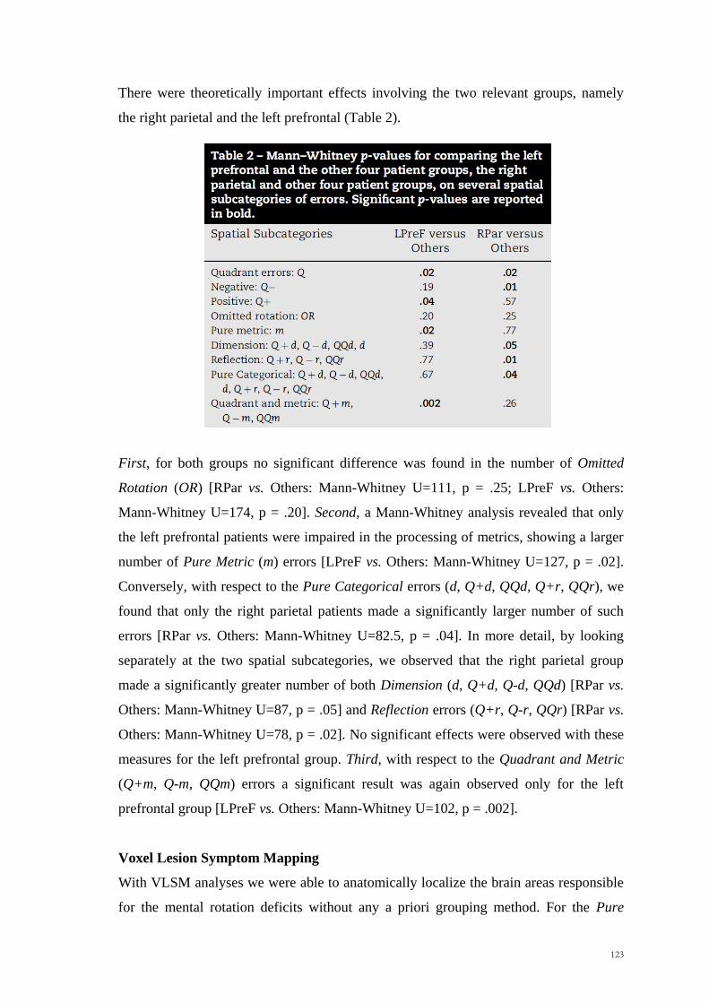
There were theoretically important effects involving the two relevant groups, namely
the right parietal and the left prefrontal (Table 2).
First, for both groups no significant difference was found in the number of Omitted
Rotation (OR) [RPar vs. Others: Mann-Whitney U=111, p = .25; LPreF vs. Others:
Mann-Whitney U=174, p = .20]. Second, a Mann-Whitney analysis revealed that only
the left prefrontal patients were impaired in the processing of metrics, showing a larger
number of Pure Metric (m) errors [LPreF vs. Others: Mann-Whitney U=127, p = .02].
Conversely, with respect to the Pure Categorical errors (d, Q+d, QQd, Q+r, QQr), we
found that only the right parietal patients made a significantly larger number of such
errors [RPar vs. Others: Mann-Whitney U=82.5, p = .04]. In more detail, by looking
separately at the two spatial subcategories, we observed that the right parietal group
made a significantly greater number of both Dimension (d, Q+d, Q-d, QQd) [RPar vs.
Others: Mann-Whitney U=87, p = .05] and Reflection errors (Q+r, Q-r, QQr) [RPar vs.
Others: Mann-Whitney U=78, p = .02]. No significant effects were observed with these
measures for the left prefrontal group. Third, with respect to the Quadrant and Metric
(Q+m, Q-m, QQm) errors a significant result was again observed only for the left
prefrontal group [LPreF vs. Others: Mann-Whitney U=102, p = .002].
Voxel Lesion Symptom Mapping
With VLSM analyses we were able to anatomically localize the brain areas responsible
for the mental rotation deficits without any a priori grouping method. For the Pure
124
Categorical Errors patients with lesions in the right inferior parietal cortex showed a
significant involvement. On the other hand, the common area for the Pure Metric and
Quadrant and Metric errors was the left insula verging on the putamen. All these
anatomical loci survived Bonferroni corrections.
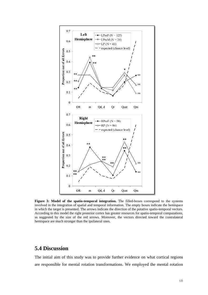
Monte-Carlo simulation
In order to test whether the qualitative impairments observed in the right parietal and in
the left prefrontal groups truly reflected a mental transformation deficit and were not
just the effect of random selection of locations within the square, we additionally
performed a Monte Carlo simulation study to obtain chance levels. We generated
random positions within the square as responses to each of the 33 stimuli that have been
really administered, and repeated this procedure 10,000 times. On each of the 10,000
samples, we applied the same error classification procedure that was applied to real data
from patients. For each spatial subcategory we compared the probability of an error
occurring by chance (expected probability) with the observed probability. The binomial
tests revealed that the error proportions in the Pure Metric and Quadrant and Metric
subcategories were significantly lower than chance in the left prefrontal group.
Conversely, the observed proportions of Pure Categorical errors were more frequent
than expected by chance in the right parietal group. These findings clearly indicate that
the incorrect responses of patients in theoretically important regions did not occur by
random selection of points in the square (Figure 5).
125
Figure 3: Model of the spatio-temporal integration. The filled-boxes correspond to the systems
involved in the integration of spatial and temporal information. The empty boxes indicate the hemispace
in which the target is presented. The arrows indicate the direction of the putative spatio-temporal vectors.
According to this model the right posterior cortex has greater resources for spatio-temporal computations,
as suggested by the size of the red arrows. Moreover, the vectors directed toward the contralateral
hemispace are much stronger than the ipsilateral ones.
5.4 Discussion
The initial aim of this study was to provide further evidence on what cortical regions
are responsible for mental rotation transformations. We employed the mental rotation
126
task developed by Bricolo et al. (2000), but we used an anatomically based group study
approach rather than a single-case method. Each patient was assigned to one of six
groups, namely left prefrontal, right prefrontal, left premotor, right premotor, left
parietal, right parietal. A broad analysis on the number of error responses in the rotation
conditions revealed that the six groups performed in a significantly different way. We
used a modification of the procedure adopted by Stuss et al. (2005) to determine
candidate impaired groups. This procedure selected the left prefrontal and right parietal
groups, which did not differ significantly from each other for the overall number of
errors, as candidate impaired groups; the other four groups were treated collectively as a
patient control group. The appropriateness of this candidate categorisation was
supported by the analyses carried out on the qualitative nature of the errors, which
revealed that the impairments in the left prefrontal and right parietal groups were
significantly different in a number of ways from the other patient groups combined.
These include findings on the direction of errors, namely the positive Quadrant errors
for the left prefrontal group and the negative Quadrant errors for the right parietal
group. In addition if one considers the qualitative error classification, there were again
significant effects for the left prefrontal group with respect to Pure Metric and
Quadrant and Metric and for the right parietal group with respect to Dimension and
Reflection errors.
Right parietal group
The analysis of the overall error rate indicated that patients with a lesion centered on the
right parietal cortex made a significantly larger number of errors with respect to the
other four patient groups combined. Particularly, in the two rotation conditions about
44% of all trials were errors, which is definitely a sizeable effect. This result supports
the widely accepted claim that the right parietal cortex is specifically involved in mental
rotation transformations, which is consistent with previous neuropsychological, EEG,
TMS and neuroimaging researches (Ratcliff, 1979; Inoue et al., 1998; Harris et al.,
2000; Harris and Miniussi, 2003). In detail, by looking at the qualitative nature of these
errors we observed that the right parietal patients were specifically impaired in the
processing of categorical spatial information. Indeed, they produced a significant
number of Pure Categorical errors, which occur when one ignores the qualitative
spatial cues without any metric impairment. If a patient operates correctly metrically
with respect to a landmark, say a corner, but chooses an incorrect neighbouring corner
127
for the operation, this produces a Dimension error. The patient‘s performance is
metrically correct, but categorically incorrect. One subset of such errors (Q-d)
corresponds to rotating the square in the incorrect direction. The right parietal group
made significantly more Dimension errors than the other four control patient groups
combined. If a patient takes a reflection of the position of the target with respect to the
horizontal, the vertical, or both axes of the square, this is a Reflection error. S/he places
the mark in a complementary horizontal or vertical position in an incorrect quadrant,
failing to take into account the categorical representation of the target. Right parietal
patients also produced significantly more such errors than the other four patient groups
combined. These results were confirmed by a subsequent simulation study, which
showed that the proportion of categorical error responses were significantly greater than
would be expected by random selection of locations in the square.
Moreover, we observed that unlike the patient control groups the right parietal patients
showed a greater tendency to rotate the square in the wrong direction (Q- errors). We
believe that this behavior reflects a deficit, which is specifically qualitative in nature.
One could argue that this significant frequency of Q- errors might instead reflect lack of
precision in applying the appropriate spatial transformations (angles). However, if this
hypothesis holds true, then it would remain unexplained why in the right parietal group
both the Pure Metric and the Quadrant & Metric error rates were not statistically
different from those in patient control groups, or even from chance (Monte Carlo
simulation). In fact errors clustered in categorically important positions of the square.
An alternative hypothesis is that the findings observed in the right parietal group might
be explained in terms of neglect. In a study performed by Kerkhoff and Zoelch in 1998,
it has been observed that when asked to orient an oblique line (―target‖) to match a
horizontal, vertical or 45° reference line, neglect patients with a right hemisphere lesion
showed a significant anticlockwise tilt of the target. In the present study, signs of
neglect on the Star Cancellation task (Wilson et al., 1987) were observed in three out of
nine right parietal patients. All three were in the subset of five patients making the
larger number of categorical errors. Of the other two patients in this subset, one
obtained a perfect score on Star Cancellation and the other had a poor but not
lateralized performance. However, with respect to our rotation task, it is not likely that
the pattern of results shown by right parietal patients can be explained just in terms of
an indirect effect of neglect. If neglect had had a major role, one would have predicted a
128
sizable difference in performance according to the direction – clockwise (CL45°) vs.
anticlockwise (AC45°), of the required rotation. Following Kerkhoff and Zoelch
(1998), neglect should induce a bias towards performing anticlockwise rotations,
resulting in more frequent errors in those trials where the opposite rotation is required,
i.e. the CL45° condition. The same prediction is derived by another possible scenario
related to neglect: in the CL45° condition the thick side is in the left half of the tilted
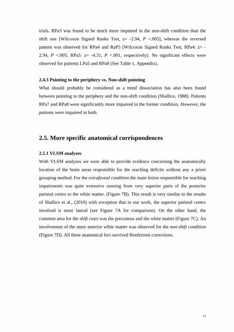
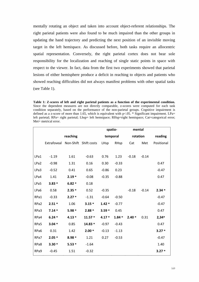
stimulus; failure to detect the thick side would induce random selection of rotation
direction, with consequent Q- errors being more frequent in this CL45° than in the
AC45° condition. However, no such effect was found, with right parietal patients who
make roughly comparable numbers of errors in the two conditions. Indeed, they made
more errors than patient controls in rotating leftwards when presented with a CL45°
stimulus, and rightwards when an AC45° stimulus was displayed. In other words, they
had an increased tendency to rotate in the wrong direction, whichever direction was
required on a trial. A possible partial role of neglect in the categorical errors remains a
possibility.
Martin et al. (2008) have argued that both hemispheres are involved in coding both
metric and categorical positions for a Continuous Spatial Coding hypothesis in which
both hemispheres are implicated in both types of spatial relation coding. They found
some degree of hemispheric specialization, not related to the categorical/metric nature
of the task, but to the processing load involved. Thus for instance, they found a right
hemisphere advantage in the inferior parietal lobule and the angular gyrus. In many
respects our evidence fits well with their findings: we also obtained greater involvement
of the right parietal than the left parietal cortex in the task. However unlike in the work
of Martin et al., we did not find any deficits in right parietal patients in carrying out
metric operations per se. This dissociation – a categorical deficit without a metric
deficit – is difficult to reconcile with the Continuous Spatial Code hypothesis, which
had explained Martin et al.‘s findings well. This model assumes that categorical and
metric relations differ on a complexity continuum, with metric encoding being
generally more complex than categorical encoding. In this case, however, one would
expect that a lesion would produce the complementary dissociation, with categorical
relations being relatively spared.
129
It is plausible that the difference between the present study and Martin et al.‘s is due to
the different type of task used. Martin et al. used a working memory task, in which
rotation was not involved. In such a task, metric accuracy is likely to be more stressed
than categorical accuracy, as no transformation is required. By contrast, our rotation
task is more heavily loaded on categorical operations than on metric accuracy, both
because of the need to rotate, and because the absolute metric error allowed was quite
large (1.5 cm).
In summary, we agree with Martin et al. on the likely involvement of both hemispheres
in both metric and categorical operations. However, the existence of an above chance
rate of error types such as Reflection and Dimension strongly suggests that the two
types of operation can be separately impaired. Indeed in Reflection and Dimension
errors, responses are very close to simple geometrical transformations of the correct
position. Thus a gross categorical mistake and a fine metric analysis are simultaneously
observed. Such a dissociation is even more convincing if other error types involving
metric-based inaccuracies do not occur at above chance levels. This profile was
previously reported in an individual case study of Bricolo et al. (2000) and is also the
case for the right parietal group in the current study.
How might such errors be explained? A typical analogue rotation process (à la Shepard
& Meltzer) would predict very different error patterns from the ones we observed. It
should be noted that our task, while corresponding to operations often made in the daily
life, is very different from the tasks standardly used in ―mental rotation‖ experiments.
Indeed, it allows another strategy in addition to the analogue rotation procedure.
Suppose that the spatial analysis of the figure is carried out in two main steps, (i)
categorical operations are carried out to relate parts of the figure to an object-centred
reference frame – known to be important, for instance, in neglect (Behrmann and
Moscovitch, 1994; Driver, 1998; Humphreys et al., 1996, Humphreys and Riddoch,
1995), and subsequently (ii) metric operations are carried out with respect to crucial
parts of the figure. It would then follow that our task allows subjects the much easier
possibility of not actually carrying out an analogue rotation operation. Instead, the
subject might store the categorical and metric encodings from the first square, and
reproduce them on the second square. This would only be possible if subjects could
categorically organise the figure in terms of an object-centred reference frame. The
130
gross spatial agnosia shown clinically by many right parietal patients (e.g. Warrington,
1969) suggests that this may not be possible in some patients of this group. In this case
Reflection and Dimension errors would correspond to a failure of one of the categorical
operations stages of the process.
More specifically, we suggest that poor performance in our mental rotation task could
be explained by an impairment of one or more steps of the following procedure:
1. Implement a correct object-centred reference frame on the first (tilted)
square.
2. Carry out a categorical encoding of the position of the dot.
3. Carry out a metric encoding of the position of the dot.
4. (Following presentation of the upright empty square), retrieve the
object-centred reference frame.
5. Retrieve the appropriate categorical representation.
6. Retrieve the metric representation.
Our proposal is that a lesion of the right parietal cortex may disrupt the object-centred
system of reference, the categorical spatial representation of the target, or both. The
account is motivated by the need to explain the qualitative impairments we observed in
our clinical population. New investigations would be needed to test whether other
predictions of the model are correct.
Left prefrontal group
A second group of patients was impaired in the performance of our mental rotation task,
namely the left prefrontal group. More detailed determination of the anatomical locus
involved was limited by characteristics of our patient series, namely a lack of patients
with tumours involving the more superior parts of prefrontal cortex.
The left prefrontal group had a different type of mental transformation deficit with
respect to right parietal patients. They produced a significant increase in the number of
metrically incorrect responses both in the correct and in the incorrect quadrants. This
finding is in agreement with the study of Martin et al. (2008), who found a strong
recruitment of the attentional and executive processes, especially when metric coding
was required. In addition, with respect to the other four groups combined the left
131
prefrontal group was the only one to produce a relatively large number of errors where
rotation was too far in the correct direction (Q+). They also made a similar number of
errors of rotation in the wrong direction (Q-). The specific mental rotation impairment
of the left prefrontal patients might be explained in different ways.
One possible explanation is that the deficits found in the left prefrontal patients are due
to impairments in the short-term retention of spatial information. Indeed activity in the
DLPFC has been often observed in both humans and primates in tasks which require the
retention of spatial information for a limited period of time (Wilson et al., 1993,
Courtney et al., 1996, 1998; Owen et al., 1996; Levy and Goldman-Rakic, 2000; Wager
and Smith, 2003). However, lesions to the right prefrontal cortex impair spatial working
memory more than ones to the left (Bor et al. 2006), so this makes this account less
plausible for a specifically left prefrontal deficit.
A second account would be in terms of a difficulty in producing the appropriate
amplitude for the motion response. Desmurget et al. (2004) presented results that are
clearly supportive of a role of the basal ganglia in advance planning of movement
extent. Patients with Parkinson disease were found to be selectively impaired in using
advance information about movement amplitude. Moreover, in a subsequent PET
experiment increased neural activation in the rostral and caudal portions of the bilateral
putamen was specifically observed in a task requiring amplitude planning. The results
found for some of the patients placed in our left prefrontal group would fit well with
damage or a disconnection of the putamen (three patients), but this would be a less
satisfactory account for patients with a more specifically prefrontal damage. Moreover,
a hypothetical amplitude planning deficit could well affect the baseline condition too
(0°, no rotation); however our left prefrontal group was not specifically impaired in
such a condition.
One related question is why no sign of any such impairment was found in the right
prefrontal group. As reported in the overall error analysis, the performance of the right
prefrontal patients was not statistically different from that observed for the other groups
combined. The absence of effects cannot be a problem of lack of statistical power.
Indeed the sample size was similar for right prefrontal (N = 12) and left prefrontal (N =
132
14) groups and the difference in the overall number of errors was sizeable and
significant (averaging at, respectively, 5.7 and 11 out of 33; Mann-Whitney p =.041).
Since the subjects almost gave their responses with their right hand, it might be
suggested that the greater impairment of the left and in the right prefrontal patients
simply reflect their using of the right hand. This possibility cannot be ruled out.
However, if a lateralized hand effect would contribute to the results, then one might
expect a greater impairment in the left premotor group, which was not found.
As a third possible explanation we suggest that the pattern of performance found in the
left prefrontal group arises from a set of processes related to acquiring action
operations. This are the so-called task setting operations (Stuss et al., 1995; Alexander
et al., 2005; Shallice et al., 2008a, 2008b) specifically impaired in left prefrontal
lesions. Task-setting is the collective name for the processes involved in learning in
going from a novel set of operations when the subject is initially faced by a new task to
their smooth well-learned execution after repeated practice. A left prefrontal lesion
would be expected to increased error rates early in task performance because of
impairments in task-setting. In our study the task was very short requiring only 5
minutes to be completed. Thus the errors occurred before the task was over-learned. We
propose that the failure on the task of the left prefrontal patients arises because they do
not acquire the specific categorical and metric operations listed in the section above
[―Right Parietal group‖ (the six-step procedure)]; instead they would fall back on a
rough rotation operation, with little control over its correct angular size, failing to carry
out a proper metric or a categorical encoding. This hypothesis would explain the
specific pattern of performance of the left prefrontal patients and in particular the
relatively large number of quadrant errors in the same direction as that of the rotation
required (Q+ errors), and the high incidence of metric errors.
133
Chapter 6
Phonological dyslexia following left and right parietal lesions
134
135
6.1 Introduction
The development of models of the reading process has been intimately linked to the
investigation of the acquired dyslexias. Thus the argument of Marshall & Newcombe
(1973) that there are two different routes through which letter strings can be read was
based on the contrast between the properties of deep and surface dyslexia. The
assumption that there are three routes for reading aloud not two, while originally
suggested by Coltheart (1978) on the basis of studies in normal subjects, received a
major boost from the identification of reading without semantics by Schwartz et al.
(1979). In this syndrome the patient has no understanding of words irregular in their
spelling-to-sound correspondences but they can be read aloud well.
More recently, with the development of models of reading aloud, such as those of Plaut
et al. (1996), Coltheart et al. (1993, 2001), Zorzi, Houghton & Butterworth (1998) and
Perry, Ziegler & Zorzi (2007) a major bone of contention between the protagonists of
the different theories has concerned their ability to account for the existence of
individual neuropsychological syndromes, and in particular for reading without
semantics and phonological alexia (see e.g. Coltheart, 2006; Woollams et al., 2007).
Phonological alexia, originally described by Beauvois & Derouesne (1979), is the
disorder in which the patient can read words well but is impaired at reading
orthographically and phonologically legal non-words. That the syndrome was
theoretically important for understanding the mechanisms of normal reading was argued
very early by Funnell (1983) on the basis of the existence of a patient of this type who
had little comprehension of words. The disorder was held to provide evidence for the
existence of separate routes for lexical and non-lexical reading aloud, both being
distinct from a semantic route. Clearly the existence of this pattern of performance is
easily explained if three routes exist, as in the models of Coltheart et al. (2001) and
Zorzi et al. (1998); the pattern can then arise from the selective impairment of a non-
lexical reading route. However, proponents of connectionist two-route models had an