PARP-1 transcriptional activity is regulated by sumoylation upon heat shock Nadine Martin 1,5 , Klaus Schwamborn 1,6,9 , Vale ´ rie Schreiber 2,9 , Andreas Werner 1,7 , Christelle Guillier 3,8 , Xiang-Dong Zhang 4 , Oliver Bischof 1 , Jacob-S Seeler 1, * and Anne Dejean 1, * 1 Department of Cell Biology and Infection, Nuclear Organisation and Oncogenesis Unit, INSERM U579, Institut Pasteur, Paris, France, 2 IREBS- FRE3211, CNRS, Universite ´ de Strasbourg, ESBS, Illkirch, France, 3 Plate- forme prote ´omique, Institut de Biologie Mole ´culaire et Cellulaire, CNRS, Strasbourg, France and 4 Department of Biochemistry and Molecular Biology, Johns Hopkins University, Bloomberg School of Public Health, Baltimore, MD, USA Heat shock and other environmental stresses rapidly induce transcriptional responses subject to regulation by a variety of post-translational modifications. Among these, poly(ADP-ribosyl)ation and sumoylation have received growing attention. Here we show that the SUMO E3 ligase PIASy interacts with the poly(ADP-ribose) polymerase PARP-1, and that PIASy mediates heat shock-induced poly-sumoylation of PARP-1. Furthermore, PIASy, and hence sumoylation, appears indispensable for full activa- tion of the inducible HSP70.1 gene. Chromatin immunopre- cipitation experiments show that PIASy, SUMO and the SUMO-conjugating enzyme Ubc9 are rapidly recruited to the HSP70.1 promoter upon heat shock, and that they are subsequently released with kinetics similar to PARP-1. Finally, we provide evidence that the SUMO-targeted ubi- quitin ligase RNF4 mediates heat-shock-inducible ubiquiti- nation of PARP-1, regulates the stability of PARP-1, and, like PIASy, is a positive regulator of HSP70.1 gene activity. These results, thus, point to a novel mechanism for regulat- ing PARP-1 transcription function, and suggest crosstalk between sumoylation and RNF4-mediated ubiquitination in regulating gene expression in response to heat shock. The EMBO Journal (2009) 28, 3534–3548. doi:10.1038/ emboj.2009.279; Published online 24 September 2009 Subject Categories: chromatin & transcription; proteins Keywords: heat shock; PARP-1; SUMO Introduction The cellular response to sudden environmental stress is characterized by a rapid activation phase, which is invariably followed by attenuation of the response, despite the persis- tent presence of the inducing signal. Many transcriptional regulatory mechanisms for this involve post-translational modifications, because their usually transient nature permits both rapid amplification and subsequent extinction of the transduced signals. The heat-shock response represents a well-characterized model system for the study of transcrip- tional responses to environmental stress. In mammals, a major consequence of heat shock is the activation of a number of heat-shock factors (HSFs) that drive the trans- criptional activation of heat-shock protein (HSP) genes that encode protein chaperones involved in protecting cellular functions from the deleterious effects of misfolded, aggre- gated, or mislocalized proteins (Morimoto, 1998, 2008). The factors that impinge on the regulation of HSP genes are, therefore, a subject of intense scrutiny. Among the proteins now known to play a key role in this regulation is the cellular sensor of DNA damage, poly(ADP- ribose) polymerase 1 (PARP-1, reviewed by Schreiber et al, 2006). PARP-1 is the most abundant and founding member of a super-family of proteins defined by their homology to the catalytic domain of PARP-1 that is responsible for the synth- esis of linear or branched polymers of ADP-ribose (PAR) from nicotinamide adenine dinucleotide (NAD þ ; Schreiber et al, 2006; Hakme et al, 2008). Poly(ADP-ribosyl)ation, besides being strongly induced by DNA-damaging agents, such as reactive oxygen (e.g. H 2 O 2 ), has been shown to exert major effects on chromatin structure and hence on the regulation of, particularly, transcriptionally active loci (for review, see Kraus, 2008). In Drosophila polytene chromosomes, for ex- ample, these PAR-containing loci are readily visible as puffs of de-compacted chromatin, thus providing perhaps the most striking evidence for the association of poly(ADP-ribosyl) ation with chromatin de-condensation (Tulin and Spradling, 2003). The concomitant rapid nucleosome loss, even prior to transcriptional onset, from the Drosophila HSP70 promoter region, has been shown to require PARP activity (Petesch and Lis, 2008). On nucleosomal DNA templates, PARP-1 was shown to occupy a position between nucleosomes, consistent with in vivo results showing that PARP-1 and linker histone H1 occupy distinct and mutually exclusive chromosomal regions (Kim et al, 2004; Krishnakumar et al, 2008). Ouararhni et al (2006) have extended these findings by showing that on the HSP70.1 promoter, DNA-bound PARP-1 is held in place and is enzymatically inactive by interaction with the variant histone macroH2A (mH2A). Perturbation of this interaction results in rapid PARP-1 activation and PARP-1 clearance from the HSP70.1 promoter. The precise mechan- ism for this release, however, is still unclear. In eukaryotes, modification by the ubiquitin (Ub)-like SUMO proteins has been shown to exert profound effects Received: 13 March 2009; accepted: 27 August 2009; published online: 24 September 2009 *Corresponding authors. J-S Seeler or A Dejean, BCI-ONO-INSERM U579, Institut Pasteur, 28, rue du Dr Roux, 75724 Paris Cedex 15, France. Tel.: +33 45 6880 86; Fax: +33145 6889 43; E-mail: [email protected] or Tel.: +33145 6888 86; Fax: +33 145 6889 43; E-mail: [email protected]5 Present address: Cell Proliferation Group, MRC Clinical Sciences Centre, London W120NN, UK 6 Present address:Pepscan Therapeutics BV, Lelystad 8219 PK, The Netherlands 7 Present address: ZMBH, University Heidelberg, Heidelberg 69120, Germany 8 Present address: UMR INRA/CNRS, Universite ´ de Bourgogne, Dijon 21 065, France 9 These authors contributed equally to this work The EMBO Journal (2009) 28, 3534–3548 | & 2009 European Molecular Biology Organization | All Rights Reserved 0261-4189/09 www.embojournal.org The EMBO Journal VOL 28 | NO 22 | 2009 & 2009 European Molecular Biology Organization EMBO THE EMBO JOURNAL THE EMBO JOURNAL 3534
Transcript
PARP-1 transcriptional activity is regulatedby sumoylation upon heat shock
Nadine Martin1,5, Klaus Schwamborn1,6,9,Valerie Schreiber2,9, Andreas Werner1,7,Christelle Guillier3,8, Xiang-Dong Zhang4,Oliver Bischof1, Jacob-S Seeler1,* andAnne Dejean1,*1Department of Cell Biology and Infection, Nuclear Organisation andOncogenesis Unit, INSERM U579, Institut Pasteur, Paris, France, 2IREBS-FRE3211, CNRS, Universite de Strasbourg, ESBS, Illkirch, France, 3Plate-forme proteomique, Institut de Biologie Moleculaire et Cellulaire, CNRS,Strasbourg, France and 4Department of Biochemistry and MolecularBiology, Johns Hopkins University, Bloomberg School of Public Health,Baltimore, MD, USA
Heat shock and other environmental stresses rapidly
induce transcriptional responses subject to regulation by
a variety of post-translational modifications. Among these,
poly(ADP-ribosyl)ation and sumoylation have received
growing attention. Here we show that the SUMO E3 ligase
PIASy interacts with the poly(ADP-ribose) polymerase
PARP-1, and that PIASy mediates heat shock-induced
poly-sumoylation of PARP-1. Furthermore, PIASy, and
hence sumoylation, appears indispensable for full activa-
tion of the inducible HSP70.1 gene. Chromatin immunopre-
cipitation experiments show that PIASy, SUMO and the
SUMO-conjugating enzyme Ubc9 are rapidly recruited to
the HSP70.1 promoter upon heat shock, and that they are
subsequently released with kinetics similar to PARP-1.
Finally, we provide evidence that the SUMO-targeted ubi-
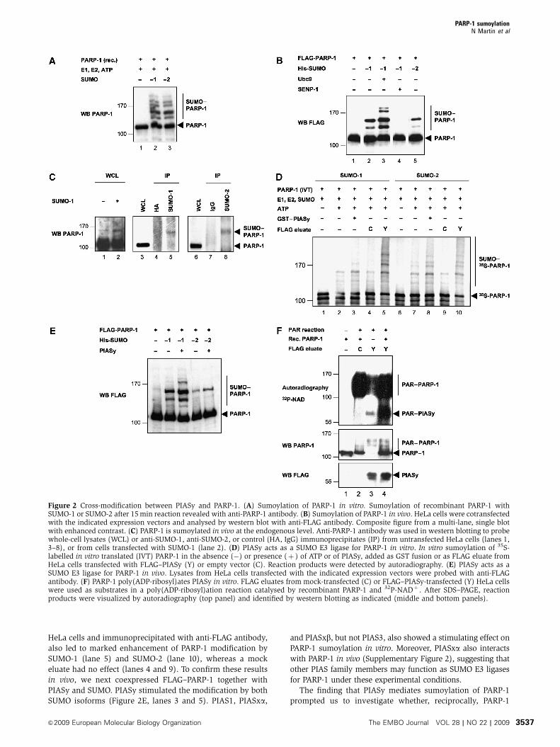
SUMO-1 modification of PARP-1. Western blotting of extracts
from HeLa cells overexpressing SUMO-1, or left untransfected,
showed that endogenous PARP-1 also is SUMO-modified
(Figure 2C, lanes 1 and 2). Similarly, immunoprecipitation of
untransfected HeLa cell extracts with anti-SUMO-1 (lane 5) and
anti-SUMO-2 (lane 8) antibodies, but not control antibodies
(lanes 4 and 7), revealed the presence of endogenously
SUMO-modified PARP-1.
To then test whether PIASy acts as a SUMO E3 ligase for
PARP-1, we added bacterially produced GST–PIASy to an
in vitro sumoylation reaction. As shown in Figure 2D,
GST–PIASy enhanced the sumoylation of PARP-1 by both
SUMO-1 (compare lanes 2 and 3) and SUMO-2 (compare
lanes 7 and 8). Similarly, use of FLAG–PIASy, expressed in
Figure 1 PIASy interacts with PARP-1. (A) Endogenous PIASy and PARP-1 interact in vivo. HeLa cell lysates were immunoprecipitated (IP)with mouse anti-PARP-1 or control (IgG) antibodies and probed with anti-PARP-1 and anti-PIASy antibodies. WCL, whole-cell lysate, 2% ofamount used in IP. (B) PIASy and PARP-1 interact in vitro. Pull-down experiment with GST, GST–PIASy, and 35S-labelled in vitro translatedPARP-1 and luciferase or recombinant PARP-1. Bound proteins revealed by autoradiography or by anti-PARP-1 antibody. Input: 20% of amountused in binding assays. (C) PIASy interacts with both unmodified and poly(ADP-ribosyl)ated PARP-1 in vitro. GST pull down with 35S-labelledin vitro translated PARP-1 (WT, wild type; E988K, catalytically inactive) after incubation of PARP-1 in a poly(ADP-ribosyl)ation reactioncontaining (þ ) or not (�) NAD. Bound proteins revealed by autoradiography. Input: 20% of amount used in the binding reaction. PAR–PARP-1, poly(ADP-ribosyl)ated PARP-1. (D) Major protein domains of PIASy and PARP-1. SAP, SAF-A/B, Acinus, and PIAS domain; SP-RING, Siz/PIAS-RING domain; AD, acidic domain; ZnF, zinc fingers; BRCT, BRCA1 C-terminus domain. (E) Integrity of PIASy SP-RING domain is requiredfor PARP-1 interaction. Co-immunoprecipitation (IP) with endogenous PARP-1 and WT or C342F (mut) FLAG–HA–PIASy expressed in HeLacells; WCL, whole-cell lysate, 5% of amount used in IP. (F) PARP-1 N-terminus and auto-modification domains interact with PIASy.Immobilized GST or GST–PARP-1 domains, expressed in HeLa cells, were incubated with 35S-Met-labelled, in vitro-translated PIASy. Boundproteins were revealed by autoradiography (top panel) and purified GSTor GST–PARP-1 proteins were detected with anti-GSTantibody (bottompanel). Input: 20% of amount used in binding reactions.
PARP-1 sumoylationN Martin et al
The EMBO Journal VOL 28 | NO 22 | 2009 &2009 European Molecular Biology Organization3536
HeLa cells and immunoprecipitated with anti-FLAG antibody,
also led to marked enhancement of PARP-1 modification by
SUMO-1 (lane 5) and SUMO-2 (lane 10), whereas a mock
eluate had no effect (lanes 4 and 9). To confirm these results
in vivo, we next coexpressed FLAG–PARP-1 together with
PIASy and SUMO. PIASy stimulated the modification by both
SUMO isoforms (Figure 2E, lanes 3 and 5). PIAS1, PIASxa,
and PIASxb, but not PIAS3, also showed a stimulating effect on
PARP-1 sumoylation in vitro. Moreover, PIASxa also interacts
with PARP-1 in vivo (Supplementary Figure 2), suggesting that
other PIAS family members may function as SUMO E3 ligases
for PARP-1 under these experimental conditions.
The finding that PIASy mediates sumoylation of PARP-1
prompted us to investigate whether, reciprocally, PARP-1
Figure 2 Cross-modification between PIASy and PARP-1. (A) Sumoylation of PARP-1 in vitro. Sumoylation of recombinant PARP-1 withSUMO-1 or SUMO-2 after 15 min reaction revealed with anti-PARP-1 antibody. (B) Sumoylation of PARP-1 in vivo. HeLa cells were cotransfectedwith the indicated expression vectors and analysed by western blot with anti-FLAG antibody. Composite figure from a multi-lane, single blotwith enhanced contrast. (C) PARP-1 is sumoylated in vivo at the endogenous level. Anti-PARP-1 antibody was used in western blotting to probewhole-cell lysates (WCL) or anti-SUMO-1, anti-SUMO-2, or control (HA, IgG) immunoprecipitates (IP) from untransfected HeLa cells (lanes 1,3–8), or from cells transfected with SUMO-1 (lane 2). (D) PIASy acts as a SUMO E3 ligase for PARP-1 in vitro. In vitro sumoylation of 35S-labelled in vitro translated (IVT) PARP-1 in the absence (�) or presence (þ ) of ATP or of PIASy, added as GST fusion or as FLAG eluate fromHeLa cells transfected with FLAG–PIASy (Y) or empty vector (C). Reaction products were detected by autoradiography. (E) PIASy acts as aSUMO E3 ligase for PARP-1 in vivo. Lysates from HeLa cells transfected with the indicated expression vectors were probed with anti-FLAGantibody. (F) PARP-1 poly(ADP-ribosyl)ates PIASy in vitro. FLAG eluates from mock-transfected (C) or FLAG–PIASy-transfected (Y) HeLa cellswere used as substrates in a poly(ADP-ribosyl)ation reaction catalysed by recombinant PARP-1 and 32P-NADþ . After SDS–PAGE, reactionproducts were visualized by autoradiography (top panel) and identified by western blotting as indicated (middle and bottom panels).
PARP-1 sumoylationN Martin et al
&2009 European Molecular Biology Organization The EMBO Journal VOL 28 | NO 22 | 2009 3537
could poly(ADP-ribosyl)ate PIASy. For this, we incubated a
FLAG–PIASy eluate with 32P-labelled NADþ and DNAseI-
treated DNA in the presence or absence of recombinant
PARP-1. As seen in Figure 2F, PARP-1 was efficiently auto-
poly(ADP-ribosyl)ated under these conditions (top and
middle panels, lanes 2 and 4). Addition of FLAG–PIASy eluate
led to the appearance of a second major band corresponding
in size to (ADP-ribosyl)ated PIASy (top panel, lane 4).
Remarkably, even in the absence of added recombinant
PARP-1, weaker signals corresponding to (ADP-ribosyl)ated
PIASy (lane 3) and PARP-1 (top and middle panels, lane 3)
could be detected, suggesting the presence of endogenous
PARP-1 activity in the FLAG–PIASy eluate. Finally, a GST–
PIASy fusion protein could also be (ADP-ribosyl)ated by
recombinant PARP-1 (data not shown). Taken together,
these results indicate that PIASy and PARP-1 cross modify
each other, suggesting a possible interplay between these two
types of protein modifications.
Lysine 486 and 203 are the principal SUMO-acceptor
sites of PARP-1
Inspection of the human PARP-1 amino-acid sequence
revealed the presence of numerous (420) putative sumoyla-
tion sites, of which five conformed most faithfully to the
classical CKxE motif (Rodriguez et al, 1999; Figure 3A). One
of these (K486) could be confirmed by mass spectroscopy
analysis (data not shown). Mutation of these lysine residues
to arginine showed two of these, K486 and K203, to be critical
for PARP-1 sumoylation, although mutation of either alone,
or both (2KR), failed to abolish PARP-1 sumoylation entirely,
both in vitro (Figure 3B and Supplementary Figure S3) and
in vivo (Figure 3C). Of note, in vitro modification with SUMO-
1 (Figure 3B, odd-numbered lanes) revealed the existence
of additional sites, whereas modification with SUMO-2
additionally led to the formation of high-molecular-weight
(MW) polymeric SUMO-2 chains (lanes 6 and 8). Taken
together, these results show PARP-1 to be SUMO-modified
Figure 3 Mapping of the SUMO-acceptor sites of PARP-1. (A) Five most probable CKxE sumoylation consensus motifs of human PARP-1protein. (B) In vitro sumoylation of 35S-labelled in vitro translated WT, K203R, K486R, or K203R/K486R (2KR) PARP-1 with SUMO-1 or SUMO-2revealed by autoradiography after long reaction time (60 min). (C) Sumoylation of FLAG–PARP-1 WTor 2KR mutant in vivo. Lysates from HeLacells transfected with the indicated expression vectors were probed with anti-FLAG antibody.
Figure 4 Heat shock induces PIASy-dependent sumoylation of PARP-1. (A) Heat shock induces preferential SUMO-2 modification of PARP-1.Whole-cell lysates of HeLa cells transfected as indicated, untreated or heat shocked (30 min, 431C), were western blotted with anti-FLAGantibody. (B) Heat shock induces poly-modification of PARP-1 by SUMO-2. Whole-cell lysates of HeLa cells transfected as indicated, untreatedor heat shocked (30 min, 431C), were western blotted with anti-FLAG antibody. Composite figure from a multi-lane, single blot with enhancedcontrast. (C) High sumoylation of PARP-1 upon heat shock is impaired by the K203R/K486R (2KR) mutation. Whole-cell lysates of HeLa cellstransfected as indicated, untreated or heat shocked (30 min, 431C), were western blotted with anti-FLAG antibody. (D) Modified PARP-1partitions to the detergent-insoluble fraction. HeLa cells transfected as indicated, untreated, or heat shocked (30 min, 431C) were either lyseddirectly in SDS sample buffer (TCE, total cell extract, left panel), or extracted in NP-40-containing Chris buffer, separated into soluble (Sol.) andinsoluble (Insol.) fractions by centrifugation (right panels), and western blotted with anti-FLAG antibody. (E) Heat shock induces accumulationof modified forms of endogenous PARP-1 in HeLa cells. Whole-cell lysates of HeLa cells, untreated or heat shocked (30 min, 431C), werewestern blotted with anti-PARP-1 antibody. (F) Heat shock induces sumoylation of endogenous PARP-1 in HeLa cells. FLAG (control) or PARP-1immunoprecipitates from untransfected HeLa cells untreated or heat shocked (30 min, 431C) were analysed by western blot using anti-SUMO-2antibody (right panel). The corresponding whole-cell lysates were analysed using anti-PARP-1 antibody (left panel). (G) PIASy enhances heat-shock-induced PARP-1 sumoylation. HeLa cells were transfected as indicated and left untreated or heat shocked (30 min, 431C). Whole-celllysates were probed with the indicated antibodies. (H) PIASy is required for the heat-shock-induced increase in PARP-1 sumoylation. HeLa cellswere transfected with scrambled control (scr) or PIASy siRNA and re-transfected 24 h later with FLAG–PARP-1, SUMO-2, and Ubc9. After 24 h,cells were either left untreated or heat shocked (30 min, 431C). Whole-cell lysates were probed with the indicated antibodies.
PARP-1 sumoylationN Martin et al
The EMBO Journal VOL 28 | NO 22 | 2009 &2009 European Molecular Biology Organization3538
on lysine 486 and 203, as well as on other, non-consensus or
promiscuous modification sites.
Heat shock induces PARP-1 sumoylation
Environmental stresses such as heat shock, osmotic, or
oxidative stress are known to induce the preferential con-
jugation of SUMO-2/SUMO-3 to numerous target proteins
(Saitoh and Hinchey, 2000). In addition, PARP-1 was shown
to regulate the expression of the heat-shock-inducible
HSP70.1 gene (Ouararhni et al, 2006). These findings
prompted us to examine whether heat shock could induce
the sumoylation of PARP-1. Consistent with recently pub-
lished results (Blomster et al, 2009; Golebiowski et al, 2009),
coexpression of FLAG–PARP-1 and Ubc9 together with either
SUMO-1 or SUMO-2 in HeLa cells exposed to heat shock
(431C, 30 min) resulted in the appearance of slower migrating
PARP-1 species in the presence of SUMO-2 but not of SUMO-1
(Figure 4A). In contrast, simultaneous coexpression of
PARP-1 sumoylationN Martin et al
&2009 European Molecular Biology Organization The EMBO Journal VOL 28 | NO 22 | 2009 3539
SENP6, a de-sumoylating enzyme with specificity for poly-
SUMO chains (Mukhopadhyay et al, 2006), led to disappear-
ance of these high-MW PARP-1 species, demonstrating that
heat shock promotes the formation of PARP-1–poly-SUMO-2
conjugates (Figure 4B, compare lanes 3 and 4), the abun-
dance of which was significantly reduced when the PARP-1
2KR mutant was expressed instead of WT (Figure 4C).
Fractionation of cell extracts from PARP-1-, Ubc9-, and
SUMO-2-overexpressing cells further revealed enhanced as-
sociation of modified PARP-1 with the detergent (Nonidet
P-40 (NP-40))-insoluble fraction under heat shock
(Figure 4D), suggesting that the induced sumoylation of
PARP-1 is preferentially associated with the chromatin
and/or nuclear matrix compartment. Non-transfected HeLa
cells similarly displayed accumulation of modified endogen-
ous PARP-1 species upon heat shock (Figure 4E).
Immunoprecipitation with anti-PARP-1 antibody (Figure 4F,
lanes 4 and 6), or anti-FLAG control antibody (lanes 3 and 5),
from extracts of unstressed or heat shocked HeLa
cells confirmed that these endogenous higher-MW PARP-1
species correspond to polymeric or multiply modified
PARP-1–SUMO-2 conjugates.
Consistent with previous in vitro results, overexpression of
PIASy (Figure 4G), PIASxa, or PIASxb (Supplementary Figure
S2B, lanes 10 and 11) stimulated heat-shock-induced PARP-1
sumoylation under cotransfection conditions. Conversely,
siRNA-mediated knockdown of PIASy expression in HeLa
cells almost completely abolished the heat-shock-induced
sumoylation of PARP-1 (Figure 4H, compare lanes 5 and 6),
whereas cells transfected with a scrambled control siRNA
behaved like mock-transfected cells (compare lanes 4 and 5),
suggesting that PIASy occupies a privileged position as a
SUMO E3 ligase for PARP-1 under heat shock. Taken together,
these results show that heat shock strongly upregulates
PARP-1 sumoylation, in both quantity as well as quality
(SUMO-2 polymers), and further, that PIASy appears to play
a critical role in this process in vivo.
Role of PARP-1 sumoylation in HSP70.1-promoter
activation
Given the role of PARP-1 in the transcriptional regulation of
the HSP70.1 gene, we next asked whether PARP-1 sumoyla-
tion could affect HSP70.1 transcription. To this end, we
matched WT (PARPþ /þ ) cells were infected with empty
vector as control. These cell populations were subsequently
heat shocked and HSP70.1 gene expression was monitored by
quantitative RT–PCR. As seen in Figure 5A, PARP-1 and
PARP-12KR were expressed at similar levels in restored
PARP-1�/� MEFs, although still less than that in the
PARPþ /þ control cells. In the absence of PARP-1, HSP70.1
gene transcription was significantly reduced and could be
restored by expression of WT PARP-1, albeit only partially
(due to the lower exogenous expression level achieved).
Remarkably, the enhancement attributable to PARP-1 was
reduced by 60% when the PARP-12KR mutant was used
instead, suggesting that PARP-1 sumoylation plays a measur-
able role in HSP70.1-promoter activation. The fact that PARP-
1 sumoylation could not be completely abrogated in the
PARP-12KR mutant (Figures 3B and C, and 4C) may, in part,
explain the residual activity of this mutant in this rescue
experiment. Non-heat shocked cells displayed a similar acti-
vation profile, further suggesting that PARP-1 sumoylation
also affects transcriptional activity under non-stress condi-
tions. Differential poly(ADP-ribosyl)ation activity of PARP-
12KR versus PARP-1 WT is unlikely to account for their
differential transactivation capacity, since both possess the
same enzymatic activity in vitro (Supplementary Figure S4).
Also, a possible role for other modifications targeting lysine
203 or 486 cannot be formally ruled out.
Given that PIASy appears responsible for much, if not all,
heat-shock-induced sumoylation of PARP-1 (Figure 4H), we
next sought to determine whether suppression of PIASy
expression would affect transcription of the HSP70.1
gene under heat shock. For this, HeLa cells were transfected
with siRNA oligonucleotides directed against PIASy or
scrambled control to monitor the expression of the HSP70.1
gene in response to heat shock. As shown in Figure 5B,
PIASy knockdown reduced heat-shock induction of the
endogenous HSP70.1 gene expression by more than 50%.
A similar reduction was obtained upon PIASy depletion
using a transfected HSP70.1 promoter–luciferase reporter
construct (Figure 5C), indicating that presence of PIASy is
necessary for the full HSP70.1 transcriptional response
under heat shock. The incomplete inhibition of PIASy
expression in cells transfected with the specific PIASy
siRNA (Figure 4H), or its possibly redundant role vis-a-vis
other PIAS SUMO E3 ligases (Supplementary Figure S2), may,
in part, account for the residual induction observed upon
heat shock.
These findings, thus, raised the question of whether PIASy-
mediated sumoylation of PARP-1 may occur and exert its role
directly on the HSP70.1 promoter. A direct mechanism would
predict the co-occupancy of components of the SUMO ma-
chinery together with PARP-1 on the HSP70.1 promoter. To
test this, we carried out chromatin immunoprecipitation
(ChIP) experiments using antibodies against PIASy, PARP-1,
Ubc9, and SUMO-2. All four antibodies, but not control
antibodies, immunoprecipitated detectable amounts of
HSP70.1 promoter fragments (Figure 5D), indicating that
the corresponding proteins are bound to the promoter in
normal conditions. To next examine the effect of heat shock
on the promoter occupancy of these proteins, we carried out a
ChIP time-course experiment upon prolonged heat-shock
treatment. Consistent with previously published results
(Ouararhni et al, 2006), we observed mild and transient
enrichment of PARP-1 during the first 5 min of heat shock,
followed by marked release from the promoter thereafter
(Figure 5E). By contrast, both PIASy (Figure 5F) and Ubc9
(Figure 5G), present at low levels at the outset, exhibited very
pronounced recruitment to the HSP70.1 promoter within the
first 5 min, with a 23-fold and five-fold increase in promoter-
associated PIASy and Ubc9, respectively. Upon longer heat-
shock treatment, the amount of PIASy and Ubc9 bound to
HSP70.1 promoter significantly decreased. Altogether, these
findings support a key role of PIASy and of PARP-1 sumoyla-
tion in HSP70.1-promoter activation. To further characterize
other possible effects on HSP70.1-promoter activation, we
also tested the interaction of PIASy with other factors known
to be present on this promoter. These included the DNA-
repair factors Ku70 and Ku80, the arginine methyltransferase
PARP-1 sumoylationN Martin et al
The EMBO Journal VOL 28 | NO 22 | 2009 &2009 European Molecular Biology Organization3540
PRMT5 (Ouararhni et al, 2006), and the tumour suppressor
MEN1, a homologue of the Drosophila Menin protein, that is
recruited to the HSP70 promoter upon heat shock
(Papaconstantinou et al, 2005). Indeed, all four of these
proteins interacted with PIASy in vivo (Supplementary
Figure S5A–C), with Ku70, Ku80 (Gocke et al, 2005;
Yurchenko et al, 2006), as well as PRMT5 (Supplementary
Figure S5D) also being SUMO substrates themselves. These
results suggest that PIASy and, by extension, sumoylation
target several other factors, besides PARP-1, present on the
HSP70.1 promoter.
Involvement of the SUMO-targeted Ub ligase RNF4
in HSP70.1 gene activation
The observation that PARP-1 is modified by multiple and
polymeric SUMO molecules upon heat shock (Figure 4),
Figure 5 PARP-1 sumoylation is necessary for full HSP70.1-promoter activation. (A–C) Impairment of PARP-1 sumoylation reduces HSP70.1gene transcription. (A) PARP-1þ /þ and PARP-1�/� MEFs were infected with retroviruses expressing WT PARP-1, K203R/K486R mutant (2KR)PARP-1, or control (C, empty vector). Cells were left untreated or heat shocked (1 h, 431C). After recovery at 371C for 30 min, HSP70.1 geneexpression, normalized against GAPDH, was determined by quantitative RT–PCR. HSP70.1 transcript levels±s.e. were then plotted relative tocontrol cells before heat shock. Protein expression was checked by western blotting using the indicated antibodies. (B, C) PIASy knockdownreduces HSP70.1 gene transcription. (B) HeLa cells were transfected with scrambled control (scr) or PIASy siRNA. Heat shock andquantification of HSP70.1 gene expression were as described in panel A. (C) HeLa cells were transfected with siRNA oligos as in panel Band re-transfected 24 h later with HSP70.1 promoter–luciferase reporter and CMV–b-gal plasmid for an additional 24 h, and then heat-shocked(1 h, 431C), or left untreated, as indicated. After a further 12 h, luciferase and b-gal activities were determined. Values for luciferase activitywere corrected for b-gal activity. Plotted values in panels B and C represent means±s.e. for three independent experiments, with non-heatshocked scrambled control value set to 1. (D–F) Occupancy of the HSP70.1 promoter by PARP-1 and the SUMO machinery. (D) ChIP fromJurkat cells with antibodies against PARP-1, PIASy, Ubc9, SUMO-2, or isotypic control antibodies (IgG) using primers targeting the HSP70.1 orGAPDH (control) gene promoters. (E–G) Time course of the association of PARP-1, PIASy and Ubc9 with the HSP70.1 promoter upon heatshock. Jurkat cells heat shocked at 431C for the indicated times were subjected to ChIP using antibodies against PARP-1 (E), PIASy (F), or Ubc9(G). ChIP products were analysed by semi-quantitative PCR with primers against the HSP70.1 promoter. Bar graphs: Densitometry of ChIP PCRnormalized to input with time zero set to 1. Results representative of several independent experiments are given.
PARP-1 sumoylationN Martin et al
&2009 European Molecular Biology Organization The EMBO Journal VOL 28 | NO 22 | 2009 3541
raised the possibility that sumoylated PARP-1 could be tar-
geted by the poly-SUMO-specific Ub E3 ligase RNF4 and
subsequently tagged for degradation by the Ub proteasome
system. To explore this possibility, we, thus, tested whether
PARP-1 and RNF4 interact in vivo. For this, HeLa cells were
cotransfected with vectors expressing FLAG–PARP-1, SUMO-
2, and either WT or RING-finger mutant (mut; C136/139/
177/180S; Hakli et al, 2005) FLAG–RNF4, or appropriate
empty vectors. As shown in Figure 6A, detectable amounts
of WT (row d, lane 4) or mut (row d, lane 8) RNF4 co-
immunoprecipitated with an anti-PARP-1 antibody, demon-
strating that RNF4 interacts with PARP-1 in vivo and that, as
shown for PML (Hakli et al, 2005), this interaction does not
require the integrity of the RNF4 RING domain. Moreover,
this experiment also demonstrated that overexpression of
WT, but not mut FLAG-RNF4, reduced the amount of PARP-
1, both in whole-cell extracts and in anti-PARP-1 immuno-
precipitates (compare lanes 4 and 8 in rows a and c),
indicating that RNF4 induces PARP-1 degradation in a man-
ner dependent on its Ub E3 ligase activity. To further rule out
that this observed disappearance of PARP-1 was possibly due
to RNF4-induced apoptosis and the consequent cleavage of
PARP-1 (a hallmark of apoptosis; Soldani and Scovassi,
2002), we carried out a similar experiment to also monitor
the amount of cleaved PARP-1. As before, expression of WT
(but not mut) FLAG–RNF4 led to consistent disappearance of
suggesting that the modified and unmodified forms of
PARP-1 show differential stability in an RNF4-dependent
manner in vivo.
To next examine whether RNF4 mediates the ubiquitina-
tion of PARP-1 or its SUMO conjugated forms, we coex-
pressed Myc–His-tagged Ub and PARP-1 for subsequent
purification of Ub conjugates by nickel-ion affinity chromato-
graphy. As shown in Figure 6G, expression of PARP-1 and
Myc–His–Ub alone yielded no detectable PARP-1–Ub conju-
gates (lane 2). Addition of RNF4, however, led to the appear-
ance of a characteristic smear corresponding to PARP-1–Ub
conjugates (lane 3). Further addition of SUMO-2 extended
this smear to even higher MW species (lane 5), an effect that
could not be seen in the absence of added RNF4 (lane 4),
Figure 6 Involvement of RNF4 in SUMO-dependent PARP-1 ubiquitination and heat-shock-promoter activation. (A) RNF4 interacts with PARP-1in vivo. Co-immunoprecipitation (IP) experiment with the indicated expression vectors (mut FLAG-RNF4: C136/139/177/180S mutant)transfected in HeLa cells. WCL, whole-cell lysate, 5% of amount used in IP. (B) RNF4 induces PARP-1 degradation through its Ub E3 ligaseactivity. Whole-cell lysates of HeLa cells transfected as indicated were analysed by western blot with the indicated antibodies. Cleaved PARP-1,PARP-1 N-terminal fragment produced by caspase-dependent cleavage. (C) RNF4-induced PARP-1 degradation is dependent on the proteasome.HeLa cells transfected as indicated were left untreated or treated with 50 mM MG132 for 8 h. Protein levels were then checked by western blotusing the indicated antibodies. (D) Preferential binding of SUMO-2-modified PARP-1 to RNF4. GST pull down with 35S-labelled PARP-1 in vitromodified by SUMO-1 and SUMO-2. Input: 5% of modified PARP-1 used in the binding reactions. (E) RNF4 reduces PARP-1-SUMO-2 conjugatelevels. Western blots of extracts from HeLa cells transfected as indicated. (F) RNF4 knockdown enhances heat-shock-induced PARP-1 conjugatelevels. Western blots of HeLa cells transfected with scramble (scr) or RNF4 siRNA, left untreated, or heat shocked (30 min, 431C) 48 h later.(G) RNF4 acts as a Ub E3 ligase for PARP-1. HeLa cells overexpressing FLAG–PARP-1 together with the indicated proteins were treated at 40 hpost transfection with 50mM MG132 for 8 h and whole-cell extracts (WCE) were prepared under denaturing conditions. His–Ub conjugates wereaffinity purified by nickel-ion chromatography and probed with anti-PARP-1 antibody (top panel). WCE, cleared of guanidine by TCAprecipitation, were probed with the indicated antibodies (middle and bottom panels). Positions and sizes (in kDa) of MW marker proteins areindicated. (H) Heat shock induces ubiquitination of endogenous PARP-1 in HeLa cells. FLAG (control) or PARP-1 immunoprecipitates fromuntransfected HeLa cells untreated or heat shocked (30 min, 431C) were analysed by western blot using anti-Ub antibody (right panel). Thecorresponding whole-cell lysates were analysed using anti-PARP-1 antibody (left panel). (I, J) RNF4 knockdown attenuates HSP70.1 geneexpression. (I) HeLa cells were transfected with scrambled control (scr) or RNF4 siRNA and left untreated or heat shocked (1 h, 431C) 48 h later.After recovery at 371C for 30 min, HSP70.1 gene expression, normalized against GAPDH, was determined by quantitative RT–PCR. (J) Hela cellswere transfected with scrambled control (scr) or RNF4 siRNA and re-transfected 24 h later with HSP70.1 promoter–luciferase reporter andCMV–b-gal control plasmid. They were then left untreated or heat shocked (1 h, 431C) after an additional 24 h. After a further 12 h, luciferaseand b-gal activities were determined. Values for luciferase activity were corrected for b-gal activity. Plotted values in panels E and F representmeans±s.e. for three independent experiments, with non-heat shocked scrambled control value set to 1.
PARP-1 sumoylationN Martin et al
The EMBO Journal VOL 28 | NO 22 | 2009 &2009 European Molecular Biology Organization3542
indicating that RNF4 and SUMO-2 enhance the ubiquitination
of PARP-1.
The finding that heat shock greatly enhances PARP-1
sumoylation and that RNF4 acts as a Ub E3 ligase for
PARP-1, predicts that heat shock would similarly enhance
PARP-1 ubiquitination. To test this at the endogenous protein
level, we carried out an anti-PARP-1 (or anti-FLAG control)
immunoprecipitation from extracts of unstressed or heat
shocked HeLa cells. As shown in Figure 6H, probing such
immunoprecipitates with an anti-Ub antibody revealed the
characteristic high-MW poly-Ub smear from extracts of heat
shocked, but not unshocked, cells (compare lanes 4 and 6).
To next evaluate the role of RNF4 in HSP70.1 gene activity
in response to heat shock, we used siRNA to ablate RNF4
PARP-1 sumoylationN Martin et al
&2009 European Molecular Biology Organization The EMBO Journal VOL 28 | NO 22 | 2009 3543
expression in HeLa cells. As seen in Figure 6I, RNF4 knock-
down led to consistent, albeit modest, reduction (25%) in
basal and heat-shock-induced activity of the endogenous
HSP70.1 gene. A similar, but more pronounced, result
(50%) was obtained using instead an HSP70.1 promoter–
luciferase reporter construct (Figure 6J), indicating that, like
PIASy (Figure 5B and C), RNF4 appears to be necessary for
full activity of this heat-shock-inducible promoter.
Taken together, these results support the involvement of
the SUMO-specific Ub E3 ligase RNF4 in regulating both the
abundance of SUMO-modified PARP-1 and the activity of the
heat-shock-inducible HSP70.1 promoter.
Discussion
In this report, we have described the association of the SUMO
E3 ligase PIASy with the poly(ADP-ribosyl)polymerase PARP-
1 and explored its functional consequences in the regulation
of the heat-shock-inducible HSP70.1 gene. As discussed
below, our results are consistent with a model whereby
heat shock induces rapid PARP-1 multi- and poly-sumoyla-
tion, which leads to RNF4 recruitment, ubiquitination, and
subsequent degradation, thus likely contributing to PARP-1
clearance from a heat-shock-inducible promoter (Figure 7).
Poly(ADP-ribosyl)ation and sumoylation
Together with three recent reports (Blomster et al, 2009;
Golebiowski et al, 2009; Messner et al, 2009), the present
work adds sumoylation to the list of post-translational mod-
ifications affecting the activity of PARP-1. Besides poly(ADP-
ribosyl)ation, previous work has also shown that PARP-1 is
subject to acetylation (Hassa et al, 2005; Messner et al, 2009),
phosphorylation (Kauppinen et al, 2006), and K48-linked
ubiquitination (Wang et al, 2008). Indeed, Messner et al
(2009) have shown that PARP-1 mono-sumoylation at K486
inhibits p300-mediated acetylation at lysines proximal to this
modification site, confirming the existence of cross-talk me-
chanisms between these different modifications. Our demon-
stration here that PIASy may be poly(ADP-ribosyl)ated,
besides confirming the physical interaction with PARP-1,
could furthermore suggest that the activity of PIASy, like
that of PARP-1 itself, is regulated by poly(ADP-ribosyl)ation.
This could, for example, affect the DNA or chromatin binding
of PIASy, as has been shown for p53 (Mendoza-Alvarez and
Alvarez-Gonzalez, 2001), or its SUMO E3 ligase activity.
Conversely, recent in vitro results suggest that sumoylation
does not affect poly(ADP-ribosyl)ation of PARP-1 (Messner
et al, 2009). Nonetheless, given our finding that poly(ADP-
ribosyl)ated PARP-1 exhibits reduced binding to PIAS in vivo,
but not in vitro, it will be interesting to further explore
the possible interplay between poly(ADP-ribosyl)ation and
sumoylation.
The steady-state level of sumoylated PARP-1 in non-
stressed cells is very low. For this reason, perhaps, Messner
et al (2009) report on only mono-sumoylated PARP-1 under
their experimental conditions. Unlike these authors, we
found PIASy to stimulate PARP-1 sumoylation in both un-
stressed, as well as heat shocked cells. This role for the
members of the PIAS family proteins in the stimulation of
sumoylation under thermal stress appears to be evolutiona-
rily conserved, as it has also been described in plants (Kurepa
et al, 2003; Yoo et al, 2006; Miura et al, 2007; Saracco et al,
2007). Heat shock greatly stimulates the formation of high-
MW PARP-1 species, which consist principally of poly-SUMO-
2/3 conjugates (this work and Blomster et al, 2009;
Figure 7 Role of PARP-1 sumoylation and ubiquitination in theregulation of HSP70.1-promoter activation. PARP-1, present on theHSP70.1 promoter under normal conditions, but repressed bysequestration in the mH2A1.1 complex, is poly(ADP-ribosyl)atedupon heat shock (not shown; Ouararhni et al, 2006). In parallel,Ubc9 and PIASy are rapidly recruited to catalyse PARP-1 poly- andmulti-sumoylation, in turn recruiting RNF4, which catalyses ubi-quitination and subsequent degradation of PARP-1, thereby con-tributing to gene activation by PARP-1 clearance from promoter.For clarity, other possible modifications and chromatin factorsdiscussed in the text have been omitted.
PARP-1 sumoylationN Martin et al
The EMBO Journal VOL 28 | NO 22 | 2009 &2009 European Molecular Biology Organization3544
Golebiowski et al, 2009). Like for arsenic-induced hyper-
sumoylation of PML (Lallemand-Breitenbach et al, 2008;
Tatham et al, 2008), or that of other proteins under different
stresses (Saitoh and Hinchey, 2000), the effectors regulating
the sumoylation of PARP-1 and of numerous other proteins
under heat shock remain to be identified.
PARP-1 ubiquitination and degradation
Our finding that heat shock induces the hyper-sumoylation of
PARP-1, principally by SUMO-2/3, raised the possibility that
PARP-1 is targeted by the E3 Ub ligase RNF4. In support of
this, we found that RNF4 overexpression enhances PARP-1
ubiquitination and proteasome-mediated degradation.
Furthermore, consistent with a role of RNF4, the highly
poly-sumoylated forms of PARP-1 displayed reduced stability,
whereas conversely, RNF4 depletion led to their stabilization.
Finally, we show that PARP-1 ubiquitination, like sumoyla-
tion, is strongly enhanced by heat shock. Altogether, these
results link the sumoylation and ubiquitination of PARP-1
and, moreover, provide evidence for a novel, caspase-inde-
pendent pathway for PARP-1 degradation.
RNF4, in possessing four SIMs, has been shown to target
only poly-SUMO-2/3-modified substrates with high affinity
(Tatham et al, 2008). That PARP-1 likely possesses many
more possible sumoylation sites besides the two principal
sites described here (K203 and K486), raises the possibility
that not only poly-sumoylation, but also multi-sumoylation
of PARP-1, could lead to RNF4 recruitment, even by the
non-chain forming SUMO-1. Such a mechanism has been
suggested recently (Ulrich, 2008) and may account for our
finding that RNF4 leads to the ubiquitination of PARP-1 even
without heat shock (Figure 6G), that is, under conditions
in which formation of poly-SUMO-2 chains is presumably
minimal.
Transcriptional regulation
Sumoylation of transcription factors and cofactors is gener-
ally associated with repression mechanisms (for reviews, see
Verger et al, 2003; Girdwood et al, 2004; Muller et al, 2004;
Gill, 2005). Where sumoylation has been shown to contribute
to activation, as in the case of p53 (Gostissa et al, 1999;
Rodriguez et al, 1999; Muller et al, 2004; Bischof et al, 2006)
or Tcf4 (Yamamoto et al, 2003), the mechanisms involved
remain obscure. In this context, members of the HSF (heat
shock factor) family of transcription factors are modified by
SUMO (Goodson et al, 2001; Hong et al, 2001; Hietakangas
et al, 2003, 2006; Hilgarth et al, 2004; Anckar et al, 2006). The
precise function, here, of sumoylation in activating or repres-
sing gene transcription, however, appears to be complex and
may involve regulation of response duration or intensity,
rather than simple on/off switching (Hietakangas et al, 2003).
Similarly, the role of PARP-1 and poly(ADP-ribosyl)ation in
transcriptional regulation is multi-faceted and context-depen-
dent. In some cases, such as in NF-kB-mediated activation,
poly(ADP-ribosyl)ation appears dispensable (Hassa et al,
2003) or may even repress activity (Meisterernst et al,
1997). Where it does contribute to activation, it is generally
seen as leading to chromatin decompaction (Poirier et al,
1982; Kim et al, 2004; Wacker et al, 2007), possibly mediated
by electrostatic repulsion between poly(ADP-ribosyl)ated
proteins (e.g. PARP-1 and histones) and the DNA. The finding
that PARP-1 poly(ADP-ribosyl)ation activity is held in check
by interaction with the variant histone mH2A (Ouararhni
et al, 2006; Nusinow et al, 2007), has also provided further
evidence that PARP-1 and poly(ADP-ribosyl)ation exert their
function in a context-dependent manner. In the case of a
constitutively silent promoter, such as that of an inactive X
(Xi)-linked transgene, PARP-1 is indispensable for silencing
(Nusinow et al, 2007), whereas for the HSP70.1 promoter, it is
required for inducible activation (Ouararhni et al, 2006).
Yet, even in the absence of PARP-1, heat shock promotes
significant promoter activation, thus suggesting the existence
of PARP-1-independent mechanisms. Nevertheless, PIASy,
sumoylation, and RNF4 appear critically involved, as redu-
cing their activity also reduces PARP-1-dependent promoter
activation. That this occurs also in the absence of heat
shock may suggest that the sumoylation of PARP-1 plays a
similar role under basal conditions, but that, in absolute
terms, sumoylation exerts its most significant effect upon
heat shock.
PARP and poly(ADP-ribosyl)ation have been shown to be
required for rapid nucleosome remodelling that precedes
transcriptional onset upon heat shock in Drosophila cells
(Petesch and Lis, 2008). Yet interestingly, poly(ADP-ribosy-
l)ation by itself does not appear to be sufficient for the release
of PARP-1 from the condensed mH2A1.1 chromatin
(Ouararhni et al, 2006), suggesting that additional factors,
such as chromatin remodellers (e.g. SWI/SNF; Pavri et al,
2005), sumoylation, or other post-translational modifications,
are critically required. The results obtained to date do not
provide sufficient temporal resolution to unravel the order, if
any, with which poly(ADP-ribosyl)ation and sumoylation
occur upon heat shock, but it is highly likely that sumoyla-
tion, like poly(ADP-ribosyl)ation, plays an important role in
the removal of PARP-1 from the promoter. Consistent with
this, we show that PARP-1, PIASy, and Ubc9 leave the
promoter with similar kinetics upon prolonged heat shock.
Moreover, our finding that sumoylated PARP-1 is associated
with the insoluble cellular fraction is again consistent with a
role of sumoylation in the differential localization of the protein.
Sumoylation-coupled ubiquitination and degradation may
also be necessary for the enhanced or prolonged clearance of
PARP-1 from a heat-shock-induced promoter in that sustained
transcriptional activation or its rapid extinction upon stimu-
lus withdrawal may require the rapid recycling of PARP-1.
A similar model has also been invoked for the sumoylation of
PEA3 during synergistic activation of target genes with CBP
(Guo and Sharrocks, 2009). Our finding that other factors
associated with HSP promoters are sumoylated or are PIASy-
binding partners (e.g. MEN1, Ku70/80, and PRMT5) suggests
that SUMO-triggered, RNF4-mediated ubiquitination may
similarly play a wider role by regulating the activity of
other proteins besides PARP-1.
Materials and methods
Plasmids and siRNAsFLAG–HA–PIASy was inserted into the pcDNA3 vector (Invitrogen);T7-PIAS1, T7-PIAS3, T7-PIASxa, T7-PIASxb, and T7-PIASy into thepSG5 vector (Stratagene); PARP-1 into pFLAG-CMV-6c (Sigma),pSG5, pBS, and pBABE vectors; and RNF4 into the pGEX2T(GE Healthcare) and pcDNA3 vectors by standard procedures.Point mutant derivatives of PARP-1 (K203R, K249R, K486R, K512R,K798R, E988K, and K203R/K486R double mutant) were constructedby site-directed mutagenesis (QuikChange XL kit; Stratagene).
PARP-1 sumoylationN Martin et al
&2009 European Molecular Biology Organization The EMBO Journal VOL 28 | NO 22 | 2009 3545
GST–PIASy, FLAG–HA–PIASy WT, and mut (C342F); GST–PARP-1,SUMO-1, His–SUMO-1, SUMO-2, His–SUMO-2, Ubc9, SENP-1, T7-PIASy, and CMV–b-galactosidase plasmids were described pre-viously (Masson et al, 1998; Sachdev et al, 2001; Bischof et al,2006). Plasmid for His–Myc–Ub was kindly provided by C Neuveut;for FLAG–RNF4 WT and mut (C136/139/177/180S) by J Palvimo;for SENP-6 by R Hay; for VSV-MEN1 by CX Zhang; forFLAG–PRMT5 by C Sardet; and for HSP70.1 promoter–luciferasereporter by O Bensaude. All constructions were verified by DNAsequencing. siRNAs used were as follows: PIASy sense sequence:CAAGACAGGUGGAGUUGAUUU; RNF4 sense sequence: GAAUGGACGUCUCAUCGUUUU, as well as scrambled controls (Dharmacon).
Cell culture, infection, transfection, and reporter assaysHeLa cells and PARP-1þ /þ and PARP-1�/� MEFs were grown inDMEM and Jurkat cells in RPMI medium under standard cultureconditions. Poly(ADP-ribosyl)ation (without heat shock) wasinduced by treatment with 1 mM H2O2 (Gifrer) for 10 min and/orinhibited with 30 mM DPQ (Alexis Biochemicals) for 1.5 h. Proteinstability was analysed by treating the cells with 50 mM MG-132(Sigma) for 8 h or 50 mg/ml cycloheximide (Sigma) for the timesindicated. Infections of MEFs by retrovirus-mediated gene transferwere performed with Phoenix packaging cells. At 24 h post-infection, cells were selected with 4 mg/ml puromycin for 4 days.Transfections of plasmids and siRNAs in HeLa cells were performedwith Lipofectamine and with Oligofectamine (Invitrogen), respec-tively. Five days after the end of selection, or 48 h after transfection,cells were heat shocked at 431C if needed and protein or RNAextraction was performed. For some in vivo sumoylation assaysand for reporter assays, HeLa cells were transfected with siRNAsand re-transfected 24 h later with expression vectors or HSP70.1promoter–luciferase reporter and CMV–b-gal control plasmids.Cells were heat shocked at 431C after a further 24 h, either lyseddirectly for in vivo sumoylation assays or 12 h later for luciferaseand b-gal assays. Luciferase and b-gal activities were determinedusing the Luciferase reporter assay system (Promega) and theGalacto-star kit (Tropix).
Protein extraction, immunoprecipitation, and His pull downFor sumoylation and ubiquitination studies, cells were washed inPBS supplemented with 10 mM N-ethylmaleimide (NEM; Sigma).For direct western blots, cells were lysed directly in sample buffercontaining 2% sodium dodecyl sulphate (SDS). For co-immuno-precipitation of PIAS with PARP-1, MEN1, Ku70/Ku80, and PRMT5,cells were scraped in PBS and lysed in Chris buffer (50 mM Tris, pH8.0, 0.5% NP-40, 200 mM NaCl, 0.1 mM EDTA, 10% glycerol, andprotease inhibitors (Complete EDTA-free; Roche)). For co-immuno-precipitation of PARP-1 with RNF4, cells were scraped in PBS andlysed in RIPA buffer (50 mM Tris, pH 8.0, 1% Triton X-100, 150 mMNaCl, 0.5% sodium deoxycholate, 0.1% SDS, 1 mM EDTA, proteaseinhibitors, 10 mM NEM). For immunoprecipitation of PARP-1conjugates under untreated or heat-shock conditions, cells werelysed in SDS sample buffer, diluted 10-fold in Chris buffer. Total celllysates were then incubated for 2 h at 41C with the appropriateantibody and immune complexes were collected by incubation for1 h at 41C with Protein G plus/Protein A agarose (Calbiochem) andwashed three times in lysis buffer. In some cases, bound proteinswere then eluted by incubating the beads for 45 min at 201C withFLAG peptide (Sigma). His pull downs from transfected HeLa cellswere carried out as described previously (Kirsh et al, 2002).
Immunoblotting and antibodiesWestern blots were prepared on Hybond C-extra membranes(Amersham) and revealed using CDP-Star (Tropix). Antibodiesused were as follows: mouse anti-PARP-1 (C2-10; Trevingen), rabbitanti-PARP-1 (H-250; Santa Cruz), mouse anti-poly(ADP-ribose)(10H; Alexis), mouse anti-HA (16B12; Covance), mouse anti-GST(B-14; Santa Cruz), mouse anti-T7 (Novagen), mouse anti-FLAG(M2; Sigma), rabbit anti-FLAG (Sigma), mouse anti-VSV (P5D4;Sigma), mouse anti-Ku70 (N3H10; Abcam), mouse anti-Ku80 (111;Abcam), mouse anti-b-actin (Sigma), mouse and rabbit IgGs(Upstate), mouse anti-Ubc9 (50; Pharmingen), mouse anti-Ub(FK2; Biomol), rabbit anti-PIASy (Bischof et al, 2006), mouseanti-SUMO-1 (Zymed), mouse anti-SUMO-2 (8A2; Zhang et al,
2008), and rabbit anti-RNF4 (a gift from J Palvimo; Hakli et al,2005).
RNA isolation and RT–PCR analysisTotal RNA was extracted using the RNeasy RNA isolation kit(Qiagen) and reverse-transcribed using the High Capacity cDNAReverse Transcription kit (Applied Biosystems). cDNAs were addedto the SYBR Green PCR master mix (Applied Biosystems) using thefollowing oligonucleotide pairs: 50-CCAAGGTGCAGGTGAACTACAA-30 and 50-CAGCACCATGGACGAGATCTC-30 for HSP70.1 and 50-GCAAAGTGGAGATTGTTGCCA-30 and 50-ATTTGCCGTGAGTGGAGTCAT-30 for GAPDH. Real-time quantitative PCR was performed withthe ABI PRISM 7900HT Sequence Detection System (AppliedBiosystems) and normalized to GAPDH signal.
Chromatin immunoprecipitationChIP was carried out as previously described (Bischof et al, 2006).Chromatin immunoprecipitated DNA was analysed by PCR with thefollowing primers: 50-GGCGAAACCCCTGGAATATTCCCGA-30 and50-AGCCTTGGGACAACGGGAG-30 for HSP70.1 promoter and 50-GGACCTGACCTGCCGTCTAGAA-30 and 50-GGTGTCGCTGTTGAAGTCAGAG-30 for GAPDH promoter.
Protein expression and in vitro sumoylation assaysGST, GST–PIASy, and GST–RNF4 were produced in BL21(DE3)pLysS cells and PARP-1 in Sf9 cells and purified under nativeconditions using standard protocols. 35S-methionine-labelled,in vitro translated proteins were prepared using the T7 or Sp6TNT-coupled reticulocyte lysate kit (Promega). In vitro sumoylationassays were carried out by incubating recombinant or 35S-methionine-labelled in vitro translated PARP-1 or PRMT5 withrecombinant Aos1/Uba2 (370 nM), Ubc9 (630 nM), and SUMO(7mM) in 30 mM Tris, 5 mM ATP, 10 mM MgCl2, pH 7.5, at 331C aspreviously described (Kirsh et al, 2002). Recombinant GST–PIASy(at a final concentration of 500 nM), a FLAG eluate, or an in vitrotranslated PIAS was added in this reaction.
In vitro poly(ADP-ribosyl)ation assaysFor in vitro poly(ADP-ribosyl)ation of PARP-1, unlabelled, or 35S-methionine-labelled, in vitro translated PARP-1 was incubated in 20mlof activity buffer (50 mM Tris, pH 7.5, 4mM MgCl2, 200mMdithiothreitol (DTT), 0.1mg/ml BSA, 4ng/ml DNaseI-activated calfthymus DNA, and 400mM NADþ ). To test PIASy poly(ADP-ribosyl)ation by PARP-1, FLAG eluates or GST fusion proteins were incubatedwith 100ng of recombinant PARP-1 in activity buffer supplementedwith 1mCi 32P-NADþ . After 2min at 201C, reactions were stopped bydilution in SDS sample buffer, resolved by gel electrophoresis, andtransferred to nitrocellulose membrane for visualization of (ADP-ribosyl)ated products by autoradiography or western blot.
GST pull downRecombinant PARP-1, 35S-methionine-labelled, in vitro translatedproteins, or products of an in vitro sumoylation or poly(ADP-ribosyl)ation assay were incubated with the relevant GST-fusionprotein bound to 10 ml of glutathione–Sepharose beads (Amer-sham). After 4 h incubation at 41C and five washes in GST bindingbuffer (50 mM Tris, pH 7.5, 250 mM NaCl, 0.1% Triton-X100, 10%glycerol, 1 mM DTT, and protease inhibitors), bound proteins wereeluted with SDS sample buffer, resolved by gel electrophoresis, andvisualized by immunoblotting with PARP-1 antibody or by directautoradiography.
Supplementary dataSupplementary data are available at The EMBO Journal Online(http://www.embojournal.org).
Acknowledgements
We thank Jorma Palvimo, Olivier Bensaude, Rudolf Grosschedl, RonHay, Chang-Xian Zhang, Claude Sardet and Christine Neuveut forgenerous gifts of reagents. We gratefully acknowledge Ali Hamicheand Marie-Claude Geoffroy for helpful discussions and reagents, andPavan Kumar, Agnes Marchio, and Delphine Cougot for technicalexpertise as well as Selina Raguz and Jesus Gil for their support.This work was supported by grants from EEC 6th FP (Rubicon), LaLigue Nationale Contre le Cancer (Equipe Labellisee), and l’AgenceNationale pour la Recherche. NM was supported by the Ecole NormaleSuperieure de Lyon, Pasteur-Weizman Foundation, INSERM, and the
PARP-1 sumoylationN Martin et al
The EMBO Journal VOL 28 | NO 22 | 2009 &2009 European Molecular Biology Organization3546
Association pour la Recherche sur le Cancer; KS by the Association forInternational Cancer Research; and AW by the Fondation pour laRecherche Medicale.
Conflict of interest
The authors declare that they have no conflict of interest.
References
Anckar J, Hietakangas V, Denessiouk K, Thiele DJ, Johnson MS,Sistonen L (2006) Inhibition of DNA binding by differentialsumoylation of heat shock factors. Mol Cell Biol 26: 955–964
Bischof O, Schwamborn K, Martin N, Werner A, Sustmann C,Grosschedl R, Dejean A (2006) The E3 SUMO ligase PIASyis a regulator of cellular senescence and apoptosis. Mol Cell 22:783–794
Blomster HA, Hietakangas V, Wu J, Kouvonen P, Hautaniemi S,Sistonen L (2009) Novel proteomics strategy brings insight intothe prevalence of SUMO-2 target sites. Mol Cell Proteomics 8:1382–1390
Burgess RC, Rahman S, Lisby M, Rothstein R, Zhao X (2007) TheSlx5–Slx8 complex affects sumoylation of DNA repair proteinsand negatively regulates recombination. Mol Cell Biol 27:6153–6162
Geiss-Friedlander R, Melchior F (2007) Concepts in sumoylation:a decade on. Nat Rev Mol Cell Biol 8: 947–956
Gill G (2005) Something about SUMO inhibits transcription. CurrOpin Genet Dev 15: 536–541
Girdwood DW, Tatham MH, Hay RT (2004) SUMO and transcrip-tional regulation. Semin Cell Dev Biol 15: 201–210
Gocke CB, Yu H, Kang J (2005) Systematic identification andanalysis of mammalian small ubiquitin-like modifier substrates.J Biol Chem 280: 5004–5012
Golebiowski F, Matic I, Tatham MH, Cole C, Yin Y, Nakamura A,Cox J, Barton GJ, Mann M, Hay RT (2009) System-wide changesto SUMO modifications in response to heat shock. Sci Signal 2:ra24, published online 26 May 2009; doi: 101126/sci-signal2000282
Goodson ML, Hong Y, Rogers R, Matunis MJ, Park-Sarge OK, SargeKD (2001) Sumo-1 modification regulates the DNA bindingactivity of heat shock transcription factor 2, a promyelocyticleukemia nuclear body associated transcription factor. J BiolChem 276: 18513–18518
Gostissa M, Hengstermann A, Fogal V, Sandy P, Schwarz SE,Scheffner M, Del Sal G (1999) Activation of p53 byconjugation to the ubiquitin-like protein SUMO-1. EMBO J 18:6462–6471
Guo B, Sharrocks AD (2009) Extracellular signal-regulated kinasemitogen-activated protein kinase signaling initiates a dynamicinterplay between sumoylation and ubiquitination to regulate theactivity of the transcriptional activator PEA3. Mol Cell Biol 29:3204–3218
Hakli M, Karvonen U, Janne OA, Palvimo JJ (2005) SUMO-1promotes association of SNURF (RNF4) with PML nuclear bodies.Exp Cell Res 304: 224–233
Hakme A, Wong HK, Dantzer F, Schreiber V (2008) The expandingfield of poly(ADP-ribosyl)ation reactions. ‘Protein Modifications:Beyond the Usual Suspects’ Review Series. EMBO Rep 9:1094–1100
Hassa PO, Buerki C, Lombardi C, Imhof R, Hottiger MO (2003)Transcriptional coactivation of nuclear factor-kappaB-dependentgene expression by p300 is regulated by poly(ADP)-ribose poly-merase-1. J Biol Chem 278: 45145–45153
Hassa PO, Haenni SS, Buerki C, Meier NI, Lane WS, Owen H,Gersbach M, Imhof R, Hottiger MO (2005) Acetylation ofpoly(ADP-ribose) polymerase-1 by p300/CREB-binding proteinregulates coactivation of NF-kappaB-dependent transcription.J Biol Chem 280: 40450–40464
Hay RT (2005) SUMO: a history of modification. Mol Cell 18:1–12
Hietakangas V, Ahlskog JK, Jakobsson AM, Hellesuo M, SahlbergNM, Holmberg CI, Mikhailov A, Palvimo JJ, Pirkkala L, SistonenL (2003) Phosphorylation of serine 303 is a prerequisite for thestress-inducible SUMO modification of heat shock factor 1. MolCell Biol 23: 2953–2968
Hietakangas V, Anckar J, Blomster HA, Fujimoto M, Palvimo JJ,Nakai A, Sistonen L (2006) PDSM, a motif for phosphorylation-
dependent SUMO modification. Proc Natl Acad Sci USA 103:45–50
Hilgarth RS, Murphy LA, O’Connor CM, Clark JA, Park-Sarge OK,Sarge KD (2004) Identification of Xenopus heat shock transcrip-tion factor-2: conserved role of sumoylation in regulating deox-yribonucleic acid-binding activity of heat shock transcriptionfactor-2 proteins. Cell Stress Chaperones 9: 214–220
Hong Y, Rogers R, Matunis MJ, Mayhew CN, Goodson ML,Park-Sarge OK, Sarge KD (2001) Regulation of heat shocktranscription factor 1 by stress-induced SUMO-1 modification.J Biol Chem 276: 40263–40267
Ii T, Mullen JR, Slagle CE, Brill SJ (2007) Stimulation of in vitrosumoylation by Slx5–Slx8: evidence for a functional interactionwith the SUMO pathway. DNA Repair (Amst) 6: 1679–1691
Kauppinen TM, Chan WY, Suh SW, Wiggins AK, Huang EJ,Swanson RA (2006) Direct phosphorylation and regulation ofpoly(ADP-ribose) polymerase-1 by extracellular signal-regulatedkinases 1/2. Proc Natl Acad Sci USA 103: 7136–7141
Kim MY, Mauro S, Gevry N, Lis JT, Kraus WL (2004) NAD+-dependent modulation of chromatin structure and transcriptionby nucleosome binding properties of PARP-1. Cell 119:803–814
Kirsh O, Seeler JS, Pichler A, Gast A, Muller S, Miska E, Mathieu M,Harel-Bellan A, Kouzarides T, Melchior F, Dejean A (2002) TheSUMO E3 ligase RanBP2 promotes modification of the HDAC4deacetylase. EMBO J 21: 2682–2691
Kosoy A, Calonge TM, Outwin EA, O’Connell MJ (2007) Fissionyeast Rnf4 homologs are required for DNA repair. J Biol Chem282: 20388–20394
Kraus WL (2008) Transcriptional control by PARP-1: chromatinmodulation, enhancer-binding, coregulation, and insulation.Curr Opin Cell Biol 20: 294–302
Krishnakumar R, Gamble MJ, Frizzell KM, Berrocal JG, Kininis M,Kraus WL (2008) Reciprocal binding of PARP-1 and histone H1 atpromoters specifies transcriptional outcomes. Science 319:819–821
Kurepa J, Walker JM, Smalle J, Gosink MM, Davis SJ, Durham TL,Sung DY, Vierstra RD (2003) The small ubiquitin-like modifier(SUMO) protein modification system in Arabidopsis.Accumulation of SUMO1 and -2 conjugates is increased by stress.J Biol Chem 278: 6862–6872
Lallemand-Breitenbach V, Jeanne M, Benhenda S, Nasr R, Lei M,Peres L, Zhou J, Zhu J, Raught B, de The H (2008) Arsenicdegrades PML or PML-RARalpha through a SUMO-triggeredRNF4/ubiquitin-mediated pathway. Nat Cell Biol 10: 547–555
Martin N, Schwamborn K, Urlaub H, Gan B, Guan JL, Dejean A(2008) Spatial interplay between PIASy and FIP200 in the regula-tion of signal transduction and transcriptional activity. Mol CellBiol 28: 2771–2781
Masson M, Menissier-de Murcia J, Mattei MG, de Murcia G,Niedergang CP (1997) Poly(ADP-ribose) polymerase interactswith a novel human ubiquitin conjugating enzyme: hUbc9.Gene 190: 287–296
Masson M, Niedergang C, Schreiber V, Muller S, Menissier-deMurcia J, de Murcia G (1998) XRCC1 is specifically associatedwith poly(ADP-ribose) polymerase and negatively regulates itsactivity following DNA damage. Mol Cell Biol 18: 3563–3571
Meisterernst M, Stelzer G, Roeder RG (1997) Poly(ADP-ribose)polymerase enhances activator-dependent transcription in vitro.Proc Natl Acad Sci USA 94: 2261–2265
Mendoza-Alvarez H, Alvarez-Gonzalez R (2001) Regulation of p53sequence-specific DNA-binding by covalent poly(ADP-ribosyl)a-tion. J Biol Chem 276: 36425–36430
Messner S, Schuermann D, Altmeyer M, Kassner I, Schmidt D,Schar P, Muller S, Hottiger MO (2009) Sumoylation of poly(ADP-ribose) polymerase 1 inhibits its acetylation and restrains tran-scriptional coactivator function. FASEB J, published online 21 July2009; doi:101096/fj09-137695fj.09-137695
PARP-1 sumoylationN Martin et al
&2009 European Molecular Biology Organization The EMBO Journal VOL 28 | NO 22 | 2009 3547
Miura K, Jin JB, Lee J, Yoo CY, Stirm V, Miura T, Ashworth EN,Bressan RA, Yun DJ, Hasegawa PM (2007) SIZ1-mediated sumoy-lation of ICE1 controls CBF3/DREB1A expression and freezingtolerance in Arabidopsis. Plant Cell 19: 1403–1414
Morimoto RI (1998) Regulation of the heat shock transcriptionalresponse: cross talk between a family of heat shock factors,molecular chaperones, and negative regulators. Genes Dev 12:3788–3796
Morimoto RI (2008) Proteotoxic stress and inducible chaperonenetworks in neurodegenerative disease and aging. Genes Dev 22:1427–1438
Mukhopadhyay D, Ayaydin F, Kolli N, Tan SH, Anan T, Kametaka A,Azuma Y, Wilkinson KD, Dasso M (2006) SUSP1 antagonizesformation of highly SUMO2/3-conjugated species. J Cell Biol 174:939–949
Mukhopadhyay D, Dasso M (2007) Modification in reverse: theSUMO proteases. Trends Biochem Sci 32: 286–295
Mullen JR, Brill SJ (2008) Activation of the Slx5–Slx8 ubiquitinligase by poly-small ubiquitin-like modifier conjugates. J BiolChem 283: 19912–19921
Muller S, Ledl A, Schmidt D (2004) SUMO: a regulator of geneexpression and genome integrity. Oncogene 23: 1998–2008
Nusinow DA, Hernandez-Munoz I, Fazzio TG, Shah GM, Kraus WL,Panning B (2007) Poly(ADP-ribose) polymerase 1 is inhibitedby a histone H2A variant, MacroH2A, and contributes tosilencing of the inactive X chromosome. J Biol Chem 282:12851–12859
Ouararhni K, Hadj-Slimane R, Ait-Si-Ali S, Robin P, Mietton F, Harel-Bellan A, Dimitrov S, Hamiche A (2006) The histone variantmH2A1.1 interferes with transcription by down-regulating PARP-1 enzymatic activity. Genes Dev 20: 3324–3336
Papaconstantinou M, Wu Y, Pretorius HN, Singh N, Gianfelice G,Tanguay RM, Campos AR, Bedard PA (2005) Menin is a regulatorof the stress response in Drosophila melanogaster. Mol Cell Biol25: 9960–9972
Pavri R, Lewis B, Kim TK, Dilworth FJ, Erdjument-Bromage H,Tempst P, de Murcia G, Evans R, Chambon P, Reinberg D (2005)PARP-1 determines specificity in a retinoid signaling pathway viadirect modulation of mediator. Mol Cell 18: 83–96
Petesch SJ, Lis JT (2008) Rapid, transcription-independent loss ofnucleosomes over a large chromatin domain at Hsp70 loci. Cell134: 74–84
Poirier GG, de Murcia G, Jongstra-Bilen J, Niedergang C, Mandel P(1982) Poly(ADP-ribosyl)ation of polynucleosomes causesrelaxation of chromatin structure. Proc Natl Acad Sci USA 79:3423–3427
Prudden J, Pebernard S, Raffa G, Slavin DA, Perry JJ, Tainer JA,McGowan CH, Boddy MN (2007) SUMO-targeted ubiquitinligases in genome stability. EMBO J 26: 4089–4101
Rodriguez MS, Desterro JM, Lain S, Midgley CA, Lane DP, Hay RT(1999) SUMO-1 modification activates the transcriptional re-sponse of p53. EMBO J 18: 6455–6461
Sachdev S, Bruhn L, Sieber H, Pichler A, Melchior F, Grosschedl R(2001) PIASy, a nuclear matrix-associated SUMO E3 ligase,represses LEF1 activity by sequestration into nuclear bodies.Genes Dev 15: 3088–3103
Saitoh H, Hinchey J (2000) Functional heterogeneity of smallubiquitin-related protein modifiers SUMO-1 versus SUMO-2/3.J Biol Chem 275: 6252–6258
Saracco SA, Miller MJ, Kurepa J, Vierstra RD (2007) Geneticanalysis of SUMOylation in Arabidopsis: conjugation of SUMO1
and SUMO2 to nuclear proteins is essential. Plant Physiol 145:119–134
Schreiber V, Dantzer F, Ame JC, de Murcia G (2006) Poly(ADP-ribose): novel functions for an old molecule. Nat Rev Mol Cell Biol7: 517–528
Sharrocks AD (2006) PIAS proteins and transcriptional regulation—more than just SUMO E3 ligases? Genes Dev 20: 754–758
Soldani C, Scovassi AI (2002) Poly(ADP-ribose) polymerase-1cleavage during apoptosis: an update. Apoptosis 7: 321–328
Sun H, Leverson JD, Hunter T (2007) Conserved function of RNF4family proteins in eukaryotes: targeting a ubiquitin ligase toSUMOylated proteins. EMBO J 26: 4102–4112
Tatham MH, Geoffroy MC, Shen L, Plechanovova A, Hattersley N,Jaffray EG, Palvimo JJ, Hay RT (2008) RNF4 is a poly-SUMO-specific E3 ubiquitin ligase required for arsenic-induced PMLdegradation. Nat Cell Biol 10: 538–546
Tatham MH, Jaffray E, Vaughan OA, Desterro JM, Botting CH,Naismith JH, Hay RT (2001) Polymeric chains of SUMO-2 andSUMO-3 are conjugated to protein substrates by SAE1/SAE2 andUbc9. J Biol Chem 276: 35368–35374
Tulin A, Spradling A (2003) Chromatin loosening by poly(ADP)-ribose polymerase (PARP) at Drosophila puff loci. Science 299:560–562
Ulrich H (2008) The fast-growing business of SUMO chains. Mol Cell32: 301–305
Uzunova K, Gottsche K, Miteva M, Weisshaar SR, Glanemann C,Schnellhardt M, Niessen M, Scheel H, Hofmann K, Johnson ES,Praefcke GJ, Dohmen RJ (2007) Ubiquitin-dependentproteolytic control of SUMO conjugates. J Biol Chem 282:34167–34175
Verger A, Perdomo J, Crossley M (2003) Modification with SUMO.A role in transcriptional regulation. EMBO Rep 4: 137–142
Wacker DA, Ruhl DD, Balagamwala EH, Hope KM, Zhang T, KrausWL (2007) The DNA binding and catalytic domains of poly(ADP-ribose) polymerase 1 cooperate in the regulation of chromatinstructure and transcription. Mol Cell Biol 27: 7475–7485
Wang T, Simbulan-Rosenthal CM, Smulson ME, Chock PB, YangDCH (2008) Polyubiquitylation of PARP-1 through ubiquitin K48is modulated by activated DNA, NAD+, and dipeptides. J CellBiochem 104: 318–328
Wang Z, Jones GM, Prelich G (2006) Genetic analysis connects SLX5and SLX8 to the SUMO pathway in Saccharomyces cerevisiae.Genetics 172: 1499–1509
Xie Y, Kerscher O, Kroetz MB, McConchie HF, Sung P, HochstrasserM (2007) The yeast Hex3.Slx8 heterodimer is a ubiquitinligase stimulated by substrate sumoylation. J Biol Chem 282:34176–34184
Yamamoto H, Ihara M, Matsuura Y, Kikuchi A (2003) Sumoylation isinvolved in beta-catenin-dependent activation of Tcf-4. EMBOJ 22: 2047–2059
Yoo CY, Miura K, Jin JB, Lee J, Park HC, Salt DE, Yun DJ, BressanRA, Hasegawa PM (2006) SIZ1 small ubiquitin-like modifier E3ligase facilitates basal thermotolerance in Arabidopsis indepen-dent of salicylic acid. Plant Physiol 142: 1548–1558
Yurchenko V, Xue Z, Sadofsky MJ (2006) SUMO modification ofhuman XRCC4 regulates its localization and function in DNAdouble-strand break repair. Mol Cell Biol 26: 1786–1794
Zhang XD, Goeres J, H Z, Yen TJ, Porter AC, Matunis MJ (2008)SUMO-2/3 modification and binding regulate the association ofCENP-E with kinetochores and progression through mitosis. MolCell 29: 729–741
PARP-1 sumoylationN Martin et al
The EMBO Journal VOL 28 | NO 22 | 2009 &2009 European Molecular Biology Organization3548


![Splicing Factor RBM20 Regulates Transcriptional Network of ...by RNA-binding splicing factors to produce protein isoforms in a tissue-specific and developmental- regulated manner [2].](https://static.documents.pub/doc/80x56/5f0254687e708231d403bc67/splicing-factor-rbm20-regulates-transcriptional-network-of-by-rna-binding-splicing.jpg)

![The therapeutic potential of targeting brain tumour ... · mutated in meningioma [23]. Like many enzymes, LDHA post-transcriptional activity is regulated by phosphorylation and acetylation](https://static.documents.pub/doc/80x56/5e5f5a01b72cf77a2b21d432/the-therapeutic-potential-of-targeting-brain-tumour-mutated-in-meningioma-23.jpg)


