Partial Resuscitative Endovascular Balloon Occlusion of the Aorta in Swine Model of Hemorrhagic Shock Rachel M Russo, MD, Lucas P Neff, MD, Christopher M Lamb, FRCS, Jeremy W Cannon, MD, Joseph M Galante, MD, Nathan F Clement, MD, J Kevin Grayson, DVM, PhD, Timothy K Williams, MD BACKGROUND: Complete resuscitative endovascular balloon occlusion of the aorta (C-REBOA) increases proximal mean arterial pressure (MAP) at the cost of distal organ ischemia, limiting the dura- tion of intervention. We hypothesized that partial aortic occlusion (P-REBOA) would main- tain a more physiologic proximal MAP and reduce distal tissue ischemia. We investigated the hemodynamic and physiologic effects of P-REBOA vs C-REBOA. STUDY DESIGN: Fifteen swine were anesthetized, instrumented, splenectomized, and subjected to rapid 25% blood volume loss. They were randomized to C-REBOA, P-REBOA, or no intervention (controls). Par- tial REBOA was created by partially inflating an aortic balloon catheter to generate a 50% blood pressure gradient across the balloon. Hemodynamics were recorded and serum makers of ischemia and inflammation were measured. After 90 minutes of treatment, balloons were deflated to eval- uate the immediate effects of reperfusion. End organs were histologically examined. RESULTS: Complete REBOA produced supraphysiologic increases in proximal MAP after hemorrhage compared with more modest augmentation in the P-REBOA group (p < 0.01), with both groups significantly greater than controls (p < 0.01). Less rebound hypotension after balloon deflation was seen in the P-REBOA compared with C-REBOA groups. Complete REBOA resulted in higher serum lactate than both P-REBOA and controls (p < 0.01). Histology revealed early necrosis and disruption of duodenal mucosa in all C-REBOA animals, but none in P-REBOA animals. CONCLUSIONS: In a porcine hemorrhagic shock model, P-REBOA resulted in more physiologically tolerable hemodynamic and ischemic changes compared with C-REBOA. Additional work is needed to determine whether the benefits associated with P-REBOA can both extend the duration of intervention and increase survival. (J Am Coll Surg 2016;223:359e368. Published by Elsevier Inc. on behalf of the American College of Surgeons. This is an open access article un- der the CC BY-NC-ND license [http://creativecommons.org/licenses/by-nc-nd/4.0/].) Disclosure Information: Nothing to disclose. Support: Funding for this study was provided by The Clinical Investigation Facility, David Grant USAF Medical Center, Travis Air Force Base, Fair- field, CA. Awarded first prize in basic science at the American College of Surgeons Re- gion 13 Resident Trauma Paper Competition and the American College of Surgeons Committee on Trauma Resident Trauma Paper Competition, San Diego, CA, March 2016. Disclaimer: The animals involved in this study were procured, maintained, and used in accordance with the Laboratory Animal Welfare Act of 1966, as amended, and NIH 80-23, Guide for the Care and Use of Laboratory An- imals, National Research Council. The views expressed in this material are those of the authors, and do not reflect the official policy or position of the US Government, the Department of Defense, the Department of the Air Force, or the University of California Davis. The work reported herein was performed under United States Air Force Surgeon General approved Clinical Investigation No. FDG20140038A. Presented at the Military Surgical Symposium, Nashville, TN, April 2015; the 2015 Military Health System Research Symposium, Ft Lauderdale, FL, August 2015; the 2015 American College of Surgeons Clinical Congress, Chicago, IL, October 2015; and the American College of Surgeons Com- mittee on Trauma Meeting, San Diego, CA, March 2016. Received April 18, 2016; Accepted April 19, 2016. From the Department of Surgery, UC Davis Medical Center, Sacramento (Russo, Neff, Galante), Clinical Investigation Facility (Russo, Neff, Lamb, Grayson, Williams), Departments of General Surgery (Neff, Cannon), Vascular and Endovascular Surgery (Lamb, Williams), and Pa- thology (Clement), David Grant USAF Medical Center, Travis Air Force Base, Fairfield, CA, Department of Surgery, Uniformed Services University of the Health Sciences, Bethesda, MD (Neff, Cannon), Academic Depart- ment of Military Surgery and Trauma, Royal Centre for Defence Medicine, Birmingham, UK (Lamb), and Department of Surgery, Perelman School of Medicine at the University of Pennsylvania, Philadelphia, PA (Cannon). Correspondence address: Timothy K Williams, MD, Department of Vascular and Endovascular Surgery, David Grant Medical Center, Travis Air Force Base, Fairfield, CA 94535. email: [email protected]359 Published by Elsevier Inc. on behalf of the American College of Surgeons. This is an open access article under the CC BY-NC-ND license (http://creativecommons.org/licenses/by-nc-nd/4.0/). http://dx.doi.org/10.1016/j.jamcollsurg.2016.04.037 ISSN 1072-7515/16
Transcript
Partial Resuscitative Endovascular BalloonOcclusion of the Aorta in Swine Model of
Hemorrhagic Shock
Rachel M Russo, MD, Lucas P Neff, MD, Christopher M Lamb, FRCS, Jeremy W Cannon, MD,Joseph M Galante, MD, Nathan F Clement, MD, J Kevin Grayson, DVM, PhD, Timothy K Williams, MD
BACKGROUND: Complete resuscitative endovascular balloon occlusion of the aorta (C-REBOA) increasesproximal mean arterial pressure (MAP) at the cost of distal organ ischemia, limiting the dura-tion of intervention. We hypothesized that partial aortic occlusion (P-REBOA) would main-tain a more physiologic proximal MAP and reduce distal tissue ischemia. We investigated thehemodynamic and physiologic effects of P-REBOA vs C-REBOA.
STUDY DESIGN: Fifteen swinewere anesthetized, instrumented, splenectomized, and subjected to rapid 25%bloodvolume loss. Theywere randomized toC-REBOA, P-REBOA, or no intervention (controls). Par-tial REBOA was created by partially inflating an aortic balloon catheter to generate a 50% bloodpressure gradient across the balloon.Hemodynamicswere recorded and serummakers of ischemiaand inflammation were measured. After 90 minutes of treatment, balloons were deflated to eval-uate the immediate effects of reperfusion. End organs were histologically examined.
RESULTS: Complete REBOA produced supraphysiologic increases in proximal MAP after hemorrhagecompared with more modest augmentation in the P-REBOA group (p< 0.01), with both groupssignificantly greater than controls (p< 0.01). Less rebound hypotension after balloon deflationwasseen in the P-REBOA compared with C-REBOA groups. Complete REBOA resulted in higherserum lactate than both P-REBOA and controls (p < 0.01). Histology revealed early necrosisand disruption of duodenal mucosa in all C-REBOA animals, but none in P-REBOA animals.
CONCLUSIONS: In a porcine hemorrhagic shock model, P-REBOA resulted in more physiologically tolerablehemodynamic and ischemic changes compared with C-REBOA. Additional work is neededto determine whether the benefits associated with P-REBOA can both extend the durationof intervention and increase survival. (J Am Coll Surg 2016;223:359e368. Published byElsevier Inc. on behalf of the American College of Surgeons. This is an open access article un-der the CC BY-NC-ND license [http://creativecommons.org/licenses/by-nc-nd/4.0/].)
Disclosure Information: Nothing to disclose.
Support: Funding for this study was provided by The Clinical InvestigationFacility, David Grant USAF Medical Center, Travis Air Force Base, Fair-field, CA.
Awarded first prize in basic science at the American College of Surgeons Re-gion 13 Resident Trauma Paper Competition and the American College ofSurgeons Committee on Trauma Resident Trauma Paper Competition, SanDiego, CA, March 2016.
Disclaimer: The animals involved in this study were procured, maintained,and used in accordance with the Laboratory Animal Welfare Act of 1966, asamended, and NIH 80-23, Guide for the Care and Use of Laboratory An-imals, National Research Council. The views expressed in this material arethose of the authors, and do not reflect the official policy or position of theUS Government, the Department of Defense, the Department of the AirForce, or the University of California Davis. The work reported hereinwas performed under United States Air Force Surgeon General approvedClinical Investigation No. FDG20140038A.
Presented at the Military Surgical Symposium, Nashville, TN, April 2015;the 2015 Military Health System Research Symposium, Ft Lauderdale, FL,
August 2015; the 2015 American College of Surgeons Clinical Congress,Chicago, IL, October 2015; and the American College of Surgeons Com-mittee on Trauma Meeting, San Diego, CA, March 2016.
Received April 18, 2016; Accepted April 19, 2016.From the Department of Surgery, UC Davis Medical Center, Sacramento(Russo, Neff, Galante), Clinical Investigation Facility (Russo,Neff, Lamb, Grayson, Williams), Departments of General Surgery (Neff,Cannon), Vascular and Endovascular Surgery (Lamb, Williams), and Pa-thology (Clement), David Grant USAF Medical Center, Travis Air ForceBase, Fairfield, CA, Department of Surgery, Uniformed Services Universityof the Health Sciences, Bethesda, MD (Neff, Cannon), Academic Depart-ment of Military Surgery and Trauma, Royal Centre for Defence Medicine,Birmingham, UK (Lamb), and Department of Surgery, Perelman School ofMedicine at the University of Pennsylvania, Philadelphia, PA (Cannon).Correspondence address: Timothy K Williams, MD, Department ofVascular and Endovascular Surgery, David Grant Medical Center, TravisAir Force Base, Fairfield, CA 94535. email: [email protected]
359Published by Elsevier Inc. on behalf of the American College of Surgeons.
This is an open access article under the CC BY-NC-ND license
C-REBOA ¼ complete resuscitative endovascular balloonocclusion of the aorta
IL ¼ interleukinMAP ¼ mean arterial pressureP-REBOA ¼ partial resuscitative endovascular balloon
occlusion of the aortaREBOA ¼ resuscitative endovascular balloon occlusion of
the aorta
360 Russo et al Partial Aortic Occlusion in Shocked Swine J Am Coll Surg
Hemorrhage is one of the leading causes of death incivilian and military trauma,1-3 and mortality increases7% for every 15 minutes that passes without definitivehemorrhage control.4 However, transport times to reachtrauma facilities frequently exceed 1 hour, and combatscenarios can require prolonged periods of prehospitalfield care.5
Resuscitative endovascular balloon occlusion of theaorta (REBOA) has emerged as a less invasive alternativeto resuscitative thoracotomy with aortic cross-clampingfor the treatment of patients in extremis from noncom-pressible hemorrhage. The less invasive nature of thiscatheter-based approach, coupled with the ability to pro-actively use this intervention before hemodynamiccollapse, has resulted in a survival benefit over open aorticcross-clamping in early translational research and clinicalexperience.6,7 Although in the United States REBOA isused primarily in fully resourced Level I trauma centers,this technique has the potential to be adapted for use inmore-austere environments and for longer periods oftime.8
Yet, the advantage of earlier intervention with REBOAis limited by the consequences of prolonged aortic occlu-sion.9-12 Although REBOA can confer a short-term sur-vival advantage by preventing exsanguination andaugmenting perfusion of the heart, lungs, and brain, itis also associated with substantial morbidity fromischemia distal to the balloon.7,13 Periods of occlusionexceeding 40 minutes can result in irreversible organinjury and death.9-11 Additionally, supraphysiologicincreases in blood pressure proximal to the occlusionballoon during complete REBOA (C-REBOA) cancontribute to cardiac failure and exacerbation of traumaticbrain injury.14-17
The morbidity associated with C-REBOA has led tothe search for alternate endovascular approaches that stillachieve effective hemorrhage control and mitigate theadverse effects of proximal hypertension and distalischemia.18 Intermittent balloon deflation regimens toperfuse distal tissue beds and limit ischemia have offered
little benefit over C-REBOA in animal models and inclinical practice.9,12 An alternative to this binary approachto aortic blood flow (ie complete occlusion alternatingwith no occlusion) is continuous, low-volume, distalperfusion achieved through partial aortic occlusion. Thisapproach, termed partial REBOA (P-REBOA), is basedon a previously described neurointerventional radiologytechnique of using partially occlusive balloon cathetersto augment cerebral perfusion in stroke patients, andhas only recently been attempted in the presence ofnoncompressible torso hemorrhage.19-24 The physiologicimpact of P-REBOA for sustained therapy in hemorrhag-ic shock has not been fully characterized. In an effort totest this effect, we hypothesized P-REBOA would pre-serve proximal aortic mean arterial pressure (MAP) closerto normal physiologic levels and concurrently reducedistal ischemia and systemic metabolic injury comparedwith C-REBOA in a porcine hemorrhagic shock model.
METHODS
Overview
This study was approved by the Institutional Animal Careand Use Committee at David Grant USAF Medical Cen-ter, Travis Air Force Base, Fairfield, CA. All animal careand use was in strict compliance with the Guide for theCare and Use of Laboratory Animals in a facility accreditedby the Association for the Assessment and Accreditationof Laboratory Animal Care International. Healthy adult,castrate male and nonpregnant female Yorkshire-crossswine (Sus scrofa) obtained from the University of Califor-nia, Davis, were acclimated for a minimum of 7 days. Atthe time of experimentation, animals were between 5 and7 months of age, with a mean weight of 102 kg (�5 kg).
Animal preparation
Animals were premedicated with 6.6 mg/kg tiletamine/zolazepam (Telazol; Fort Dodge Animal Health) intra-muscularly. After isoflurane induction and endotrachealintubation, maintenance anesthesia consisted of 2% iso-flurane in 100% oxygen. Animals were mechanicallyventilated with tidal volumes of 7 to 10 mL/kg and a res-piratory rate of 10 to 15 breaths per minute sufficient tomaintain end tidal CO2 at 40 � 5 mmHg. The pigs wereplaced on a warming blanket set at 39�C to maintainbody temperature.Both carotid arteries were exposed through a midline
neck incision, and the femoral arteries were accessedthrough separate oblique groin incisions. Arterial accesswas obtained for controlled hemorrhage, hemodynamicmonitoring, and to facilitate endovascular intervention.
Vol. 223, No. 2, August 2016 Russo et al Partial Aortic Occlusion in Shocked Swine 361
Proximal aortic MAP was measured via a 6F 30-cm intro-ducer sheath (Super Sheath, Boston Scientific Corpora-tion) inserted in the left common carotid artery, withthe tip positioned under fluoroscopic guidance at the levelof the proximal descending thoracic aorta. Distal aorticpressure was similarly obtained via a 6F 30-cm introducersheath (Super Sheath) inserted retrograde via the rightfemoral artery, with the tip residing within the infrarenalabdominal aorta. A 6F 13-cm sheath was inserted retro-grade into the right common carotid artery (SuperSheath) to enable controlled hemorrhage. Additionally,a 12F 13-cm introducer sheath (Cook Incorporated)was inserted retrograde into the left femoral artery,through which the occlusion balloon catheter wasintroduced.Concurrently, a laparotomy was performed. A splenec-
tomy was completed to minimize hemodynamic variationfrom autotransfusion, and a Foley catheter was insertedinto the bladder via cystotomy. A 5F 10-cm micropunc-ture sheath (Cook Incorporated) was inserted retrogradeinto a distal jejunal branch of the superior mesentericartery for visceral arterial pressure monitoring. Proceduralblood loss was quantified and the abdomen was closedbefore initiation of hemorrhage. An aortogram was per-formed to measure the aortic diameter at the level ofthe diaphragm using a radiopaque sizing catheter (Accu-Vue; Angiodynamics).
Hemorrhage
During a 20-minute period, 25% of the estimated totalblood volume (66 mL/kg � 0.25 ¼ 16.5 mL/kg) wasevacuated from the right carotid intravascular sheath.To overcome the vasodilatory effects of general anesthesia,an intravenous infusion of norepinephrine (0.01 mg/kg/h)was initiated. Normal saline was then administered at 10mL/kg/h. At the conclusion of hemorrhage, a repeat aor-togram was performed to measure the change in aorticdiameter induced by hypovolemic shock.
Intervention
Animals were then assigned to control, C-REBOA, orP-REBOA via a block randomization scheme. Resuscita-tive endovascular balloon occlusion of the aorta was per-formed using a noncompliant 80-mm balloon catheter(Armada; Abbott Laboratories Vascular Enterprises).The balloon was advanced over a stiff guide wire intothe descending thoracic aorta, and its position at the levelof the diaphragm was confirmed by fluoroscopy. Theballoon catheter was secured in position, but was notinflated in the control group. In the C-REBOA group,a balloon with a nominal diameter 2 mm greater thanthat of the shock-state aorta was inflated until pulsatile
distal aortic pressure was lost. In the P-REBOA group,a balloon with a nominal diameter equal to that of theshock-state aorta was partially inflated until a 50% prox-imal to distal aortic pressure gradient was achieved. Thisaortic pressure gradient was measured and maintainedcontinuously throughout the experiment by manualadjustment of balloon inflation volume. Balloon inflationwas performed with a standard balloon inflation device(Encore Advantage Kit; Boston Scientific Corporation).The moment of balloon inflation was designated astime zero. Balloons remained inflated for 90 minutes inthe intervention groups and uninflated in the controlgroup. After the 90-minute intervention period, balloonsin the intervention groups were incrementally deflated for2 minutes. After balloon deflation, animals were moni-tored for an additional 15 minutes before being eutha-nized to examine the immediate effects of reperfusion.Physiologic data were continuously captured throughoutthe experiment using a multichannel recorder (MP150;Biopac Systems Incorporated). Analyzed data includedproximal and distal aortic pressures, visceral arterial pres-sure, core body temperature, and ECG monitoring.Arterial blood gas measurements were performed at
baseline (immediately pre hemorrhage), every 15 minutesfor 90 minutes, and then every 5 minutes to the end of thestudy. Complete blood counts, basic metabolic assays,and serum cytokine analysis (tumor necrosis factor-a,interleukin [IL] 6, IL8, IL10, and heat shock protein90; Quantikine ELISA; R&D Systems) were performedat baseline and then every 30 minutes for 90 minutes.All animals underwent necropsy with samples fromhind-limb adductor muscles, duodenum, renal paren-chyma, myocardium, and brain preserved in 10% buff-ered formalin solution, embedded in paraffin, sectioned,and stained with hematoxylin and eosin for histopatho-logic evaluation by a pathologist blinded to theintervention.
Data analysis
Data are presented as mean � SEM, unless otherwisespecified, and were analyzed using repeated measuresANOVA with post-hoc pairwise comparisons, whenindicated, using standard statistical software (STATA,version 13.0, Stata Corp). Statistical significance was setat p < 0.05.
RESULTSBaseline characteristics of animals in the C-REBOA,P-REBOA, and control groups are shown in Table 1.There were no significant differences in baselineanatomic, hemodynamic, and physiologic values among
Table 1. Baseline Physiologic and Hematologic Parameters by Treatment Group
Value
Resuscitative endovascular balloonocclusion of the aorta
Control p ValueComplete Partial
n 5 5 5
Weight, kg, mean � SEM 92 � 7.6 100 � 10.7 115 � 6.9 0.20
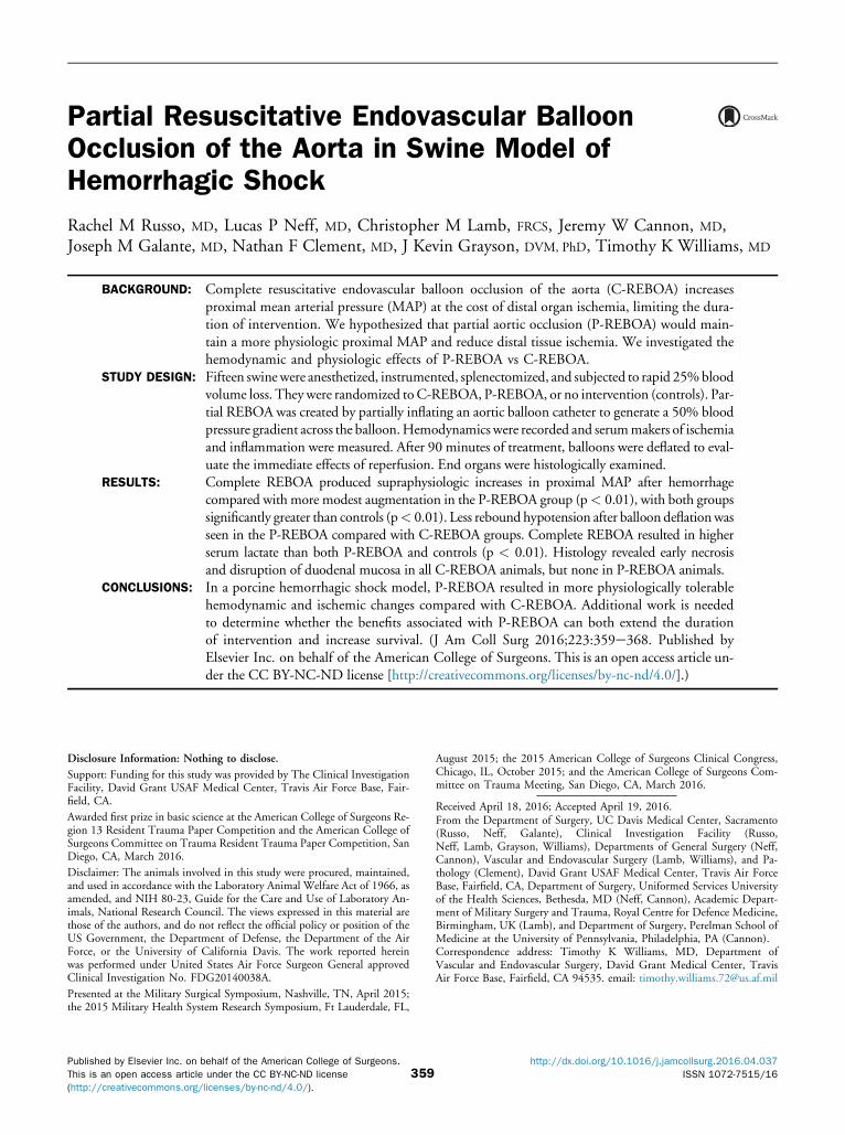
Figure 1. Comparison of differential proximal (cranial to balloon)and distal (caudal to balloon) aortic systolic blood pressure differ-ential among complete resuscitative endovascular balloon occlu-sion of the aorta, partial resuscitative endovascular balloonocclusion of the aorta, and control groups. Differential pressureswere significantly different among the groups (p < 0.01). H, onset ofhemorrhage.
362 Russo et al Partial Aortic Occlusion in Shocked Swine J Am Coll Surg
the groups. Consistent pressure gradients were main-tained between proximal and distal systolic pressures ineach treatment group, averaging 90% in the C-REBOA,50% in the P-REBOA, and 10% in control groups(Fig. 1).At 60 minutes, P-REBOA resulted in a proximal aortic
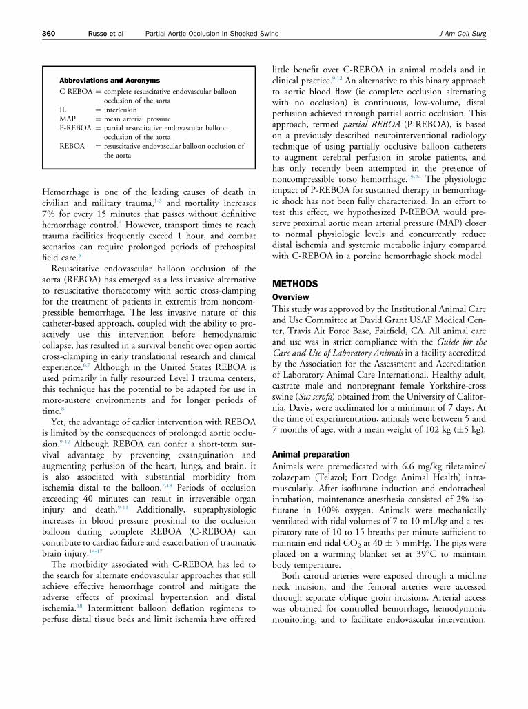
MAP closer to baseline than either the C-REBOA or con-trol group (þ13.5 vs þ69.0 and �20.2 mmHg, respec-tively; p < 0.01) (Fig. 2A). Distal MAP duringintervention in the P-REBOA group was equivalent tocontrols (p ¼ 0.47), and distal MAP in the C-REBOAgroup was significantly decreased compared with eitherthe P-REBOA or control groups (p < 0.01) (Fig. 2B).Similarly, visceral MAP during intervention in theP-REBOA group was equivalent to controls (p ¼ 0.62),and visceral MAP in the C-REBOA group was signifi-cantly lower than either the P-REBOA or control groups(p ¼ 0.01) (Fig. 2C). On balloon deflation, aortic pres-sure gradients rapidly dissipated in both the C-REBOAand P-REBOA groups.Serum lactate levels increased significantly (p < 0.05)
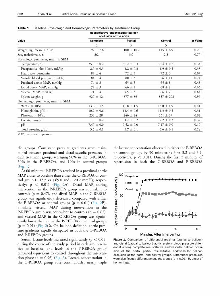
during the course of the study period in each group rela-tive to baseline, and levels in the P-REBOA groupremained equivalent to control throughout the interven-tion phase (p ¼ 0.96) (Fig. 3). Lactate concentration inthe C-REBOA group rose continuously, nearly triple
the lactate concentration observed in either the P-REBOAor control groups by 90 minutes (9.3 vs 3.2 and 3.2,respectively; p < 0.01). During the first 5 minutes ofreperfusion in both the C-REBOA and P-REBOA
Figure 2. (A) Comparison among mean arterial pressure (MAP) in the proximal (cranial to balloon) aorta in completeresuscitative endovascular balloon occlusion of the aorta (C-REBOA), partial resuscitative endovascular balloonocclusion of the aorta (P-REBOA), and control groups. Mean arterial pressures were significantly different among thegroups (p < 0.01). (B) Comparison of MAP in the distal (caudal to balloon) aorta among C-REBOA, P-REBOA, andcontrol groups. Mean arterial pressures were significantly different between P-REBOA and control (p ¼ 0.03), andboth were significantly different from C-REBOA (p < 0.01). (C) Comparison of MAP in a standardized jejunal branch ofthe superior mesenteric artery (caudal to balloon) among C-REBOA, P-REBOA, and control groups. Mean arterialpressures were equivalent between P-REBOA and control (p ¼ 0.12), and both were significantly different fromC-REBOA (p < 0.01). H, onset of hemorrhage. Dashed line, baseline MAP. Vertical line, balloon deflation.
Vol. 223, No. 2, August 2016 Russo et al Partial Aortic Occlusion in Shocked Swine 363
arms, serum lactate concentration increased above thehighest levels reached during the intervention phase(Fig. 3). As washout of distal tissue beds continued,lactate levels quickly stabilized in the P-REBOA group,but continued to rise in the C-REBOA group. Serumlactate concentration peaked at 13.3 mmol/L in theC-REBOA group during reperfusion, which wasmore than double the concentrations seen in theP-REBOA group or control group by the end of the study(p < 0.01).Additional findings among the groups at 90 minutes
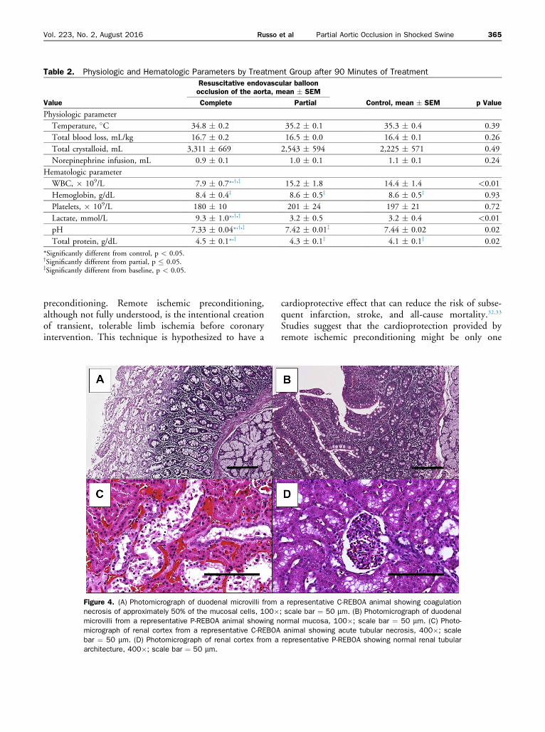
include a more severe acidosis and a profound leukopeniain the C-REBOA group compared with the P-REBOAgroup or control group (p < 0.01) Within each group,WBC, hemoglobin concentration, platelet count, pH,and total protein concentration decreased significantly(Table 2). Production of inflammatory cytokines
increased in all 3 groups from baseline to the end of inter-vention, but none reached statistical significance exceptfor IL6 and IL10 in the C-REBOA group (p < 0.01and p ¼ 0.02, respectively). There were no significant dif-ferences in cytokine concentration across the groups atany time point.Histologic examination of the duodenum showed
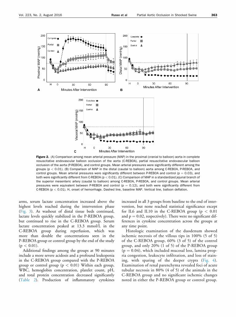
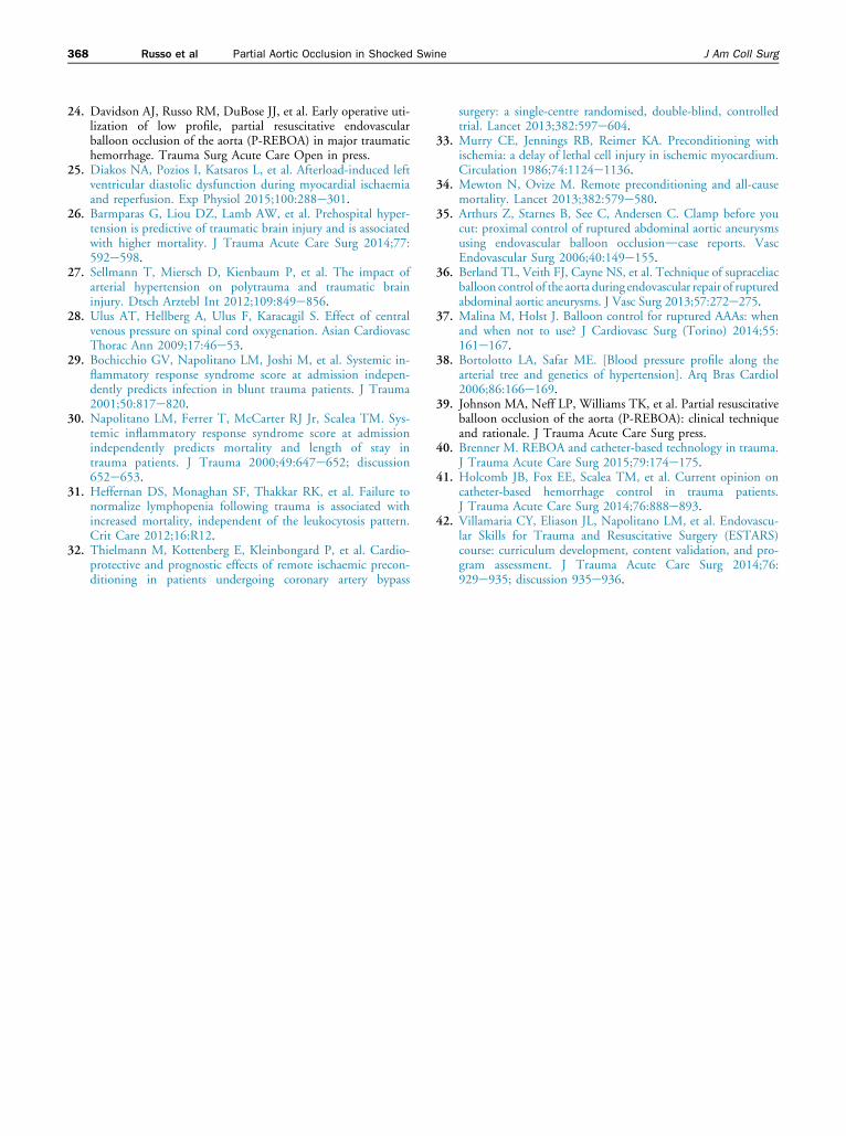
ischemic necrosis of the villous tips in 100% (5 of 5)of the C-REBOA group, 60% (3 of 5) of the controlgroup, and only 20% (1 of 5) of the P-REBOA group(p ¼ 0.04), which included mucosal loss, lamina prop-ria congestion, leukocyte infiltration, and loss of stain-ing, with sparing of the deeper crypts (Fig. 4).Examination of renal parenchyma revealed foci of acutetubular necrosis in 80% (4 of 5) of the animals in theC-REBOA group and no significant ischemic changesnoted in either the P-REBOA group or control group.
Figure 3. Blood lactate concentrations at various time pointsamong complete resuscitative endovascular balloon occlusion ofthe aorta (C-REBOA), partial resuscitative endovascular balloonocclusion of the aorta (P-REBOA), and control groups. Blood lactateconcentrations were significantly higher in the C-REBOA thanP-REBOA and control groups (p < 0.01), but were statisticallyequivalent in the P-REBOA and control groups (p ¼ 0.66). H, lactateconcentration at the onset of hemorrhage.
364 Russo et al Partial Aortic Occlusion in Shocked Swine J Am Coll Surg
All but one animal survived the duration of the exper-iment. The only death occurred in the C-REBOA groupimmediately after balloon deflation at 90 minutes.
DISCUSSIONResuscitative endovascular balloon occlusion of the aortais emerging as a viable alternative to open aortic cross-clamping for hemorrhagic shock. This technique hasbeen embraced by several Level I trauma centersthroughout the United States and is being used interna-tionally. Although REBOA is effective at limiting exsan-guination and restoring proximal perfusion pressure tothe heart, brain, and lungs, it results in a cumulative phys-iologic insult over time that can have detrimental conse-quences.10 The supraphysiologic proximal pressures andafterload seen with REBOA can result in cardiac dysfunc-tion or exacerbation of traumatic brain injury.14,16 Addi-tionally, intervention times in excess of 40 to 60minutes can result in irreversible ischemia to distal tissues,with profound ischemia-reperfusion injury.9-11 PartialREBOA can mitigate the adverse effects of sustained com-plete aortic occlusion and increase the maximal durationof intervention through the early restoration of distalblood flow.In this study, we have demonstrated that P-REBOA
maintained proximal MAP at more physiologicallynormal levels during intervention and after balloon defla-tion, and avoided the hemodynamic extremes seen with
C-REBOA. Partial REBOA also simultaneously main-tained enough distal perfusion to minimize organischemia and the systemic burden of ischemia-reperfusion injury compared with C-REBOA.Maintaining proximal MAP within a normal physio-
logic range with P-REBOA can reduce the incidence ofcardiac dysfunction, cerebral edema, and respiratory fail-ure compared with sustained aortic occlusion.9,10 Begin-ning at balloon inflation, C-REBOA producessupraphysiologic augmentation of proximal aortic MAPwith dramatic increases in cardiac afterload, even in thecontext of hypovolemic shock. Proximal MAP as highas 222 mmHg was seen in the C-REBOA arm of ourstudy. Supraphysiologic MAP >110 mmHg persisted inall animals in the C-REBOA arm as long as the balloonremained inflated. This dramatic increase in aortic after-load might be detrimental to cardiac performance, partic-ularly in the context of an already ischemic or injuredmyocardium.15,16,25 The downtrend in proximal MAPseen in the C-REBOA arm might represent decliningcardiac function during the course of the experiment.This trend was not seen in the P-REBOA or controlarms. Additionally, these supraphysiologic proximal pres-sures can exacerbate concomitant blunt aortic, brain, orpulmonary injuries.17,25-28 Therapies aimed at preservinghemodynamics within a normal physiologic range canresult in less end-organ damage and increase the potentialnumber of patients that can benefit from these treatments.Partial REBOA can also offer a way to extend the dura-
tion of intervention beyond what is currently possiblewith C-REBOA. Partial REBOA reduced the systemicmetabolic burden of ischemia and inflammationcompared with the C-REBOA group. Buildup of lacticacid and resultant metabolic acidosis were less severe inthe P-REBOA group than the C-REBOA group, bothfrom the lessened tissue ischemia and a continuouswashout of metabolites provided by preserved distalperfusion. Additionally, white cell sequestration inischemic distal tissue beds created a profound leukopeniain the C-REBOA group that worsened as the study pro-gressed and was not seen in either the P-REBOA groupor control group. Although this sequestration phenome-non is not fully understood, leukopenia in traumapatients has been associated with worse outcomes.29-31 Inaddition, P-REBOA preserved distal blood flow sufficientto avoid the significant duodenal and kidney ischemiaseen in the C-REBOA group.Unexpectedly, there was less duodenal ischemia in the
P-REBOA group than in the control group, despiteequivalent visceral MAP. This finding suggests thatP-REBOA can induce resistance to ischemic organinjury at the cellular level, similar to remote ischemic
Table 2. Physiologic and Hematologic Parameters by Treatment Group after 90 Minutes of Treatment
Value
Resuscitative endovascular balloonocclusion of the aorta, mean � SEM
*Significantly different from control, p < 0.05.ySignificantly different from partial, p � 0.05.zSignificantly different from baseline, p < 0.05.
Vol. 223, No. 2, August 2016 Russo et al Partial Aortic Occlusion in Shocked Swine 365
preconditioning. Remote ischemic preconditioning,although not fully understood, is the intentional creationof transient, tolerable limb ischemia before coronaryintervention. This technique is hypothesized to have a
Figure 4. (A) Photomicrograph of duodenal microvilli fromnecrosis of approximately 50% of the mucosal cells, 100�microvilli from a representative P-REBOA animal showing nmicrograph of renal cortex from a representative C-REBOAbar ¼ 50 mm. (D) Photomicrograph of renal cortex from aarchitecture, 400�; scale bar ¼ 50 mm.
cardioprotective effect that can reduce the risk of subse-quent infarction, stroke, and all-cause mortality.32,33
Studies suggest that the cardioprotection provided byremote ischemic preconditioning might be only one
a representative C-REBOA animal showing coagulation; scale bar ¼ 50 mm. (B) Photomicrograph of duodenalormal mucosa, 100�; scale bar ¼ 50 mm. (C) Photo-animal showing acute tubular necrosis, 400�; scalerepresentative P-REBOA showing normal renal tubular
366 Russo et al Partial Aortic Occlusion in Shocked Swine J Am Coll Surg
aspect of a much larger effect, preparing the bodyto defend against global ischemia and reperfusioninjury.34
When sustained periods of aortic occlusion are neces-sary, P-REBOA can result in less rebound hypotensionthan C-REBOA. Balloon deflation precipitates rapidredistribution of circulating blood into distal vascularbeds dilated from ischemia. This abrupt decrease incardiac afterload combined with washout of ischemicmetabolites further suppresses myocardial function andweakens vascular tone.35 Taken together, these effectscan cause abrupt hemodynamic collapse.36 The need forrepeat aortic occlusion to counter this collapse has beenseen in both large animal models and clinically.12,35-37 Inthe current study, the C-REBOA group demonstratedprofound decreases in central MAP after balloon defla-tion, and even resulted in the death of one animal beforethe end of the study. In contrast, the P-REBOA groupexperienced smaller decreases in central aortic pressureafter balloon deflation, with all animals maintaining anaortic MAP above the pre-hemorrhage baseline for theremainder of the study. Maintaining even a small amountof perfusion distal to the balloon can ameliorate tissueischemia, rebound hypotension after balloon deflationand reperfusion injury, resulting in decreased morbidityand mortality.
Limitations
Our model of P-REBOA using a conventional noncom-pliant balloon catheter to achieve partial aortic occlusionis not clinically applicable in its current form. This type ofballoon architecture has a narrow diameter range thatrequires using fluoroscopy to size the balloon to the indi-vidual aorta. However, it was intentionally chosen toovercome an important limitation of more commonlyused compliant spherical aortic occlusion balloons, whereprecise control of aortic pressure gradients is challenging ifnot impossible.18,24 The elongated, cylindrical shape of thenoncompliant balloon approximates the wall of the aortaacross a longer length, which provides precise control overthe degree of aortic occlusion. We were able to reproducespecified aortic pressure gradients with a high degree offidelity in this study. Complete aortic occlusion generateda 90% proximal to distal aortic pressure gradient, ratherthan a 100% gradient, in the C-REBOA group. Thisobservation might indicate a small amount of nonpulsa-tile distal aortic flow entering through collateral pathwaysor the inherent back pressure of the aorta below the pointof occlusion. The 10%, rather than 0%, pressure gradientobserved in the control group might represent a smallamount of aortic occlusion created by the uninflatedballoon catheter or the normal physiologic decrease in
MAP that is known to occur along the length of theaorta.38
The 50% gradient chosen for P-REBOA was selectedas a starting point for our proof-of-concept study.However, it remains unclear how the pressure gradientachieved in this study relates to tissue perfusion at theorgan level. Given the dynamic nature of bloodpressure and flow, pressure gradient alone might notbe a sufficient metric to ensure distal aortic bloodflow within a desirable range in a true clinical sce-nario.18,24,39 More work is needed to determine theoptimal parameters to guide control of aortic bloodflow to minimize hemorrhage and maintain distal tissueperfusion.Our study duration was too short to examine the inter-
mediate and long-term effects of C-REBOA vs P-REBOAin terms of ongoing resuscitation requirements and multi-organ dysfunction reported in other studies with short-term survival end points.11-13 The short duration of thestudy also likely contributed to the limited histologicevidence of end-organ ischemia in all but the duodenumand kidneys, as well as the limited differences in inflam-matory cytokine production we observed.This study was conducted in a controlled hemorrhage
model that does not assess the ability of P-REBOA toconfer these benefits in the presence of uncontrolled,multifocal, or ongoing hemorrhage. In our previouslypublished study of P-REBOA in uncontrolled hemor-rhage, we saw that P-REBOA prevented the hemody-namic extremes associated with C-REBOA, but thepresence of ongoing hemorrhage limited our ability toobserve any benefits from preserved distal perfusion.22
Partial REBOA might be suitable when control of majorsources of hemorrhage has been achieved. Additionally,P-REBOA might be more practical when access to bloodproducts and surgical capabilities is readily available.Additional development and refinement are neededbefore this technique is feasible outside of well-resourced hospitals.Finally, the need for arterial access, radiographic imag-
ing to confirm catheter position, and limited physicianexperience with endovascular techniques all stand asobstacles to the widespread use of REBOA.7,40-42
CONCLUSIONSIn our proof-of-concept study in a swine model of hem-orrhagic shock, P-REBOA maintained normal physiologybetter than C-REBOA, lessened the systemic impact ofdistal organ ischemia, and reduced hemodynamic insta-bility, providing the potential for longer periods of inter-vention than are currently recommended with
Vol. 223, No. 2, August 2016 Russo et al Partial Aortic Occlusion in Shocked Swine 367
C-REBOA. This study provides support for additionaldevelopment of this technique in an effort to overcomethe current limitations of C-REBOA. Overcoming thedeleterious effects of prolonged aortic occlusion couldfurther revolutionize the management of noncompressibletorso hemorrhage, particularly closer to the point ofinjury and before hemodynamic collapse. Additionaltechnological innovation is warranted to address thelimitations of current catheter designs to facilitate imple-mentation of P-REBOA in the clinical environment.
Author Contributions
Study conception and design: Russo, Neff, Lamb,Cannon, Galante, Grayson, Williams
Acquisition of data: Russo, Neff, Lamb, Clement,Grayson, Williams
Analysis and interpretation of data: Russo, Lamb,Cannon, Galante, Clement, Grayson, Williams
Drafting of manuscript: Russo, Neff, Lamb, Neff,Galante, Clement, Grayson, Williams
Critical revision: Russo, Neff, Lamb, Cannon, Galante,Clement, Grayson, Williams
Acknowledgment: The authors thank SSG Kelly Caneen, MrCarl Gibbins, Ms Sally Knode, SSgt Elaine Spotts, and SrAGeoffrey O’Hair for their outstanding technical assistance,and the other staff of the Clinical Investigations FacilityDavid Grant USAF Medical Center for their support.
REFERENCES
1. Blackbourne LH, Czarnik J, Mabry R, et al. Decreasing killedin action and died of wounds rates in combat wounded.J Trauma 2010;69[Suppl 1]:S1eS4.
2. Eastridge BJ, Hardin M, Cantrell J, et al. Died of wounds onthe battlefield: causation and implications for improving com-bat casualty care. J Trauma 2011;71[Suppl]:S4eS8.
3. Eastridge BJ, Mabry RL, Seguin P, et al. Death on the battle-field (2001e2011): implications for the future of combatcasualty care. J Trauma Acute Care Surg 2012;73[Suppl 5]:S431eS437.
4. Alarhayem A, Myers J, Dent D, Eastridge B. No time to bleed:the impact of time from injury to the operating room on sur-vival in patients with hemorrhage from blunt abdominaltrauma. Presented at the 74th Annual Meeting of American As-sociation for the Surgery of Trauma and Clinical Congress ofAcute Care Surgery, September 9-12, 2015, Las Vegas, NV.
5. NAEMT. PHTLS Prehospital Trauma Life Support: MilitaryVersion. 6th ed. New York: Elsevier Science Health Science Di-vision; 2006.
6. White JM, Cannon JW, Stannard A, et al. Endovascularballoon occlusion of the aorta is superior to resuscitative tho-racotomy with aortic clamping in a porcine model of hemor-rhagic shock. Surgery 2011;150:400e409.
7. Brenner ML, Moore LJ, DuBose JJ, et al. A clinical series ofresuscitative endovascular balloon occlusion of the aorta for
hemorrhage control and resuscitation. J Trauma Acute CareSurg 2013;75:506e511.
8. DuBose JJ, Scalea TM, Brenner M, et. al. The AAST Prospec-tive Aortic Occlusion for Resuscitation in Trauma and AcuteCare Surgery (AORTA) Registry: Data on contemporary utili-zation and outcomes of aortic occlusion and resuscitativeballoon occlusion of the aorta (REBOA) [published onlineahead of print April 5, 2016]. J Trauma Acute Care Surg.doi: 10.1097/TA.0000000000001079.
9. Saito N, Matsumoto H, Yagi T, et al. Evaluation of the safetyand feasibility of resuscitative endovascular balloon occlusionof the aorta. J Trauma Acute Care Surg 2015;78:897e903;discussion 904.
10. Norii T, Crandall C, Terasaka Y. Survival of severe blunttrauma patients treated with resuscitative endovascular balloonocclusion of the aorta compared with propensity score-adjusted untreated patients. J Trauma Acute Care Surg2015;78:721e728.
11. Avaro JP, Mardelle V, Roch A, et al. Forty-minute endovascu-lar aortic occlusion increases survival in an experimental modelof uncontrolled hemorrhagic shock caused by abdominaltrauma. J Trauma 2011;71:720e725; discussion 725e726.
12. Morrison JJ, Ross JD, Houston R, et al. Use of resuscitativeendovascular balloon occlusion of the aorta in a highly lethalmodel of noncompressible torso hemorrhage. Shock 2014;41:130e137.
13. Markov NP, Percival TJ, Morrison JJ, et al. Physiologic toler-ance of descending thoracic aortic balloon occlusion in a swinemodel of hemorrhagic shock. Surgery 2013;153:848e856.
14. Long KN, Houston R, Watson JD, et al. Functional outcomeafter resuscitative endovascular balloon occlusion of the aortaof the proximal and distal thoracic aorta in a swine model ofcontrolled hemorrhage. Ann Vasc Surg 2015;29:114e121.
15. Stokland O, Miller M, Ilebekk A, Kiil F. Mechanism of hemo-dynamic responses to occlusion of the descending thoracicaorta. Am J Physiol Heart Circul Physiol 1980;238:H423eH429.
16. Dunn E, Prager RL, Fry W, Kirsh MM. The effect of abdom-inal aortic cross-clamping on myocardial function. J Surg Res1977;22:463e468.
17. Annecke T, Kubitz JC, Langer K, et al. Lung injury followingthoracic aortic occlusion: comparison of sevoflurane and pro-pofol anaesthesia. Acta Anaesthesiol Scand 2008;52:977e986.
18. Russo RM, Neff LP, Johnson MA, Williams TK. Emergingendovascular therapies for non-compressible torso hemorrhage[published online ahead of print May 11, 2016]. Shock. doi:10.1097/SHK.0000000000000641.
19. Hammer M, Jovin T, Wahr JA, Heiss WD. Partial occlusionof the descending aorta increases cerebral blood flow in a non-stroke porcine model. Cerebrovasc Dis 2009;28:406e410.
20. Liebeskind DS. Aortic occlusion for cerebral ischemia: fromtheory to practice. Curr Cardiol Rep 2008;10:31e36.
22. Russo RM, Williams TK, Grayson JK, et al. Extending thegolden hour: partial resuscitative endovascular balloon occlu-sion of the aorta in a highly lethal swine liver injury model.J Trauma Acute Care Surg 2016;80:372e380.
23. Horer TM, Cajander P, Jans A, Nilsson KF. A case of partialaortic balloon occlusion in an unstable multi-trauma patient.Trauma 2016;18:150e154.
368 Russo et al Partial Aortic Occlusion in Shocked Swine J Am Coll Surg
24. Davidson AJ, Russo RM, DuBose JJ, et al. Early operative uti-lization of low profile, partial resuscitative endovascularballoon occlusion of the aorta (P-REBOA) in major traumatichemorrhage. Trauma Surg Acute Care Open in press.
25. Diakos NA, Pozios I, Katsaros L, et al. Afterload-induced leftventricular diastolic dysfunction during myocardial ischaemiaand reperfusion. Exp Physiol 2015;100:288e301.
26. Barmparas G, Liou DZ, Lamb AW, et al. Prehospital hyper-tension is predictive of traumatic brain injury and is associatedwith higher mortality. J Trauma Acute Care Surg 2014;77:592e598.
27. Sellmann T, Miersch D, Kienbaum P, et al. The impact ofarterial hypertension on polytrauma and traumatic braininjury. Dtsch Arztebl Int 2012;109:849e856.
28. Ulus AT, Hellberg A, Ulus F, Karacagil S. Effect of centralvenous pressure on spinal cord oxygenation. Asian CardiovascThorac Ann 2009;17:46e53.
29. Bochicchio GV, Napolitano LM, Joshi M, et al. Systemic in-flammatory response syndrome score at admission indepen-dently predicts infection in blunt trauma patients. J Trauma2001;50:817e820.
30. Napolitano LM, Ferrer T, McCarter RJ Jr, Scalea TM. Sys-temic inflammatory response syndrome score at admissionindependently predicts mortality and length of stay intrauma patients. J Trauma 2000;49:647e652; discussion652e653.
31. Heffernan DS, Monaghan SF, Thakkar RK, et al. Failure tonormalize lymphopenia following trauma is associated withincreased mortality, independent of the leukocytosis pattern.Crit Care 2012;16:R12.
32. Thielmann M, Kottenberg E, Kleinbongard P, et al. Cardio-protective and prognostic effects of remote ischaemic precon-ditioning in patients undergoing coronary artery bypass
surgery: a single-centre randomised, double-blind, controlledtrial. Lancet 2013;382:597e604.
33. Murry CE, Jennings RB, Reimer KA. Preconditioning withischemia: a delay of lethal cell injury in ischemic myocardium.Circulation 1986;74:1124e1136.
34. Mewton N, Ovize M. Remote preconditioning and all-causemortality. Lancet 2013;382:579e580.
35. Arthurs Z, Starnes B, See C, Andersen C. Clamp before youcut: proximal control of ruptured abdominal aortic aneurysmsusing endovascular balloon occlusiondcase reports. VascEndovascular Surg 2006;40:149e155.
36. Berland TL, Veith FJ, Cayne NS, et al. Technique of supraceliacballoon control of the aorta during endovascular repair of rupturedabdominal aortic aneurysms. J Vasc Surg 2013;57:272e275.
37. Malina M, Holst J. Balloon control for ruptured AAAs: whenand when not to use? J Cardiovasc Surg (Torino) 2014;55:161e167.
38. Bortolotto LA, Safar ME. [Blood pressure profile along thearterial tree and genetics of hypertension]. Arq Bras Cardiol2006;86:166e169.
39. Johnson MA, Neff LP, Williams TK, et al. Partial resuscitativeballoon occlusion of the aorta (P-REBOA): clinical techniqueand rationale. J Trauma Acute Care Surg press.
40. Brenner M. REBOA and catheter-based technology in trauma.J Trauma Acute Care Surg 2015;79:174e175.
41. Holcomb JB, Fox EE, Scalea TM, et al. Current opinion oncatheter-based hemorrhage control in trauma patients.J Trauma Acute Care Surg 2014;76:888e893.
42. Villamaria CY, Eliason JL, Napolitano LM, et al. Endovascu-lar Skills for Trauma and Resuscitative Surgery (ESTARS)course: curriculum development, content validation, and pro-gram assessment. J Trauma Acute Care Surg 2014;76:929e935; discussion 935e936.