16
Particle size and shape characterization by Static Image Analysis

Particle size and shape characterization by Static Image Analysis

www.microtrac.com | 2 – 3
Microtrac MRB is your reliable partner for the
characterization of disperse systems. Using high-
end technology, we support our customers in
consistently achieving excellent results. Innovation
and quality form the basis of our success.
Microtrac MRB has three product lines with
competence centers on three continents:
Scattered light analysis: Microtrac MRB is a
leading provider of laser diffraction instruments
(static light scattering), a versatile technique
for particle size measurement. The portfolio
also includes instruments for the reliable
characterization of nanoparticles by means of
dynamic light scattering. The production and
development of this product line are located in
Pennsylvania, USA.
Image analysis: With the devices of the CAMSIZER
series, Microtrac MRB offers high-quality systems
for determining particle size and particle shape
using imaging methods. We develop and
manufacture the image analysis instruments at our
facilities in Haan, Germany.
Surface and porosity measurement:
Specific surface area, BET value and porosity of
powders are determined via gas adsorption.
The competence center for surface analysis is
located in Osaka, Japan.
As a member of the Verder Scientific Group, we
provide worldwide support through numerous
agencies and subsidiaries.
Microtrac MRB – Particle Characterization at Its BestComprehensive analysis of powders, granulates and suspensions

With the ever-increasing demands for product
quality, accurate evaluation of raw materials,
intermediates and final products has become
indispensable. Automation, high sample
throughput and short measurement times make
the CAMSIZER systems ideal tools for routine
analyses and quality control. A sophisticated
evaluation software offers a wealth of valuable
information making the analyzers ideally suited for
use in research and development.
Microtrac MRB has set the standard in dynamic
image analysis with the development of the
CAMSIZER X2 and CAMSIZER P4. The unique dual
camera technology implemented in both instruments
permits rapid analysis with highest accuracy and
reproducibility and an extremely wide measuring
range.
The latest innovation, CAMSIZER M1, is based on
static image analysis. This technique is particularly
suitable for analysis of particle size and shape in the
low micron range with utmost precision.
.
CAMSIZER X2: Dynamic Image Analysis for powders and suspensions
CAMSIZER P4: Dynamic Image Analysis for free-flowing bulk materials and granulates
CAMSIZER M1: Static Image Analysis for very fine powders
CAMSIZER Series High-resolution image analysis of particle size
and particle shape
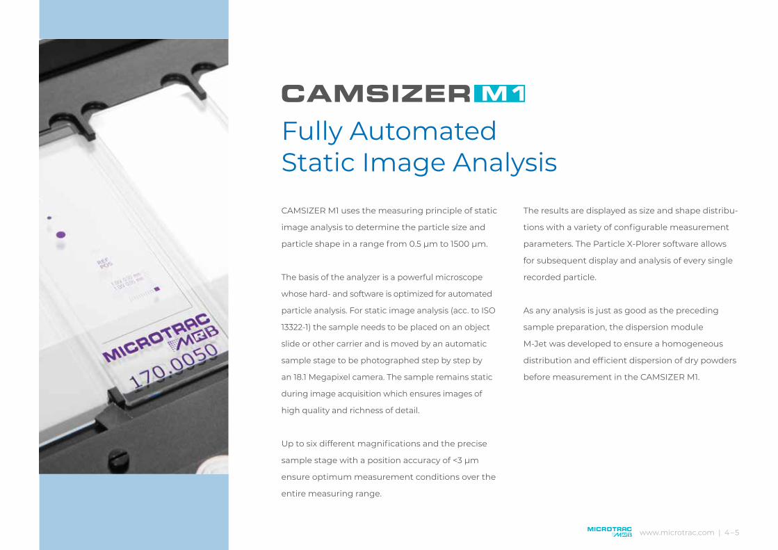
CAMSIZER M1 uses the measuring principle of static
image analysis to determine the particle size and
particle shape in a range from 0.5 µm to 1500 µm.
The basis of the analyzer is a powerful microscope
whose hard- and software is optimized for automated
particle analysis. For static image analysis (acc. to ISO
13322-1) the sample needs to be placed on an object
slide or other carrier and is moved by an automatic
sample stage to be photographed step by step by
an 18.1 Megapixel camera. The sample remains static
during image acquisition which ensures images of
high quality and richness of detail.
Up to six different magnifications and the precise
sample stage with a position accuracy of <3 µm
ensure optimum measurement conditions over the
entire measuring range.
The results are displayed as size and shape distribu-
tions with a variety of configurable measurement
parameters. The Particle X-Plorer software allows
for subsequent display and analysis of every single
recorded particle.
As any analysis is just as good as the preceding
sample preparation, the dispersion module
M-Jet was developed to ensure a homogeneous
distribution and efficient dispersion of dry powders
before measurement in the CAMSIZER M1.
www.microtrac.com | 4 – 5
Fully Automated Static Image Analysis

Particle Analyzer CAMSIZER M1: High-resolution static image analysis (ISO 13322-1) from 0.5 µm to 1500 µm
18.1 Megapixel color camera
Upto6magnifications
Accurate analysis of very small particles
Excellent image quality
Highly precise shape analysis
Dispersion module M-Jet
Automatic measurement, easy operation
Stitching function
Comprehensive evaluation possibilities with Particle X-Plorer

ADVANTAGE CAMSIZER M1:
The sample stage is controlled
with a three-axis joystick with four
programmable function keys.
ADVANTAGE CAMSIZER M1:
The highly efficient dispersion module
M-Jet ensures perfect preparation of
powder samples within seconds.
ADVANTAGE CAMSIZER M1:
An 18.1 Megapixel color camera captures particle
images with excellent resolution for highly
accurate analysis results.
ADVANTAGE CAMSIZER M1:
The sample can be analyzed in
transmitted or incident light.
Powerful LED light sources guarantee
homogeneous illumination and
excellent image quality.
ADVANTAGE CAMSIZER M1:
The highly precise sample stage has a wide
traversing range of 225 mm x 76 mm with a
position accuracy of <3 µm and a positional
repeatability of <1 µm. Thus, it is possible to
analyze an area which corresponds to eight
standard object slides.
Superiority in Detail
www.microtrac.com | 6 – 7
ADVANTAGE CAMSIZER M1:
The analyzer is equipped with five
objectives with magnifications from
2.5x to 50x. An additional objective
(e. g. 100x) can be added on request.

2
1 2
3 4
1. Objectives with large working distance ensure reliable results
2. Sample preparation with M-Jet
3. Wide selection of accessories
4. Intuitive software, including Particle X-Plorer

www.microtrac.com | 8– 9
The CAMSIZER M1 software records a variety of
different measurement parameters and offers
comprehensive evaluation possibilities. The particle
size is ascertained based on over 50 different
parameters such as maximum or minimum Feret
length, chord length or area equivalent diameter.
Available shape parameters include, for example,
aspect ratio, circularity, symmetry, roundness
and convexity. All measurement and evaluation
parameters can be saved as standard operating
procedures which makes operation of the CAMSIZER
M1 particularly safe and comfortable. The Master
Operation allows for automated subsequent analysis
of several samples with optimum settings.
Results are conveniently displayed as diagrams or
tables. A newly developed data manager facilitates
administration of the measurement data.
A special feature of the software is the integrated
Particle X-Plorer which permits detailed evaluation
and presentation of all recorded particle images,
even after the measurement. Thus, it is easily
possible to find and quantify particles with specific
properties or combinations of properties.
Intuitive Measurement and Evaluation SoftwareDetailed analysis and presentation with Particle X-Plorer

2
1 2
3 4
1. Display of particle properties in a „filter cube“ facilitates evaluation
2. Clearly structured and intuitive user interface
3. Statistics function allows to compare different samples
4. Display of particle images and corresponding size and shape values

www.microtrac.com | 10 – 11
The CAMSIZER M1 is perfectly suitable for analysis of pharmaceutical ingredients and
excipients. Particle size and particle shape are precisely measured and clearly presented.
Thus, changes in product quality become immediately apparent.
The example shows the evaluation of a Paracetamol sample. Measuring time: 9 minutes,
number of images: 1600, number of measured particles: 160 000.
Pharmaceutical ingredients and excipients
Toner for laser printers usually contains particles sized 5 microns and larger, with a very
narrow size distribution. High-resolution image analyzers like the CAMSIZER M1 provide
higher accuracy than alternative methods such as laser diffraction which measures particle
size indirectly based on the evaluation of a diffraction pattern. Moreover, only imaging
methods are suitable for describing the morphology of very small particles.
Toner
Analysis of Paracetamol with CAMSIZER M1. Upper left: picture taken with 10x objective, transmitted light. Upper right: the particles on the picture are recognized and all relevant size and shape parameters are identified. Lower left: Clearly structured presentation of the size distribution. Lower right: shape analysis shows the aspect ratio for various particle sizes.
x_area [µm]20 40 60 80 100 1200
10
20
30
40
50
60
70
80
90
Q3 [%]
0
1
2
3
4
5
6
7
8
9
p3 [%]
Particle size [µm]
Cu
mu
lati
ve d
istr
ibu
tion
Q3
[%]
Frac
tion
s p3
[%]
x [µm]10 20 30 40 50 60 70 80 900
0.1
0.2
0.3
0.4
0.5
0.6
0.7
0.8
b/l3
Particle size [µm]
Asp
ect
rati
o
xc_min [µm]2 4 6 8 10 12 14 16 18 200
5
10
15
20
25
30
q3 [%/µm]
Particle size [µm]
Cu
mu
lati
ve d
istr
ibu
tion
Q3
[%/
µm
]
The example shows the results of measuring five toner samples with the CAMSIZER M1. Four samples have a mean particle size of approx. 6 µm, one sample has considerably larger particles and a wider size distribution.

Fine grinding and polishing media consist of, for example, corundum, carbides or industrial
diamonds. The relevant standards stipulate the use of sediment analysis for quality control.
However, this method is very time-consuming and imprecise. The CAMSIZER M1 is able to
generate more exact and comprehensive information which allows for profound evaluation
of the material in question.
Industrial Diamonds
A polymer sample was ground with two different mill types, a rotor
mill and a ball mill. Both samples have an almost identical median of
approx. 47 µm. However, the size distribution of the ball milled sample
is more symmetrical and uniform. The rotor mill produces more fines
and more oversized particles. The CAMSIZER M1 analysis also shows
that the particles ground in the ball mill are more spherical.
Polymers
Size distribution of two industrial diamond samples measured with the CAMSIZER M1. The mean values are 3.1 µm and 20.3 µm. The black curves represent the result of laser diffraction analysis. The size is accurately measured even for the finest particles. High-resolution images illustrate the particle shape.
Polymer sample ground in a rotor mill (red curve) and in a ball mill (blue curve). Size distribution (left) and shape parameter „roundness“ (right).
xc_min [µm]20 40 60 80 100 1200
10
20
30
40
50
60
70
80
90
Q3 [%]
0
5
10
15
20
25
30
35
40
45
p3 [%]
Both samples:Median ~ 47 µm
Particle size [µm]
Cu
mu
lati
ve d
istr
ibu
tion
Q3
[%]
Frac
tion
s p3
[%]
x [µm]0 10 20 30 40 50 60 70 80
0.25
0.30
0.35
0.40
0.45
0.50 Sample 1 has lowerroundness than Sample 2
Particle size [µm]
Rou
nd
nes
s
x_area [µm]5 10 15 20 25 300
10
20
30
40
50
60
70
80
90
Q3 [%]
Particle size [µm]
Cu
mu
lati
ve d
istr
ibu
tion
Q3
[%]
5 µm

www.microtrac.com | 12 – 13
The new dispersion unit M-Jet ensures optimum sample
preparation on various carriers for microscopic analysis.
The function principle is based on negative pressure,
compressed air supply is not required. Thanks to the
unique nozzle geometry even finest powders are effectively
separated and distributed uniformly on the object plate.
Benefits at a glance:
Dispersion by negative pressure 10 kPa – 70 kPa
Variety of dispersion nozzles
Storage of eight SOPs possible
Comfortable operation via touch pad and
control knob
Perfect Powder Dispersion with M-JetEfficient sample preparation for microscopic analysis

3 4
1. Easy changing of sample holder
2. Sample holder for Camsizer M1
3. Position calibration
4. Reproduction scale calibration
2
1
The wide range of accessories available for CAMSIZER M1 provides everything
you need for successful particle analysis. Four different sample holders can be
quickly and easily inserted into the sample stage and ensure a high degree of
flexibility.
Holder for eight standard object plates, 76 x 28 mm
Holder for four object plates, 76 x 52 mm
Glass plate, 147 mm x 313 mm
Holder for round filter
In addition to the standard objectives with magnifications of 2.5x, 5x, 10x, 20x
and 50x, one more objective can be used, for example 1.25 or 100x.

Particle characterization can be performed by Static
or Dynamic Image Analysis. For Dynamic Image
Analysis (ISO 13322-2), a particle stream is generated
which is photographed and evaluated by cameras.
Static Image Analysis (ISO 13322-1) is based on mi-
croscopy; the particles are placed on an object plate
which is photographed step by step. Hence, the dif-
ference lies in the particle movement: for Dynamic
Image Analysis the particles are captured in motion,
for Static Image Analysis, they remain motionless.
Dynamic Image Analysis is ideally suited for routine
measurements of bulk goods, powders, granules
and suspensions. This method is characterized by
high sample throughput, reliability and excellent
reproducibility. It is often used as an alternative to
conventional sieve analysis.
Static Image Analysis is predominantly used for
measuring narrow size distributions with a focus
on the characterization of very fine particles. This
method provides high resolution and pin-sharp
particle images which allow for size and shape
description with utmost accuracy. This technique is
widely used in research & development applications.
With the CAMSIZER series Microtrac MRB offers
powerful analysis systems for both measurement
techniques. We are happy to advise you on which
method is best suited for your requirements.
Static or Dynamic Image Analysis?
www.microtrac.com | 14 – 15

CAMSIZER M1 and M-Jetat a glance
Technical Data
CAMSIZER® M1Measurement range 0.5 µm – 1500 µm
(Stitching algorithm for measuring elongated particles)
Measurement principle Static Image Analysis (ISO 13322-1)
Objective 2.5 x – 5 x – 10 x – 20 x – 50 x (Standard); 1.25 x or 100 x (optional)
Camera 18.1 MPixel, color
Max. digital resolution 35 nm
Sample stage Precision up to 3 μm / repeatability up to 1 μm
Illumination LED / Measurement with transmitted or incident light or both
Measurement parameters Particle size Smallest diameter, length, mean diameter etc.
Particle shape Aspect ratio, circularity, symmetry, roundness, convexity etc.
Instrument dataCamsizer M1
Dimensions (W x H x D)) 450 mm x 540 mm x 550 mm
Weight (without PC) 45 kg
Control PC with Windows 10, programmable Joystick
Connections USB 2.0 (analyzer); USB 3.0 (camera)
Power supply 100 - 230 V / 50/60 Hz
M-Jet dispersion unit Dimensions (B x H x T) 350 mm x 250 mm x 140 mm
Weight approx. 10 kg
Pressure range Negative pressure -10 bis -70 kPa
The CAMSIZER M1 is CE-certified and complies with the relevant regulations and standards.
Dynamic Image Analysis
Dynamic Image Analysis (DIA) is a modern, powerful method for the characterization of particle size and particle shape of powders, granules and suspensions.
The analyzers CAMSIZER P4 and CAMSIZER X2 operate based on Dynamic Image Analysis. Thanks to the unique dual camera technology, the two instruments each cover a very wide dynamic measurement range from 20 µm to 30 mm and 0.8 µm to 8 mm respectively. The two cameras share the work: one camera photographs the fine particles, the other records the large particles with high resolution and detection efficiency. Dynamic Image Analysis is employed in many industries for quality and process control of powders and granules. Thanks to its superior performance, DIA frequently replaces conventional methods such as sieve analysis or laser diffraction.
For more information please visitwww.microtrac.com

Sub
ject
to te
chn
ical
mod
ific
atio
ns
and
err
ors
• Cop
yrig
ht
© b
y M
icro
trac
MR
B •
980.
2019
/E-0
2-20
20
Microtrac Retsch GmbH Retsch-Allee 1-5 · 42781 Haan · Germany Phone +49 2104 2333-300 · [email protected]
Microtrac Inc. 215 Keystone Drive · PA-18936 Montgomeryville · USA Phone 1-888-643-5880 · [email protected]
MicrotracBEL Corp. 8-2-52 Nanko-Higashi · Suminoe-ku · Osaka 559-0031 · Japan Phone +81-6-7166-2161 · [email protected]
www.microtrac.com


