Paternal alcohol exposure in mice alters brain NGFand BDNF and increases ethanol-elicited preference inmale offspring
Mauro Ceccanti1*, Roberto Coccurello2*, Valentina Carito2, Stefania Ciafrè3,Giampiero Ferraguti4, Giacomo Giacovazzo2, Rosanna Mancinelli5, Paola Tirassa2,George N. Chaldakov6, Esterina Pascale7, Marco Ceccanti8, Claudia Codazzo1 & Marco Fiore2
Centro Riferimento Alcologico Regione Lazio1, Department of Cellular Biotechnologies and Hematology4, Department of Medical-Surgical Sciences andBiotechnologies7, Department of Neurology and Psychiatry8, Sapienza University of Rome, Italy, Institute of Cell Biology and Neurobiology (IBCN)/IRCCS S. LuciaFoundation, Italy2, Institute of Translational Pharmacology (IFT), National Research Council of Italy (C.N.R.), Italy3, Centro Nazionale Sostanze Chimiche, InstitutoSuperiore di Sanità, Rome, Italy5 and Laboratory of Cell Biology, Medical University, Bulgaria6
ABSTRACT
Ethanol (EtOH) exposure during pregnancy induces cognitive and physiological deficits in the offspring. However, therole of paternal alcohol exposure (PAE) on offspring EtOH sensitivity and neurotrophins has not received muchattention. The present study examined whether PAE may disrupt nerve growth factor (NGF) and/or brain-derivedneurotrophic factor (BDNF) and affect EtOH preference/rewarding properties in the male offspring. CD1 sire mice werechronically addicted for EtOH or administered with sucrose. Their male offsprings when adult were assessed for EtOHpreference by a conditioned place preference paradigm. NGF and BDNF, their receptors (p75NTR, TrkA and TrkB),dopamine active transporter (DAT), dopamine receptors D1 and D2, pro-NGF and pro-BDNF were also evaluated inbrain areas. PAE affected NGF levels in frontal cortex, striatum, olfactory lobes, hippocampus and hypothalamus. BDNFalterations in frontal cortex, striatum and olfactory lobes were found. PAE induced a higher susceptibility to the EtOHrewarding effects mostly evident at the lower concentration (0.5 g/kg) that was ineffective in non-PAE offsprings.Moreover, higher ethanol concentrations (1.5 g/kg) produced an aversive response in PAE animals and a significantpreference in non-PAE offspring. PAE affected also TrkA in the hippocampus and p75NTR in the frontal cortex. DAT wasaffected in the olfactory lobes in PAE animals treated with 0.5 g/kg of ethanol while no differences were found onD1/D2 receptors and for pro-NGF or pro-BDNF. In conclusion, this study shows that: PAE affects NGF and BDNFexpression in the mouse brain; PAE may induce ethanol intake preference in the male offspring.
Correspondence to: Marco Fiore, Institute of Cell Biology and Neurobiology (IBCN)-CNR, Via del Fosso di Fiorano 64, Roma 00143, Italy. E-mail:[email protected]
INTRODUCTION
A great body of evidence suggests that maternal alcoholexposure in utero may be associated with a wide variety ofanomalies in the offspring, such as pre- and postnatalgrowth retardation, a distinctive facial dysmorphology,damage to several organs, including heart, kidney andskeleton, and central nervous system dysfunction, result-ing in cognitive deficits, hyperactivity, sleep disturbance,etc. (Jones 1975; Streissguth & O’Malley 2000; O’Malley &
Nanson 2002; Abel 2009). A clinically diagnosablepattern of these effects is termed fetal alcohol syndrome(FAS) that should not be confused with fetal alcoholspectrum disorders (FASDs), a condition that describes acontinuum of permanent birth defects caused by mater-nal consumption of alcohol during pregnancy, whichincludes FAS (Astley 2004). Based also on the four-digitmethod of diagnosis (Astley 2013), there are also anumber of other subtypes with evolving nomenclatureand definitions settled on partial expressions of FAS,
including partial fetal alcohol syndrome, alcohol-relatedneurodevelopmental disorder, alcohol-related birthdefects, and fetal alcohol effect (Bakoyiannis et al. 2014).
Contrary to the great attention given to the influencethat maternal factors have on the outcome of pregnancy,little is currently known about the possible role played bypaternal factors, particularly about the influence of pater-nal alcohol exposure (PAE) on the neurobehavioral anddevelopmental features of offspring (Abel 2004). It hasbeen hypothesized that about 75 percent of children withFAS have heavy drinkers or alcoholic biological fathers(Abel 2004). These data suggest that the anomalies in theoffspring attributed to the influence of teratogenic effectsof maternal drinking are also the result of the PAE, so theanomalies may be due to or are exacerbated by paternaldrinking. It has been demonstrated that offsprings sired byalcohol-exposed fathers have some difficulty in certainlearning tasks, e.g. passive avoidance (Abel & Dintcheff1986). Recent studies demonstrated also that PAE canincrease the percentage of fetuses with the human equiva-lent of low birth weight (Abel 2004). Other studies estab-lished that animals sired by alcohol-treated fathersshowed an increase in major malformations or averagefetal or birth weight (Randall et al. 1982; Bielawski & Abel1997; Abel 1999). It has been also shown that paternalchronic vapor ethanol reduced ethanol preference andconsumption, enhanced sensitivity to the anxiolytic andmotor-enhancing effects of ethanol compared withcontrol-sired male offspring (Finegersh & Homanics2014). Unlike the effects observed as a result of maternalalcohol exposure, which are probably due to the directeffect and disruption of normal morphogenesis in thedeveloping embryo and fetus (Goodlett et al. 1989;Goodlett & Horn 2001), the effects observed after PAE aredifficult to explain. In this regard, three possible mecha-nisms have been proposed involving non-genetic, geneticand epigenetic factors (Abel 2004, 2009).
As for maternal ethanol exposure, important effectshave been shown in brain regions inducing neuronal celldeath in the offspring (Servais et al. 2007), with signifi-cant changes at the neurotrophin signaling pathwayslevel (Aloe & Tirassa 1992; Aloe, Bracci-Laudiero &Tirassa 1993; Aloe 2006; Fiore et al. 2009b,c; Ceccantiet al. 2012, 2013). Similar to what occurs as a result ofmaternal prenatal alcohol exposure, paternal alcoholconsumption could affect biological mediators, such asneurotrophins, molecules known to play a crucial role inthe survival, development and function of nervous system(Sofroniew, Howe & Mobley 2001; Chao, Rajagopal & Lee2006). Among these, nerve growth factor (NGF) andbrain-derived neurotrophic factor (BDNF) are prominentgrowth factors that play a critical role in the physiopathol-ogy of the brain and cardiometabolic system (Chao 2000;Fiore, Chaldakov & Aloe 2009a; Yanev et al. 2013). NGF
was the first neurotrophin discovered, about 60 years agoby Rita Levi-Montalcini and colleagues, as a solubleprotein that promotes the survival and growth of sympa-thetic and sensory neurons during development (Cohen,Levi-Montalcini & Hamburger 1954; Levi-Montalcini1987). Several data then showed that NGF exerts trophiceffects on NGF-responsive neurons, not only during thedevelopment but also in adulthood (Alleva, Aloe & Bigi1993; Aloe 2001; Aloe, Alleva & Fiore 2002). Despite theNGF action was once considered limited to central andperipheral nervous systems, more studies have widelydemonstrated that NGF actions also extended to severalnon-neuronal cells. In this regard, NGF can affect thefunctioning of the immune-hematopoietic system and itplays an important role in the maintenance of a balancebetween neuroendocrine, immune and metabolic systems(Aloe et al. 1997; Bonini et al. 2002; Fiore et al. 2009a;Yanev et al. 2013). Furthermore, BDNF is a growth factorexpressed in a range of tissue and cell types, such as thecentral and peripheral nervous system, retina and adiposetissue (Allen & Dawbarn 2006; Chao et al. 2006; Yanevet al. 2013). BDNF plays an important role in dendriticbranching and dendritic spine plasticity associated withlearning and memory processes (He et al. 2013;Bekinschtein, Cammarota & Medina 2014). There is alsoevidence showing that the precursors to NGF or BDNF,pro-NGF and pro-BDNF, may also play important roles dueto their ‘mixed’ apoptotic and trophic properties(Hempstead 2014). Both NGF and BDNF are widelyknown to play an important role within the pathogenesisof developmental alcohol exposure since it has beenshown that alcohol exposure during pregnancy disruptsneurotrophin pathways that in turn affect brain cellgrowth and development (Aloe 2006; Fiore et al. 2009b)with or without the contribution of other mechanismsleading to FASD onset: i.e. the FASD glutamatergic hypoth-esis (Tsai, Gastfriend & Coyle 1995).
The biological effects of these two neurotrophins aremediated through activation of the members of thetropomyosin-related kinase (Trk) family of receptortyrosine kinases: NGF activates TrkA, BDNF bind to TrkB(Freund-Michel & Frossard 2008). Both NGF and BDNFare able to activate the low affinity p75 neurotrophinreceptor (p75NTR) (Chao & Hempstead 1995; Huang &Reichardt 2003).
Thus, the aim of the present work was to investigate inthe mouse the effects of PAE on the NGF and BDNF levelsin the offspring brain and on the expression of theirreceptors, TrkA, TrkB and p75NTR. In our study, weassessed PAE offspring as EtOH-induced preference todetermine whether or not paternal chronic alcohol expo-sure affects the postnatal experiences on processing ofrewarding and aversive stimuli. We also investigated theexpression of pro-NGF, pro-BDNF and the expression of
the dopamine active transporter (DAT) and dopaminereceptors D1 and D2 known to have a subtle role inalcohol dependence and associated rewarding mecha-nisms (Tabakoff & Hoffman 2013).
MATERIALS AND METHODS
Animals
CD1 outbred male mice were used in this experiment. Allanimals were 3 months old and housed singularly at thebeginning of the experiments in Plexiglas cages(33 × 13 × 14 cm) under standardized conditions withpellet food (enriched standard diet purchased fromMucedola, Settimo Milanese, Italy). Food (Purina LabChow #5015) and water were available ad libitum. A12L:12D lighting regime was used.
Ten male CD1 mice were randomly divided into twogroups: a group of mice (n = 5) received ad libitum, as onlysource of liquid, after an habituation period of 10 days,ethanol 11 percent dissolved in water for 60 days; anothergroup of mice (n = 5) received sucrose dissolved in waterat equivalent caloric intake of the ethanol group and wasused as control group. Ethanol used for the preparation ofthe drinking solution was obtained from Sigma-Aldrich(St. Louis, MO, USA) and was of analytical grade.The dailyconsumption of ethanol solution was measured day byday. Two months after treatment, males were allowed tobreed with non-treated females (n = 10), for 2 hours,without any source of liquid. The presence of a copulationplug in female mice was presumed to indicate mating. Atbirth, all litters were reduced to four males only (Cirulli,Adriani & Laviola 1997). Pups remained with their ownmother. The experiments were carried out only on themale offspring (20 offsprings sired by control fathers and20 offsprings sired by alcohol-exposed fathers). To investi-gate ethanol consumption preference in the offspring,60-day-old offsprings were treated i.p. with 0.5 or1.5 g/kg of ethanol or vehicle (saline solution) to generatesix experimental groups, namely: PAE-Veh; PAE-0.5 EtOH;PAE-1.5 EtOH; non-PAE-Veh; non-PAE-0.5 EtOH; non-PAE-1.5 EtOH (n = 6 for each experimental group). Allefforts were made to minimize and reduce animal suffer-ing and for limiting the number of animals used. All ani-mal experiments were carried out following the proceduredescribed in the guidelines of the European CommunityCouncil Directive of 24 November 1986 (86/609/EEC).
Place conditioning
The procedure for evaluating EtOH-induced conditionedplace preference (CPP) has been described previously(Ventura et al. 2013). The place conditioning apparatuscomprised two Plexiglas chambers (15 × 15 × 20 cm),one white with a large grid floor and the other black with
a narrow grid floor and a central alley (15 × 5 × 20 cm).Two sliding doors (4 × 20 cm) connected the alley to thechambers. In each chamber, two triangular parallelepi-peds (5 × 5 × 20 cm) made of black Plexiglas andarranged in different patterns (always covering thesurface of the chamber) were used as conditioned stimuli.The luminance of the chambers was adjusted so thatvisual and tactile cues would not induce unbiased prefer-ences for a specific chamber. Briefly, on day 1 (pre-conditioning), PAE or non-PAE mice were placed betweenthe two chambers free to explore the entire apparatus for20 minutes. During the following 8 days (conditioningphase), each mouse was confined daily for 40 minutesalternately in one of the two chambers. During EtOH con-ditioning, animals were randomly assigned to receiveEtOH (0, 0.5 or 1.5 g/kg, prepared from 20 percentethanol solution in isotonic saline, v/v). For all threegroups (0, 0.5 and 1.5 g/kg), pairings were counterbal-anced so that for half of each group the unconditionedstimulus (EtOH) was paired with one of the patterns andfor half with the other (saline). Testing for the expressionof CPP was conducted on day 10 (post-conditioning)using the pretest procedure during which the total timespent in each chamber was recorded. Behavioral datawere collected and analyzed by the ‘Etho-Vision’ (Noldus,the Netherlands) fully automated video tracking system.The experimental system is recorded by a digital videocamera and the signal digitized (by a hardware devicecalled ‘frame grabber’) and transmitted to computer’s datastorage. Next, the digital data were analyzed by means ofthe Etho-Vision software to obtain the ‘time spent’(seconds) that was used for each subject as raw data forpreference scores in each sector of the apparatus. CPP wasdetermined by plotting the time spent in the EtOH-pairedcompartment during the post-conditioning phase(day 10).
Blood and brain tissue dissection
Animals were sacrificed by a guillotine 7 days after the endof the behavioral experiments. The blood was collected inheparin vials and quickly centrifuged at 10 000 rpm for15 minutes. The brain was quickly removed and tissuesdissected out using a mouse brain matrix (ASI Instru-ments, Inc. Co., Warren, MI, USA) (Cuello 1983) andstored at −80°C until used. Brain tissues were thenhomogenized and centrifuged at 8500 rpm and the super-natant used for NGF and BDNF determination andWestern blot analysis.
Blood ethanol levels by gas chromatography/head space(HS) procedure
Gas chromatography/HS is applied in this research todetermine blood alcohol concentration in whole blood
samples. In this research, we used a Clarus 600 Gas Chro-matography Perkin Elmer and a TurboMatrix 40 TrapHeadSpace Sampler Perkin Elmer with FID detector. Ana-lytical conditions were set up and the method was vali-dated by a previous study (Macchia et al. 1995). Fromeach blood sample is collected 100 μl of whole blood witha micropipette and transferred this volume into a gaschromatography (GC) vial. The gasses that are formedinside GC vial will be collected in GC to be analyzed. If thesample is not analyzed in the same day as its collection, itis important to firmly close the vial to prevent the evapo-ration of ethanol during time and to conserve the vialinside of a refrigerator. Standard solutions were set up forcalibration curve at 100, 50, 25, 12.5, 6.2 and 3.1 mg%of pure ethanol and were obtained by consequent dilu-tions of pure ethanol in distilled water.
NGF and BDNF determination
NGF and BDNF were measured following indicationspreviously released (De Nicolo et al. 2013) in thehippocampus, frontal cortex, olfactory lobes, hypothala-mus, liver and kidney (n = 4). NGF/BDNF evaluation wascarried out with ELISA kits ‘NGF EmaxTM ImmunoAssaySystem number G7631’ and ‘BDNF EmaxTMImmunoAssay System number G7611’ by Promega(Madison, WI, USA) following the instructions providedby the manufacturer. The NGF sensitivity of the assay wasabout 3 pg/g of wet tissue and cross-reactivity with otherrelated neurotrophic factor (BDNF, neurotrophin-3 andneurotrophin-4) was less than 3 percent. The BDNF sen-sitivity of the assay was about 15 pg/ml of wet tissue andcross-reactivity with other related neurotrophic factor(NGF, neurotrophin-3 and neurotrophin-4) was less than3 percent. Data are represented as pg/mg total proteinsand all assays were performed in duplicate which wereaveraged for statistical comparison.
Western blotting analyses for TrkA, TrkB, p75NTR, DAT,D1, D2, pro-NGF and pro-BDNF
TrkA, TrkB, p75NTR, DAT, D1, D2, pro-NGF and pro-BDNFwere measured in the hippocampus, hypothalamus, olfac-tory lobes and frontal cortex (n = 4) following methodspreviously described (De Nicolo et al. 2013). Aliquots(30 mg) from total tissue lysate proteins were resolvedon 10 percent SDS-PAGE gels and analyzed byimmunoblotting with the following antibodies: anti-TrkA1:1000 (provided by Santa Cruz, CA, USA, catalognumber: 763:sc-118), anti-TrkB 1:1000 (provided by BDBiosciences Pharmingen, San Jose, CA, USA, catalognumber: 610102), anti-p75NTR 1:1000 (provided byCell Signaling, Danvers, MA, USA, catalog number:8238), anti-DAT (provided by Millipore, Billerica, MA,USA, catalog number: AB1591P), anti-D1 1:1000
(provided by Abcam, Cambridge, UK, catalog number:AB20066), anti-D2 1:1000 (provided by Merck Millipore,Billerica, MA, USA, catalog number: AB5084P), anti-pro-NGF 1:1000 (provided by Alomone Labs, Israel, catalognumber: ANT-005) or anti pro-BDNF 1:2000 (provided byMillipore, catalog number: AB5613P). The secondaryantibodies used were as follows: horseradish peroxidase-conjugated anti-rabbit IgG or anti-mouse IgG (Cell Signal-ling, Beverly, MA, USA). The blots were developedwith enhanced chemiluminescence assay (AmershamBioscience, Amersham, UK), following the instructions ofthe manufacturer.
Statistical analysis
Data were analyzed by two-way ANOVAs considering asfactors the paternal ethanol exposure and the ethanoltreatment. Post hoc comparisons were performed usingthe Bonferroni’s test.
RESULTS
General description of the PAE mouse model
Ethanol drinking did not elicit changes in the body weightof fathers, indeed at the time of mating there were notsignificant differences in body weight between the ethanolgroup of sires and the sucrose group (49.33 ± 2.21 versus47.97 ± 1.33, respectively). Liquid and food consumptionof ethanol-exposed fathers and control fathers were alsosimilar between groups. In the ethanol group of fathers, acareful day-by-day measurement of liquid consumptionindicated that values of ethanol intake are comprised of0.15 and 0.26 ml with a mean daily consumption of0.20 ± 0.018 ml. The blood ethanol levels in the ethanol-exposed sires 1 day after mating, expressed as mg/100 mlof mouse blood, were comprised of 3.5 and 21.5 mg/100 ml. As for the PAE offsprings, no differences in perina-tal mortality, numbers of pups (litter size) and numbers ofdead-born pups were observed. Quite interestingly, PAEoffsprings had significant lower values of body weight asshown at the time of mouse sacrifice when compared withthe body weight values of controls offspring without anyeffects of ethanol treatment (42.88 ± 0.65 versus47.24 ± 0.69 g; P < 0.05 in the ANOVA for the effect ofethanol administration).
NGF determination
Figure 1 shows the results of the ELISA for NGF(expressed as pg/mg total protein) in the offspring sired byalcohol-treated fathers or sucrose control fathers. In thecortex, data show lower NGF levels in PAE-0.5 EtOH buthighest levels in non-PAE-Vehicle (Ps < 0.05 for the
effects of PAE, ethanol treatment and PAE × ethanoltreatment interaction; see post hocs for differencesbetween groups). In the hippocampus, ANOVA revealedthe effect of PAE due to the low NGF levels in PAE-0.5EtOH mice (P < 0.01 for the PAE effect, P < 0.05 in posthocs). In the olfactory lobes, data showed effects of bothPAE and ethanol administration (Ps < 0.01 for the effectsof PAE and ethanol treatment; see post hocs for differencesbetween groups). In the hypothalamus, ANOVA revealedthe effect of PAE due to the low NGF levels in both PAE-0.5EtOH and PAE-1.5 EtOH mice (P < 0.01 for the PAE effect,
P < 0.05 in post hocs). Quite interestingly, in the liver NGFlevels were higher in PAE-1.5 EtOH animals when com-pared with the respective non-PAE (P < 0.05 for theinteraction PAE × ethanol treatment; see post hoc for dif-ferences between groups). Similar results were found inthe kidney with a NGF potentiation in PAE-1.5 EtOH micecompared with their non-PAE (Ps < 0.01 for the effects ofethanol treatment and PAE × ethanol treatment interac-tion; see post hocs for differences between groups). Nosignificant differences between groups were observed inthe testis (data not shown).
Figure 1 NGF levels expressed as pg/g proteins in selected areas (frontal cortex, hippocampus, olfactory lobes hypothalamus, kidney andliver) of male mice sired by control fathers or alcohol-exposed fathers and treated i.p. with 0.5 or 1.5 g/kg of ethanol or vehicle (PAE-Veh;PAE-0.5 EtOH; PAE-1.5 EtOH; controls-Veh; controls-0.5 EtOH; controls-1.5 EtOH).The vertical lines in the figure indicate pooled standarderror means (SEM) derived from appropriate error mean square in the ANOVA. Asterisks indicate significant differences between groups(*P < 0.05)
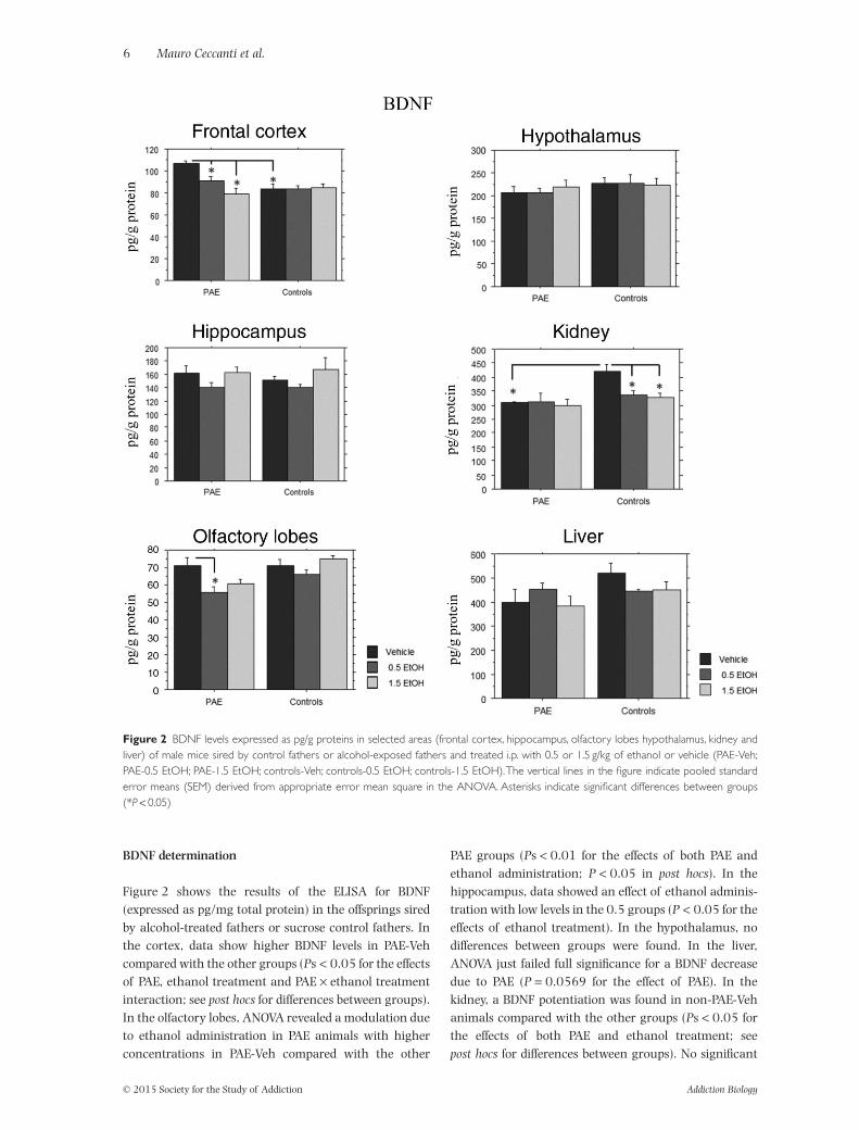
Figure 2 shows the results of the ELISA for BDNF(expressed as pg/mg total protein) in the offsprings siredby alcohol-treated fathers or sucrose control fathers. Inthe cortex, data show higher BDNF levels in PAE-Vehcompared with the other groups (Ps < 0.05 for the effectsof PAE, ethanol treatment and PAE × ethanol treatmentinteraction; see post hocs for differences between groups).In the olfactory lobes, ANOVA revealed a modulation dueto ethanol administration in PAE animals with higherconcentrations in PAE-Veh compared with the other
PAE groups (Ps < 0.01 for the effects of both PAE andethanol administration; P < 0.05 in post hocs). In thehippocampus, data showed an effect of ethanol adminis-tration with low levels in the 0.5 groups (P < 0.05 for theeffects of ethanol treatment). In the hypothalamus, nodifferences between groups were found. In the liver,ANOVA just failed full significance for a BDNF decreasedue to PAE (P = 0.0569 for the effect of PAE). In thekidney, a BDNF potentiation was found in non-PAE-Vehanimals compared with the other groups (Ps < 0.05 forthe effects of both PAE and ethanol treatment; seepost hocs for differences between groups). No significant
Figure 2 BDNF levels expressed as pg/g proteins in selected areas (frontal cortex, hippocampus, olfactory lobes hypothalamus, kidney andliver) of male mice sired by control fathers or alcohol-exposed fathers and treated i.p. with 0.5 or 1.5 g/kg of ethanol or vehicle (PAE-Veh;PAE-0.5 EtOH; PAE-1.5 EtOH; controls-Veh; controls-0.5 EtOH; controls-1.5 EtOH).The vertical lines in the figure indicate pooled standarderror means (SEM) derived from appropriate error mean square in the ANOVA. Asterisks indicate significant differences between groups(*P < 0.05)
differences between groups were observed in the testis(data not shown).
Western blotting analyses for TrkA, TrkB, p75NTR, DAT,D1, D2, pro-NGF and pro-BDNF
Protein tissue extracts have been used to analyze (byWestern blot) the expression of NGF and BDNF receptors(TrkA, TrkB and p75NTR), pro-NGF/pro-BDNF, dopaminereceptors (D1 and D2) and the dopamine transporter(DAT) in different brain areas, such as: hippocampus,hypothalamus, frontal cortex and olfactory lobes. Thedata obtained by densitometric analysis in the offspringsired by alcohol-treated fathers or sucrose control fathersshowed no significant differences in the protein expressionof NGF and BDNF receptors, except for TrkA in thehippocampus and p75NTR in the frontal cortex (Fig. 3). Inthe hippocampus (Fig. 3a), data showed higher TrkAexpression in all PAE groups compared with respectivenon-PAE groups (P < 0.05 for the effects of PAE in theANOVA, see post hocs in the table for differences betweengroups). In the frontal cortex (Fig. 3b), data evidenced lowp75NTR expression in non-PAE-1.5 EtOH, in PAE-Veh andin PAE-0.5 EtOH when compared with non-PAE-Veh(P = 0.01 in the ANOVA for the interaction PAE × Ethanoladministration; see post hocs comparison in the table forthe differences between groups). As for the expression ofDAT (Fig. 3c), ANOVA revealed that the administration of0.5 of ethanol elicited a strong DAT potentiation in theolfactory lobes (P < 0.01 for the effect of ethanol admin-istration; see post hocs in the table for differences betweengroups). No differences were found in the protein expres-sion of D1 and D2 receptors in brain tissues or for pro-NGFand pro-BDNF (data not shown).
Place conditioning
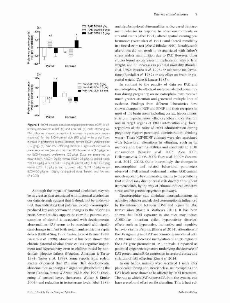
The effects of EtOH-elicited preference in PAE or non-PAEmale offsprings are shown in Fig. 4a and b, respectively.There was no statistically significant difference in prefer-ence for one chamber of the apparatus in control or EtOHgroups during the pretest session (data not shown). Forpost-conditioning session in PAE offspring, the repeatedmeasures ANOVA evidenced a significant EtOH × pairinginteraction (P < 0.01). Post hoc analysis revealed the exist-ence of a significant preference for the EtOH-paired com-partment only in mice conditioned with the lower dose ofEtOH (0.5 mg/kg; Fig. 4a), while mice conditioned to thehigher dose of EtOH (1.5 mg/kg) showed a significantincrease in preference toward the EtOH unpaired (Fig. 4a)and therefore no preference when the compartment waspaired to the higher dose tested. For post-conditioningsession in non-PAE offspring, the repeated measureANOVA showed a significant EtOH × pairing interaction
(P < 0.001). Post hoc analysis further revealed a signifi-cant preference for the EtOH-paired compartment only inthe non-PAE offspring conditioned with the higher dose ofEtOH (1.5 mg/kg; Fig. 4b), while the offspring conditionedto the lower dose of EtOH (0.5 mg/kg) showed no differ-ence as compared with the control group (Fig. 4b). Finally,there were no significant differences in locomotionbetween control and EtOH-conditioned mice during theconditioning phase (data not shown).
DISCUSSION
This is the first study to demonstrate that PAE affects NGF(Fig. 1) and BDNF (Fig. 2) levels in targeted brain regionsand peripheral tissues from mice offspring and enhancesthe sensitivity to the rewarding effects of EtOH adminis-tration in male progeny. Our data provide evidence thatEtOH-sired male offspring displayed a CPP to an environ-ment paired with a non-reinforcing dose of EtOH (0.5 g/kg). The PAE-induced lowering of reward threshold toEtOH effects in mice offspring is further underlined by theaversive response induced by the higher EtOH dose(1.5 g/kg). Thus, the EtOH dose eliciting a maximal placeconditioning effect in the offspring of non-PAE mice pro-duced a conditioned place aversion in the offspring of PAEmice, while a subthreshold dosage in non-PAE mice off-spring produced a rewarding effect in male offspring ofalcoholic fathers.
Our study provides evidence that PAE may affect thelevels of key trophic factors as NGF and BDNF in theoffspring brain, affecting also TrkA in the hippocampusand p75NTR in the frontal cortex. Our data show thatNGF is a neurotrophin particularly vulnerable to alcohol-induced transgenerational neuronal damage. We found amarked decrease in NGF expression in the frontal cortexof male offspring from PAE mice, as compared with off-spring from non-PAE animals, regardless of the dose ofEtOH administrated (Fig. 1a). However, NGF levels alsoshowed higher susceptibility to EtOH administration athippocampal, hypothalamic and olfactory lobe levels(Fig. 1b, c and e). Indeed, 8 days of EtOH administrationwas sufficient to reduce the NGF expression in these brainareas below the level showed by the untreated offspringfrom PAE mice. Notably, the lower the dose of EtOHadministered, the greater the reduction of NGF expres-sion detected. In this regard, the increased sensitivity torewarding effects of EtOH administration appearsinversely related to the levels of NGF expressed in thefrontal cortex, hippocampus, hypothalamus and olfac-tory lobes. By contrast, BDNF levels were increased onlyin the frontal cortex of PAE offspring that did not receiveany EtOH treatment. Higher levels of BDNF mRNAexpression have been detected in the amygdala andventral tegmental area of alcohol preferring animals
(Raivio, Miettinen & Kiianmaa 2014) and chronic EtOHvapor-induced PAE was shown to increase the expressionof BDNF in the ventral tegmental area of the offspring(Finegersh & Homanics 2014). Our results appear to
support an increase in BDNF expression in the frontalcortex of PAE offsprings, while EtOH administrationreduced the BDNF expression to levels comparable tothose showed by the non-PAE male offspring.
TrkA hippocampus
p75 frontal cortex
DAT olfactory lobes
Control PAE
EtOH 0 0.5 1.5 0 0.5 1.5
TrkA
β-ac�n
Control PAE
p75
β-ac�n
EtOH 0 0.5 1.5 0 0.5 1.5
(a)
(b)
(c)
Control PAE
EtOH 0 0.5 1.5 0 0.5 1.5
DAT
β-ac�n
EtOH sal
EtOH 0.5
EtOH 1.5
Band
den
sity
0
0.5
1
Control
Band
den
sity
PAE
0
0.2
0.4
0.6
Control
Band
den
sity
PAE
*
EtOH sal
EtOH 0.5
EtOH 1.5
EtOH sal
EtOH 0.5
EtOH 1.5
**
0
0.1
0.2
PAEControl
** *
Figure 3 Densitometric analysis and gel expression of TrkA in the hippocampus (a), p75NTR in the frontal cortex (b), and DAT in the olfactorylobes (c) of male mice sired by control fathers or alcohol-exposed fathers and treated i.p. with 0.5 or 1.5 g/kg of ethanol or vehicle. Geldensitometric analysis has been carried out by using the public domain ‘Image J’ software (http://imagej.nih.gov/ij/).The vertical lines in the figureindicate pooled standard error means (SEM) derived from appropriate error mean square in the ANOVA. Asterisks indicate significantdifferences between groups (*P < 0.05)
Although the impact of paternal alcoholism may notbe as great as that associated with maternal alcoholism,our data strongly suggest that it should not be underval-ued, thus indicating that paternal alcohol consumptionproduced key and permanent changes in the offspring’sbrain. Several studies support the view that paternal con-sumption of alcohol is associated with developmentalabnormalities. PAE seems to be associated with signifi-cant changes in infant birth weight and ventricular septaldefects (Little & Sing 1987; Tarter, Jacob & Bremer 1989;Passaro et al. 1998). Moreover, it has been shown thatchronic paternal alcohol abuse causes cognitive impair-ment and hyperactivity, even in children raised by non-drinker adoptive fathers (Hegedus, Alterman & Tarter1984; Tarter et al. 1989). Some reports from rodentstudies evidenced that PAE may elicit developmentalabnormalities, as changes in organ weights including thebrain (Tanaka, Suzuki & Arima 1982; Abel 1993), thick-ening of cortical layers (Jamerson, Wulser & Kimler2004), and reduction in testosterone levels (Abel 1989)
and also behavioral abnormalities as decreased displace-ment behavior in response to novel environments orstressful events (Abel 1991), altered spatial learning per-formances (Wozniak et al. 1991), and altered immobilityin a forced swim test (Abel & Bilitzke 1990). Notably, suchalterations did not result to be associated with father’sstress and/or malnutrition due to PAE. However, otherstudies found no decreases in implantation sites or fetalweight, and no increases in prenatal mortality (Randallet al. 1982; Passaro et al. 1998) or soft tissue malforma-tions (Randall et al. 1982) or any effect on brain or pla-cental weight (Cake & Lenzer 1985).
In contrast to the paucity of data on PAE andneurotrophins, the effects of maternal alcohol consump-tion during pregnancy on neurotrophins have receivedmuch greater attention and generated multiple lines ofevidence. Findings from different laboratories haveshown changes in NGF and BDNF and their receptors inmost of the brain areas including cortex, hippocampus,striatum, hypothalamus, olfactory lobes and cerebellumand in target organs of EtOH intoxication (e.g. liver),regardless of the route of EtOH administration duringpregnancy (vapor; parenteral administration; drinkingwater). These NGF/BDNF changes were also associatedwith behavioral alterations in offspring, such as inmemory and learning abilities and sensitivity to EtOHconsumption (Naassila et al. 2003; Aloe 2006;Hellemans et al. 2008, 2009; Fiore et al. 2009b; Ceccantiet al. 2012, 2013). Quite interestingly, the changes inneurotrophins and related behavioral parametersobserved in PAE animal models and in other FASD animalmodels appear to be comparable, leading to the possibilitythat ethanol may disrupt brain cells directly, throughoutits metabolites, by the way of ethanol-induced oxidativestress and/or genetic-epigenetic pathways.
Neurotrophins can modulate neuroadaptations inaddictive behavior and alcohol consumption is influencedby the interaction between BDNF and dopamine (DA)transmission (Bosse & Mathews 2011). It has beenshown that EtOH exposure in sire mice may induceADHD-like (attention deficit hyperactivity disorder)effects such as hyperactive, inattentive and impulsivebehaviors in the offspring (Kim et al. 2014). Alterations ofthe DA signaling and DAT are commonly associated withADHD, and an increased methylation of a CpG region ofthe DAT gene promoter in PAE animals is reported aspotential epigenetic signature underlying the decrease ofDAT protein and mRNA expression in cerebral cortex andstriatum of PAE offspring (Kim et al. 2014).
In our hands, animals were sacrificed 1 week afterplace conditioning and, nevertheless, neurotrophins andDAT levels were shown to be affected by EtOH treatment.The rate at which DAT removes DA from the synapse canhave a profound effect on DA signaling. This is best evi-
Figure 4 EtOH-induced conditioned place preference (CPP) is dif-ferently modulated in PAE (a) and non-PAE (b) male offspring. (a)PAE offspring showed a significant increase in preference scores(seconds) for the EtOH-paired side (0.5 g/kg) while a significantincrease in preference scores (seconds) for the EtOH-unpaired side(1.5 g/kg). (b) Non-PAE offspring showed a significant increase inpreference scores (seconds) for the EtOH-paired side (1.5 g/kg) butno EtOH-induced preference (0.5 g/kg). Data are presented asmean ± SEM. *EtOH 0 g/kg versus EtOH 0.5 g/kg (a, paired side);*EtOH 0 g/kg versus EtOH 1.5 g/kg (b, paired side); #EtOH 0.5 g/kgversus EtOH 1.5 g/kg (a and b, paired side); °EtOH 0 g/kg versusEtOH 0.5 g/kg or 1.5 g/kg (a, unpaired side). Tukey’s post hoc test(P < 0.05)
denced by the severe cognitive deficits and hyperactivityof mice with no DA transporters (Van der Kooij &Glennon 2007). We found a potential inverse relation-ship between EtOH doses and levels of DAT expression inolfactory lobes of male offspring from PAE mice (Fig. 3c).Thus, DAT expression was increased in animals thatreceived 0.5 g/kg EtOH, although the magnitude of DATexpression did not differ from that showed by offspringfrom non-PAE mice treated with the same EtOH dose.Despite the lack of difference between DAT expressions inoffspring from PAE or non-PAE animals, these effectsappear the consequence of the tendency toward anincrease in DAT expression in offspring from PAE micethat did not receive any EtOH administration. DAT func-tion is implicated in a number of DA-related disorders,including clinical depression and alcoholism (Getachewet al. 2010). DA neurotransmission plays an importantrole in EtOH consumption and alcohol-seeking behavior(for review, see Söderpalm & Ericson 2013). EtOH intakepotentiates DA release from ventral tegmental areadopaminergic (DAergic) neurons and increases DAextracellular levels in the nucleus accumbens (Koob2014). Moreover, DAergic agents and lesion of the DAsystem modify EtOH self-administration, thus confirminga specific role for DA in EtOH reward (Di Chiara 1997).Olfactory lobes are a key limbic structure and the greatestsensitivity to variations in the total amount of DATprotein was observed with the same low dose eliciting theEtOH-induced rewarding effects. Consequently, thesedata support the view that EtOH reward and EtOH-seeking behavior of PAE male offspring may be positivelyassociated with the amount of DAT protein and, likely,with reduced DA in the olfactory bulb. In this regard, thereduction in DA levels is reminding of the withdrawalsyndrome hypothesis and withdrawal-induced decreasein DA transmission, as the neural correlate of increasingmotivation to EtOH-seeking behavior (Koob 2014).Hence, adult male offsprings from PAE seem to exhibitsome of the neuroadaptations (i.e. changes in NGF, BDNFand DA signaling) known to produce changes in sensitiv-ity to EtOH-mediated effects (withdrawal symptoms) fol-lowing chronic exposure and discontinuation to alcoholintake.
As previously observed, the findings of the presentstudy indicate that alcohol drinking behavior may beepigenetically transferred through the father lineage. Ourworking hypothesis posits that alcohol is an epimutagendetermining specific long-lasting changes affecting thenext generations and their alcohol abuse, although theethanol mechanism of action as epimutagen is not fullyknown at least in PAE. However, several FASD studiesindicate that this spectrum of disorders due to prenatalethanol arises from a complex interplay of genetic andepigenetic factors (reviewed in Mead & Sarkar 2014). The
present results demonstrate a direct impact of PAE onbasal NGF and BDNF levels in target tissues of EtOHintoxication and the lowering of the threshold to EtOH-elicited rewarding effects in male offspring although alimitation of the present study may be represented by thesmall number of experimental subjects used in thetestings (as in any animal model study) that could have insome way affected the results.
In conclusion, since EtOH consumption before and atthe time of procreation is a key factor in healthy childdevelopment, the present study may represent a stepforward in the attempt to disclose some biomolecular pro-cesses and behavioral responses to rewarding outcomesunderlying the transgenerational effects of alcoholintoxication. Therefore, these results may be of interestfor the investigation of human addiction and in particu-lar for those interested in the epigenetic programming viathe paternal lineage.
Acknowledgements
We thank Sara De Nicolo for her valuable contribution inmanaging PAE mice. This work was supported by CNR,Regione Lazio and CRARL Lazio. The funders had no rolein study design, data collection and analysis, decision topublish or preparation of the manuscript.
Disclosure/Conflict of Interest
The authors declare no conflict of interest.
Authors Contribution
MauC and MF were responsible for the study concept anddesign. RC, VC, SC, GF, GG, PT, CC and MF contributed tothe acquisition of animal data. VC, SC, GF, PT, CC, EP, RMand MarC performed the biomolecular analyses. MauC,RC, VC and MF contributed to data analysis and interpre-tation of findings. RC, VC and MF drafted the manuscript.GC provided critical revision of the manuscript for impor-tant intellectual content. All authors critically reviewedcontent and approved final version for publication.
References
Abel E (2004) Paternal contribution to fetal alcohol syndrome.Addict Biol 9:127–133, discussion 135–6.
Abel EL (1989) Paternal and maternal alcohol consumption:effects on offspring in two strains of rats. Alcohol Clin Exp Res13:533–541.
Abel EL (1991) Paternal alcohol consumption affects groomingresponse in rat offspring. Alcohol Fayettev N 8:21–23.
Abel EL (1993) Rat offspring sired by males treated with alcohol.Alcohol Fayettev N 10:237–242.
Abel EL (1999) What really causes FAS? Teratology 59:4–6.Abel EL (2009) Fetal alcohol syndrome: same old, same old.
Abel EL, Bilitzke P (1990) Paternal alcohol exposure: paradoxicaleffect in mice and rats. Psychopharmacology (Berl) 100:159–164.
Abel EL, Dintcheff BA (1986) Effects of prenatal alcohol expo-sure on behavior of aged rats. Drug Alcohol Depend 16:321–330.
Allen SJ, Dawbarn D (2006) Clinical relevance of theneurotrophins and their receptors. Clin Sci 110:175–191.
Alleva E, Aloe L, Bigi S (1993) An updated role for nerve growthfactor in neurobehavioural regulation of adult vertebrates.Rev Neurosci 4:41–62.
Aloe L (2001) Nerve growth factor and neuroimmuneresponses: basic and clinical observations. Arch PhysiolBiochem 109:354–356.
Aloe L (2006) Alcohol intake during prenatal life affectsneuroimmune mediators and brain neurogenesis. Ann IstSuper Sanita 42:17–21.
Aloe L, Alleva E, Fiore M (2002) Stress and nerve growth factor:findings in animal models and humans. Pharmacol BiochemBehav 73:159–166.
Aloe L, Bracci-Laudiero L, Bonini S, Manni L (1997) The expand-ing role of nerve growth factor: from neurotrophic activity toimmunologic diseases. Allergy 52:883–894.
Aloe L, Bracci-Laudiero L, Tirassa P (1993) The effect of chronicethanol intake on brain NGF level and on NGF-target tissues ofadult mice. Drug Alcohol Depend 31:159–167.
Aloe L, Tirassa P (1992) The effect of long-term alcohol intakeon brain NGF-target cells of aged rats. Alcohol 9:299–304.
Astley S (2004) Diagnostic Guide for Fetal Alcohol SpectrumDisorders: The 4-Digit Diagnostic Code, Third—2004, Seattle,WA: FAS Diagnostic and Prevention Network University ofWashington.
Bakoyiannis I, Gkioka E, Pergialiotis V, Mastroleon I,Prodromidou A, Vlachos GD, Perrea D (2014) Fetal alcoholspectrum disorders and cognitive functions of young children.Rev Neurosci 25:631–639.
Bielawski DM, Abel EL (1997) Acute treatment of paternalalcohol exposure produces malformations in offspring.Alcohol Fayettev N 14:397–401.
Bonini S, Lambiase A, Lapucci G, Properzi F, Bresciani M,Bracci Laudiero ML, Mancini MJ, Procoli A, Micera A,Sacerdoti G, Bonini S, Levi-Schaffer F, Rasi G, Aloe L (2002)Nerve growth factor and asthma. Allergy 57 (Suppl. 72):13–15.
Bosse KE, Mathews TA (2011) Ethanol-induced increasesin extracellular dopamine are blunted in brain-derivedneurotrophic factor heterozygous mice. Neurosci Lett489:172–176.
Cake H, Lenzer I (1985) On effects of paternal ethanol treatmenton fetal outcome. Psychol Rep 57:51–57.
Ceccanti M, De Nicolo S, Mancinelli R, Chaldakov G, Carito V,Ceccanti M, Laviola G, Tirassa P, Fiore M (2013) NGF andBDNF long-term variations in the thyroid, testis and adrenalglands of a mouse model of fetal alcohol spectrum disorders.Ann Ist Super Sanita 49:383–390.
Ceccanti M, Mancinelli R, Tirassa P, Laviola G, Rossi S, Romeo M,Fiore M (2012) Early exposure to ethanol or red wine andlonglasting effects in aged mice. A study on nerve growthfactor, brain-derived neurotrophic factor, hepatocyte growth
factor, and vascular endothelial growth factor. NeurobiolAging 33:359–367.
Chao MV (2000) Trophic factors: an evolutionary cul-de-sac ordoor into higher neuronal function? J Neurosci Res 59:353–355.
Chao MV, Hempstead BL (1995) p75 and Trk: a two-receptorsystem. Trends Neurosci 18:321–326.
Chao MV, Rajagopal R, Lee FS (2006) Neurotrophin signallingin health and disease. Clin Sci 110:167–173.
Cirulli F, Adriani W, Laviola G (1997) Sexual segregation ininfant mice: behavioural and neuroendocrine responsesto d-amphetamine administration. Psychopharmacol Berl.134:140–152.
Cohen S, Levi-Montalcini R, Hamburger V (1954) A nervegrowth-stimulating factor isolated from sarcomas 37 and180. Proc Natl Acad Sci U S A 40:1014–1018.
Cuello AC (1983) Brain Microdissection Techniques, Chichester:Wiley.
De Nicolo S, Tarani L, Ceccanti M, Maldini M, Natella F, Vania A,Chaldakov GN, Fiore M (2013) Effects of olive polyphenolsadministration on nerve growth factor and brain-derivedneurotrophic factor in the mouse brain. Nutrition 29:681–687.
Di Chiara G (1997) Alcohol and dopamine. Alcohol Health ResWorld 21:108–114.
Finegersh A, Homanics GE (2014) Paternal alcohol exposurereduces alcohol drinking and increases behavioral sensitivityto alcohol selectively in male offspring. PLoS ONE 9:e99078.
Fiore M, Chaldakov GN, Aloe L (2009a) Nerve growth factoras a signaling molecule for nerve cells and also for theneuroendocrine-immune systems. Rev Neurosci 20:133–145.
Fiore M, Laviola G, Aloe L, di Fausto V, Mancinelli R, Ceccanti M(2009b) Early exposure to ethanol but not red wine atthe same alcohol concentration induces behavioral andbrain neurotrophin alterations in young and adult mice.Neurotoxicology 30:59–71.
Fiore M, Mancinelli R, Aloe L, Laviola G, Sornelli F, Vitali M,Ceccanti M (2009c) Hepatocyte growth factor, vascularendothelial growth factor, glial cell-derived neurotrophicfactor and nerve growth factor are differentially affected byearly chronic ethanol or red wine intake. Toxicol Lett188:208–213.
Freund-Michel V, Frossard N (2008) The nerve growth factorand its receptors in airway inflammatory diseases. PharmacolTher 117:52–76.
Getachew B, Hauser SR, Taylor RE, Tizabi Y (2010) Alcohol-induced depressive-like behavior is associated with corticalnorepinephrine reduction. Pharmacol Biochem Behav96:395–401.
Goodlett CR, Gilliam DM, Nichols JM, West JR (1989) Geneticinfluences on brain growth restriction induced by develop-ment exposure to alcohol. Neurotoxicology 10:321–334.
Goodlett CR, Horn KH (2001) Mechanisms of alcohol-induceddamage to the developing nervous system. Alcohol Res Health25:175–184.
He Y-Y, Zhang X-Y, Yung W-H, Zhu J-N, Wang J-J (2013) Role ofBDNF in central motor structures and motor diseases. MolNeurobiol 48:783–793.
Hegedus AM, Alterman AI, Tarter RE (1984) Learning achieve-ment in sons of alcoholics. Alcohol Clin Exp Res 8:330–333.
Hellemans KG, Sliwowska JH, Verma P, Weinberg J (2009) Pre-natal alcohol exposure: fetal programming and later life vul-nerability to stress, depression and anxiety disorders. NeurosciBiobehav Rev 34:791–807.
Huang EJ, Reichardt LF (2003) Trk receptors: roles in neuronalsignal transduction. Annu Rev Biochem 72:609–642.
Jamerson PA, Wulser MJ, Kimler BF (2004) Neurobehavioraleffects in rat pups whose sires were exposed to alcohol. BrainRes Dev Brain Res 149:103–111.
Jones KL (1975) The fetal alcohol syndrome. Addict Dis 2:79–88.
Kim P, Choi CS, Park JH, Joo SH, Kim SY, Ko HM, Kim KC, JeonSJ, Park SH, Han SH, Ryu JH, Cheong JH, Han JY, Ko KN, ShinCY (2014) Chronic exposure to ethanol of male mice beforemating produces attention deficit hyperactivity disorder-likephenotype along with epigenetic dysregulation of dopaminetransporter expression in mouse offspring. J Neurosci Res92:658–670.
Mead EA, Sarkar DK (2014) Fetal alcohol spectrum disordersand their transmission through genetic and epigenetic mecha-nisms. Front Genet 5:154.
Naassila M, Pierrefiche O, Beauge FJ, Sebire N, Daoust M (2003)Chronic ethanol exposure differentially regulates NOS1mRNA levels depending on rat brain area. Neurosci Lett338:221–224.
O’Malley KD, Nanson J (2002) Clinical implications of a linkbetween fetal alcohol spectrum disorder and attention-deficithyperactivity disorder. Can J Psychiatry 47:349–354.
Passaro KT, Little RE, Savitz DA, Noss J (1998) Effect of paternalalcohol consumption before conception on infant birthweight. ALSPAC Study Team. Avon Longitudinal Study ofPregnancy and Childhood. Teratology 57:294–301.
Raivio N, Miettinen P, Kiianmaa K (2014) Innate BDNF expres-sion is associated with ethanol intake in alcohol-preferring
AA and alcohol-avoiding ANA rats. Brain Res 1579:74–83.
Randall CL, Burling TA, Lochry EA, Sutker PB (1982) The effectof paternal alcohol consumption on fetal development inmice. Drug Alcohol Depend 9:89–95.
Servais L, Hourez R, Bearzatto B, Gall D, Schiffmann SN, CheronG (2007) Purkinje cell dysfunction and alteration of long-term synaptic plasticity in fetal alcohol syndrome. Proc NatlAcad Sci USA 104:9858–9863.
Söderpalm B, Ericson M (2013) Neurocircuitry involved in thedevelopment of alcohol addiction: the dopamine system andits access points. Curr Top Behav Neurosci. 13:127–161.
Streissguth AP, O’Malley K (2000) Neuropsychiatric implica-tions and long-term consequences of fetal alcohol spectrumdisorders. Semin Clin Neuropsychiatry 5:177–190.
Tabakoff B, Hoffman PL (2013) The neurobiology of alcoholconsumption and alcoholism: an integrative history.Pharmacol Biochem Behav 113:20–37.
Tanaka H, Suzuki N, Arima M (1982) Experimental studies onthe influence of male alcoholism on fetal development. BrainDev 4:1–6.
Tarter RE, Jacob T, Bremer DL (1989) Specific cognitive impair-ment in sons of early onset alcoholics. Alcohol Clin Exp Res13:786–789.
Tsai G, Gastfriend DR, Coyle JT (1995) The glutamatergic basis ofhuman alcoholism. Am J Psychiatry 152:332–340.
Van der Kooij MA, Glennon JC (2007) Animal models concern-ing the role of dopamine in attention-deficit hyperactivity dis-order. Neurosci Biobehav Rev 31:597–618.
Ventura R, Coccurello R, Andolina D, Latagliata EC, Zanettini C,Lampis V, Battaglia M, D’Amato FR, Moles A (2013) Postnatalaversive experience impairs sensitivity to natural rewards andincreases susceptibility to negative events in adult life. CerebCortex 23:1606–1617.
Wozniak DF, Cicero TJ, Kettinger L, Meyer ER (1991) Paternalalcohol consumption in the rat impairs spatial learning per-formance in male offspring. Psychopharmacology (Berl)105:289–302.
Yanev S, Aloe L, Fiore F, Chaldakov GN (2013) Neurotrophicand metabotrophic potential of nerve growth factor andbrain-derived neurotrophic factor: linking cardiometabolicand neuropsychiatric diseases. World J Pharmacol 2:92–99.