Pathogenesis and management ofgastrointestinal inflammation and fibrosis:from inflammatory bowel diseases toendoscopic surgeryKentaro Iwata1,2†, Yohei Mikami1*† , Motohiko Kato1,2, Naohisa Yahagi2 and Takanori Kanai1*
Abstract
Gastrointestinal fibrosis is a state of accumulated biological entropy caused by a dysregulated tissue repair response.Acute or chronic inflammation in the gastrointestinal tract, including inflammatory bowel disease, particularly Crohn’sdisease, induces fibrosis and strictures, which often require surgical or endoscopic intervention. Recent technicaladvances in endoscopic surgical techniques raise the possibility of gastrointestinal stricture after an extended resection.Compared to recent progress in controlling inflammation, our understanding of the pathogenesis of gastrointestinalfibrosis is limited, which requires the development of prevention and treatment strategies. Here, we focus ongastrointestinal fibrosis in Crohn’s disease and post-endoscopic submucosal dissection (ESD) stricture, and we reviewthe relevant literature.
Keywords: Gastrointestinal fibrosis, Crohn’s disease, Endoscopic surgery
BackgroundGastrointestinal stricture is the pathological thickeningof the wall of the gastrointestinal tract, characterized byexcessive accumulation of extracellular matrix (ECM)and expansion of the population of mesenchymal cells.Gastrointestinal stricture leads to blockage of the gastro-intestinal tract, which significantly reduces a patient’squality of life. Upper gastrointestinal stricture may causenausea, vomiting, anorexia, and abdominal pain becauseof food stagnation. In addition to these obstructivesymptoms, lower gastrointestinal stricture may cause in-testinal perforation, intra-abdominal abscess, and fistu-lizing disease because of increased pressure in the regionof the inflamed intestinal tract.Malignant and benign processes cause gastrointestinal
stricture as well as inflammation and the healing of
surgical wounds. Fibrostenosis of the gastrointestinaltract, in particular, is a frequent complication of Crohn’sdisease. Further, a recent highly significant advance inendoscopic treatment enables resection of premalignantand early-stage gastrointestinal cancers. This proceduredoes not involve surgical reconstruction of the gastro-intestinal tract, although fibrotic stricture after endo-scopic treatment is an emerging clinical problem. Here,we focus on post-endoscopic scarring and Crohn’s dis-ease, which cause artificial and spontaneous fibrosis ofthe gastrointestinal tract, and we review shared andunique mechanisms of pathogenesis.
Current endoscopic treatment and challengesEndoscopic mucosal resection (EMR) and ESD are endo-scopic techniques for resecting epithelial tumours withlow risk of metastasis. EMR is a conventional method toresect relatively small and superficial tumours. A metalring (named snare forceps) is used to capture the lesionthat is excised using a high-frequency electric current.
* Correspondence: [email protected]; [email protected]†Kentaro Iwata and Yohei Mikami contributed equally to this work.1Division of Gastroenterology and Hepatology, Department of InternalMedicine, Keio University School of Medicine, Tokyo, JapanFull list of author information is available at the end of the article
Inflammation and RegenerationIwata et al. Inflammation and Regeneration (2021) 41:21 https://doi.org/10.1186/s41232-021-00174-7
EMR is simple and safe; however, it is limited to rela-tively small (e.g. less than approximately 20-mm diam-eter) lesions.ESD was first reported in 1999 by Gotoda et al. [1].
Unlike EMR, ESD enables secure resection, regardless oflesion size or location, through precise dissection of thesubmucosal layer. The costs of ESD of the stomach wereinitially covered by health insurance in Japan in 2006, in2008 for the oesophagus, and in 2011 for the colon.With the widespread use of screening endoscopy, thechances of early detection of cancer have increased [2–4], and it is a standard treatment worldwide. Moreover,when applied to gastric cancer, ESD achieves higher en-bloc resection rates and lower local recurrence [5, 6] foroesophageal [7] and duodenal cancers [7].An advantage of ESD is its ability to securely resect le-
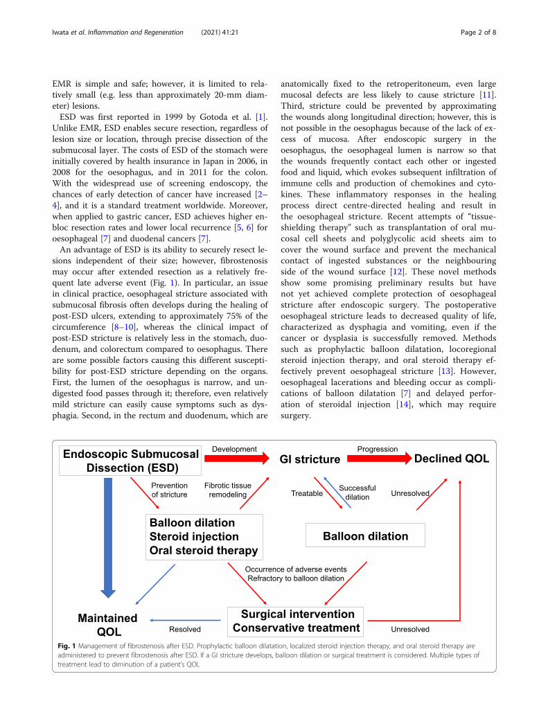
sions independent of their size; however, fibrostenosismay occur after extended resection as a relatively fre-quent late adverse event (Fig. 1). In particular, an issuein clinical practice, oesophageal stricture associated withsubmucosal fibrosis often develops during the healing ofpost-ESD ulcers, extending to approximately 75% of thecircumference [8–10], whereas the clinical impact ofpost-ESD stricture is relatively less in the stomach, duo-denum, and colorectum compared to oesophagus. Thereare some possible factors causing this different suscepti-bility for post-ESD stricture depending on the organs.First, the lumen of the oesophagus is narrow, and un-digested food passes through it; therefore, even relativelymild stricture can easily cause symptoms such as dys-phagia. Second, in the rectum and duodenum, which are
anatomically fixed to the retroperitoneum, even largemucosal defects are less likely to cause stricture [11].Third, stricture could be prevented by approximatingthe wounds along longitudinal direction; however, this isnot possible in the oesophagus because of the lack of ex-cess of mucosa. After endoscopic surgery in theoesophagus, the oesophageal lumen is narrow so thatthe wounds frequently contact each other or ingestedfood and liquid, which evokes subsequent infiltration ofimmune cells and production of chemokines and cyto-kines. These inflammatory responses in the healingprocess direct centre-directed healing and result inthe oesophageal stricture. Recent attempts of “tissue-shielding therapy” such as transplantation of oral mu-cosal cell sheets and polyglycolic acid sheets aim tocover the wound surface and prevent the mechanicalcontact of ingested substances or the neighbouringside of the wound surface [12]. These novel methodsshow some promising preliminary results but havenot yet achieved complete protection of oesophagealstricture after endoscopic surgery. The postoperativeoesophageal stricture leads to decreased quality of life,characterized as dysphagia and vomiting, even if thecancer or dysplasia is successfully removed. Methodssuch as prophylactic balloon dilatation, locoregionalsteroid injection therapy, and oral steroid therapy ef-fectively prevent oesophageal stricture [13]. However,oesophageal lacerations and bleeding occur as compli-cations of balloon dilatation [7] and delayed perfor-ation of steroidal injection [14], which may requiresurgery.
Fig. 1 Management of fibrostenosis after ESD. Prophylactic balloon dilatation, localized steroid injection therapy, and oral steroid therapy areadministered to prevent fibrostenosis after ESD. If a GI stricture develops, balloon dilation or surgical treatment is considered. Multiple types oftreatment lead to diminution of a patient’s QOL
Iwata et al. Inflammation and Regeneration (2021) 41:21 Page 2 of 8
Recently, implantation of oral mucosal epithelial cellsheets [15], PGA-felt and fibrin gluing [16], and bio-degradable stents [17], although useful, are notemployed in routine clinical practice because of theircost, time required, and technical problems. Further,although tissue biopsy is important for definitive diag-nosis before administering ESD, submucosal fibrosisoften develops after biopsy. Unfortunately, progress inincreasing our understanding of the pathogenesis ofgastrointestinal fibrosis and efforts to develop preven-tion and treatment methods lag behind the advancesin ESD technology.
Healing of oesophageal ulcers after endoscopyAfter endoscopic treatment, fibrosis terminates in 4weeks, and infiltration of inflammatory cells occurs inthe submucosa 2–4 days after the creation of a so-called“artificial ulcer”. After 7 days, epithelial cells proliferate,the number of inflammatory cells in the submucosal tis-sue decreases, and fibrous tissue associated with angio-genesis proliferates. After 28 days, fibrous tissue replacesthe lesion. In the oesophagus, oesophageal glands or mu-cosal fascia are not observed after the completion of epi-thelialization of the artificial ulcer, and the epitheliumand submucosal layer are thinner than usual [18].Nonaka et al. [19] found that spindle-shaped myofi-
broblasts, which express α-SMA, are present in thebase of the ulcer 1 week after the creation of an arti-ficial ulcer, which contributes to the formation of thestricture. In contrast, spindle-shaped myofibroblastsare irregularly located in the fibrous region of therepaired tissue after topical steroid injection forprophylaxis of oesophageal stricture. Further, kera-tinocyte growth factor (KGF), hepatocyte growth fac-tor (HGF), prostaglandin E-prostanoid 2 receptors,cAMP, and cAMP response element-binding proteincontribute to the repair of the oesophageal epithelium[19–22].However, few reports employ animal models of
oesophageal fibrostenosis compared with those of thesmall and large intestines. This is partially attributed tothe technical difficulties involved in approaching theoesophagus of a small animal. Further, the lack of suit-able transgenic animals (e.g. mice) makes it difficult todetermine the contributions of certain cell subsets, cyto-kines, chemokines, growth factors, and other effectors.Available models of fibrostenosis of the oesophagus,such as those employing the 100% acetic acid-inducedoesophagitis model in Sprague-Dawley rats [23], andpost-ESD oesophageal ulceration model in pigs [24] anddogs [25]. Thus, small animal models of stableoesophageal fibrostenosis help identify the mechanismof pathogenesis of oesophageal fibrosis.
Clinical features and epidemiology of chronicinflammatory conditions that cause strictures inthe gastrointestinal tractBenign oesophageal strictures are caused by different ae-tiologies. Gastroesophageal reflux disease and eosino-philic gastritis have been two major causes of theoesophageal strictures, but recent technological advancesin cancerous treatment strategies including radiationand endoscopic surgery highlight the rapid increase iniatrogenic or secondary strictures after treatment [26]. Itis of note that most of the aetiologies are associated withthe inflammatory process followed by stenosis and it isimportant to understand both inflammatory and remod-elling phages of the gastrointestinal tract. Among themultiple aetiologies of oesophageal strictures, Crohn’sdisease is a rare but important condition known to causestrictures in the small and large intestines [26, 27]. Wehere summarize the aetiology and pathologies of Crohn’sdisease because it is one of the fields where the fibrosismechanisms have been extensively studied and theknowledge in the Crohn’s strictures may be sharedacross the entire gastrointestinal tract. The incidence ofCrohn’s disease in Japan shows a clear, recently increas-ing trend. Crohn’s disease is characterized by chronicgranulomatous inflammation that may involve any re-gion of the gastrointestinal tract, predominantly the ter-minal ileum and adjacent colon, and presents with asegmental, asymmetric distribution [28, 29]. The mainsymptoms are abdominal pain, diarrhoea, fistula, anal le-sions, and systemic symptoms differing in severity [30].Crohn’s disease frequently manifests extraintestinal com-plications such as nodular erythema, necrotizing pyo-derma, polymorphic exudative erythema, iritis, andvaginitis [31]. Recurrence of this progressive diseaseleads to major complications. Despite recent advances intreatment, these intra- and extra-intestinal complicationsimpair the quality of life.Crohn’s disease is characterized by discontinuous skip
lesions that are observed during endoscopy [32]. Intes-tinal stricture is a common complication of Crohn’s dis-ease, affecting approximately 33% of patients within 10years of onset. Treatment of Crohn’s disease aims toachieve sustained clinical and endoscopic remission(“low entropy”) and to interrupt the naturally progressivedestructive disease course that culminates in intestinalfailure and associated complications (“high entropy”).Although multiple clinical, environmental, serological,genetic, and epigenetic markers are potential predictorsof fibrostenotic Crohn’s disease (Table 1) [33–35], welack specific and reliable markers that represent the stateof gut fibrosis and predict stricturing.Behçet’s disease, sometimes referred to as the “Silk
Route disease” because of its elevated frequencies in theMiddle East and far-eastern Asia [36–38] which are
Iwata et al. Inflammation and Regeneration (2021) 41:21 Page 3 of 8
traditionally considered endemic areas. HLA-B51 is arisk factor for Behçet’s disease [36, 37]. The most pro-nounced symptoms of Behçet’s disease are associatedwith the intestine. Intestinal Behçet’s disease typicallyforms a round to oval swell-like ulcer in the terminalileum. Ulcers may form in the entire gastrointestinaltract, although oesophageal lesions are infrequent [39].Cases of intestinal Behçet’s disease with an aphthousulcer may be difficult to differentiate from Crohn’s dis-ease because the morphology of the former is similar tothat of early Crohn’s disease. In addition, Crohn’s diseaseand Behçet’s disease can both affect the entire gastro-intestinal tract and cause ulcers because of chronic auto-immune inflammation.The differences in endoscopic morphology between
these otherwise similar diseases include the characteris-tics of the ulcer base, depth, shape, and margin. A soli-tary deep oval ulcer with a thick exudative necrotic layerat the ulcer bottom serves as a signature of intestinalBehçet’s disease compared with the ulcer of Crohn’s dis-ease. Further, plasma cells in the granulation tissue ofBehçet’s disease accumulate to abnormally high levels.Inflammation surrounding the ulcer margin and ulcerbed is milder and more localized than in Crohn’s dis-ease. Epitheloid granuloma is detected in approximately50% of patients with Crohn’s disease who undergo surgi-cal resection of the intestine. Moreover, focal cryptitis,basal plasmacytosis, lymphoid aggregates, and nervefibre hyperplasia are detected in Crohn’s disease.
Pathophysiology of Crohn’s diseaseGastrointestinal bacterial species among healthy individ-uals are diverse, and this diversity may be significantly
influenced by dietary and drug-induced factors. Dysbiosisis involved in the onset and exacerbation [40] of Crohn’sdisease [41, 42]. The diversity of bacterial species repre-senting the phyla Firmicutes and Bacteroidetes is reducedin patients with Crohn’s disease [43–45]. However, a re-cent large-scale multiomics analysis conducted as a com-ponent of the Integrative Human Microbiome Project(HMP2) found that metagenomic species differ signifi-cantly between patients with Crohn’s disease and controls[46]. These features of dysbiosis remain to be establishedas causes or consequences of Crohn’s disease.Previous studies mainly focus on the microbiota of the
faeces, which widely differs from that of the small intes-tine. More recent studies employing endoscopy of thesmall bowel show that the microbiome of the mucosal tis-sues of the small intestine harbours several bacterial spe-cies that are closely associated with Crohn’s disease [47].The small intestine is covered with a single layer of a
simple columnar epithelium. Goblet cells are present inthe intestinal villi and secret a mucus biofilm to protectthe mucosa [43]. In Crohn’s disease, the expression ofmucin-1 (MUC1) in the inflamed epithelium at the ter-minal ileus suggests that the mucin cover is insufficient[48]. Paneth cells defend the mucosa by secreting anti-microbial peptide granules, such as a-defensins, and con-trol the composition of the bacterial flora. Paneth cellsfrom patients with Crohn’s disease that harbour mutationsin the autophagy gene ATG16L have fewer granules, ex-hibit morphological abnormalities, and are functionallyimpaired compared with wild-type mice [49].Further, epithelial cells attach to neighbouring cells
through tight-junction proteins such as claudin [50]. InCrohn’s disease, this tight junction becomes leaky because ofchanges in the expression of tight-junction proteins. This al-teration increases cell permeability; luminal antigens accessthe lamina propria which leads to the accumulation of innateimmune cells that produce inflammatory cytokines that acti-vate the adaptive immune system.Crohn’s disease is characterized by an imbalance be-
tween effector T cells and innate regulatory T cells [51].For example, Th1 and Th17 effector T cells protect themucosa from bacteria and fungi by secreting IFN-γ,TNF-α, IL-17, and IL-22. Treg cells secrete IL-10 andTGF-β to inhibit the proliferation of dendritic cells andlymphoid cells and induce immune tolerance [52]. Thesetwo main opposing phenotypes, Th17 and Treg, origin-ate from CD4+ T cells under stringent negative regula-tion by the transcription factors RORγt and FOXP3 [53,54]. Further, the generation of peripherally inducedTregs is influenced by the local microenvironment suchas the microbiota and its metabolites, bile acids, andneural stimulation [55–57].Transforming growth factor (TGF)- β signalling in-
duces Treg differentiation and is required for Th17 cell
Table 1 Predictors of fibrostenosing Crohn’s disease
Clinical Age at diagnosis < 40 years
Perianal disease at diagnosis
Need for steroids during first flare
Early use of azathioprine or anti-TNF
Small bowel disease location
Prior appendectomy
Environmental Smoking
Endoscopic Deep mucosal laceration
Genetic Janus-associated kinase 2 (JAK2)
ATG16L1
NOD2/CARD15 mutations on both chromosomes
TNF superfamily 15 (TNFSF15) in Asians
5T5T in the MMP3 gene
rs1363670
Serological Antimicrobial antibodies
Anti-Saccharomyces cerevisiae (ASCA) IgA in Asians
Iwata et al. Inflammation and Regeneration (2021) 41:21 Page 4 of 8
differentiation. Th17 cells are induced to differentiatefrom naive CD4+ T cells into Th17 cells. This processrequires TGF-β and IL-6 signalling, which activatesSTAT3 to induce the synthesis of RORγt, which is re-quired for the proliferation and survival of Th17 cells.More than 200 genes are associated with susceptibilityto IBD, including ulcerative colitis and Crohn's disease[58]. NOD2 encodes a sensor molecule for bacterial con-stituent proteins, and variants of MMP3 contribute tofibrostenosing Crohn’s disease [59]. Furthermore, anti-bodies to ECM molecules, growth factors, and microbialcomponents may be associated with the development ofIBD and intestinal fibrosis.
Cellular and molecular mechanisms ofgastrointestinal fibrosisFibrosis of the gastrointestinal tract is caused by exces-sive production of ECM components by activated mes-enchymal cells (Fig. 2). After endoscopic treatment,inflammatory cells invade the submucosal layer subse-quent to thermal injury and exposure to digestive fluid.In Crohn’s disease, inflammatory cells are inducedthrough the activation of adaptive immunity by intestinalbacteria, as described above. Inflammation potently in-duces TGF-β signalling, activates ECM-producing cells,and induces tissue fibrosis [60, 61]. ECM-producingcells, which are mainly fibroblasts, comprise a diverse
population of cells with diverse origins, including epithe-lial cells, endothelial cells, astrocytes, and bone marrow-derived stem cells [62].Fibroblasts are classically characterized through their
expression of the cytoskeletal proteins α-SMA, vimentin,CD90 (Thy1), PDGFRα, Sca-1, integrin-α8, CD34, andCD26 (DPP4) [63]. However, recent single-cell omics ana-lyses reveal the functional heterogeneity and tissue specifi-city of fibroblasts [64–66]. Mesenchymal cells areactivated by pathways induced through autocrine andparacrine signalling and microbe-associated and damage-associated molecular patterns. Inhibition of TGF-β signal-ling leads to prolonged inflammation, because TGF-β,which is induced by inflammation, serves as a mediator offibrosis and plays a role in the immunomodulation of Tregcells as an inhibitory cytokine [67–69].Other fibrotic factors include activins, connective tis-
sue growth factor (CTGF), platelet-derived growth fac-tor, insulin-like growth factors (IGF) 1 and 2, epidermalgrowth factor (EGF), endothelins, and IL-13 which areinduced by intense inflammation. However, anti-inflammatory drugs only suppress the generation of in-flammatory factors but not fibrotic factors. Thus, evi-dence indicates that fibrosis is an independent factor ofinflammation [70].Furthermore, the progression of fibrosis is affected by
the turnover of ECM components. The generation and
Fig. 2 Pathophysiology of GI fibrosis. Crohn’s disease activates innate and adaptive immunity because of genetic abnormalities and the intestinalmicroflora. Endoscopic treatment activates innate immune cells through thermal injury, exposure to digestive fluid, and submucosal injection,which are strong triggers for ECM-producing cells that cause GI stricture
Iwata et al. Inflammation and Regeneration (2021) 41:21 Page 5 of 8
degradation of the ECM are balanced by MMPs andMMP inhibitors, and fibrosis occurs when ECM produc-tion increases and exceeds its rate of degradation [71,72]. Recent studies using animal models of fibrosis sug-gest that pirfenidone, currently approved by the FDA forthe treatment of idiopathic pulmonary fibrosis (IPF), ananti-matrix metalloproteinase 9 (MMP9) antibody,OGR1 (pH-sensing ovarian cancer G-protein-coupledreceptor 1), and BCL2 inhibitors may prevent fibrosis as-sociated with IBD [73].
Conclusion and future prospectsAlthough prophylaxis and treatment have been inten-sively investigated for preventing and managing gastro-intestinal stricture, this condition imposes a greatburden on patients and may cause deterioration of theirquality of life. Post-endoscopic ulcers cause tissue dam-age to the submucosa through similar as well as distinctmechanisms responsible for the stricture of Crohn’s dis-ease. The environment of the oesophagus differs fromthat of the small and large intestines, where there is asmall diversity of microbiota, covered with a layer ofstratified squamous epithelium, with no immune relaytissues such as those comprising Paneth cells. These en-vironmental tissue factors contribute to pathogenesisand tissue-specific phenotypes of the fibroblasts in theoesophagus and the intestine.To further dissect tissue specificity of fibroblasts, stud-
ies of analogues derived from the skin that share thestructural features of the stratified squamous epitheliummay help to understand the features of the stroma in theoesophagus that were previously unpredictable. Identifi-cation of the tissue-specific roles of fibroblasts in thegastrointestinal tract and identification of common anddistinct mechanisms underlying gastrointestinal fibrosisacross organs will contribute to our understanding offibrostenosis under inflammatory and non-inflammatoryconditions.
AcknowledgementsNot applicable.
Authors’ contributionsI.K. and Y.M. wrote the manuscript with input from the other authors. M.K.,N.Y., and T.K. provided supervision, editing, and support. The authors readand approved the final manuscript.
Authors’ informationKI is a board-certified internal medicine physician and gastroenterologist. Hehas graduated from Keio University School of Medicine and received M.D.,followed by completing residency in internal medicine and in gastroenter-ology and hepatology. He is currently a Ph.D. student in the IBD research fel-low programme under the supervision of Dr. Takanori Kanai at KeioUniversity. He is interested in the unknown pathogenic mechanism of IBDand trying to develop novel treatment strategies against IBD.
FundingThis study was funded by Japan Society for the Promotion of Science (JSPS)KAKENHI (B) 20H03666 for YM, and (A) 20H00536 for TK; Advanced Research
and Development Programs for Medical Innovation (AMED-CREST;16gm1010003h0001 for TK and 20gm1210001h0002 for YM, the PracticalResearch Project for Rare/Intractable Disease; 21ek0109556h0001 for Y.M.);Takeda Science Foundation; Kanae Foundation for The Promotion of MedicalScience; Mishima Kaiun Memorial Foundation Research Grant; Yakult Bio-Science Foundation; Keio University Medical Fund. We thank Edanz Group(https://en-author-services.edanz.com/ac) for editing a draft of thismanuscript.
Availability of data and materialsNot applicable.
Declarations
Ethics approval and consent to participateNot applicable.
Consent for publicationNot applicable.
Competing interestsThe authors declare no competing interests.
Author details1Division of Gastroenterology and Hepatology, Department of InternalMedicine, Keio University School of Medicine, Tokyo, Japan. 2Division ofResearch and Development for Minimally Invasive Treatment, Cancer Center,Keio University School of Medicine, Tokyo, Japan.
Received: 15 February 2021 Accepted: 30 June 2021
References1. Gotoda T, Kondo H, Ono H, Saito Y, Yamaguchi H, Saito D, et al. A new
endoscopic mucosal resection procedure using an insulation-tippedelectrosurgical knife for rectal flat lesions: report of two cases. GastrointestEndosc. 1999;50(4):560–3. https://doi.org/10.1016/S0016-5107(99)70084-2.
2. Moore JS, Aulet TH. Colorectal Cancer Screening. Surg Clin North Am. 2017;97(3):487–502. https://doi.org/10.1016/j.suc.2017.01.001.
3. Pasechnikov V, Chukov S, Fedorov E, Kikuste I, Leja M. Gastric cancer:prevention, screening and early diagnosis. World J Gastroenterol. 2014;20(38):13842–62. https://doi.org/10.3748/wjg.v20.i38.13842.
4. di Pietro M, Canto MI, Fitzgerald RC. Endoscopic management of earlyadenocarcinoma and squamous cell carcinoma of the esophagus:screening, diagnosis, and therapy. Gastroenterology. 2018;154(2):421–36.https://doi.org/10.1053/j.gastro.2017.07.041.
5. Kato M, Nishida T, Yamamoto K, Hayashi S, Kitamura S, Yabuta T, et al.Scheduled endoscopic surveillance controls secondary cancer after curativeendoscopic resection for early gastric cancer: a multicentre retrospectivecohort study by Osaka University ESD study group. Gut. 2013;62(10):1425–32. https://doi.org/10.1136/gutjnl-2011-301647.
6. Nishizawa T, Yahagi N. Endoscopic mucosal resection and endoscopicsubmucosal dissection: technique and new directions. Curr OpinGastroenterol. 2017;33(5):315–9. https://doi.org/10.1097/MOG.0000000000000388.
7. Tsujii Y, Nishida T, Nishiyama O, Yamamoto K, Kawai N, Yamaguchi S, et al.Clinical outcomes of endoscopic submucosal dissection for superficialesophageal neoplasms: a multicenter retrospective cohort study.Endoscopy. 2015;47(9):775–83.
8. Katada C, Muto M, Manabe T, Boku N, Ohtsu A, Yoshida S. Esophagealstenosis after endoscopic mucosal resection of superficial esophageallesions. Gastrointest Endosc. 2003;57(2):165–9. https://doi.org/10.1067/mge.2003.73.
9. Ono S, Fujishiro M, Niimi K, Goto O, Kodashima S, Yamamichi N, et al.Predictors of postoperative stricture after esophageal endoscopicsubmucosal dissection for superficial squamous cell neoplasms. Endoscopy.2009;41(8):661–5. https://doi.org/10.1055/s-0029-1214867.
10. Mizuta H, Nishimori I, Kuratani Y, Higashidani Y, Kohsaki T, Onishi S.Predictive factors for esophageal stenosis after endoscopic submucosaldissection for superficial esophageal cancer. Dis Esophagus. 2009;22(7):626–31. https://doi.org/10.1111/j.1442-2050.2009.00954.x.
Iwata et al. Inflammation and Regeneration (2021) 41:21 Page 6 of 8
11. Ohara Y, Toyonaga T, Tanaka S, Ishida T, Hoshi N, Yoshizaki T, et al. Risk ofstricture after endoscopic submucosal dissection for large rectal neoplasms.Endoscopy. 2016;48(1):62–70. https://doi.org/10.1055/s-0034-1392514.
12. Abe S, Iyer PG, Oda I, Kanai N, Saito Y. Approaches for stricture preventionafter esophageal endoscopic resection. Gastrointest Endosc. 2017;86(5):779–91. https://doi.org/10.1016/j.gie.2017.06.025.
13. Yamamoto Y, Kikuchi D, Nagami Y, Nonaka K, Tsuji Y, Fujimoto A, et al.Management of adverse events related to endoscopic resection of uppergastrointestinal neoplasms: Review of the literature and recommendationsfrom experts. Dig Endosc. 2019;31(Suppl 1):4–20. https://doi.org/10.1111/den.13388.
14. Yamashita S, Kato M, Fujimoto A, Maehata T, Sasaki M, Inoshita N, et al.Inadequate steroid injection after esophageal ESD might cause muralnecrosis. Endosc Int Open. 2019;7(2):E115–e21. https://doi.org/10.1055/a-0781-2333.
15. Yang GP, Soetikno RM. Treatment of oesophageal ulcerations usingendoscopic transplantation of tissue-engineered autologous oral mucosalepithelial cell sheets in a canine model. Gut. 2007;56(3):313–4. https://doi.org/10.1136/gut.2006.100073.
16. Iizuka T, Kikuchi D, Yamada A, Hoteya S, Kajiyama Y, Kaise M. Polyglycolicacid sheet application to prevent esophageal stricture after endoscopicsubmucosal dissection for esophageal squamous cell carcinoma.Endoscopy. 2015;47(4):341–4. https://doi.org/10.1055/s-0034-1390770.
17. Saito Y, Tanaka T, Andoh A, Minematsu H, Hata K, Tsujikawa T, et al. Novelbiodegradable stents for benign esophageal strictures following endoscopicsubmucosal dissection. Dig Dis Sci. 2008;53(2):330–3. https://doi.org/10.1007/s10620-007-9873-6.
18. Honda M, Nakamura T, Hori Y, Shionoya Y, Nakada A, Sato T, et al. Processof healing of mucosal defects in the esophagus after endoscopic mucosalresection: histological evaluation in a dog model. Endoscopy. 2010;42(12):1092–5. https://doi.org/10.1055/s-0030-1255741.
19. Baatar D, Jones MK, Pai R, Kawanaka H, Szabo IL, Moon WS, et al. Selectivecyclooxygenase-2 blocker delays healing of esophageal ulcers in rats andinhibits ulceration-triggered c-Met/hepatocyte growth factor receptorinduction and extracellular signal-regulated kinase 2 activation. Am J Pathol.2002;160(3):963–72. https://doi.org/10.1016/S0002-9440(10)64918-8.
20. Baatar D, Jones MK, Tsugawa K, Pai R, Moon WS, Koh GY, et al. Esophagealulceration triggers expression of hypoxia-inducible factor-1 alpha andactivates vascular endothelial growth factor gene: implications forangiogenesis and ulcer healing. Am J Pathol. 2002;161(4):1449–57. https://doi.org/10.1016/S0002-9440(10)64420-3.
21. Baatar D, Kawanaka H, Szabo IL, Pai R, Jones MK, Kitano S, et al. Esophagealulceration activates keratinocyte growth factor and its receptor in rats:implications for ulcer healing. Gastroenterology. 2002;122(2):458–68. https://doi.org/10.1053/gast.2002.31004.
22. Ahluwalia A, Baatar D, Jones MK, Tarnawski AS. Novel mechanisms andsignaling pathways of esophageal ulcer healing: the role of prostaglandinEP2 receptors, cAMP, and pCREB. Am J Physiol Gastrointest Liver Physiol.2014;307(6):G602–10. https://doi.org/10.1152/ajpgi.00177.2014.
23. Tsuji H, Fuse Y, Kawamoto K, Fujino H, Kodama T. Healing process ofexperimental esophageal ulcers induced by acetic acid in rats. Scand JGastroenterol Suppl. 1989;162:6–10. https://doi.org/10.3109/00365528909091112.
24. Beye B, Barret M, Alatawi A, Beuvon F, Nicco C, Pratico CA, et al. Topicalhemostatic powder promotes reepithelialization and reduces scar formationafter extensive esophageal mucosal resection. Dis Esophagus. 2016;29(6):520–7. https://doi.org/10.1111/dote.12378.
25. Dua KS, Sasikala M. Repairing the human esophagus with tissueengineering. Gastrointest Endosc. 2018;88(4):579–88. https://doi.org/10.1016/j.gie.2018.06.032.
27. Mastracci L, Grillo F, Parente P, Unti E, Battista S, Spaggiari P, et al. Nongastro-esophageal reflux disease related esophagitis: an overview with ahistologic diagnostic approach. Pathologica. 2020;112(3):128–37. https://doi.org/10.32074/1591-951X-156.
28. Strober W, Fuss I, Mannon P. The fundamental basis of inflammatory boweldisease. J Clin Invest. 2007;117(3):514–21. https://doi.org/10.1172/JCI30587.
30. Burgmann T, Clara I, Graff L, Walker J, Lix L, Rawsthorne P, et al. TheManitoba Inflammatory Bowel Disease Cohort Study: prolonged symptomsbefore diagnosis--how much is irritable bowel syndrome? Clin GastroenterolHepatol. 2006;4(5):614–20. https://doi.org/10.1016/j.cgh.2006.03.003.
31. Harbord M, Annese V, Vavricka SR, Allez M, Barreiro-de Acosta M, BobergKM, et al. The first European evidence-based consensus on extra-intestinalmanifestations in inflammatory bowel disease. J Crohns Colitis. 2016;10(3):239–54. https://doi.org/10.1093/ecco-jcc/jjv213.
32. Cheifetz AS. Management of active Crohn disease. Jama. 2013;309(20):2150–8. https://doi.org/10.1001/jama.2013.4466.
33. Rieder F, Lawrance IC, Leite A, Sans M. Predictors of fibrostenotic Crohnʼsdisease. Inflamm Bowel Dis. 2011;17(9):2000–7. https://doi.org/10.1002/ibd.21627.
34. Rieder F, Fiocchi C, Rogler G. Mechanisms, management, and treatment offibrosis in patients with inflammatory bowel diseases. Gastroenterology.2017;152(2):340–50 e6. https://doi.org/10.1053/j.gastro.2016.09.047.
35. Rieder F, Zimmermann EM, Remzi FH, Sandborn WJ. Crohnʼs diseasecomplicated by strictures: a systematic review. Gut. 2013;62(7):1072–84.https://doi.org/10.1136/gutjnl-2012-304353.
36. Verity DH, Marr JE, Ohno S, Wallace GR, Stanford MR. Behçetʼs disease, the SilkRoad and HLA-B51: historical and geographical perspectives. Tissue Antigens.1999;54(3):213–20. https://doi.org/10.1034/j.1399-0039.1999.540301.x.
37. Takeuchi M, Kastner DL, Remmers EF. The immunogenetics of Behçetʼsdisease: a comprehensive review. J Autoimmun. 2015;64:137–48. https://doi.org/10.1016/j.jaut.2015.08.013.
38. Nakamura K, Iwata Y, Asai J, Kawakami T, Tsunemi Y, Takeuchi M, et al.Guidelines for the treatment of skin and mucosal lesions in Behçetʼsdisease: a secondary publication. J Dermatol. 2020;47(3):223–35. https://doi.org/10.1111/1346-8138.15207.
39. Hatemi I, Esatoglu SN, Hatemi G, Erzin Y, Yazici H, Celik AF. Characteristics,treatment, and long-term outcome of gastrointestinal involvement inBehcetʼs syndrome: a strobe-compliant observational study from adedicated multidisciplinary center. Medicine (Baltimore). 2016;95(16):e3348.https://doi.org/10.1097/MD.0000000000003348.
40. Sommer F, Anderson JM, Bharti R, Raes J, Rosenstiel P. The resilience of theintestinal microbiota influences health and disease. Nat Rev Microbiol. 2017;15(10):630–8. https://doi.org/10.1038/nrmicro.2017.58.
41. Henderson NC, Rieder F, Wynn TA. Fibrosis: from mechanisms to medicines.Nature. 2020;587(7835):555–66. https://doi.org/10.1038/s41586-020-2938-9.
42. Kanai T, Mikami Y, Hayashi A. A breakthrough in probiotics: Clostridiumbutyricum regulates gut homeostasis and anti-inflammatory response ininflammatory bowel disease. J Gastroenterol. 2015;50(9):928–39. https://doi.org/10.1007/s00535-015-1084-x.
43. Moussata D, Goetz M, Gloeckner A, Kerner M, Campbell B, Hoffman A, et al.Confocal laser endomicroscopy is a new imaging modality for recognitionof intramucosal bacteria in inflammatory bowel disease in vivo. Gut. 2011;60(1):26–33. https://doi.org/10.1136/gut.2010.213264.
44. Frank DN, St Amand AL, Feldman RA, Boedeker EC, Harpaz N, Pace NR.Molecular-phylogenetic characterization of microbial communityimbalances in human inflammatory bowel diseases. Proc Natl Acad Sci U SA. 2007;104(34):13780–5. https://doi.org/10.1073/pnas.0706625104.
45. Willing BP, Dicksved J, Halfvarson J, Andersson AF, Lucio M, Zheng Z, et al.A pyrosequencing study in twins shows that gastrointestinal microbialprofiles vary with inflammatory bowel disease phenotypes.Gastroenterology. 2010;139(6):1844–54.e1.
46. Lloyd-Price J, Arze C, Ananthakrishnan AN, Schirmer M, Avila-Pacheco J,Poon TW, et al. Multi-omics of the gut microbial ecosystem in inflammatorybowel diseases. Nature. 2019;569(7758):655–62. https://doi.org/10.1038/s41586-019-1237-9.
47. Nagayama M, Yano T, Atarashi K, Tanoue T, Sekiya M, Kobayashi Y, et al. TH1cell-inducing Escherichia coli strain identified from the small intestinalmucosa of patients with Crohn's disease. Gut Microbes. 2020;12(1):1788898.https://doi.org/10.1080/19490976.2020.1788898.
48. Buisine MP, Desreumaux P, Debailleul V, Gambiez L, Geboes K, Ectors N, etal. Abnormalities in mucin gene expression in Crohnʼs disease. InflammBowel Dis. 1999;5(1):24–32. https://doi.org/10.1097/00054725-199902000-00004.
49. Cadwell K, Liu JY, Brown SL, Miyoshi H, Loh J, Lennerz JK, et al. A key rolefor autophagy and the autophagy gene Atg16l1 in mouse and humanintestinal Paneth cells. Nature. 2008;456(7219):259–63. https://doi.org/10.1038/nature07416.
Iwata et al. Inflammation and Regeneration (2021) 41:21 Page 7 of 8
50. Zeissig S, Bürgel N, Günzel D, Richter J, Mankertz J, Wahnschaffe U, et al.Changes in expression and distribution of claudin 2, 5 and 8 lead todiscontinuous tight junctions and barrier dysfunction in active Crohnʼsdisease. Gut. 2007;56(1):61–72. https://doi.org/10.1136/gut.2006.094375.
51. Neurath MF. Targeting immune cell circuits and trafficking in inflammatorybowel disease. Nat Immunol. 2019;20(8):970–9. https://doi.org/10.1038/s41590-019-0415-0.
52. Collison LW, Chaturvedi V, Henderson AL, Giacomin PR, Guy C, Bankoti J, etal. IL-35-mediated induction of a potent regulatory T cell population. NatImmunol. 2010;11(12):1093–101. https://doi.org/10.1038/ni.1952.
53. Zhou L, Lopes JE, Chong MM, Ivanov II, Min R, Victora GD, et al. TGF-beta-induced Foxp3 inhibits T(H)17 cell differentiation by antagonizingRORgammat function. Nature. 2008;453(7192):236–40. https://doi.org/10.1038/nature06878.
54. Hagihara Y, Yoshimatsu Y, Mikami Y, Takada Y, Mizuno S, Kanai T. Epigeneticregulation of T helper cells and intestinal pathogenicity. SeminImmunopathol. 2019;41(3):379–99. https://doi.org/10.1007/s00281-019-00732-9.
55. Song X, Sun X, Oh SF, Wu M, Zhang Y, Zheng W, et al. Microbial bile acidmetabolites modulate gut RORγ(+) regulatory T cell homeostasis. Nature.2020;577(7790):410–5. https://doi.org/10.1038/s41586-019-1865-0.
56. Campbell C, McKenney PT, Konstantinovsky D, Isaeva OI, Schizas M, Verter J,et al. Bacterial metabolism of bile acids promotes generation of peripheralregulatory T cells. Nature. 2020;581(7809):475–9. https://doi.org/10.1038/s41586-020-2193-0.
57. Teratani T, Mikami Y, Nakamoto N, Suzuki T, Harada Y, Okabayashi K, et al.The liver-brain-gut neural arc maintains the T(reg) cell niche in the gut.Nature. 2020;585(7826):591–6. https://doi.org/10.1038/s41586-020-2425-3.
58. Graham DB, Xavier RJ. Pathway paradigms revealed from the genetics ofinflammatory bowel disease. Nature. 2020;578(7796):527–39. https://doi.org/10.1038/s41586-020-2025-2.
59. Cooney R, Baker J, Brain O, Danis B, Pichulik T, Allan P, et al. NOD2stimulation induces autophagy in dendritic cells influencing bacterialhandling and antigen presentation. Nat Med. 2010;16(1):90–7. https://doi.org/10.1038/nm.2069.
60. Lawrance IC, Rogler G, Bamias G, Breynaert C, Florholmen J, Pellino G, et al.Cellular and molecular mediators of intestinal fibrosis. J Crohns Colitis. 2017;11(12):1491–503. https://doi.org/10.1016/j.crohns.2014.09.008.
61. Mikami Y, Takada Y, Hagihara Y, Kanai T. Innate lymphoid cells in organfibrosis. Cytokine Growth Factor Rev. 2018;42:27–36. https://doi.org/10.1016/j.cytogfr.2018.07.002.
62. Latella G, Rogler G, Bamias G, Breynaert C, Florholmen J, Pellino G, et al.Results of the 4th scientific workshop of the ECCO (I): pathophysiology ofintestinal fibrosis in IBD. J Crohns Colitis. 2014;8(10):1147–65. https://doi.org/10.1016/j.crohns.2014.03.008.
63. Powell DW, Mifflin RC, Valentich JD, Crowe SE, Saada JI, West AB.Myofibroblasts. II. Intestinal subepithelial myofibroblasts. Am J Phys. 1999;277(2):C183–201. https://doi.org/10.1152/ajpcell.1999.277.2.C183.
64. Kinchen J, Chen HH, Parikh K, Antanaviciute A, Jagielowicz M, Fawkner-Corbett D, et al. Structural remodeling of the human colonic mesenchymein inflammatory bowel disease. Cell. 2018;175(2):372–86.e17.
65. Croft AP, Campos J, Jansen K, Turner JD, Marshall J, Attar M, et al. Distinctfibroblast subsets drive inflammation and damage in arthritis. Nature. 2019;570(7760):246–51. https://doi.org/10.1038/s41586-019-1263-7.
66. McCarthy N, Manieri E, Storm EE, Saadatpour A, Luoma AM, Kapoor VN, etal. Distinct mesenchymal cell populations generate the essential intestinalBMP signaling gradient. Cell Stem Cell. 2020;26(3):391–402.e5.
67. Diebold RJ, Eis MJ, Yin M, Ormsby I, Boivin GP, Darrow BJ, et al. Early-onsetmultifocal inflammation in the transforming growth factor beta 1-nullmouse is lymphocyte mediated. Proc Natl Acad Sci U S A. 1995;92(26):12215–9. https://doi.org/10.1073/pnas.92.26.12215.
68. Gorelik L, Flavell RA. Abrogation of TGFbeta signaling in T cells leads tospontaneous T cell differentiation and autoimmune disease. Immunity.2000;12(2):171–81. https://doi.org/10.1016/S1074-7613(00)80170-3.
69. Kulkarni AB, Ward JM, Yaswen L, Mackall CL, Bauer SR, Huh CG, et al.Transforming growth factor-beta 1 null mice. An animal model forinflammatory disorders. Am J Pathol. 1995;146(1):264–75.
70. Wu F, Chakravarti S. Differential expression of inflammatory and fibrogenicgenes and their regulation by NF-kappaB inhibition in a mouse model ofchronic colitis. J Immunol. 2007;179(10):6988–7000. https://doi.org/10.4049/jimmunol.179.10.6988.
71. Pender SL. Do metalloproteinases contribute to tissue destruction orremodeling in the inflamed gut? Inflamm Bowel Dis. 2008;14(Suppl 2):S136–7. https://doi.org/10.1002/ibd.20630.
72. Ravi A, Garg P, Sitaraman SV. Matrix metalloproteinases in inflammatorybowel disease: boon or a bane? Inflamm Bowel Dis. 2007;13(1):97–107.https://doi.org/10.1002/ibd.20011.
73. Hutter S, van Haaften WT, Hünerwadel A, Baebler K, Herfarth N, Raselli T, etal. Intestinal activation of pH-sensing receptor OGR1 [GPR68] contributes tofibrogenesis. J Crohns Colitis. 2018;12(11):1348–58. https://doi.org/10.1093/ecco-jcc/jjy118.
Publisher’s NoteSpringer Nature remains neutral with regard to jurisdictional claims inpublished maps and institutional affiliations.
Iwata et al. Inflammation and Regeneration (2021) 41:21 Page 8 of 8