# Neurotherapeutics (2019) 16:1115–1132 REVIEW Pathogenic Mechanisms and Therapy Development for C9orf72 Amyotrophic Lateral Sclerosis/Frontotemporal Dementia Jie Jiang 1 & John Ravits 2 The American Society for Experimental NeuroTherapeutics, Inc. 2019 Abstract In 2011, a hexanucleotide repeat expansion in the first intron of the C9orf72 gene was identified as the most common genetic cause of amyotrophic lateral sclerosis (ALS) and frontotemporal dementia (FTD). The proposed disease mechanisms include loss of C9orf72 function and gain of toxicity from the bidirectionally transcribed repeat-containing RNAs. Over the last few years, substantial progress has been made to determine the contribution of loss and gain of function in disease pathogenesis. The extensive body of molecular, cellular, animal, and human neuropathological studies is conflicted, but the predominance of evidence favors gain of toxicity as the main pathogenic mechanism for C9orf72 repeat expansions. Alterations in several downstream cellular functions, such as nucleocytoplasmic transport and autophagy, are implicated. Exciting progress has also been made in therapy development targeting this mutation, such as by antisense oligonucleotide therapies targeting sense transcripts and small molecules targeting nucleocytoplasmic transport, and these are now in phase 1 clinical trials. Key Words C9orf72 . amyotrophic lateral sclerosis . frontotemporal dementia . antisense oligonucleotide . RNA foci . dipeptide repeat proteins . nucleocytoplasmic transport Introduction Amyotrophic lateral sclerosis (ALS) and frontotemporal de- mentia (FTD) are two devastating, adult-onset neurodegenera- tive disorders with distinct clinical presentations. ALS, also known as Lou Gehrig’ s disease, is the most common motor neuron disease and is invariably fatal. Its rapid progression is caused by the loss of upper and lower motor neurons and leads to muscle weakness and paralysis [1]. FTD is the second com- mon dementia in people under 65 years of age. Only Alzheimer’ s disease is more prevalent. In FTD, the degenera- tion of neurons in the frontal and temporal lobes leads to be- havioral and personality changes, deficits in executive func- tions, and language impairments [2]. It is now increasingly recognized that ALS and FTD belong to a continuous disease spectrum with clinical, pathological, and genetic overlaps. In clinic, about 15% of FTD patients show symptoms of ALS, and up to 50% of ALS patients have detectable cognitive impair- ment [3]. Pathologically, more than 97% of ALS and about 50% of FTD cases accumulate inclusions of the RNA- binding protein TAR DNA–binding protein 43 (TDP-43) in neuron and glia. Genetically, mutations in several genes cause either ALS or FTD [4]. In 2011, a hexanucleotide repeat ex- pansion in the C9orf72 (abbreviated from its genomic loci in chromosome 9 open reading frame 72) was identified as the most common genetic cause of both ALS and FTD [5–7], and accounts for 40% of familial ALS, 5–10% of sporadic ALS, 12–25% of familial FTD, and 6–7% of sporadic FTD cases [8]. Identification of the C9orf72 locus added ALS/FTD (re- ferred to as c9ALS/FTD thereafter) to the increasing number of repeat expansion disorders (e.g., myotonic dystrophy, Huntington’ s disease, and several spinocerebellar ataxias) [9]. Based on lessons learned from these other repeat expan- sion disorders and initial pathological characterization of C9orf72 postmortem tissues, three disease mechanisms have been proposed: 1) loss of C9orf72 function; 2) toxicity from the bidirectionally transcribed repeat-containing RNAs, such as mediated through sequestration of key RNA-binding * Jie Jiang [email protected]* John Ravits [email protected]1 Department of Cell Biology, Emory University, Atlanta GA 30322 USA 2 Department of Neurosciences, University of California at San Diego, La Jolla CA 92093 USA https://doi.org/10.1007/s13311-019-00797-2 Published online: 30 October 2019
Transcript
#
Neurotherapeutics (2019) 16:1115–1132
REVIEW
Pathogenic Mechanisms and Therapy Development for C9orf72Amyotrophic Lateral Sclerosis/Frontotemporal Dementia
Jie Jiang1& John Ravits2
The American Society for Experimental NeuroTherapeutics, Inc. 2019
AbstractIn 2011, a hexanucleotide repeat expansion in the first intron of the C9orf72 gene was identified as the most common geneticcause of amyotrophic lateral sclerosis (ALS) and frontotemporal dementia (FTD). The proposed disease mechanisms include lossof C9orf72 function and gain of toxicity from the bidirectionally transcribed repeat-containing RNAs. Over the last few years,substantial progress has been made to determine the contribution of loss and gain of function in disease pathogenesis. Theextensive body of molecular, cellular, animal, and human neuropathological studies is conflicted, but the predominance ofevidence favors gain of toxicity as the main pathogenic mechanism for C9orf72 repeat expansions. Alterations in severaldownstream cellular functions, such as nucleocytoplasmic transport and autophagy, are implicated. Exciting progress has alsobeen made in therapy development targeting this mutation, such as by antisense oligonucleotide therapies targeting sensetranscripts and small molecules targeting nucleocytoplasmic transport, and these are now in phase 1 clinical trials.
Amyotrophic lateral sclerosis (ALS) and frontotemporal de-mentia (FTD) are two devastating, adult-onset neurodegenera-tive disorders with distinct clinical presentations. ALS, alsoknown as Lou Gehrig’s disease, is the most common motorneuron disease and is invariably fatal. Its rapid progression iscaused by the loss of upper and lower motor neurons and leadsto muscle weakness and paralysis [1]. FTD is the second com-mon dementia in people under 65 years of age. OnlyAlzheimer’s disease is more prevalent. In FTD, the degenera-tion of neurons in the frontal and temporal lobes leads to be-havioral and personality changes, deficits in executive func-tions, and language impairments [2]. It is now increasingly
recognized that ALS and FTD belong to a continuous diseasespectrum with clinical, pathological, and genetic overlaps. Inclinic, about 15% of FTD patients show symptoms of ALS, andup to 50% of ALS patients have detectable cognitive impair-ment [3]. Pathologically, more than 97% of ALS and about50% of FTD cases accumulate inclusions of the RNA-binding protein TAR DNA–binding protein 43 (TDP-43) inneuron and glia. Genetically, mutations in several genes causeeither ALS or FTD [4]. In 2011, a hexanucleotide repeat ex-pansion in the C9orf72 (abbreviated from its genomic loci inchromosome 9 open reading frame 72) was identified as themost common genetic cause of both ALS and FTD [5–7], andaccounts for 40% of familial ALS, 5–10% of sporadic ALS,12–25% of familial FTD, and 6–7% of sporadic FTD cases [8].
Identification of the C9orf72 locus added ALS/FTD (re-ferred to as c9ALS/FTD thereafter) to the increasing numberof repeat expansion disorders (e.g., myotonic dystrophy,Huntington’s disease, and several spinocerebellar ataxias)[9]. Based on lessons learned from these other repeat expan-sion disorders and initial pathological characterization ofC9orf72 postmortem tissues, three disease mechanisms havebeen proposed: 1) loss of C9orf72 function; 2) toxicity fromthe bidirectionally transcribed repeat-containing RNAs, suchas mediated through sequestration of key RNA-binding
proteins (RBPs) into RNA foci; and 3) toxicity from produc-tion of dipeptide repeat (DPR) proteins through a noncanoni-cal repeat-associated non-AUG-dependent (RAN) translation.Although the C9orf72 mutation was only identified 8 yearsago, research progress has been rapid, and interestingly, in-sights from studying C9orf72 are significantly influencing theway we think about neurodegeneration and therapy. In thisreview, we will discuss the current understanding of the path-ogenic mechanisms caused by the C9orf72 repeat expansionsand therapy development.
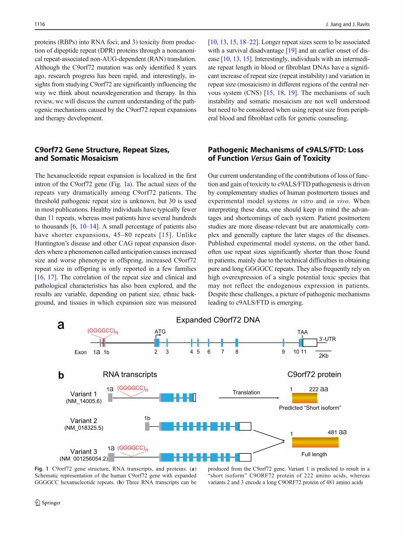
The hexanucleotide repeat expansion is localized in the firstintron of the C9orf72 gene (Fig. 1a). The actual sizes of therepeats vary dramatically among C9orf72 patients. Thethreshold pathogenic repeat size is unknown, but 30 is usedin most publications. Healthy individuals have typically fewerthan 11 repeats, whereas most patients have several hundredsto thousands [6, 10–14]. A small percentage of patients alsohave shorter expansions, 45–80 repeats [15]. UnlikeHuntington’s disease and other CAG repeat expansion disor-ders where a phenomenon called anticipation causes increasedsize and worse phenotype in offspring, increased C9orf72repeat size in offspring is only reported in a few families[16, 17]. The correlation of the repeat size and clinical andpathological characteristics has also been explored, and theresults are variable, depending on patient size, ethnic back-ground, and tissues in which expansion size was measured
[10, 13, 15, 18–22]. Longer repeat sizes seem to be associatedwith a survival disadvantage [19] and an earlier onset of dis-ease [10, 13, 15]. Interestingly, individuals with an intermedi-ate repeat length in blood or fibroblast DNAs have a signifi-cant increase of repeat size (repeat instability) and variation inrepeat size (mosaicism) in different regions of the central ner-vous system (CNS) [15, 18, 19]. The mechanisms of suchinstability and somatic mosaicism are not well understoodbut need to be considered when using repeat size from periph-eral blood and fibroblast cells for genetic counseling.
Pathogenic Mechanisms of c9ALS/FTD: Lossof Function Versus Gain of Toxicity
Our current understanding of the contributions of loss of func-tion and gain of toxicity to c9ALS/FTD pathogenesis is drivenby complementary studies of human postmortem tissues andexperimental model systems in vitro and in vivo. Wheninterpreting these data, one should keep in mind the advan-tages and shortcomings of each system. Patient postmortemstudies are more disease-relevant but are anatomically com-plex and generally capture the later stages of the diseases.Published experimental model systems, on the other hand,often use repeat sizes significantly shorter than those foundin patients, mainly due to the technical difficulties in obtainingpure and long GGGGCC repeats. They also frequently rely onhigh overexpression of a single potential toxic species thatmay not reflect the endogenous expression in patients.Despite these challenges, a picture of pathogenic mechanismsleading to c9ALS/FTD is emerging.
Fig. 1 C9orf72 gene structure, RNA transcripts, and proteins. (a)Schematic representation of the human C9orf72 gene with expandedGGGGCC hexanucleotide repeats. (b) Three RNA transcripts can be
produced from the C9orf72 gene. Variant 1 is predicted to result in aBshort isoform^ C9ORF72 protein of 222 amino acids, whereasvariants 2 and 3 encode a long C9ORF72 protein of 481 amino acids
1116 J. Jiang and J. Ravits
Loss of C9orf72 Function Alone Is Insufficientto Cause ALS/FTD
Reduced C9orf72 Expression in c9ALS/FTD Patients
In humans, C9orf72 is transcribed into three major RNA iso-forms (Fig. 1b). In variants 1 (NM_145005.6) and 3(NM_001256054.2), the expanded GGGGCC repeat is locat-ed in an intron between two alternatively spliced exons, and invariant 2 (NM_018325.5), the repeat is located in thepromoter region. Variant 2 expresses at higher level thanvariants 1 and 3 in CNS tissues [23]. The presence of anenormous GGGGCC repeat expansion in the intron regionmay cause early abortion during transcription, leading to theincreased transcripts containing upstream of the repeats andthe decreased transcripts containing downstream of the repeatsin C9orf72 pat ien ts [24] . In addi t ion , cy tos inehypermethylation of CG dinucleotides in CpG islands(regions with significantly higher frequency of the CGsequence) is an important epigenetic modification that couldlead to gene silencing. There is a CpG island at the 5′ ofGGGGCC repeats, and the repeat expansion itself alsoprovides many more CpG islands. In C9orf72 patients, theCpG island 5′ of the GGGGCC repeats is hypermethylated[25], as is the repeat itself [26]. Finally, trimethylation ofhistones H3 and H4 at the C9orf72 locus was detected inC9orf72 patient blood, suggesting another mechanism fortranscription regulation [27]. All of these could contribute todownregulation of C9orf72 gene expression and loss ofC9orf72 protein function (Fig. 2a). Many independent groupshave shown reduced one or more of the C9orf72 RNA tran-scripts in induced pluripotent stem cell (iPSC)-derived neu-rons [28–30] and patient tissues, including lymphoblasts,frontal cortex, and cerebellum [6, 7, 31]. Demonstrating re-duced C9orf72 protein levels has been more challenging, dueto the low abundance of C9orf72 protein and a lack of goodantibodies for its detection. However, several studies suggestthat the expression levels of C9orf72 proteins are reduced inthe frontal and temporal cortices in patients [32–34].
Physiological Function of C9orf72 Protein
Transcript variants 2 and 3 encode the full-length C9orf72protein with 481 amino acids, and variant 1 produces a pre-dicted Bshort isoform^ protein of 222 amino acids with aunique lysine residue at the C-terminus(Fig. 1b). Whetherthe predicted short isoform C9orf72 is expressed in nature iscontroversial. Xiao et al. [35] developed an antibody that se-lectively detects the predicted short isoform C9orf72 proteinand showed that its expression levels on nuclear membraneswere lower in c9ALS cases than in controls. However, usingan antibody against the N-terminal region of C9orf72 thatcould recognize both the full-length and the predicted short
isoform C9orf72, we demonstrated that C9orf72 is predomi-nantly, if not exclusively, expressed as the full-length proteinin human and mouse tissues [34]. This is further confirmed byanother study using novel knockout-validated C9orf72 mono-clonal rat and mouse antibodies [33].
Nevertheless, the physiological function of the full-lengthC9orf72 protein is emerging. Bioinformatic analysis showsthat C9orf72 protein has high homologywith the differentiallyexpressed in normal and neoplastic cell (DENN) protein fam-ily, which functions as guanine nucleotide exchange factor(GEF) proteins to activate RAB GTPase and regulate mem-brane trafficking [36, 37]. In agreement with this, C9orf72interacts with Smith–Magenis syndrome chromosomal regioncandidate gene 8 (SMCR8) andWD repeat-containing protein41 (WDR41), and this complex possesses GEF activity fordifferent Rab proteins and plays a role in multiple steps ofautophagy/lysosomal pathways [38–45]. Several Rab proteinsare substrates of C9orf72, including Rab1, Rab3, Rab5, Rab7,Rab8a, Rab11, and Rab39 [40, 41, 43–45]. KnockdownC9orf72 in human cell lines and primary neurons inhibitsautophagy induction, leading to accumulation of P62 [40,45]. Accumulation of autophagy substrates, including P62,is also observed in spleens of C9orf72 knockout mice and iniPSC–derived neurons from C9orf72 patients [45, 46]. iPSneurons from C9orf72 patients also showed increased sensi-tivity to autophagy inhibitions, suggesting that reductions inC9orf72 levels contribute to cellular stress [29]. In agreementwith this, C9orf72 protein is necessary for the formation ofstress granules [47].
Loss of C9orf72 Function in ALS/FTD Pathogenesis
Is reduced C9orf72 function as the main disease driver forALS/FTD caused by the C9orf72 repeat expansions? In hu-man, only one sporadic ALS patient has ever been found tocarry a splice site mutation potentially causing a heterozygousloss of function, and it is unclear whether this mutation is thecause of disease or variant of undetermined significance [48,49]. In addition, one would expect that patients homozygousfor C9orf72 repeat expansions (and thus with less C9orf72than heterozygotes) will have more severe symptoms of dis-ease, but this is not the case [50]. Although not conclusive,these human studies do not support a loss of function as thedisease-driving mechanism.
Studies in lower species model systems suggest thatC9orf72 is critical for the development of motor systems andmotor functions. In zebrafish, blocking C9orf72 translation orsplicing with antisense morpholino oligonucleotides resultedin axonopathy and reduced mobility [51]. Overexpressing ofC9orf72 that lacks the complete DENN protein domain alsophenocopied the morpholino experiment, suggesting the in-dispensability of the complete DENN protein domain [52].Similarly, deletion of C9orf72 orthologue in Caenorhabditis
Pathogenic Mechanisms and Therapy Development for C9orf72 Amyotrophic Lateral Sclerosis/Frontotemporal... 1117
elegans led to age-dependent motility deficits, paralysis, andincreased susceptibility to environmental osmotic stress [53].Primary hippocampal neurons cultured from C9orf72 knock-out mice have reduced dendritic arborization and spinal den-sity, suggesting its role in neuronal morphogenesis [54].However, sustained reduction of mouse C9orf72 to 30–40%of its normal level throughout the CNS using antisense oligo-nucleotides (ASOs) is well tolerated, producing no behavioralor pathological features characteristic of ALS/FTD [55].Several ubiquitous or neuronal/glialcell–specific C9orf72knockout mice have also been characterized. None of thesemice produced any ALS/FTD-like pathological or behavioralphenotypes [41, 42, 56–60], indicating that C9orf72 loss-of-function alone is not themainALS/FTD disease driver inmice.Interestingly, global C9orf72 knockout mice develop aproinflammatory/autoimmune phenotype with enlarged spleenand lymph nodes, consistent with the observation that C9orf72ALS/FTD patients also show an increased prevalence of auto-immune disease [61]. These findings are particularly notewor-thy, given growing appreciation of the role of the immunesystem in a variety of neurodegenerative diseases [62].
Overall, current available data suggest that C9orf72 loss offunction is insufficient to precipitate disease. Instead, hyperme-thylation of the mutant C9orf72 allele (one of the mechanismsleading to reduced C9orf72 expression) might actually be pro-tective, potentially by decreasing the transcription of toxic repeat-
containing C9orf72 RNAs [63–65]. However, given the role ofC9orf72 in autophagy and microglia, pathways implicated inALS and FTD, haploinsufficiencymight contribute to the diseaseprocess in synergy with the gain-of-toxicity mechanism. Thiswill be discussed in the section BSynergistic Mechanisms.^
Gain of Toxicity Plays a Central Role in DiseasePathogenesis
RNAs containing sense GGGGCC and antisense CCCCGGrepeats are bidirectionally transcribed from the C9orf72 mu-tation. These repeat-containing RNAs can exert toxic gain-of-function and drive neurodegeneration (Fig. 2b). First, theymay form complicated secondary structures, such as hairpins,i-motifs, and G-quadruplexes [66–68], which, in turn, seques-ter RBPs into intranuclear RNA foci that can be experimen-tally visualized using fluorescence in situ hybridization withprobes against the repeats. These RNA foci lead to depletionof the normal function of the sequestered RBPs and neuronaldeath. The RBP sequestration model is best demonstrated inthe neuromuscular disease myotonic dystrophy 1, where themuscleblind family proteins are sequestered by the CTG re-peats in the DMPK gene and cause downstream splicingchanges [69]. Secondly, despite being in an intronic region,the repeat of the C9orf72 RNAs can remarkably be translatedthrough an unusual mechanism (RAN translation) in every
Fig. 2 Proposed pathogenic mechanisms in C9orf72 ALS/FTD. (a) Thepresence of expanded GGGGCC repeats potentially causes abortivetranscription from exon 1a and hypermethylation of both DNA andhistones to reduce C9orf72 RNA transcription, leading to a loss ofC9orf72 protein function. (b) Bidirectionally transcribed repeat-
containing RNAs cause neuronal toxicity by sequestration of RNA-binding proteins into RNA foci or production of at least 5 aberrant dipep-tide repeat proteins [poly(GA), poly(GP), poly(GR), poly(PR), andpoly(PA)] through a novel repeat-associated non-AUG-dependent trans-lation mechanism
1118 J. Jiang and J. Ravits
reading frame to produce five different DPR proteins thatmight be detrimental to neurons. More specifically,poly(GA) and poly(GR) are uniquely translated from thesense GGGGCC RNA; poly(PR) and poly(PA) are uniquelyfrom the antisense CCCCGG RNA, and poly(GP) is fromeither direction.
In c9ALS/FTD patients, both sense and antisense RNAfoci were detected in fibroblast, lymphoblast, iPSC-derivedneurons, and several CNS regions of c9ALS/FTD patients[28, 55, 70–73]. RNA foci are most abundant in neurons butalso occur in astrocytes, microglia, and oligodendrocytes [55,71, 72]. Similarly, accumulations of DPR proteins have beendetected using either immunohistochemical assays for DPRinclusions or immunoblotting and ELISA-based assays forthe soluble form [71, 73–76]. DPR inclusions are widely dis-tributed throughout the brains of c9ALS/FTD and, interesting-ly, are less frequent in the spinal cord [76–80]. Based on thestaining patterns, they can be categorized into neuronal cyto-plasmic inclusions (NCIs), intranuclear inclusions (INIs), dys-trophic neurites (DNs), and diffuse dendritic or cytoplasmicstaining. So far, no DPR inclusions have been reported inastrocyte, microglia, and oligodendrocytes. Of the differenttypes, the sense-strand RNA-encoded poly(GA), poly(GP),and poly(GR) inclusions are far more frequent than theantisense-strand RNA-encoded poly(PA) and poly(PR), andthey can co-occur in the same neuron [74]. SDS-solublepoly(GA) and poly(GP) can also be detected in CSF and bloodby immunoassay [81–83]. This will be further discussed in thesection BBiomarkers.^
To determine the gain-of-toxicity mechanism from theC9orf72 repeats, several studies have overexpressedGGGGCC repeats with variable sizes and demonstrated dele-terious phenotypes in cell culture [84], C. elegans [85],zebrafish [86], and Drosophila [87–89]. The gain-of-toxicitymechanism is also supported by studies expressing individualDPR proteins (discussed below), as well as studies usingiPSC-derived neurons. Short or interrupted repeats are gener-ally used, given the technical difficulties to obtain long andpure repeats. Adeno-associated viruses expressing 66 or 149GGGGCC repeats were injected intraventrically into newbornmice to induce somatic transgenesis. These mice accumulatednuclear RNA foci and inclusions of DPR proteins and conse-quently exhibited hyperactivity, anxiety, antisocial behavior,and motor deficits [90, 91]. Four different groups, includingours, also generated transgenic mice expressing a bacterialartificial chromosome (BAC) that can accommodate genomicDNA from c9ALS/FTD patients to express long repeats (up to~1000 repeats) in the context of the human gene. All thesemouse models produced sense and antisense RNA foci, aswell as DPR proteins from the sense strand. Two transgenicmodels failed to produce neurodegeneration [92, 93], and ourmice, expressing exons 1–5 of the human C9orf72 gene and~450 repeats, developed increased age-dependent anxiety and
cognitive deficits, accompanied by the loss of hippocampalneurons, but no unusual motor phenotype [94]. In the lastmodel expressing the full-length C9orf72 gene and ~500 re-peats, a subset of female mice developed an acute motor phe-notype, including paralysis and decreased survival. Other fe-males and males showed slower progression [95]. The reasonsfor differences in these mouse models are unknown. Theyfeature different genetic backgrounds, repeat sizes, transgeneinsertion sites, and expression levels. An in-depth comparisonof all BAC models especially the accumulation of RNA fociand DPR proteins will provide insights to the toxic speciesleading to motor and/or cortical neuron death.
In summary, expressing GGGGCC repeats in model sys-tems clearly recapitulates both cellular pathology (RNA fociand DPR proteins) and neuronal degeneration as in patients,even though unphysiologically high levels of expression wereused. It is also challenging to dissect the relative contributionsof RNA foci or DPR proteins to disease pathogenesis sincethese repeats will produce both RNA foci and DPR proteins,and the relative contributions of sense and antisense repeat–containing RNAs remain unclear.
RNA Foci-Mediated Toxicity
Few neuropathological studies have attempted to correlate theabundance and distribution of RNA foci with c9ALS/FTDclinical features. Mizielinska et al. [72] showed that patientswith more sense RNA foci had an earlier age of symptomonset in a small number of C9orf72 FTD patients. Antisensefoci showed a similar trend [72]. In agreement with this, anti-sense RNA foci are associated with nucleoli andmislocalization of TDP-43 [70, 96]. However, another studywith larger patient population reported a contradictory obser-vation. Patients with more antisense RNA foci in middle fron-tal gyrus (cortical layers 3–6) neurons showed a later age ofonset [97].
Expressing C9orf72 GGGGCC repeats in primary neurons[84],Drosophila [87, 98], and zebrafish [86] suggests that thetoxicity arises from RNA foci because no DPR proteins weredetected. However, the inability to detect DPR proteins inthese studies is insufficient to exclude a role for these proteins.Others argue against the toxicity from RNA foci. In two im-portant studies, researchers generated Drosophila expressingeither pure GGGGCC repeats or interrupted repeats of similarsize, but with stop codons inserted in each reading frame toprevent the translation of the repeats into DPR proteins [88,99]. Both pure and interrupted repeats form similar levels ofRNA foci. Expression of pure repeats caused toxicity andearly lethality whereas the interrupted ones had no effect. Inanother study, Drosophila expressing 160 GGGGCC repeatsin the intron formed abundant sense RNA foci in the nucleusbut produced little DPR protein and no neurodegeneration,again suggesting that nuclear sense RNA foci are not
Pathogenic Mechanisms and Therapy Development for C9orf72 Amyotrophic Lateral Sclerosis/Frontotemporal... 1119
sufficient to drive neurodegeneration in this model [100].There are, however, some caveats for interpreting these exper-iments, including whether the interrupted repeat RNAs havesimilar secondary structures to sequester the same key RBPsas the pure repeats, and whether RNA foci in humans maysequester RBPs that are not expressed in Drosophila.
A key to validating RNA foci–mediated toxicity is to iden-tify the RBP(s) that are sequestered and thus lose function.Many proteins are proposed to interact with short, synthesizedC9orf72 repeat RNAs in vitro, including hnRNPA1,hnRNPA3, hnRNP-H, hnRNP-H1/F, nucleolin, Pur-α,double-stranded RNA-specific editase B2 (ADARB2),RanGAP1, THO complex subunit 4 (ALYREF), serine/arginine-rich splicing factor 1 (SRSF1), SRSF2, and Zfp106[24, 28, 87, 101–105]. In c9ALS/FTD brain tissues, sense andantisense RNA foci colocalize with hnRNPA1, hnRNP-H/F,SRSF2, and ALYREF [101, 102, 106]. But most of theseRNA-binding proteins appear to colocalize with RNA foci ata low frequency; only hnRNP-H colocalizes with 70% of allsense foci detected [102]. Nevertheless, key data to supportRNA foci–mediated toxicity are missing (i.e., demonstratingthat the loss of function of any of these RBPs leads to C9orf72diseases and upregulation of their functions can rescue pheno-types caused by C9orf72 repeat expansions).
DPR Protein-Mediated Toxicity
To gain insights for whether and/or which DPR proteins arethe primary culprit of c9ALS/FTD, several clinicopathologi-cal studies have been carried out. Most of these studies raisedsuspicions about the pathogenicity of DPR inclusions, includ-ing 1) regions with the highest burden of DPR inclusions arecerebellum, hippocampus, and neocortex, where no signifi-cant loss of neurons occurs; 2) DPR inclusions do not differin the neuroanatomical distributions in the brain between FTDand ALS cases; 3) DPR inclusions are rarely observed inlower motor neurons in the spinal cord; and most importantly,4) no obvious correlation between the abundance levels ofDPR inclusions with neurodegeneration [77, 78, 107, 108].However, a few neuropathological studies support DPRprotein–mediated toxicity. Mackenzie et al. showed moderateassociations for the amount of poly(GA)-positive DNs withlocal degeneration in the frontal cortex [77]. In another study,more abundant poly(GA) NCIs in cortical, hippocampal, andmotor regions were associated with earlier age at symptomonset [107]. Poly(GR) inclusions, but not the other DPR in-clusions, correlated with areas of neurodegeneration inC9orf72 ALS and were unique in colocalizing with TDP-43pathology in a small sample of brains that were obtained short-ly after death [34]. In a larger cohort of 40 c9ALS/FTD cases,middle frontal gyrus poly(GR) inclusions were strongly cor-related with neurodegeneration in this brain region [109].Notably, these studies detect only aggregated, insoluble
proteins, and it is possible that soluble species may mediatetoxicity. Alternatively, CNS regions that have extensive DPRpathology but are unaffected by neurodegenerationmight con-tain protective factors.
To move pathology to pathogenesis, several groups haveexpressed individual DPR proteins in isolation in yeast [110],mammalian cells [84, 111–117], Drosophila [84, 88, 89, 112,118, 119], zebrafish [86, 120, 121], and mice [122–127]. Ofall the DPR proteins, poly(GR) and poly(PR) are most toxic.Synthetic 20-mer poly(GR) and poly(PR) can cause rapiddeath of U2OS cells and cultured human astrocytes whenadded exogenously [128]. These DPR proteins also consis-tently show toxicity when overexpressed in several differentcell lines, such as HEK293T cells, NSC-34 cells, and iPSC-derived neurons [84, 111, 112]. In Drosophila, targeted ex-pression of poly(GR) or (PR) resulted in eye degeneration,motor deficits, and reduced survival [84, 88, 89, 112, 118,119]. Finally, AAV-mediated or transgenic expression ofpoly(GR) and poly(PR) in mice caused age-dependent neuro-degeneration, brain atrophy, and motor/memory deficits[124–127]. Poly(GA) also exerts toxicity. Syntheticpoly(GA) exogenously applied to human cells or primary neu-rons is toxic [129]. Poly(GA) overexpression in cultured cells[115, 116], primary neurons [115, 116], zebrafish [120, 121],Drosophila [88], or mouse brains [122, 123] leads to toxicity.The remaining DPR proteins [poly(PA) and poly(GP)] are lesslikely to be the toxic species. These studies provide compel-ling evidence that certain DPR proteins are toxic. However,since high overexpression levels of individual DPR proteinswere used, a key question remains to determine whether(which) DPR proteins are the main toxic species in c9ALS/FTD when expressed at the endogenous levels in patients.Another important issue is whether different DPR proteins,and repeat RNA and DPR proteins act synergistically, poten-tially in conjunction with the loss of function of C9orf72, toelicit downstream effects.
Synergistic Mechanisms
In c9ALS/FTD patients, pathological evidence exists for bothloss of C9orf72 function and gain of toxicity. Whether loss ofC9orf72 function synergizes with gain of toxicity to exacer-bate ALS/FTD disease is not known. Mice with loss ofC9orf72 do not develop ALS/FTD-related behavioral deficits,but additional stressors might be required as second hits toinduce neurodegeneration in these mouse models. Shi et al.reported that loss of C9orf72 in human iPSC–derived motorneurons impacts lysosomal biogenesis and vesicular traffick-ing and exacerbates toxicity to poly(GR) and poly(PR) expo-sure [30]. Similarly, loss of C9orf72 protein sensitized cells totoxicity induced by expression of 30 and 60 GGGGCC re-peats [47]. Shao et al. [130] bred C9orf72 knockout mice withone BAC transgenic model expressing the repeat-expanded
1120 J. Jiang and J. Ravits
full-length human C9orf72 gene to determine the synergism.One would doubt such strategy since transgene overexpres-sion would compensate for loss of the mouse C9orf72 endog-enous protein. But surprisingly, the transgenic mouse linedoes not express full-length C9orf72 proteins from the trans-gene and C9orf72 haploinsufficiency exacerbate motor be-havior deficits in a dose-dependent manner [130]. It is prema-ture to say that C9orf72 loss of function induces neurodegen-eration together with toxic RNA and DPR proteins. Furtherinvestigations crossing C9orf72 knockout mice with transgen-ic mice that do not express the full-length C9orf72 proteins orinjecting AAV virus expressing GGGGCC repeats intoC9orf72 knockout mice are needed to address this question.In addition, poly(GA) recruits poly(GR) into inclusions andreduces poly(PR) toxicity when co-expressed in human cellsand Drosophila [117, 118]. Whether interaction betweenindividual DPR proteins enhances or suppresses theirtoxicity is thus a point of interest. Poly(GA) can also inducenuclear RNA foci in cells expressing 80 GGGGCC repeatsand in patient fibroblast cells, suggesting a positive feedbackloop between RNA foci and DPR proteins [131]. Finally,translational frameshifting has been suggested to occur incells expressing short C9orf72 repeats [132] and other repeatexpansions [133], which might produce chimeric repeatpeptides. However, it is unknown whether such a frameshiftoccurs in C9orf72 mutation carriers and, if it does, thecontribution of chimeric repeat peptides to diseasepathogenesis.
Downstream Molecular PathwayDysfunctions
In addition to identifying the major toxic species, research hasfocused on determining cellular dysfunctions that result fromthe C9orf72 repeat expansions. These events provide a basicunderstanding of disease pathophysiology and new therapeu-tic targets (Fig. 3).
Nucleocytoplasmic Transport Deficits
Active transport of proteins and RNAs through nuclearpore complexes (NPCs), a process called nucleocytoplasmictransport (NCT), is essential for cellular functions [134].Zhang et al. [98] showed that RanGAP1 binds toGGGGCC repeat RNA in vitro and is sequestered intosense RNA foci. RanGAP1 activates Ran small GTPase,which plays an important role in regulating the interactionsbetween cargo molecules and transport receptors, such asthe importin and exportin family of proteins [134]. In bothDrosophila overexpressing 30 GGGGCC repeats andc9ALS iPSC–derived neurons, the nucleocytoplasmic Rangradient is decreased, and the nuclear import of proteins and
nuclear export of RNAs are compromised. In addition, therepeat toxicity can be readily suppressed by increased activityof RanGAP1, suggesting that RNA foci–mediated NCT def-icit is a fundamental pathway in c9ALS/FTD [98]. In parallel,unbiased genetic screenings for phenotypic modifiers ofDrosophila expressing GGGGCC repeats [89] or poly(PR)[119], yeast [110], and human cells expressing poly(GR) orpoly(PR) [135] identified that many proteins involved in NCTmitigate or exacerbate disease phenotypes. Mechanistically,poly(GR) and poly(PR) interact with nucleopore proteins(Nups), including the central channel of the nucleopore, toreduce trafficking [136]. Poly(GA) also sequestersnucleocytoplasmic transport proteins, such as HR23, and in-duces an abnormal distribution of RanGAP1 in mice [122].The primary cause for the NCT dysfunction in c9ALS/FTD isunknown. NCT defects have also been reported in other neu-rodegenerative disorders, such as Alzheimer’s disease [137]and Huntington’s disease [138, 139], suggesting that it mayplay a more global role in the neurodegenerative process.
Nucleolar Dysfunction
Nucleolus plays an important role in ribosomal RNA biogen-esis. When applied exogenously to astrocyte cultures,poly(GR) and poly(PR) 20-mers accumulate in nucleolus,leading to splicing changes and impaired ribosomal RNAmat-uration [128]. Overexpressed poly(GR) and poly(PR) also co-localize with nucleoli in cultured cells, primary neurons,iPSC-derived neurons, and Drosophila, leading to abnormalnucleolar morphology [84]. However, poly(GR) and poly(PR)do not localize within the nucleolus in C9orf72 patient brains.Instead, sense RNA foci can bind nucleolin, a principle com-ponent of the nucleolus, and aberrant subcellular localizationof nucleolin was observed in C9orf72 iPSC–derived motorneurons and patient motor cortex [24]. Contrary to this, anti-sense RNA foci associated with nucleoli were identifiedneuropathologically to be associated with disease [96].Nucleolin mislocalization was also found in C9orf72 BACtransgenic mice without significant changes in ribosomalRNA processing or splicing [92]. Interestingly, C9orf72 pa-tient brains exhibit bidirectional nucleolar volume changes,with smaller nucleoli overall but enlarged nucleoli in neuronscontaining poly(GR) inclusions [140].
DNA Damage
Nucleolar stress induces DNA damage, and postmitotic neu-rons are particularly susceptible to DNA damage. An age-dependent increase in DNA damage and oxidative stresswas reported in iPSC-derived motor neurons of patients withC9orf72 repeat expansions [141]. DNA damage is also in-creased in spinal cord neurons of C9orf72 ALS patients[142]. Such DNA damage is partially caused by poly(GR)
Pathogenic Mechanisms and Therapy Development for C9orf72 Amyotrophic Lateral Sclerosis/Frontotemporal... 1121
since expression of poly(GR) in iPSC-derived control neurons,neuronal cell lines or in mice in vivo is sufficient to induce
DNA damage and toxicity [126]. Mechanistically, poly(GR)binds to Atp5a1 and compromises mitochondrial function,
Fig. 3 Cellular processes impaired by the C9orf72 repeat expansions andpotential therapeutic interventions. A wide range of cellular pathwayshave been implicated in c9ALS/FTD, including DNA damage,nucleolar stress, nucleocytoplasmic transport deficits, ER stress,
autophagy dysfunction, translational inhibition, proteasome inhibition,and altered stress granule dynamics. Therapies targeting these deficits,as well as directly targeting repeat expanded C9orf72 DNA/RNA andDPR proteins, are also highlighted
1122 J. Jiang and J. Ravits
leading to increased oxidative stress and an overactivatedKu80-dependent DNA repair pathway [126, 143].Introducing ectopic Atp5a1 expression or reduction in oxida-tive stress by treatment with antioxidants partially rescuesDNA damage in motor neurons from C9orf72 carriers andneurons expressing poly(GR). As a result of DNA damage,the levels of phosphorylated ATM and P53 and other down-stream proapoptotic proteins, such as PUMA, Bax, andcleaved caspase-3, are significantly increased in C9orf72 pa-tient neurons. Partial loss of Ku80 function in these neuronsthrough CRISPR/Cas9-mediated ablation or small RNA-mediated knockdown suppresses the apoptotic pathway.Thus, poly(GR)-mediated DNA damage contributes to ALS/FTD pathogenesis, and partial inhibition of overactivatedKu80-dependentDNA repair pathway is a promising therapeu-tic target.
Alteration in Stress Granules
Eukaryotic cells have evolved sophisticated strategies to com-bat unexpected cellular stress. Cytoplasmic ribonucleoprotein(RNP) granules, such as processing bodies (P-bodies) andstress granules (SGs), assemble quickly under stress to se-quester messenger RNAs (mRNAs), translation initiation fac-tors, 40S ribosomes, and many other RNA-binding proteins.In this way, only essential proteins needed for survival areproduced. These granular structures can either dissemble uponrelease of cellular stress or be degraded by the autophagypathway. Dysregulated formation or clearance of SGs hasbeen proposed as a key pathogenic mechanism for ALS/FTD [144]. In recent years, groundbreaking work from sever-al groups showed that SG-associated RNA-binding proteins,such as FUS and hnRNP1/2, undergo liquid–liquid-phase sep-aration (LLPS) to form droplets in vitro and have the propen-sity to further fibrilize into irreversible hydrogel or even insol-uble amyloid structures [145–148]. One characteristic of theseRBPs is that they all contain prion-like, intrinsically disor-dered low-complexity domains (LCDs). Poly(GR) andpoly(PR) interact with these LCD proteins, impair LLPS,and disrupt the dynamics of stress granules [149]. Consistentwith this, primary cortical neurons overexpressing poly(PR)showed a reduction in cytoplasmic P-bodies with larger sizesand an increase in stress granule formation [84], and knock-down of several of the LCD proteins modifies the eye degen-eration phenotype in Drosophila expressing poly(GR) [112].Activation of the SG response or exposure to either poly(GR)or poly(PR) was also sufficient to disrupt nucleocytoplasmictransport by promoting the sequestration of several nuclearpore proteins into SGs [150]. Interestingly, poly(GR) andpoly(PR) themselves undergo LLPS at high concentrations,and GGGGCC repeat RNA also undergoes gel transition itself[151] and promotes the phase transition of RNA granule pro-teins in vitro and in cells [152].
Translation Inhibition and Ubiquitin ProteasomeSystem
In addition to regulating stress granule dynamics andribosomal RNA synthesis, overexpression of poly(GR) and(PR) blocked global translation in an in vitro translation assayand in cell lines [114]. This was attributed to direct binding ofpoly(GR) and poly(PR) to mRNA, thereby blocking access tothe translational machinery. Poly(PR) and poly(GR) also in-teract with translation initiation and elongation factors andribosome subunits in pull-down assays [114]. In addition,GGGCC repeat–containing RNAs sequester ribosomal sub-units, trigger stress granule formation, and inhibit translation[152, 153]. Interestingly, 26S proteasome complexes, compo-nents of the ubiquitin proteasome system (UPS), are seques-tered within poly(GA) aggregates in cells as revealed by pro-tein cryo-electron tomography technology, which allows 3Dimaging of the cell interior in close-to-native conditions [154].These results provide new insight into the mechanism bywhich poly(GA) deposition promotes UPS impairment andregulate protein homeostasis.
Therapeutics
Currently, no cure is available for ALS or FTD. The identifi-cation of C9orf72 as the most common genetic cause and fastprogress in research unveiling disease mechanisms has in-spired multiple therapeutic interventions for patients carryingthis mutation, the prospects of which are particularly exciting(Fig. 3).
Targeting C9orf72 Repeat-Expanded RNA/DNA
Broad evidence supports a gain of toxicity from which theC9orf72 repeat RNAs play a central role in ALS/FTD patho-genesis. Thus, inhibiting the transcription or selectively reduc-ing repeat-containing RNAs is a promising strategy with an-tisense oligonucleotide therapy being the most advanced interms of clinical development. Indeed, unraveling the exactcontributions from RNA toxicity and DPR protein toxicityor the exact cellular pathways may not be necessary for suchan approach to be successful. However, unraveling the exactcontributions from sense and antisense strand transcripts isnecessary, since these must be targeted separately.
Antisense Oligonucleotide Therapy
ASOs are designed synthetic oligonucleotides or oligonucle-otide analogs that bind to target RNAs. Depending on itschemical modifications, ASOs selectively degrade mRNAsthrough endonuclease RNase H recruitment or prevent theinteraction of RNAs with RBPs, thereby modulating its
Pathogenic Mechanisms and Therapy Development for C9orf72 Amyotrophic Lateral Sclerosis/Frontotemporal... 1123
splicing/processing without degradation [155]. In recentyears, ASO therapy for neurological disorders has gained sig-nificant interest, with the FDA approval of nusinersen(Spinraza) for spinal muscular atrophy (SMA) in 2016.Nusinersen increases the expression of survival motor neuron(SMN) protein by modulating splicing of the SMN2 pre-mRNAs [156]. The ability of ASOs to selectively degrademRNAs is also advantageous if the pathogenesis of neurode-generation is caused by gain of toxicity from mutant RNA/proteins. Phase I trials have been completed with ASOstargeting superoxide dismutase 1 (SOD1) in familiar ALS[157] and mutant huntingtin gene in Huntington’s disease[158]. For C9orf72 diseases, early studies showed that ASOstargeting within or immediately upstream of the sense repeatsreduced sense RNA foci, increased survival from glutamateexcitotoxicity, and abrogated aberrant gene expression pat-terns in fibroblast and iPSC-derived neurons [28, 55, 101].These observations were further extended to in vivo mousemodels. We demonstrated that a single-dose, intraventricularadministration of ASOs targeting the sense repeat–containingRNAs led to sustained reduction of sense RNA foci and DPRproteins and mitigated the behavioral and cognitive deficits intransgenic mice expressing 450 repeats [94]. Importantly,t r a n s c r i p t v a r i a n t s 1 (NM_1 4 5 0 0 5 . 6 ) a n d 3(NM_001256054.2) that carry the repeat expansion can bespecifically targeted by ASOs without reducing variant 2(NM_018325.5) expression. Since variant 2 is expressed atmuch higher levels than variants 1 and 3, overall C9orf72abundance remained relatively intact [94]. Thus, even if lossof function is important in pathogenesis, toxic transcripts canbe reduced without further exacerbating loss. Based on theseencouraging results, a phase I clinical trial of ASOs targetingthe sense strand for C9ALS patients has started by IonisPharmaceuticals and its partner Biogen Inc. in September2018 (NCT03626012).
RNA Interference Strategy
RNA interference (RNAi) represents another alternative ap-proach against RNA/protein-mediated gain of toxicity. RNAiis a biological process in which certain double-stranded RNAmolecules inhibit the expression of target genes by destroyingthe messenger RNAs in the cytoplasm mediated through theRNA-induced silencing complex (RISC) [159]. The threemost common RNAi approaches include short interferingRNAs (siRNAs), shRNAs, or artificial microRNAs(miRNAs). Given the recent progress of using adeno-associated virus (AAV) for therapeutic gene delivery [160],RNAi therapy becomes even more attractive to treat neurode-generative disorders. The major challenge for C9orf72 ALS/FTD is to target the repeat-containing C9orf72 transcripts inthe nucleus. Indeed, one study showed that siRNA significant-ly reduced C9orf72 mRNA in patient fibroblast but did not
affect nuclear RNA foci abundance [55]. However, anotherstudy suggested that single-strand silencing RNAs reducedboth sense and antisense RNA foci by reducing mutantRNA transcript via RNAi and sterically blocking RBP bind-ing to RNAs [161]. Designed double-strand RNA targetingthe repeat region can also block RNA foci formation [162].In addition, AAV5-driving miRNAs can silence C9orf72 andreduce RNA foci in both the nucleus and cytoplasm in iPSC-derived motor neurons and in an ALS mouse model [163,164]. Further studies are needed to demonstrate the efficacyof RNAi to mitigate behavioral deficits in transgenic mice.
Small Molecules
Small molecules offer an attractive approach for targetingC9orf72 repeat DNA/RNAs given their pharmacological ad-vantages, such as a small molecular mass, which is importantfor blood–brain barrier permeability. TMPyP4, a known G-quadruplex binder, binds (GGGGCC)8 RNA in vitro, ablatesthe sequestration of RBPs [165], and rescues transport defectpathologies and neurodegeneration in Drosophila overex-pressing 30 repeats [98]. Recent progress has also been madein the design of nucleotide structure–specific small molecules.Several compounds bind the hairpin structure of GGGGCCrepeats and significantly decrease foci formation andpoly(GP) accumulation in cultured cells overexpressing 66repeats and patient iPSC-derived neurons [83, 166]. It is un-known whether these compounds also reduce poly(GR) andpoly(PR), two DPR proteins that are most toxic in modelsystems. Further experiments are also needed to demonstratethat they are beneficial in reducing cellular toxicity bothin vitro and in vivo.
Reduce Repeat-Containing RNA Transcription
The complicated secondary structure formed by GC-richC9orf72 repeat expansions suggest that specialized transcrip-tion machinery may be required to facilitate RNA polymeraseII through such long repeats. DRB sensitivity-inducing factor(DSIF) complex and the RNA polymerase II associated factor1 complex (PAF1C) are highly conserved and have nonredun-dant roles in activating RNA polymerase II during elongation[167]. SUPT4H and SUPT5H (components of DSIF) andPAF1C play critical roles in the transcriptional elongation ofC9orf72 repeat expansions [168, 169]. Targeting SUPT4Hreduces levels of sense and antisense C9orf72 repeat tran-scripts, as well as accumulation of RNA foci and DPR prod-ucts, and ameliorates neurodegeneration in C9orf72 iPSC-derived neurons and in Drosophila [168]. Similarly, inC9orf72 patients, the expression levels of PAF1 and LEO1(components of PAF1C) are upregulated, and their expressioncorrelates positively with the expression of repeat-containingtranscripts. Depletion of PAF1C reduces RNA and poly(GR)
1124 J. Jiang and J. Ravits
dipeptide production in the Drosophila-expressing GGGGCCtransgene [169]. Although exciting, careful examination ofthis therapeutic strategy is needed since depletion ofSUPT4H causes a global RNA reduction [170].
CRISPR
Recent advances in genome editing with CRISPR/Cas tech-nology have made it possible to target the mutant gene at thegenomic level. CRISPR/Cas is an abbreviation for clusteredregularly interspaced short palindromic repeats and CRISPR-associated protein. It was adapted from a naturally occurringgenome-editing system in bacteria [171]. CRISR/Cas9 can bedesigned to cut the C9orf72 repeat expansions, following generepair through nonhomologous end joining. As a proof ofconcept, genetic correction of C9orf72 repeat expansions inpatient iPSCs has been achieved [30, 126]. In addition, Pintoet al. [172] targeted enzymatically dead Cas9 to directly bindrepeat-expanded DNAs, such as CTG expansion in DM1,CTGG expansion in DM2, and GGGGCC expansion inC9ALS/FTD. This approach sterically impedes repeat RNAtranscription and leads to a drastic decrease in the productionof aberrant proteins produced from RAN translation [172].Similar to ASOs, RNA-targeting Cas9 with RNA endonucle-ase can degrade repeat expanded RNAs and reduce RNA fociand DPR protein levels in cell lines [173]. The clinical use ofCRISPR/Cas technology in ALS/FTD is still in its infancy.Ethical concerns, adequate methods of delivery and celltargeting, off-target and safety measures, and treatment effica-cy are the main issues that need to be addressed before apply-ing this method to patients.
Targeting DPR Proteins and DownstreamMechanisms
Overall, ASO, RNAi, and other abovementioned approachestarget the upstream of disease mechanism and also positivelyaffect different downstream pathways. For example, Zhanget al. [98] showed that ASOs targeting C9orf72 sense RNAor TMPyP4 treatment to inhibit RBPs bindings to repeatRNAs can rescue NCT deficits. Nevertheless, strategies di-rectly targeting DPR proteins and downstream mechanismshave also been pursued.
Target DPR Proteins
Aggregates of DPR proteins are a pathological hallmark ofC9orf72 ALS/FTD, and DPR proteins seem to exert toxicityin model systems. In addition to inhibiting RAN translation aswe learn more about its mechanisms [153, 174, 175], one wayto directly mitigate DPR protein–mediated toxicity is to in-crease toxic protein turnover rate. To this end, overexpressingthe small heat shock protein HSPB8 facilitated the autophagy-mediated disposal of a large variety of classical misfolded
aggregation-prone proteins and significantly decreased the ac-cumulation of most DPR insoluble species [176]. Antibodyimmunization against DPR proteins represents another noveltherapeutic approach. In other neurodegenerative disorders,active and passive immunization have been explored to targettoxic proteins that transmit from cells to cells, such asamyloid-β, tau, and α-synuclein [177]. The prion-like propa-gation of misfolded/aggregated proteins is also a pathogenicprinciple in ALS/FTD [178, 179]. Using coculture cell assays,poly(GA) was shown to possess cell-to-cell transmissionproperties [180] and so do poly(GP) and poly(PA) [131].Interestingly, poly(GA) antibodies reduce intracellularpoly(GA) aggregations and seeding activity of C9orf72 pa-tient brain extracts [131]. Since poly(GR) and poly(PR) aremore toxic than poly(GA), poly(GP), and poly(PA) in exper-imental models, further studies are needed to determine whichDPR proteins and conformations to target. The specificity ofantibodies against DPR proteins is also a concern as similarshort repetitive sequences are present in human proteome.
Target Downstream Mechanisms
Other therapeutic approaches aim to correct downstream cel-lular pathways affected by C9orf72 repeat expansions.Reducing nuclear export by targeting the nuclear export fac-tors SRSF1 or exportin 1 ameliorates the toxicity mediated byC9orf72 repeat expansion in Drosophila [98]. This could bedue to the reduced export of toxic repeat-containing RNA tothe cytoplasm and thus less DPR proteins [181] or through amore general mechanism to reverse alterations in NCT andconsequent mislocalization of RBPs, such as TDP-43 [98].In addition to the genetic tools, selective inhibitors of nuclearexport (SINEs) mitigate NCT deficits and neurodegenerationin a Drosophila model [98]. SINE compounds also improveprimary neuron survival and partially improve motor functionin rats overexpressing mutant TDP-43 [182]. Currently,Karyopharm Therapeutics and its partner Biogen Inc. are tak-ing one of the SINE compounds, KPT-350, into clinical trials.Zhang et al. [150] also showed that inhibition of stress granuleassembly by using ASOs targeting ataxin 2 is sufficient toabrogate NCT dysfunction and neurodegeneration in patient-derived neurons and in vivo. As reducing ataxin 2 significantlyextend the lifespan of TDP-43 mice, this strategy holds greatpromise [183].
Biomarkers
One of the critical themes that has emerged over the last twodecades of clinical trials in ALS relates to biomarkers. There isno clear diagnostic biomarker for diagnosis, tracking diseaseprogression, or determining if a putative therapy engages itstherapeutic target. In C9orf72-associated diseases, effectivebiomarkers could also identify phenoconversion of
Pathogenic Mechanisms and Therapy Development for C9orf72 Amyotrophic Lateral Sclerosis/Frontotemporal... 1125
asymptomatic genetic carriers. Clinical, neurophysiological,and imaging biomarkers are difficult to employ and/or insen-sitive. Recently, progress has emerged for biomarkers in theblood and CSF. Neurofilament proteins herald the onset ofdisease in presymptomatic carriers and appear to track withdisease progression, although these biomarkers are nonspecif-ic to C9orf72 diseases and are sensitive to damage caused byfree radicals [184]. Significantly, the DPR protein poly(GP)was detected in CSF and in peripheral blood mononuclearcells from c9ALS patients. Thus, tracking poly(GP) in CSFprovides a direct readout of target engagement for ASO andother clinical trials [82].
Conclusions and Future Perspectives
Since the discovery of C9orf72 repeat expansions, an impres-sive effort has been made to investigate disease mechanisms.Many disease models were developed to determine the loss offunction and/or gain of toxicity and downstream cellular func-tion alterations. Therapeutic approaches and biomarkers havealso been explored. Excitingly, antisense oligonucleotide ther-apy to selectively degrade repeat-containing RNAs has goneinto a phase I clinical trial for C9orf72 ALS patients. With acontinuing fast pace of research on these devastating diseases,a promising therapy should soon be on the horizon.
Acknowledgements JR is supported by grants from ALS Association(5356S3), Target ALS (20134792), the National Institute ofNeurological Diseases and Stroke (NIH R01NS088578 andNS047101), and Pam Golden and is involved in Biogen-sponsored clin-ical trials. JJ is a recipient of career development grant from MuscularDystrophy Association (479769).
Required Author Forms Disclosure forms provided by the authors areavailable with the online version of this article.
References
1. Taylor JP, Brown RH, Jr., Cleveland DW. Decoding ALS: fromgenes to mechanism. Nature. 2016;539(7628):197–206.
3. Ng AS, Rademakers R, Miller BL. Frontotemporal dementia: abridge between dementia and neuromuscular disease. Ann N YAcad Sci. 2015;1338:71–93.
4. Ling SC, Polymenidou M, Cleveland DW. Converging mecha-nisms in ALS and FTD: disrupted RNA and protein homeostasis.Neuron. 2013;79(3):416–38.
5. Renton AE, Majounie E, Waite A, Simon-Sanchez J, Rollinson S,Gibbs JR, et al. A hexanucleotide repeat expansion in C9ORF72 isthe cause of chromosome 9p21-linked ALS-FTD. Neuron.2011;72(2):257–68.
6. DeJesus-Hernandez M, Mackenzie IR, Boeve BF, Boxer AL,Baker M, Rutherford NJ, et al. Expanded GGGGCChexanucleotide repeat in noncoding region of C9ORF72 causeschromosome 9p-linked FTD and ALS. Neuron. 2011;72(2):245–56.
7. Gijselinck I, Van Langenhove T, van der Zee J, Sleegers K,Philtjens S, Kleinberger G, et al. A C9orf72 promoter repeat ex-pansion in a Flanders-Belgian cohort with disorders of thefrontotemporal lobar degeneration-amyotrophic lateral sclerosisspectrum: a gene identification study. Lancet Neurology.2012;11(1):54–65.
8. Nguyen HP, Van Broeckhoven C, van der Zee J. ALSGenes in theGenomic Era and their Implications for FTD. Trends Genet.2018;34(6):404–23.
9. La Spada AR, Taylor JP. Repeat expansion disease: progress andpuzzles in disease pathogenesis. Nature Reviews Genetics.2010;11(4):247–58.
10. Beck J, Poulter M, Hensman D, Rohrer JD, Mahoney CJ,Adamson G, et al. Large C9orf72 hexanucleotide repeat expan-sions are seen in multiple neurodegenerative syndromes and aremore frequent than expected in the UK population. AmericanJournal of Human Genetics. 2013;92(3):345–53.
11. BuchmanVL, Cooper-Knock J, Connor-RobsonN, HigginbottomA, Kirby J, Razinskaya OD, et al. Simultaneous and independentdetection of C9ORF72 alleles with low and high number ofGGGGCC repeats using an optimised protocol of Southern blothybridisation. Molecular Neurodegeneration. 2013;8:12.
12. Dobson-Stone C, Hallupp M, Loy CT, Thompson EM, Haan E,Sue CM, et al. C9ORF72 repeat expansion in Australian andSpanish frontotemporal dementia patients. PLoS One. 2013;8(2):e56899.
13. Hubers A, Marroquin N, Schmoll B, Vielhaber S, Just M, MayerB, et al. Polymerase chain reaction and Southern blot-based anal-ysis of the C9orf72 hexanucleotide repeat in different motor neu-ron diseases. Neurobiology of Aging. 2014;35(5):1214.e1–6.
14. Ishiura H, Takahashi Y, Mitsui J, Yoshida S, Kihira T, Kokubo Y,et al. C9ORF72 repeat expansion in amyotrophic lateral sclerosisin the Kii peninsula of Japan. Archives of Neurology. 2012;69(9):1154–8.
15. Gijselinck I, Van Mossevelde S, van der Zee J, Sieben A,Engelborghs S, De Bleecker J, et al. The C9orf72 repeat sizecorrelates with onset age of disease, DNA methylation and tran-scriptional downregulation of the promoter. Molecular Psychiatry.2016;21(8):1112–24.
16. Proudfoot M, Gutowski NJ, Edbauer D, Hilton DA, Stephens M,Rankin J, et al. Early dipeptide repeat pathology in afrontotemporal dementia kindred with C9ORF72 mutation andintellectual disability. Acta Neuropathologica. 2014;127(3):451–8.
17. Van Mossevelde S, van der Zee J, Gijselinck I, Sleegers K, DeBleecker J, Sieben A, et al. Clinical Evidence of DiseaseAnticipation in Families Segregating a C9orf72 RepeatExpansion. JAMA Neurology. 2017;74(4):445–52.
18. Nordin A, Akimoto C, Wuolikainen A, Alstermark H, Jonsson P,Birve A, et al. Extensive size variability of the GGGGCC expan-sion in C9orf72 in both neuronal and non-neuronal tissues in 18patients with ALS or FTD. Human Molecular Genetics.2015;24(11):3133–42.
19. van Blitterswijk M, DeJesus-Hernandez M, Niemantsverdriet E,Murray ME, HeckmanMG, Diehl NN, et al. Association betweenrepeat sizes and clinical and pathological characteristics in carriersof C9ORF72 repeat expansions (Xpansize-72): a cross-sectionalcohort study. Lancet Neurology. 2013;12(10):978–88.
20. Benussi L, Rossi G, Glionna M, Tonoli E, Piccoli E, Fostinelli S,et al. C9ORF72 hexanucleotide repeat number in frontotemporal
1126 J. Jiang and J. Ravits
lobar degeneration: a genotype-phenotype correlation study.Journal of Alzheimer’s Disease: JAD. 2014;38(4):799–808.
21. Dols-Icardo O, Garcia-Redondo A, Rojas-Garcia R, Sanchez-Valle R, Noguera A, Gomez-Tortosa E, et al. Characterization ofthe repeat expansion size in C9orf72 in amyotrophic lateral scle-rosis and frontotemporal dementia. Human Molecular Genetics.2014;23(3):749–54.
22. Suh E, Lee EB, Neal D, Wood EM, Toledo JB, Rennert L, et al.Semi-automated quantification of C9orf72 expansion size revealsinverse correlation between hexanucleotide repeat number anddisease duration in frontotemporal degeneration. ActaNeuropathologica. 2015;130(3):363–72.
23. Rizzu P, Blauwendraat C, Heetveld S, Lynes EM, Castillo-LizardoM, Dhingra A, et al. C9orf72 is differentially expressed in thecentral nervous system andmyeloid cells and consistently reducedin C9orf72, MAPT and GRN mutation carriers. ActaNeuropathologica Communications. 2016;4(1):37.
25. Xi Z, Zinman L, Moreno D, Schymick J, Liang Y, Sato C, et al.Hypermethylation of the CpG island near the G4C2 repeat in ALSwith a C9orf72 expansion. American Journal of Human Genetics.2013;92(6):981–9.
26. Xi Z, Zhang M, Bruni AC, Maletta RG, Colao R, Fratta P, et al.The C9orf72 repeat expansion itself is methylated in ALS andFTLD patients. Acta Neuropathologica. 2015;129(5):715–27.
27. Belzil VV, Bauer PO, Prudencio M, Gendron TF, Stetler CT, YanIK, et al. Reduced C9orf72 gene expression in c9FTD/ALS iscaused by histone trimethylation, an epigenetic event detectablein blood. Acta Neuropathologica. 2013;126(6):895–905.
28. Donnelly CJ, Zhang PW, Pham JT, Haeusler AR, Mistry NA,Vidensky S, et al. RNA toxicity from the ALS/FTD C9ORF72expansion is mitigated by antisense intervention. Neuron.2013;80(2):415–28.
29. Almeida S, Gascon E, Tran H, Chou HJ, Gendron TF, Degroot S,et al. Modeling key pathological features of frontotemporal de-mentia with C9ORF72 repeat expansion in iPSC-derived humanneurons. Acta Neuropathologica. 2013;126(3):385–99.
30. Shi Y, Lin S, Staats KA, Li Y, Chang WH, Hung ST, et al.Haploinsufficiency leads to neurodegeneration in C9ORF72ALS/FTD human induced motor neurons. Nature Medicine.2018;24(3):313–25.
31. van Blitterswijk M, Gendron TF, Baker MC, DeJesus-HernandezM, Finch NA, Brown PH, et al. Novel clinical associations withspecific C9ORF72 transcripts in patients with repeat expansions inC9ORF72. Acta Neuropathologica. 2015;130(6):863–76.
32. Waite AJ, Baumer D, East S, Neal J, Morris HR, Ansorge O, et al.Reduced C9orf72 protein levels in frontal cortex of amyotrophiclateral sclerosis and frontotemporal degeneration brain with theC9ORF72 hexanucleotide repeat expansion. Neurobiology ofAging. 2014;35(7):1779 e5- e13.
33. Frick P, Sellier C, Mackenzie IRA, Cheng CY, Tahraoui-Bories J,Martinat C, et al. Novel antibodies reveal presynaptic localizationof C9orf72 protein and reduced protein levels in C9orf72mutationcarriers. Acta Neuropathologica Communications. 2018;6(1):72.
34. Saberi S, Stauffer JE, Jiang J, Garcia SD, Taylor AE, Schulte D,et al. Sense-encoded poly-GR dipeptide repeat proteins correlateto neurodegeneration and uniquely co-localize with TDP-43 indendrites of repeat-expanded C9orf72 amyotrophic lateral sclero-sis. Acta Neuropathologica. 2018;135(3):459–74.
35. Xiao S, MacNair L, McGoldrick P, McKeever PM, McLean JR,Zhang M, et al. Isoform-specific antibodies reveal distinct subcel-lular localizations of C9orf72 in amyotrophic lateral sclerosis.Annals of Neurology. 2015;78(4):568–83.
36. Levine TP, Daniels RD, Gatta AT, Wong LH, Hayes MJ. Theproduct of C9orf72, a gene strongly implicated in neurodegener-ation, is structurally related to DENN Rab-GEFs. Bioinformatics(Oxford, England). 2013;29(4):499–503.
37. Zhang D, Iyer LM, He F, Aravind L. Discovery of Novel DENNProteins: Implications for the Evolution of Eukaryotic IntracellularMembrane Structures and Human Disease. Frontiers in Genetics.2012;3:283.
38. Amick J, Roczniak-Ferguson A, Ferguson SM. C9orf72 bindsSMCR8, localizes to lysosomes, and regulates mTORC1 signal-ing. Molecular Biology of the Cell. 2016;27(20):3040–51.
39. Jung J, Nayak A, Schaeffer V, Starzetz T, Kirsch AK, Muller S,et al. Multiplex image-based autophagy RNAi screening identifiesSMCR8 as ULK1 kinase activity and gene expression regulator.eLife. 2017;6.
40. Sellier C, Campanari ML, Julie Corbier C, Gaucherot A, Kolb-Cheynel I, Oulad-Abdelghani M, et al. Loss of C9ORF72 impairsautophagy and synergizes with polyQ Ataxin-2 to induce motorneuron dysfunction and cell death. The EMBO Journal.2016;35(12):1276–97.
41. Sullivan PM, Zhou X, Robins AM, Paushter DH, Kim D, SmolkaMB, et al. The ALS/FTLD associated protein C9orf72 associateswith SMCR8 and WDR41 to regulate the autophagy-lysosomepathway. Acta Neuropathologica Communications. 2016;4(1):51.
42. Ugolino J, Ji YJ, Conchina K, Chu J, Nirujogi RS, Pandey A, et al.Loss of C9orf72 Enhances Autophagic Activity via DeregulatedmTOR and TFEB Signaling. PLoS Genetics. 2016;12(11):e1006443.
43. Yang M, Liang C, Swaminathan K, Herrlinger S, Lai F,Shiekhattar R, et al. A C9ORF72/SMCR8-containing complexregulates ULK1 and plays a dual role in autophagy. ScienceAdvances. 2016;2(9):e1601167.
44. Farg MA, Sundaramoorthy V, Sultana JM, Yang S, Atkinson RA,Levina V, et al. C9ORF72, implicated in amytrophic lateral scle-rosis and frontotemporal dementia, regulates endosomal traffick-ing. Human Molecular Genetics. 2014;23(13):3579–95.
45. Webster CP, Smith EF, Bauer CS, Moller A, Hautbergue GM,Ferraiuolo L, et al. The C9orf72 protein interacts with Rab1aand the ULK1 complex to regulate initiation of autophagy. TheEMBO Journal. 2016;35(15):1656–76.
46. Aoki Y, Manzano R, Lee Y, Dafinca R, Aoki M, Douglas AGL,et al. C9orf72 and RAB7L1 regulate vesicle trafficking in amyo-trophic lateral sclerosis and frontotemporal dementia. Brain: ajournal of neurology. 2017;140(4):887–97.
47. Maharjan N, Kunzli C, Buthey K, Saxena S. C9ORF72 RegulatesStress Granule Formation and Its Deficiency Impairs StressGranule Assembly, Hypersensitizing Cells to Stress. MolecularNeurobiology. 2017;54(4):3062–77.
48. Harms MB, Cady J, Zaidman C, Cooper P, Bali T, Allred P, et al.Lack of C9ORF72 coding mutations supports a gain of functionfor repeat expansions in amyotrophic lateral sclerosis.Neurobiology of Aging. 2013;34(9):2234 e13–9.
49. Liu F, Liu Q, Lu CX, Cui B, Guo XN, Wang RR, et al.Identification of a novel loss-of-function C9orf72 splice site mu-tation in a patient with amyotrophic lateral sclerosis.Neurobiology of Aging. 2016;47:219.e1-.e5.
50. Fratta P, Poulter M, Lashley T, Rohrer JD, Polke JM, Beck J, et al.Homozygosity for the C9orf72 GGGGCC repeat expansion infrontotemporal dementia. Acta Neuropathologica. 2013;126(3):401–9.
51. Ciura S, Lattante S, Le Ber I, Latouche M, Tostivint H, Brice A,et al. Loss of function of C9orf72 causes motor deficits in azebrafish model of amyotrophic lateral sclerosis. Annals ofNeurology. 2013;74(2):180–7.
Pathogenic Mechanisms and Therapy Development for C9orf72 Amyotrophic Lateral Sclerosis/Frontotemporal... 1127
52. Yeh TH, Liu HF, Li YW, LuCS, Shih HY, Chiu CC, et al. C9orf72is essential for neurodevelopment and motility mediated by CyclinG1. Experimental Neurology. 2018;304:114–24.
53. Therrien M, Rouleau GA, Dion PA, Parker JA. Deletion ofC9ORF72 results in motor neuron degeneration and stress sensi-tivity in C. elegans. PLoS One. 2013;8(12):e83450.
54. Ho WY, Tai YK, Chang JC, Liang J, Tyan SH, Chen S, et al. TheALS-FTD-linked gene product, C9orf72, regulates neuronal mor-phogenesis via autophagy. Autophagy. 2019;15(5):827–42.
55. Lagier-Tourenne C, BaughnM, Rigo F, Sun S, Liu P, Li HR, et al.Targeted degradation of sense and antisense C9orf72 RNA foci astherapy for ALS and frontotemporal degeneration. Proceedings ofthe National Academy of Sciences of the United States ofAmerica. 2013;110(47):E4530–9.
56. Koppers M, Blokhuis AM, Westeneng HJ, Terpstra ML, ZundelCA, Vieira de Sa R, et al. C9orf72 ablation in mice does not causemotor neuron degeneration or motor deficits. Annals ofNeurology. 2015;78(3):426–38.
57. Atanasio A, Decman V,White D, RamosM, Ikiz B, Lee HC, et al.C9orf72 ablation causes immune dysregulation characterized byleukocyte expansion, autoantibody production, andglomerulonephropathy in mice. Scientific Reports. 2016;6:23204.
58. Sudria-Lopez E, Koppers M, de Wit M, van der Meer C,Westeneng HJ, Zundel CA, et al. Full ablation of C9orf72 in micecauses immune system-related pathology and neoplastic eventsbut no motor neuron defects. Acta Neuropathologica.2016;132(1):145–7.
59. O’Rourke JG, Bogdanik L, Yanez A, Lall D, Wolf AJ,Muhammad AK, et al. C9orf72 is required for proper macrophageand microglial function in mice. Science (New York, NY).2016;351(6279):1324–9.
60. Burberry A, Suzuki N, Wang JY, Moccia R, Mordes DA, StewartMH, et al. Loss-of-function mutations in the C9ORF72 mouseortholog cause fatal autoimmune disease. Science TranslationalMedicine. 2016;8(347):347-93.
61. Miller ZA, Sturm VE, Camsari GB, Karydas A, Yokoyama JS,Grinberg LT, et al. Increased prevalence of autoimmune diseasewithin C9 and FTD/MND cohorts: Completing the picture.Neurology(R) Neuroimmunology & Neuroinflammation.2016;3(6):e301.
62. Molteni M, Rossetti C. Neurodegenerative diseases: The immu-nological perspective. J Neuroimmunol. 2017;313:109–15.
63. Liu EY, Russ J,WuK, Neal D, Suh E,McNally AG, et al. C9orf72hypermethylation protects against repeat expansion-associated pa-thology in ALS/FTD. Acta Neuropathologica. 2014;128(4):525–41.
64. McMillan CT, Russ J, Wood EM, Irwin DJ, Grossman M,McCluskey L, et al. C9orf72 promoter hypermethylation is neu-roprotective: Neuroimaging and Neuropathologic Evidence.Neurology. 2015;84(16):1622–30.
65. Russ J, Liu EY, Wu K, Neal D, Suh E, Irwin DJ, et al.Hypermethylation of repeat expanded C9orf72 is a clinical andmolecular disease modifier. Acta Neuropathologica. 2015;129(1):39–52.
66. Fratta P, Mizielinska S, Nicoll AJ, Zloh M, Fisher EM, ParkinsonG, et al. C9orf72 hexanucleotide repeat associated with amyotro-phic lateral sclerosis and frontotemporal dementia forms RNA G-quadruplexes. Scientific Reports. 2012;2:1016.
67. Reddy K, Zamiri B, Stanley SY, Macgregor RB, Jr., Pearson CE.The disease-associated r(GGGGCC)n repeat from the C9orf72gene forms tract length-dependent uni- and multimolecular RNAG-quadruplex structures. The Journal of Biological Chemistry.2013;288(14):9860–6.
68. Kovanda A, Zalar M, Sket P, Plavec J, Rogelj B. Anti-sense DNAd(GGCCCC)n expansions in C9ORF72 form i-motifs and proton-ated hairpins. Scientific Reports. 2015;5:17944.
69. Rahimov F, Kunkel LM. The cell biology of disease: cellular andmolecular mechanisms underlying muscular dystrophy. TheJournal of Cell Biology. 2013;201(4):499–510.
70. Cooper-Knock J, Higginbottom A, Stopford MJ, Highley JR, IncePG, Wharton SB, et al. Antisense RNA foci in the motor neuronsof C9ORF72-ALS patients are associated with TDP-43proteinopathy. Acta Neuropathologica. 2015;130(1):63–75.
71. Gendron TF, Bieniek KF, Zhang YJ, Jansen-West K, Ash PE,Caulfield T, et al. Antisense transcripts of the expandedC9ORF72 hexanucleotide repeat form nuclear RNA foci and un-dergo repeat-associated non-ATG translation in c9FTD/ALS. ActaNeuropathologica. 2013;126(6):829–44.
72. Mizielinska S, Lashley T, Norona FE, Clayton EL, Ridler CE,Fratta P, et al. C9orf72 frontotemporal lobar degeneration ischaracterised by frequent neuronal sense and antisense RNA foci.Acta Neuropathologica. 2013;126(6):845–57.
73. Zu T, Liu Y, Banez-Coronel M, Reid T, Pletnikova O, Lewis J,et al. RAN proteins and RNA foci from antisense transcripts inC9ORF72 ALS and frontotemporal dementia. Proceedings of theNational Academy of Sciences of the United States of America.2013;110(51):E4968–77.
74. Mori K, Arzberger T, Grasser FA, Gijselinck I, May S, RentzschK, et al. Bidirectional transcripts of the expanded C9orf72hexanucleotide repeat are translated into aggregating dipeptiderepeat proteins. Acta Neuropathologica. 2013;126(6):881–93.
75. Mori K,Weng SM, Arzberger T, May S, Rentzsch K, Kremmer E,et al. The C9orf72 GGGGCC repeat is translated into aggregatingdipeptide-repeat proteins in FTLD/ALS. Science (New York,NY). 2013;339(6125):1335–8.
76. Ash PE, Bieniek KF, Gendron TF, Caulfield T, Lin WL, Dejesus-Hernandez M, et al. Unconventional translation of C9ORF72GGGGCC expansion generates insoluble polypeptides specificto c9FTD/ALS. Neuron. 2013;77(4):639–46.
77. Mackenzie IR, Frick P, Grasser FA, Gendron TF, Petrucelli L,Cashman NR, et al. Quantitative analysis and clinico-pathological correlations of different dipeptide repeat protein pa-thologies in C9ORF72 mutation carriers. Acta Neuropathologica.2015;130(6):845–61.
78. Schludi MH, May S, Grasser FA, Rentzsch K, Kremmer E,Kupper C, et al. Distribution of dipeptide repeat proteins in cellu-lar models and C9orf72 mutation cases suggests link to transcrip-tional silencing. Acta Neuropathologica. 2015;130(4):537–55.
79. Gomez-Deza J, Lee YB, Troakes C, Nolan M, Al-Sarraj S, GalloJM, et al. Dipeptide repeat protein inclusions are rare in the spinalcord and almost absent from motor neurons in C9ORF72 mutantamyotrophic lateral sclerosis and are unlikely to cause their degen-eration. Acta Neuropathologica Communications. 2015;3:38.
80. Schipper LJ, Raaphorst J, Aronica E, Baas F, de Haan R, de VisserM, et al. Prevalence of brain and spinal cord inclusions, includingdipeptide repeat proteins, in patients with the C9ORF72hexanucleotide repeat expansion: a systematic neuropathologicalreview. Neuropathology and Applied Neurobiology. 2016;42(6):547–60.
81. Lehmer C, Oeckl P, Weishaupt JH, Volk AE, Diehl-Schmid J,Schroeter ML, et al. Poly-GP in cerebrospinal fluid linksC9orf72-associated dipeptide repeat expression to the asymptom-atic phase of ALS/FTD. EMBO Mol Med. 2017;9(7):859–68.
82. Gendron TF, Chew J, Stankowski JN, Hayes LR, Zhang YJ,Prudencio M, et al. Poly(GP) proteins are a useful pharmacody-namic marker for C9ORF72-associated amyotrophic lateral scle-rosis. Science Translational Medicine. 2017;9(383).
83. Su Z, Zhang Y, Gendron TF, Bauer PO, Chew J, Yang WY, et al.Discovery of a biomarker and lead small molecules to targetr(GGGGCC)-associated defects in c9FTD/ALS. Neuron.2014;83(5):1043–50.
1128 J. Jiang and J. Ravits
84. WenX, TanW,Westergard T, Krishnamurthy K,Markandaiah SS,Shi Y, et al. Antisense proline-arginine RAN dipeptides linked toC9ORF72-ALS/FTD form toxic nuclear aggregates that initiatein vitro and in vivo neuronal death. Neuron. 2014;84(6):1213–25.
85. Wang X, Hao L, Saur T, Joyal K, Zhao Y, Zhai D, et al. ForwardGenetic Screen in Caenorhabditis elegans Suggests F57A10.2 andacp-4 As Suppressors of C9ORF72 Related Phenotypes. FrontMol Neurosci. 2016;9:113.
86. Swinnen B, Bento-Abreu A, Gendron TF, Boeynaems S, BogaertE, Nuyts R, et al. A zebrafish model for C9orf72 ALS revealsRNA tox i c i t y a s a pa t hogen i c mechan i sm . Ac t aNeuropathologica. 2018;135(3):427–43.
87. Xu Z, Poidevin M, Li X, Li Y, Shu L, Nelson DL, et al. ExpandedGGGGCC repeat RNA associated with amyotrophic lateral scle-rosis and frontotemporal dementia causes neurodegeneration.Proceedings of the National Academy of Sciences of the UnitedStates of America. 2013;110(19):7778–83.
88. Mizielinska S, Gronke S, Niccoli T, Ridler CE, Clayton EL,Devoy A, et al. C9orf72 repeat expansions cause neurodegenera-tion in Drosophila through arginine-rich proteins. Science (NewYork, NY). 2014;345(6201):1192–4.
89. FreibaumBD, LuY, Lopez-Gonzalez R, KimNC, Almeida S, LeeKH, et al. GGGGCC repeat expansion in C9orf72 compromisesnucleocytoplasmic transport. Nature. 2015;525(7567):129–33.
90. Chew J, Gendron TF, Prudencio M, Sasaguri H, Zhang YJ,Castanedes-Casey M, et al. Neurodegeneration. C9ORF72 repeatexpansions in mice cause TDP-43 pathology, neuronal loss, andbehavioral deficits. Science (New York, NY). 2015;348(6239):1151–4.
91. Chew J, Cook C, Gendron TF, Jansen-West K, Del Rosso G,Daughrity LM, et al. Aberrant deposition of stress granule-resident proteins linked to C9orf72-associated TDP-43proteinopathy. Molecular Neurodegeneration. 2019;14(1):9.
92. O’Rourke JG, Bogdanik L, Muhammad A, Gendron TF, Kim KJ,Austin A, et al. C9orf72 BAC Transgenic Mice Display TypicalPathologic Features of ALS/FTD. Neuron. 2015;88(5):892–901.
93. Peters OM, Cabrera GT, Tran H, Gendron TF, McKeon JE,Metterville J, et al. Human C9ORF72 HexanucleotideExpansion Reproduces RNA Foci and Dipeptide RepeatProteins but Not Neurodegeneration in BAC Transgenic Mice.Neuron. 2015;88(5):902–9.
94. Jiang J, Zhu Q, Gendron TF, Saberi S, McAlonis-Downes M,Seelman A, et al. Gain of Toxicity from ALS/FTD-LinkedRepeat Expansions in C9ORF72 Is Alleviated by AntisenseOligonucleotides Targeting GGGGCC-Containing RNAs.Neuron. 2016;90(3):535–50.
95. Liu Y, Pattamatta A, Zu T, Reid T, Bardhi O, Borchelt DR, et al.C9orf72 BAC Mouse Model with Motor Deficits andNeurodegenerative Features of ALS/FTD. Neuron. 2016;90(3):521–34.
96. Aladesuyi Arogundade O, Stauffer JE, Saberi S, Diaz-Garcia S,Malik S, Basilim H, et al. Antisense RNA foci are associated withnucleoli and TDP-43 mislocalization in C9orf72-ALS/FTD: aquantitative study. Acta Neuropathologica. 2019;137(3):527–30.
97. DeJesus-Hernandez M, Finch NA, Wang X, Gendron TF, BieniekKF, Heckman MG, et al. In-depth clinico-pathological examina-tion of RNA foci in a large cohort of C9ORF72 expansion carriers.Acta Neuropathologica. 2017;134(2):255–69.
99. Moens TG, Mizielinska S, Niccoli T, Mitchell JS, Thoeng A,Ridler CE, et al. Sense and antisense RNA are not toxic inDrosophila models of C9orf72-associated ALS/FTD. ActaNeuropathologica. 2018;135(3):445–57.
100. Tran H, Almeida S, Moore J, Gendron TF, Chalasani U, Lu Y,et al. Differential Toxicity of Nuclear RNA Foci versus DipeptideRepeat Proteins in a Drosophila Model of C9ORF72 FTD/ALS.Neuron. 2015;87(6):1207–14.
101. Sareen D, O’Rourke JG, Meera P, Muhammad AK, Grant S,Simpkinson M, et al. Targeting RNA foci in iPSC-derived motorneurons from ALS patients with a C9ORF72 repeat expansion.Science Translational Medicine. 2013;5(208):208-149.
102. Lee YB, Chen HJ, Peres JN, Gomez-Deza J, Attig J, Stalekar M,et al. Hexanucleotide repeats in ALS/FTD form length-dependentRNA foci, sequester RNA binding proteins, and are neurotoxic.Cell Reports. 2013;5(5):1178–86.
103. Mori K, Lammich S, Mackenzie IR, Forne I, Zilow S,Kretzschmar H, et al. hnRNP A3 binds to GGGGCC repeatsand is a constituent of p62-positive/TDP43-negative inclusionsin the hippocampus of patients with C9orf72 mutations. ActaNeuropathologica. 2013;125(3):413–23.
104. Conlon EG, Lu L, Sharma A, Yamazaki T, Tang T, Shneider NA,et al. The C9ORF72 GGGGCC expansion forms RNA G-quadruplex inclusions and sequesters hnRNP H to disrupt splicingin ALS brains. eLife. 2016;5.
105. Celona B, Dollen JV, Vatsavayai SC, Kashima R, Johnson JR,Tang AA, et al. Suppression of C9orf72 RNA repeat-induced neu-rotoxicity by the ALS-associated RNA-binding protein Zfp106.eLife. 2017;6.
106. Cooper-Knock J, Walsh MJ, Higginbottom A, Robin Highley J,Dickman MJ, Edbauer D, et al. Sequestration of multiple RNArecognition motif-containing proteins by C9orf72 repeat expan-sions. Brain: a journal of neurology. 2014;137(Pt 7):2040–51.
107. Davidson YS, Barker H, Robinson AC, Thompson JC, Harris J,Troakes C, et al. Brain distribution of dipeptide repeat proteins infrontotemporal lobar degeneration and motor neurone disease as-sociated with expansions in C9ORF72. Acta NeuropathologicaCommunications. 2014;2:70.
108. Mackenzie IR, Arzberger T, Kremmer E, Troost D, Lorenzl S,Mori K, et al. Dipeptide repeat protein pathology in C9ORF72mutation cases: clinico-pathological correlations. ActaNeuropathologica. 2013;126(6):859–79.
109. Sakae N, Bieniek KF, Zhang YJ, Ross K, Gendron TF, MurrayME, et al. Poly-GR dipeptide repeat polymers correlate with neu-rodegeneration and Clinicopathological subtypes in C9ORF72-related brain disease. Acta Neuropathologica Communications.2018;6(1):63.
110. Jovicic A, Mertens J, Boeynaems S, Bogaert E, Chai N, YamadaSB, et al. Modifiers of C9orf72 dipeptide repeat toxicity connectnucleocytoplasmic transport defects to FTD/ALS. NatureNeuroscience. 2015;18(9):1226–9.
111. Tao Z, Wang H, Xia Q, Li K, Li K, Jiang X, et al. Nucleolar stressand impaired stress granule formation contribute to C9orf72 RANtranslation-induced cytotoxicity. Human Molecular Genetics.2015;24(9):2426–41.
112. Lee KH, Zhang P, Kim HJ, Mitrea DM, Sarkar M, Freibaum BD,et al. C9orf72 Dipeptide Repeats Impair the Assembly, Dynamics,and Function of Membrane-Less Organelles. Cell. 2016;167(3):774–88 e17.
113. Zu T, Gibbens B, Doty NS, Gomes-Pereira M, Huguet A, StoneMD, et al. Non-ATG-initiated translation directed bymicrosatelliteexpansions. Proceedings of the National Academy of Sciences ofthe United States of America. 2011;108(1):260–5.
114. Kanekura K, Yagi T, Cammack AJ, Mahadevan J, Kuroda M,Harms MB, et al. Poly-dipeptides encoded by the C9ORF72 re-peats block global protein translation. HumanMolecular Genetics.2016;25(9):1803–13.
Pathogenic Mechanisms and Therapy Development for C9orf72 Amyotrophic Lateral Sclerosis/Frontotemporal... 1129
translated proteins cause neurotoxicity by inducing ER stress. ActaNeuropathologica. 2014;128(4):505–24.
116. May S, Hornburg D, Schludi MH, Arzberger T, Rentzsch K,Schwenk BM, et al. C9orf72 FTLD/ALS-associatedGly-Ala di-peptide repeat proteins cause neuronal toxicity and Unc119 se-questration. Acta Neuropathologica. 2014;128(4):485–503.
117. Yamakawa M, Ito D, Honda T, Kubo K, Noda M, Nakajima K,et al. Characterization of the dipeptide repeat protein in the molec-ular pathogenesis of c9FTD/ALS. Human Molecular Genetics.2015;24(6):1630–45.
118. Yang D, Abdallah A, Li Z, Lu Y, Almeida S, Gao FB. FTD/ALS-associated poly(GR) protein impairs the Notch pathway and isrecruited by poly(GA) into cytoplasmic inclusions. ActaNeuropathologica. 2015;130(4):525–35.
119. Boeynaems S, Bogaert E, Michiels E, Gijselinck I, Sieben A,Jovicic A, et al. Drosophila screen connects nuclear transportgenes to DPR pathology in c9ALS/FTD. Scientific Reports.2016;6:20877.
120. Swaminathan A, Bouffard M, Liao M, Ryan S, Callister JB,Pickering-Brown SM, et al. Expression of C9orf72-related dipep-tides impairs motor function in a vertebrate model. HumanMolecular Genetics. 2018;27(10):1754–62.
121. Ohki Y, Wenninger-Weinzierl A, Hruscha A, Asakawa K,Kawakami K, Haass C, et al. Glycine-alanine dipeptide repeatprotein contributes to toxicity in a zebrafish model of C9orf72associated neurodegeneration. Molecular Neurodegeneration.2017;12(1):6.
122. ZhangYJ, Gendron TF, Grima JC, Sasaguri H, Jansen-West K, XuYF, et al. C9ORF72 poly(GA) aggregates sequester and impairHR23 and nucleocytoplasmic transport proteins. NatureNeuroscience. 2016;19(5):668–77.
123. Schludi MH, Becker L, Garrett L, Gendron TF, Zhou Q, SchreiberF, et al. Spinal poly-GA inclusions in a C9orf72 mouse modeltrigger motor deficits and inflammation without neuron loss.Acta Neuropathologica. 2017;134(2):241–54.
124. Zhang YJ, Gendron TF, Ebbert MTW, O’Raw AD, Yue M,Jansen-West K, et al. Poly(GR) impairs protein translation andstress granule dynamics in C9orf72-associated frontotemporal de-mentia and amyotrophic lateral sclerosis. Nature Medicine.2018;24(8):1136–42.
125. Zhang YJ, Guo L, Gonzales PK, Gendron TF, Wu Y, Jansen-WestK, et al. Heterochromatin anomalies and double-stranded RNAaccumulation underlie C9orf72 poly(PR) toxicity. Science (NewYork, NY). 2019;363(6428).
126. Choi SY, Lopez-Gonzalez R, Krishnan G, Phillips HL, Li AN,Seeley WW, et al. C9ORF72-ALS/FTD-associated poly(GR)binds Atp5a1 and compromises mitochondrial function in vivo.Nature Neuroscience. 2019;22(6):851–62.
127. Hao Z, Liu L, Tao Z, Wang R, Ren H, Sun H, et al. Motor dys-function and neurodegeneration in a C9orf72 mouse line express-ing poly-PR. Nature Communications. 2019;10(1):2906.
128. Kwon I, Xiang S, Kato M, Wu L, Theodoropoulos P, Wang T,et al. Poly-dipeptides encoded by the C9orf72 repeats bind nucle-oli, impede RNA biogenesis, and kill cells. Science (New York,NY). 2014;345(6201):1139–45.
129. Flores BN, Dulchavsky ME, Krans A, Sawaya MR, Paulson HL,Todd PK, et al. Distinct C9orf72-Associated Dipeptide RepeatStructures Correlate with Neuronal Toxicity. PLoS One.2016;11(10):e0165084.
130. Shao Q, Liang C, Chang Q, ZhangW, Yang M, Chen JF. C9orf72deficiency promotes motor deficits of a C9ALS/FTDmouse mod-el in a dose-dependent manner. Acta NeuropathologicaCommunications. 2019;7(1):32.
131. Zhou Q, Lehmer C, Michaelsen M, Mori K, Alterauge D,Baumjohann D, et al. Antibodies inhibit transmission and
aggregation of C9orf72 poly-GA dipeptide repeat proteins.EMBO Mol Med. 2017;9(5):687–702.
132. Tabet R, Schaeffer L, Freyermuth F, Jambeau M, Workman M,Lee CZ, et al. CUG initiation and frameshifting enable productionof dipeptide repeat proteins from ALS/FTD C9ORF72 transcripts.Nature Communications. 2018;9(1):152.
133. Wojciechowska M, Olejniczak M, Galka-Marciniak P,Jazurek M, Krzyzosiak WJ. RAN translat ion andframeshifting as translational challenges at simple repeatsof human neurodegenerative disorders. Nucleic AcidsResearch. 2014;42(19):11849–64.
134. Knockenhauer KE, Schwartz TU. The Nuclear Pore Complex as aFlexible and Dynamic Gate. Cell. 2016;164(6):1162–71.
135. Kramer NJ, Haney MS, Morgens DW, Jovicic A, Couthouis J, LiA, et al. CRISPR-Cas9 screens in human cells and primary neu-rons identify modifiers of C9ORF72 dipeptide-repeat-protein tox-icity. Nature Genetics. 2018;50(4):603–12.
136. Shi KY, Mori E, Nizami ZF, Lin Y, Kato M, Xiang S, et al. ToxicPRn poly-dipeptides encoded by the C9orf72 repeat expansionblock nuclear import and export. Proceedings of the NationalAcademy of Sciences of the United States of America.2017;114(7):E1111-E7.
137. Eftekharzadeh B, Daigle JG, Kapinos LE, Coyne A,Schiantarelli J, Carlomagno Y, et al. Tau Protein DisruptsNucleocytoplasmic Transport in Alzheimer’s Disease.Neuron. 2019;101(2):349.
138. Grima JC, Daigle JG, Arbez N, Cunningham KC, Zhang K,Ochaba J, et al. Mutant Huntingtin Disrupts the Nuclear PoreComplex. Neuron. 2017;94(1):93–107.e6.
139. Gasset-Rosa F, Chillon-Marinas C, Goginashvili A, AtwalRS, Artates JW, Tabet R, et al. Polyglutamine-ExpandedHuntingtin Exacerbates Age-Related Disruption of NuclearIntegrity and Nucleocytoplasmic Transport. Neuron.2017;94(1):48–57.e4.
140. Mizielinska S, Ridler CE, Balendra R, Thoeng A, Woodling NS,Grasser FA, et al. Bidirectional nucleolar dysfunction in C9orf72frontotemporal lobar degeneration. Acta NeuropathologicaCommunications. 2017;5(1):29.
141. Lopez-Gonzalez R, Lu Y, Gendron TF, Karydas A, Tran H, YangD, et al. Poly(GR) in C9ORF72-RelatedALS/FTD CompromisesMitochondrial Function and Increases Oxidative Stress and DNADamage in iPSC-Derived Motor Neurons. Neuron. 2016;92(2):383–91.
142. Farg MA, Konopka A, Soo KY, Ito D, Atkin JD. The DNA dam-age response (DDR) is induced by the C9orf72 repeat expansionin amyotrophic lateral sclerosis. Human Molecular Genetics.2017;26(15):2882–96.
143. Lopez-Gonzalez R, Yang D, Pribadi M, Kim TS, KrishnanG, Choi SY, et al. Partial inhibition of the overactivatedKu80-dependent DNA repair pathway rescues neurodegen-eration in C9ORF72-ALS/FTD. Proceedings of the NationalAcademy of Sciences of the United States of America.2019;116(19):9628–33.
144. Li YR, KingOD, Shorter J, Gitler AD. Stress granules as cruciblesof ALS pathogenesis. The Journal of Cell Biology. 2013;201(3):361–72.
145. Molliex A, Temirov J, Lee J, Coughlin M, Kanagaraj AP, Kim HJ,et al. Phase separation by low complexity domains promotes stressgranule assembly and drives pathological fibrillization. Cell.2015;163(1):123–33.
146. Murakami T, Qamar S, Lin JQ, Schierle GS, Rees E, Miyashita A,et al. ALS/FTDMutation-Induced Phase Transition of FUS LiquidDroplets and Reversible Hydrogels into Irreversible HydrogelsImpairs RNP Granule Function. Neuron. 2015;88(4):678–90.
1130 J. Jiang and J. Ravits
147. Patel A, Lee HO, Jawerth L, Maharana S, Jahnel M, Hein MY,et al. A Liquid-to-Solid Phase Transition of the ALS Protein FUSAccelerated by Disease Mutation. Cell. 2015;162(5):1066–77.
148. Kato M, Han TW, Xie S, Shi K, Du X, Wu LC, et al. Cell-freeformation of RNA granules: low complexity sequence domainsform dynamic fibers within hydrogels. Cell. 2012;149(4):753–67.
149. Boeynaems S, Bogaert E, Kovacs D, Konijnenberg A,Timmerman E, Volkov A, et al. Phase Separation of C9orf72Dipeptide Repeats Perturbs Stress Granule Dynamics.Molecular Cell. 2017;65(6):1044–55 e5.
150. Zhang K, Daigle JG, Cunningham KM, Coyne AN, RuanK, Grima JC, et al. Stress Granule Assembly DisruptsNucleocytoplasmic Transport. Cell. 2018;173(4):958–71e17.
151. Jain A, Vale RD. RNA phase transitions in repeat expansion dis-orders. Nature. 2017;546(7657):243–7.
152. Fay MM, Anderson PJ, Ivanov P. ALS/FTD-AssociatedC9ORF72 Repeat RNA Promotes Phase Transitions In Vitroand in Cells. Cell Reports. 2017;21(12):3573–84.
153. Green KM, Glineburg MR, Kearse MG, Flores BN, Linsalata AE,Fedak SJ, et al. RAN translation at C9orf72-associated repeat ex-pansions is selectively enhanced by the integrated stress response.Nature Communications. 2017;8(1):2005.
154. Guo Q, Lehmer C, Martinez-Sanchez A, Rudack T, Beck F,Hartmann H, et al. In Situ Structure of Neuronal C9orf72 Poly-GA Aggregates Reveals Proteasome Recruitment. Cell.2018;172(4):696–705 e12.
155. Bennett CF, Krainer AR, Cleveland DW. AntisenseOligonucleotide Therapies for Neurodegenerative Diseases.Annual Review of Neuroscience. 2019;42:385–406.
156. Hua Y, Vickers TA, Baker BF, Bennett CF, Krainer AR.Enhancement of SMN2 exon 7 inclusion by antisense oli-gonucleotides targeting the exon. PLoS Biology. 2007;5(4):e73.
157. Miller TM, Pestronk A, David W, Rothstein J, Simpson E, AppelSH, et al. An antisense oligonucleotide against SOD1 deliveredintrathecally for patients with SOD1 familial amyotrophic lateralsclerosis: a phase 1, randomised, first-in-man study. LancetNeurology. 2013;12(5):435–42.
158. Tabrizi SJ, Leavitt BR, Landwehrmeyer GB, Wild EJ, Saft C,Barker RA, et al. Targeting Huntingtin Expression in Patients withHuntington’s Disease. The New England Journal of Medicine.2019;380(24):2307–16.
159. Castanotto D, Rossi JJ. The promises and pitfalls of RNA-interference-based therapeutics. Nature. 2009;457(7228):426–33.
160. Wang D, Tai PWL, Gao G. Adeno-associated virus vector as aplatform for gene therapy delivery. Nature Reviews DrugDiscovery. 2019;18(5):358–78.
161. Hu J, Rigo F, Prakash TP, Corey DR. Recognition of c9orf72Mutant RNA by Single-Stranded Silencing RNAs. Nucleic AcidTher. 2017;27(2):87–94.
162. Hu J, Liu J, Li L, Gagnon KT, Corey DR. Engineering DuplexRNAs for Challenging Targets: Recognition of GGGGCC/CCCCGG Repeats at the ALS/FTD C9orf72 Locus. Chem Biol.2015;22(11):1505–11.
163. Martier R, Liefhebber JM, Garcia-Osta A, Miniarikova J,Cuadrado-Tejedor M, Espelosin M, et al. Targeting RNA-Mediated Toxicity in C9orf72 ALS and/or FTD by RNAi-Based Gene Therapy. Mol Ther Nucleic Acids. 2019;16:26–37.
164. Martier R, Liefhebber JM, Miniarikova J, van der Zon T,Snapper J, Kolder I, et al. Artificial MicroRNAs TargetingC9orf72 Can Reduce Accumulation of Intra-nuclearTranscripts in ALS and FTD Patients. Mol Ther NucleicAcids. 2019;14:593–608.
165. Zamiri B, Reddy K, Macgregor RB, Jr., Pearson CE. TMPyP4porphyrin distorts RNA G-quadruplex structures of the disease-associated r(GGGGCC)n repeat of the C9orf72 gene and blocksinteraction of RNA-binding proteins. The Journal of BiologicalChemistry. 2014;289(8):4653–9.
166. Wang ZF, Ursu A, Childs-Disney JL, Guertler R, Yang WY,Bernat V, et al. The Hairpin Form of r(G4C2)(exp) in c9ALS/FTD Is Repeat-Associated Non-ATG Translated and a Target forBioactive Small Molecules. Cell Chemical Biology. 2019;26(2):179–90 e12.
167. Van Oss SB, Cucinotta CE, Arndt KM. Emerging Insights into theRoles of the Paf1 Complex in Gene Regulation. Trends BiochemSci. 2017;42(10):788–98.
168. Kramer NJ, Carlomagno Y, Zhang YJ, Almeida S, Cook CN,Gendron TF, et al. Spt4 selectively regulates the expression ofC9orf72 sense and antisense mutant transcripts. Science (NewYork, NY). 2016;353(6300):708–12.
169. Goodman LD, Prudencio M, Kramer NJ, Martinez-RamirezLF, Srinivasan AR, Lan M, et al. Toxic expandedGGGGCC repeat transcription is mediated by the PAF1complex in C9orf72-associated FTD. Nature Neuroscience.2019;22(6):863–74.
170. Naguib A, Sandmann T, Yi F, Watts RJ, Lewcock JW, DowdleWE. SUPT4H1 Depletion Leads to a Global Reduction in RNA.Cell Reports. 2019;26(1):45–53 e4.
171. Heidenreich M, Zhang F. Applications of CRISPR-Cas sys-tems in neuroscience. Nature Reviews Neuroscience.2016;17(1):36–44.
172. Pinto BS, Saxena T, Oliveira R, Mendez-Gomez HR, Cleary JD,Denes LT, et al. Impeding Transcription of ExpandedMicrosatellite Repeats by Deactivated Cas9. Molecular Cell.2017;68(3):479–90 e5.
173. Batra R, Nelles DA, Pirie E, Blue SM, Marina RJ, WangH, et al. Elimination of Toxic Microsatellite RepeatExpans ion RNA by RNA-Ta rge t i ng Cas9 . Ce l l .2017;170(5):899–912 e10.
174. Cheng W, Wang S, Mestre AA, Fu C, Makarem A, Xian F, et al.C9ORF72 GGGGCC repeat-associated non-AUG translation isupregulated by stress through eIF2alpha phosphorylation. NatureCommunications. 2018;9(1):51.
175. Yamada SB, Gendron TF, Niccoli T, Genuth NR, GroselyR, Shi Y, et al. RPS25 is required for efficient RAN trans-lation of C9orf72 and other neurodegenerative disease-associated nucleotide repeats. Nature Neuroscience.2019;22(9):1383–8.
176. Cristofani R, Crippa V, Vezzoli G, Rusmini P, Galbiati M,Cicardi ME, et al. The small heat shock protein B8(HSPB8) efficiently removes aggregating species of dipep-tides produced in C9ORF72-related neurodegenerative dis-eases. Cell Stress & Chaperones. 2018;23(1):1–12.
177. Gallardo G, Holtzman DM. Antibody Therapeutics TargetingAbeta and Tau. Cold Spring Harbor Perspectives in Medicine.2017;7(10).
178. Hock EM, PolymenidouM. Prion-like propagation as a pathogen-ic principle in frontotemporal dementia. Journal ofNeurochemistry. 2016;138 Suppl 1:163–83.
179. Ravits J. Focality, stochasticity and neuroanatomic propagation inALS pathogenesis. Experimental Neurology. 2014;262 Pt B:121–6.
180. Chang YJ, Jeng US, Chiang YL, Hwang IS, Chen YR. TheGlyc ine -Alan ine Dipep t i de Repea t f r om C9or f72Hexanucleotide Expansions Forms Toxic Amyloids PossessingCell-to-Cell Transmission Properties. The Journal of BiologicalChemistry. 2016;291(10):4903–11.
181. Hautbergue GM, Castelli LM, Ferraiuolo L, Sanchez-Martinez A,Cooper-Knock J, Higginbottom A, et al. SRSF1-dependent
Pathogenic Mechanisms and Therapy Development for C9orf72 Amyotrophic Lateral Sclerosis/Frontotemporal... 1131
nuclear export inhibition of C9ORF72 repeat transcripts preventsneurodegeneration and associated motor deficits. NatureCommunications. 2017;8:16063.
182. Archbold HC, Jackson KL,Arora A,WeskampK, Tank EM, Li X,et al. TDP43 nuclear export and neurodegeneration in models ofamyotrophic lateral sclerosis and frontotemporal dementia.Scientific Reports. 2018;8(1):4606.
183. Becker LA, Huang B, Bieri G, Ma R, Knowles DA, Jafar-Nejad P,et al. Therapeutic reduction of ataxin-2 extends lifespan and
reduces pathology in TDP-43 mice. Nature. 2017;544(7650):367–71.
184. Floeter MK, Gendron TF. Biomarkers for Amyotrophic LateralSclerosis and Frontotemporal Dementia Associated WithHexanucleotide Expansion Mutations in C9orf72. Frontiers inNeurology. 2018;9:1063.
Publisher’s Note Springer Nature remains neutral with regard to jurisdic-tional claims in published maps and institutional affiliations.