Page 1
Pathogenic Mechanisms of Cancer Causing MLH1 MutationsFunctional Relationship between DNA Mismatch Repair and Cancer-Risk
Eddie O’DonnellLaboratory of Dr. Andrew B. Buermeyer
Department of Environmental and Molecular ToxicologyImage: Ribbon diagram of E. Coli MutL Protein (PDB)
Page 3
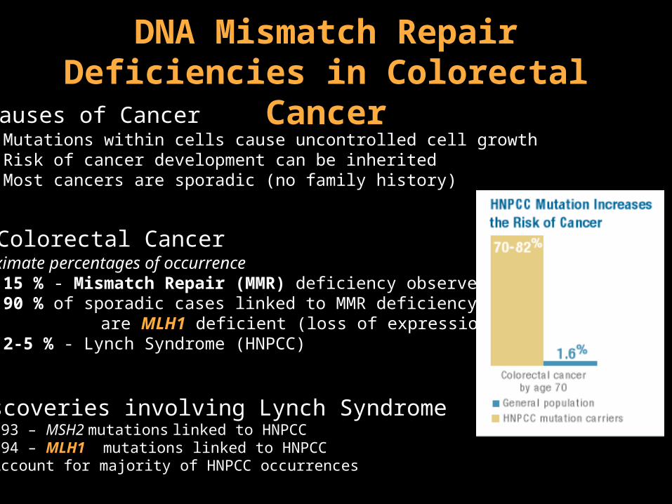
DNA Mismatch Repair Deficiencies in Colorectal Cancer
• Causes of Cancer• Mutations within cells cause uncontrolled cell growth• Risk of cancer development can be inherited • Most cancers are sporadic (no family history)
• Colorectal CancerApproximate percentages of occurrence
15 % - Mismatch Repair (MMR) deficiency observed 90 % of sporadic cases linked to MMR deficiency
are MLH1 deficient (loss of expression) 2-5 % - Lynch Syndrome (HNPCC)
•Discoveries involving Lynch Syndrome1993 – MSH2 mutations linked to HNPCC1994 – MLH1 mutations linked to HNPCC*Account for majority of HNPCC occurrences
Page 4
• DNA mismatches arise from errors during DNA Replication
• MMR corrects replication errors
• MMR Stimulates apoptosis in response to DNA damage
• Basic Mechanism:
• Mismatch recognition
•MutS familyMSH2/MSH6MSH2/MSH3
G
T
* MLH1/MLH3
• Strand choice•MutL family
MLH1/PMS2MLH1/PMS1
• Excision• Exonucleases
• PCNA• RPA
T
• Resynthesis•Replicative
DNA polymerase
A
T
Mechanism & Functions of DNA MMR
G
T
ATP Dependant
Page 5
DNA Synthesis Error Mutation
Mutations Prevented by MMR
Successful Repair
Dinucleotide Loop Insertion via slip-mispairing
ACTG
No Repair, Additional Replication
Insertion Mutation
Insertion / Deletion Loops Microsatellite Instability (MSI)
GT
No Repair, Additional Replication
AT
AT
GC
Incorrect insertion of base
Successful Repair
Base Mismatches Base Substitution Mutations
Page 6
Implications of MMR Deficiency for Cancer Screening & Treatment• Chemotherapy
• Microsatellite Instability - An Effective Screening Tool
• Clinical Relevance of MLH1: HNPCC cases without MSI?
36, 694 - 699 (2004)
D132HMLH1 amino acid site 132 changed from D (Aspartic Acid) to H (Histidine)
Loss of repeats
Normal
Tumor
Page 7
Hypothesis:
Initial DataData from recent publications
• D132H apparently associated with 5-fold increased cancer risk
• Modest decrease in ATPase function in D132H
• Increased mutation rate not dramatic enough for MSI detection
• Base substitutions more affected than microsatellites
• Apoptosis signaling function more affected than error correction
Attenuated MLH1 function of D132H increases cancer risk
Page 8
Is there an observable phenotype associated with MLH1-D132H?
Central Question
Research Goals
2. Determine in vitro repair capabilities for MLH1 mutant D132H using biochemical assays
1. Use Cellular assays to evaluate the effect of the MLH1 mutation D132H in vivo
Page 9
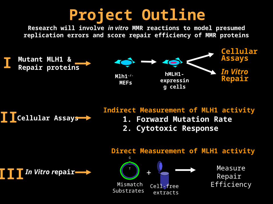
Project Outline
Mutant MLH1 & Repair proteinsІ
ІІ
ІІІ
Cellular Assays
In Vitro repair
Research will involve in vitro MMR reactions to model presumed replication errors and score repair efficiency of MMR proteins
hMLH1-expressing
cells
Mlh1-/- MEFs
Cellular Assays
Measure Repair
Efficiency
G
T
+Mismatch
Substrates
In Vitro Repair
1. Forward Mutation Rate2. Cytotoxic Response
Indirect Measurement of MLH1 activity
Direct Measurement of MLH1 activity
Cell-free extracts
Page 10
Identification of Cell Lines Expressing MLH1 Mutants
Western Blot Analysis of Extract Preparation
Cell-free extracts
Transfection
hMLH1
Neo-R
MLH1
Drug Resistance Mlh1-/- MEFs Drug Selection
D132H
-8
D132H
-9
MLH1
MLH1-
13 (+
)
MLH1-
2 (+
)
MC2A
(-)
PMS2
• Screen for MLH1 Expression with Western Blotting• Isolate and Expand Expressing Cell Lines for extract• 2 D132H Lines identified.
•Expression is less than MLH1 wildtype lines.
Page 11
Fluctuation Analysis: Forward Mutation to OuabainR
Cell Line Events/Cell/Generation (Rate)
MLH1 (-/-) 60 x 10-7
+ WT hMLH1* ~ 1 x 10⁻⁷
+ Hmlh1- D132H 0.7 ± 0.2 x 10⁻⁷ **
* - ** -
12 Cultures(1000 OuabainS cells)
Expansion, Accumulation of Mutants
Exposure to Ouabain Count number of Ouabain
Resistant Clones, Calculate Rate of mutation
* Rates in MEF cell line determined by Dr. Andrew Buermeyer, 1999. ** Assay Repeated Twice
~5 x 10⁶ cells, includes some OuabainR cells
Conclusion: Expression of D132H decreased rate of base substitution
Page 12
Response to Cytotoxic Agents: 6-Thioguanine Response
300-3000 Cells
24 Hours
6-Thioguanine 0-6 uM Doses
24 Hours
Remove 6-Thioguanine
Count Surviving Colonies
6-10 Days
Conclusion: Expression of D132H increased cytotoxic response to 6-Thioguanine
Page 13
In Vitro Mismatch Repair Assay
CT T GAG
GA G CTC
Mismatch substrateincubated with repair factors from extracts
- Mismatch Blocks activityof Restriction Endonuclease
- 3’ Nick initiates repair, facilitatesStrand choice
nick
Mismatch dependantnick directed excision
Xho1CT C GAGGA G CTC
Pvu1
Resynthesis leadsto restoration of Xho1 site Pvu1 Site used to facilitateanalysis
Page 14
Substrate preparation protocol developed in the Hay’s Laboratory, OSUGels 1% TAE 8 cm, 170V, 30’ w/Stain & w/Destain (10’,30’)
Preparation of Mismatch Substrates
Xho1CT T GAG
GA G CTC
nick
A – Closed Circular Substrate
B – Double Digest
ConclusionsSubstrate Preparation yields >95% Mismatch SubstrateSuccessful Preparation for G/T and CT Loop mismatches
Starting Plasmid
A B A B A B
G/T Mismatch
-CT- Loop
Linear (Pvu1 Cut)
Xho1 & Pvu1 CutPvu1
A
Page 15
Results & Discussion
I. Expression of D132H in MLH1 deficient cells:
1) Reduced mutation rate similar to wildtype expressing cells, suggesting good repair activity in vivo
2) Increased cytotoxic response to 6-Thioguanine with a modest decrease in response relative to wildtype expressing
cells -Protein Expression?
II. In Vitro Repair
1) Substrates Prepared, Assays in Progress
Future Work
Repair Assays Additional D132H expressing lines for cellular assays
Page 16
Acknowledgments
• Dr. Andrew Buermeyer• Buermeyer Lab Group
Xin Huo• Hays Lab Group
Pete HoffmanHuixian Wang
• Howard Hughes Medical Institute• Dr. Kevin Ahern