The pathology of human Lassa fever W. C. WINN, JR 1 & D. H. WALKER 2 Pathological findings have been described in only a small number of cases of Lassa fever since the virus was first isolated in 1969. Morphologically, eosinophilic necrosis of hepatocytes was the most frequent finding and focal necroses, often extensive, were present in most cases. These findings are similar to the lesions previously described in Argentinian and Bolivian haemorrhagic fever. Focal interstitial pneumonitis, focal tubular necrosis in the kidney, lymphocytic infiltration of the splenic veins, and partial replacement of the splenic follicles by amorphous eosinophilic material have been described, but the significance of these findings is unclear. More detailed and sophisticated investigations are required in the future ifpathogenetic mechanisms are to be unravelled. INTRODUCTION In the 6 years since the initial description of Lassa fever, outbreaks have been documented in several West African countries, and the theoretical hazard of importation into other continents has been real- ized. Information about the pathology and patho- genesis of the disease remains fragmentary, however. Descriptions have been given of post-mortem exami- nations, including gross and microscopic pathology, performed on 2 cases from the initial outbreak in Jos, Nigeria (1), 2 cases from the 1970 outbreak in Jos (2), and 3 cases from the 1972 epidemic in Liberia (3). The description of the Liberian cases was limited to the hepatic pathology. Ultrastructural findings in a case from the 1972 Sierra Leone epidemic were also limited to the liver (4). Despite the paucity of autopsied cases, the existing reports do represent a well documented sample. Lassa virus was isolated from the Sierra Leone case and in all 4 of the Nigerian autopsies. Of the 4 cases considered by Sarrat et al. (3), one was confirmed as Lassa fever by isolation of the virus and 2 were presumed to be Lassa fever, on the basis of clinical, epidemiological, and pathological findings. A fourth case from the outbreak was excluded by the authors, because the autopsy showed pathological evidence of malaria. The potential contribution of coincident disease to the pathological lesions must be faced constantly, even in virologically documented cases of Lassa fever. 1 Assistant Professor of Pathology, University of Virgi- nia Medical School, Charlottesville, VA 22901, USA. 2 Assistant Professor of Pathology, University of North Carolina Medical School, Chapel Hill, NC 27514, USA. We have reviewed the published cases and ob- tained some of the microscopic material through the courtesy of Drs Edington and Robin. In addition, Dr Jacinto Gochoco, Jr, of York Hospital, York, Pennsylvania, has made available to us material from a case of laboratory-associated infection. The pathological findings on this last case have not been reported previously; Lassa virus was isolated from blood antemortem. GROSS PATHOLOGICAL FINDINGS As in many other viral diseases, the gross patho- logy in Lassa fever is unimpressive compared to the dramatic clinical course and mortality rate. Constant but nonspecific findings include congestion of the viscera, oedema of the soft tissue, and petechiae, especially in the gastrointestinal tract. Pleural effu- sions have been present in most cases, and ascites has been noted clinically. Luminal blood in the small intestine was described in one case; massive bleeding sufficient to produce clinical shock does not appear to have been present. The kidneys of two cases were described as swollen or haemorrhagic and nodular, but microscopic evidence of tubular necrosis has been minimal. Neuropathological findings are available only on the case from York, Pennsylvania. The brain was normal in weight. The meningeal blood vessels were congested. Oedema of the true and false vocal cords was also noted; the terminal events in this case began with acute respiratory distress and probable laryngospasm, for which a tracheotomy was per- formed. 3381 _ 535 BULL. WORLD HEALTH ORGAN., Vol. 52, 1975
Transcript
The pathology of human Lassa feverW. C. WINN, JR 1 & D. H. WALKER 2
Pathologicalfindings have been described in only a small number of cases ofLassa feversince the virus was first isolated in 1969. Morphologically, eosinophilic necrosis ofhepatocytes was the most frequent finding andfocal necroses, often extensive, were presentin most cases. These findings are similar to the lesions previously described in Argentinianand Bolivian haemorrhagic fever. Focal interstitialpneumonitis, focal tubular necrosis in thekidney, lymphocytic infiltration of the splenic veins, and partial replacement of the splenicfollicles by amorphous eosinophilic material have been described, but the significance ofthese findings is unclear. More detailed and sophisticated investigations are required in thefuture ifpathogenetic mechanisms are to be unravelled.
INTRODUCTION
In the 6 years since the initial description of Lassafever, outbreaks have been documented in severalWest African countries, and the theoretical hazardof importation into other continents has been real-ized. Information about the pathology and patho-genesis of the disease remains fragmentary, however.Descriptions have been given of post-mortem exami-nations, including gross and microscopic pathology,performed on 2 cases from the initial outbreak inJos, Nigeria (1), 2 cases from the 1970 outbreak inJos (2), and 3 cases from the 1972 epidemic inLiberia (3). The description of the Liberian cases waslimited to the hepatic pathology. Ultrastructuralfindings in a case from the 1972 Sierra Leoneepidemic were also limited to the liver (4).
Despite the paucity of autopsied cases, the existingreports do represent a well documented sample.Lassa virus was isolated from the Sierra Leone caseand in all 4 of the Nigerian autopsies. Of the 4 casesconsidered by Sarrat et al. (3), one was confirmed asLassa fever by isolation of the virus and 2 werepresumed to be Lassa fever, on the basis of clinical,epidemiological, and pathological findings. A fourthcase from the outbreak was excluded by the authors,because the autopsy showed pathological evidence ofmalaria. The potential contribution of coincidentdisease to the pathological lesions must be facedconstantly, even in virologically documented cases ofLassa fever.
1 Assistant Professor of Pathology, University of Virgi-nia Medical School, Charlottesville, VA 22901, USA.
2 Assistant Professor of Pathology, University of NorthCarolina Medical School, Chapel Hill, NC 27514, USA.
We have reviewed the published cases and ob-tained some of the microscopic material through thecourtesy of Drs Edington and Robin. In addition,Dr Jacinto Gochoco, Jr, of York Hospital, York,Pennsylvania, has made available to us materialfrom a case of laboratory-associated infection. Thepathological findings on this last case have not beenreported previously; Lassa virus was isolated fromblood antemortem.
GROSS PATHOLOGICAL FINDINGS
As in many other viral diseases, the gross patho-logy in Lassa fever is unimpressive compared to thedramatic clinical course and mortality rate. Constantbut nonspecific findings include congestion of theviscera, oedema of the soft tissue, and petechiae,especially in the gastrointestinal tract. Pleural effu-sions have been present in most cases, and asciteshas been noted clinically. Luminal blood in the smallintestine was described in one case; massive bleedingsufficient to produce clinical shock does not appearto have been present. The kidneys of two cases weredescribed as swollen or haemorrhagic and nodular,but microscopic evidence of tubular necrosis hasbeen minimal.
Neuropathological findings are available only onthe case from York, Pennsylvania. The brain wasnormal in weight. The meningeal blood vessels werecongested. Oedema of the true and false vocal cordswas also noted; the terminal events in this casebegan with acute respiratory distress and probablelaryngospasm, for which a tracheotomy was per-formed.
3381 _ 535 BULL. WORLD HEALTH ORGAN., Vol. 52, 1975
W. C. WINN, JR & D. H. WALKER
MICROSCOPIC PATHOLOGY
Complete microscopic descriptions are availablefor 4 of the autopsies; the hepatic pathology hasbeen studied in an additional 4 cases.
Heart. Congestion and slight interstitial oedemawere present and in one instance slight nonspecificepicardial infiltrates were noted. No microscopicevidence of myocarditis has been described.
Lungs. Congestion and oedema without significanthaemorrhage were present. Edington (2) describedinterstitial pneumonitis with mononuclear cells andmegakaryocytes in 2 cases. In the York case therewas focal interstitial accumulation of mononuclearcells; many of these were in capillaries, however, anda classical interstitial pneumonia was not present.
Kidneys. Occasional focal tubular and glomerularnecroses were described by Edington et al. (2) in onecase. Three cases showed congestion, autolysis, andoccasional hyaline or pigment casts. Typicalischaemic nephrosis has not been described.
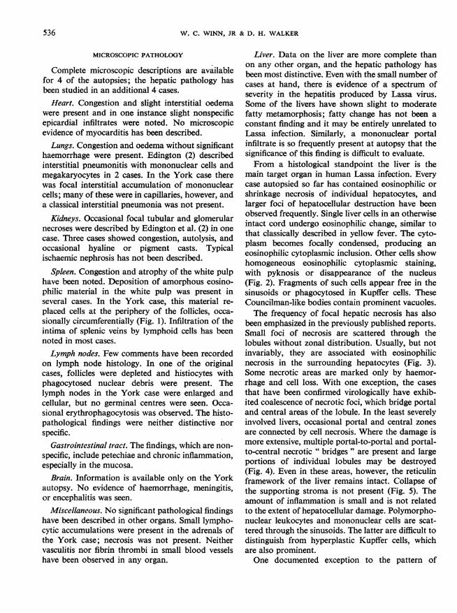
Spleen. Congestion and atrophy of the white pulphave been noted. Deposition of amorphous eosino-philic material in the white pulp was present inseveral cases. In the York case, this material re-placed cells at the periphery of the follicles, occa-sionally circumferentially (Fig. 1). Infiltration of theintima of splenic veins by lymphoid cells has beennoted in most cases.Lymph nodes. Few comments have been recorded
on lymph node histology. In one of the originalcases, follicles were depleted and histiocytes withphagocytosed nuclear debris were present. Thelymph nodes in the York case were enlarged andcellular, but no germinal centres were seen. Occa-sional erythrophagocytosis was observed. The histo-pathological findings were neither distinctive norspecific.
Gastrointestinal tract. The findings, which are non-specific, include petechiae and chronic inflammation,especially in the mucosa.
Brain. Information is available only on the Yorkautopsy. No evidence of haemorrhage, meningitis,or encephalitis was seen.
Miscellaneous. No significant pathological findingshave been described in other organs. Small lympho-cytic accumulations were present in the adrenals ofthe York case; necrosis was not present. Neithervasculitis nor fibrin thrombi in small blood vesselshave been observed in any organ.
Liver. Data on the liver are more complete thanon any other organ, and the hepatic pathology hasbeen most distinctive. Even with the small number ofcases at hand, there is evidence of a spectrum ofseverity in the hepatitis produced by Lassa virus.Some of the livers have shown slight to moderatefatty metamorphosis; fatty change has not been aconstant finding and it may be entirely unrelated toLassa infection. Similarly, a mononuclear portalinfiltrate is so frequently present at autopsy that thesignificance of this finding is difficult to evaluate.From a histological standpoint the liver is the
main target organ in human Lassa infection. Everycase autopsied so far has contained eosinophilic orshrinkage necrosis of individual hepatocytes, andlarger foci of hepatocellular destruction have beenobserved frequently. Single liver cells in an otherwiseintact cord undergo eosinophilic change, similar tothat classically described in yellow fever. The cyto-plasm becomes focally condensed, producing aneosinophilic cytoplasmic inclusion. Other cells showhomogeneous eosinophilic cytoplasmic staining,with pyknosis or disappearance of the nucleus(Fig. 2). Fragments of such cells appear free in thesinusoids or phagocytosed in Kupffer cells. TheseCouncilman-like bodies contain prominent vacuoles.The frequency of focal hepatic necrosis has also
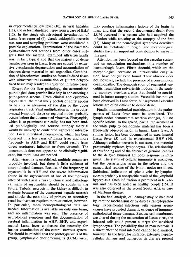
been emphasized in the previously published reports.Small foci of necrosis are scattered through thelobules without zonal distribution. Usually, but notinvariably, they are associated with eosinophilicnecrosis in the surrounding hepatocytes (Fig. 3).Some necrotic areas are marked only by haemor-rhage and cell loss. With one exception, the casesthat have been confirmed virologically have exhib-ited coalescence of necrotic foci, which bridge portaland central areas of the lobule. In the least severelyinvolved livers, occasional portal and central zonesare connected by cell necrosis. Where the damage ismore extensive, multiple portal-to-portal and portal-to-central necrotic " bridges" are present and largeportions of individual lobules may be destroyed(Fig. 4). Even in these areas, however, the reticulinframework of the liver remains intact. Collapse ofthe supporting stroma is not present (Fig. 5). Theamount of inflammation is small and is not relatedto the extent of hepatocellular damage. Polymorpho-nuclear leukocytes and mononuclear cells are scat-tered through the sinusoids. The latter are difficult todistinguish from hyperplastic Kupffer cells, whichare also prominent.One documented exception to the pattern of
536
Fig. 1. Circumferential deposition of amorphous Fig. 2. Councilman-like bodies in Lassa fever. Round-eosinophilic material in the white pulp of the ed, dense, eosinophilic cells are located in the hepaticspleen. This material did not stain with amyloid cords and sinusoids. Focal eosinophilic change instains. H & E. the cytoplasm of hepatocytes is also present. H & E.
Fig. 3. Focal hepatic necrosis associated with Fig. 4. Coalescence of hepatic necrosis to involveeosinophilia of the surrounding hepatocytes and most of the lobule. Surviving hepatocytes showedCouncilman-like bodies. There is no zonal distribu- eosinophilic change, but Councilman-like bodies aretion to the necrosis. H & E. not evident. H & E.
Fig. 5. Intact reticulin framework of the liver in an Fig. 6. Single vacuolated hepatocyte undergoing acido-area of extensive necrosis. Same field as Fig. 4 in an philic necrosis. Mononuclear sinusoidal infiltrate isadjacent section. Wilder's reticulin stain. concentrated around this cell. H & E.
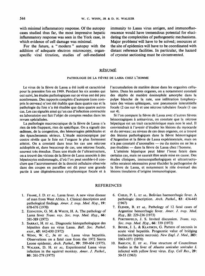
Fig. 7. Minimal hepatic damage in Lassa fever. Varia- Fig. 8. Focal hepatic necrosis. Surrounding hepato-tion in size of nuclei, binucleated hepatocytes, and cytes show vacuolation and increased cytoplasmicmitotic figures are present. H & E. density. They do not represent classical Councilman-
like bodies, but do resemble the eosinophilic cells seenin paraffin sections. Toluidine blue.
Fig. 9. Degenerating hepatocyte with dilated rough endoplasmic reticulum and flocculent densities within mito-chondria. Numerous Lassa virions are present along the cell membrane. Lead citrate-uranyl acetate.
Fig. 10. Rounded portion of an hepatocyte showing cytoplasmic degeneration. This cell probably represents one ofthe Councilman-like bodies observed in paraffin sections. Lead citrate-uranyl acetate.
U.! p,m
Fig. 11. Numerous typical Lassa virions are present in the extracellular space between the microvilli of an hepatocyte.Lead citrate-uranyl acetate.
PATHOLOGY OF HUMAN LASSA FEVER
necrosis is represented by the York autopsy. In thiscase, rare acidophilic change was observed, involv-ing single, scattered hepatocytes. Clear vacuoles,consistent with the presence of lipid, were present inthese cells also. A moderate mononuclear infiltratewas present throughout the sinusoids; in one focus,the infiltrate was accentuated around an eosinophilichepatocyte (Fig. 6). Multicellular foci of necrosiswere not observed. In contrast to the other cases,binucleated liver cells and numerous mitotic figureswere observed (Fig. 7). These findings suggest hepaticinjury and attempts at regeneration.A case of intermediate severity is included in those
reported by Sarrat et al. (3). The liver showedextensive acidophilic change without concentrationinto necrotic foci. Again there was no zonal localiza-tion. Although the case was not confirmed virologi-cally, it occurred as part of a documented Lassafever epidemic and histologically it falls into themiddle of a spectrum of damage observed in provedcases.
Ultrastructural studies of Lassa fever have beenhampered by lack of facilities for electron micro-scopy in the epidemic areas and inadequate fixationwhen attempts have been made to evaluate thespecimens in reference laboratories (2). An excel-lently preserved liver biopsy was obtained from onepatient in the Sierra Leone epidemic (4). Thicksections for light microscopy revealed foci of necro-sis, similar to those previously described, except thatclassical Councilman bodies were rarely observed(Fig. 8).Damaged cells were vacuolated and cell bounda-
ries were often blurred. Portions of a few dense cells,having the appearance of Councilman bodies, werepresent in Kupffer cells. The majority of affectedhepatocytes, however, showed some increased den-sity, but did not demonstrate the remarkable con-densation of classical acidophilic necrosis. Thesefindings were confirmed ultrastructurally. Dilatationof the endoplasmic reticulum and accumulation offlocculent electron-dense material in swollen mito-chondria were the most common findings (Fig. 9).Areas of focal cytoplasmic degeneration were fre-quently found. Rounded cells or portions of cells,showing the increased electron density associatedwith focal cellular degeneration, were also observed(Fig. 10), but the classical cytoplasmic condensationof acidophilic necrosis was observed only in a fewphagocytosed cell fragments. As had been observedwith the light microscope, undamaged hepatocyteswere often adjacent to severely damaged cells.
The most instructive finding in the ultrastructuralstudy was the demonstration of large numbers oftypical arenavirus particles. Although Lassa virushad been isolated antemortem, the demonstration ofvirions in relation to the pathological lesion con-firmed the specificity of the lesions that had beenobserved in the light microscope. Virions were al-ways found in association with hepatocytes ratherthan with Kupffer cells (Fig. 11). Interestingly, theywere more frequent in areas where cell damage wasless severe. Because Lassa virus matures by buddingfrom cell membranes, intact host cells are requiredfor production of viral particles. No ribosomalaggregates, suggestive of arenavirus inclusions, wereseen.
DISCUSSION
In a systemic viral infection, such as Lassa fever,classical histopathology often contributes to theinitial understanding of the disease, but definition ofpathogenetic mechanisms requires the addition ofcomplementary techniques. Although the number ofcases for consideration is small, they fortunatelyrepresent a select and well documented group. Theuniformity of the findings reinforces the impressionthat we are dealing with a minimum of backgroundnoise from coincident diseases. A review of thecurrently available pathological data demonstrates amajor site of tissue damage in the liver and outlinesseveral other areas that deserve further study.The similarities between the pathology of Lassa
fever and that of Bolivian and Argentinian haemor-rhagic fever (BHF and AHF) have been noted byother authors. The pathogenesis of these infectionsin man is not known, but research in each may beapplicable to the others. Comparison of the reportedpathology in the South American haemorrhagicfevers with the data on Lassa fever and with theinformation that Walker et al. (5) have obtained insubhuman primates infected with Lassa virus pro-vides interesting parallels (Table 1). Child et al. (6)described the autopsies of 8 patients with BHF. Thelivers of all 8 cases showed Kupffer cell hyperplasiawith erythrophagocytosis and acidophilic necrosis ofhepatocytes. In addition, two cases showed foci ofnecrosis, similar to the hepatic necrosis observed inthe Lassa cases. Similarly, the description of AHFby Elsner et al. (7) includes Kupffer cell hyperplasia,acidophilic Councilman-like bodies, and, in 5 out of12 cases, the presence of focal, non-zonal necrosis.The liver is undoubtedly a major target organ for
Lassa virus and the pattern of hepatic necrosis
9
541
W. C. WINN, JR & D. H. WALKER
Table 1. Histopathologic findings in human arenavirus infections
No. of times lesions observed/No. ofcases examined
Organ Lesion .LassasinBolivian Argentinefassa haemorrhagic haemorrhagicfever fever (6) fever 17)
Heart Myocarditis 0/4 not described 4/12
Lung Interstitial pneumonitis or 2/4 6/6 4/12hyaline membrane formation
Kidney Tubular necrosis 1/4 2/8 6/12
Spleen Deposition of eosinophilic 3/4 0/8 0/12material in white pulp
Brain Lymphocytic infiltrate or 0/1 6/6 5/12microglial proliferation
Multicellular foci of 6/8 2/7 5/12hepatic necrosis
produced appears to be characteristic of humanarenavirus Infections. Nevertheless, other infectionsand toxins may produce a similar type of hepaticnecrosis; the differential diagnosis has been discus-sed by Sarrat et al. (3), and an excellent set ofslides demonstrating the histopathological differen-tiation from yellow fever is available from theArmed Forces Institute of Pathology, Washington,D.C. (set No. L-15274). The non-zonal distributionof the necrosis differentiates Lassa virus hepatitisfrom the classical yellow fever lesion. We do wonder,however, as Porterfield (8) did, whether cases ofLassa fever occurring before the characterizationof the virus may have been mistaken for yellowfever. The vacuolar character of the acidophilicnecrosis mimics that of yellow fever and the lobulardistribution may be difficult to evaluate when necro-sis is extensive. Although acidophilic bodies havebeen demonstrated in viral hepatitis, the most fre-quent form of hepatocellular damage is " balloon-ing" degeneration, which is not seen in Lassa virushepatitis. The integrity of the hepatic reticulin frame-work is another potentially important difference inthe hepatic damage seen in viral hepatitis and Lassavirus hepatitis. " Bridging" eosinophilic necrosiswas seen in the more severely damaged livers ofLassa fever patients. This pattern of hepatic necrosisin viral hepatitis has been reported by Boyer &Klatskin (9) to correlate with poor prognosis and thedevelopment of chronic liver disease. In viral hepati-tis, this " bridging" necrosis is associated with col-
lapse of the reticulin framework of the liver, but inLassa fever, as in yellow fever, the reticulin remainsintact even in severely damaged lobules. One wouldpredict that the subsequent development of cirrhosisin Lassa fever would be rare, as it is in yellow fever.
Other clinical differential diagnoses, such as mala-ria and typhoid fever, are not a major problem in thepathological diagnosis. However, an additionaldifferential problem in African patients has beenintroduced by a recent case of haemorrhagic feverwith hepatic necrosis caused by Marburg virus. Theextensive eosinophilic necrosis of hepatocytes andnon-zonal distribution of the lesions suggested adiagnosis of Lassa fever before the isolation of thevirus established the diagnosis.
In several of the autopsied cases of Lassa fever,hepatic necrosis has been sufficiently extensive tosuggest hepatic failure as a major contributor to thefatal outcome. In the available cases, however, thereis no correlation of the extent or severity of hepaticdamage with the duration of disease. Extensivenecrosis is not related either to overwhelming, rapid-ly fatal infection or to prolonged infection, in whichmore extensive tissue damage might be seen histo-logically. This lack of correlation is emphasized bythe virtual absence of necrosis in the York autopsyand in the squirrel monkeys experimentally infectedwith Lassa virus.The nature of the acidophilic necrosis found in
Lassa fever requires further investigation. The ultra-structure of acidophilic necrosis has been described
542
PATHOLOGY OF HUMAN LASSA FEVER
in experimental yellow fever (10), in viral hepatitis(11), and in formalin-fixed tissue from a case ofBHF(12). In the single ultrastructural investigation ofLassa fever reported (4), classic acidophilic necrosiswas rare and sampling problems were considered apossible explanation. Examination of the haemato-xylin-eosin-stained sections from other cases sug-gests that the material examined ultrastructurallywas, in fact, typical and that the majority of densehepatocytes seen in Lassa fever are caused by exten-sive cytoplasmic degeneration and accumulation ofelectron-dense material in cell organelles. Combina-tion of histochemical studies on formalin-fixed tissuewith ultrastructural examination of glutaraldehyde-fixed tissue may resolve this question in future cases.
Except for the liver pathology, the accumulatedpathological data provide little help in constructing apathogenetic scheme. From clinical and epidemio-logical data, the most likely portals of entry appearto be cuts or abrasions of the skin or the upperrespiratory tract. Viral multiplication in the oropha-ryngeal lymphoid tissue or regional nodes probablyoccurs before the documented viraemia. Pharyngitis,which is so prominent clinically, has not been stud-ied pathologically and routine autopsy materialwould be unlikely to contribute significant informa-tion. Focal interstitial pneumonitis, which has beenobserved in a few cases of Lassa fever and morefrequently in AHF and BHF, could result fromdirect respiratory infection or from viraemia. Thepneumonitis does not appear to be clinically signifi-cant, but more data are needed.
After viraemia is established, multiple organs areprobably involved, but there is little evidence ofmorphological damage. Because of the frequency ofmyocarditis in AHF and the severe inflammationfound in the myocardium of one of the monkeysinfected with Lassa virus (5), clinical and pathologi-cal signs of myocarditis should be sought in thefuture. Tubular necrosis in the kidney is difficult toevaluate because of the concurrent hepatic necrosisand shock; the possibility of primary or secondaryrenal involvement requires more attention, however.In particular, more neuropathological data areneeded. Information is available on only one brain,and no inflammation was seen. The presence ofneurological symptons and the documentation ofcerebral inflammation in AHF, BHF, and experi-mental Lassa fever emphasize the necessity forfurther examination of the central nervous system.We should be mindful that the prototype virus of thegroup, lymphocytic choriomeningitis (LCM) virus,
may produce inflammatory lesions of the brain inman, and that the second documented death fromLCM occurred in a patient who had acquired theinfection while assisting at the autopsy of the first(13). Many of the neurological symptoms describedcould be metabolic in origin, and morphologicalstudies have an important contribution to make inthis area.
Attention has been focused on the vascular systemand on coagulation mechanisms in a number ofhaemorrhagic fevers. Fibrin thrombi, which are themorphological correlate of intravascular coagula-tion, have not yet been found. Their absence doesnot, however, exclude the presence of a consumptioncoagulopathy. The demonstration of segmental vas-culitis, resembling polyarteritis nodosa, in the squir-rel monkeys provides a clue that should be consid-ered in future autopsy studies. Vasculitis has not yetbeen observed in Lassa fever, but segmental vascularlesions are often difficult to demonstrate.
Finally, immunological participation in the patho-genesis of Lassa fever must be considered. Thelymph nodes demonstrate reactive changes, but nospecific lesions. In the spleen, partial replacement ofthe white pulp by eosinophilic material has been afrequently observed lesion in human Lassa fever. Asimilar lesion has been documented in experimentalLassa fever (5) and in experimental BHF (14).Although cellular necrosis is not seen, the materialpresumably replaces lymphocytes. The relationshipof this finding and of the absence of germinal centresto the delayed humoral antibody response is intri-guing. The status of cellular immunity is unknown,but the periarteriolar areas in the spleen and theparacortical regions of the lymph nodes are intact.Subintimal infiltration of splenic veins by lympho-cytes is probably a nonspecific result of the lymphoidhyperplasia. Such infiltration is prominent in leukae-mia and has been noted in healthy people (15). Itwas also observed in the recent South African caseof Marburg disease.
In the final analysis, cell damage may be mediatedby immune mechanisms or by direct viral cytopatho-logy. Experimental infections with various arena-viruses have provided dramatic evidence of immuno-pathological tissue damage. Because cell membranesare altered during the maturation of Lassa virus, theinfected cells could present a target for sensitizedlymphocytes. The possibility that in man necrosis isa direct effect of viral infection cannot be dismissed,however. In the liver, for instance, extensive hepato-cellular damage and numerous virions are present
543
544 W. C. WINN, JR & D. H. WALKER
with minimal inflammatory response. Of the autopsycases studied thus far, the most impressive hepaticinflammatory response was seen in the York case, inwhich evidence of cell damage was minimal.For the future, a " modem " autopsy with the
addition of adequate electron microscopy, organ-specific viral titration, studies of cell-mediated
immunity to Lassa virus antigen, and immunofluo-rescence would have tremendous potential for eluci-dating the complexities of pathogenetic mechanisms.Major problems will have to be solved; resources atthe site of epidemics will have to be coordinated withdistant reference facilities. In particular, the hazardof cryostat sectioning must be circumvented.
RtSUMt
PATHOLOGIE DE LA FIEVRE DE LASSA CHEZ L HOMME
Le virus de la fievre de Lassa a ete isole et caracterisepour la premiere fois en 1969. Pendant les six annees quiont suivi, les etudes pathologiques sur ce virus ont 6t6 peunombreuses. Des rapports complets d'autopsie (non com-pris le cerveau) n'ont ete etablis que dans quatre cas et lapathologie du foie n'a ete etudiee que dans quatre autrescas. Les cas signales ainsi qu'un cas d'infection contract6een laboratoire ont fait l'objet de comptes rendus dans lesrevues specialis6es.La pathologie macroscopique de la fievre de Lassa n'a
rien de tres frappant, rien de specifique. On a signale desced6mes, de la congestion, des he6morragies petechiales etdes epanchements serieux. L'etude microscopique parcontre revele que le foie est l'organe le plus fortementatteint. On a constat6 dans tous les cas une n6croseacidophile et, dans beaucoup de cas, une necrose focale,souvent tres etendue. Dans une etude de l'ultrastructure,on a trouve des virions de la fievre de Lassa associes a deshepatocytes endommages, d'ou l'on peut semble-t-il con-clure que l'accroissement de la densit6 cellulaire observeedans des coupes en paraffine est du pour une grandepartie a une degenerescence cytoplasmique focale et a
l'accumulation de matiere dense dans les organites cellu-laires. Dans les autres organes, on a notamment constatedes depots de matiere eosinophile amorphe dans lapulpe blanche de la rate, une infiltration lymphocy-taire des veines spleniques, une pneumonie interstitiellefocale (2 cas sur 4) et une necrose tubulaire focale (1 cassur 4).
Si l'on compare la fievre de Lassa avec d'autres fievreshemorragiques a arenavirus, on constate que la necrosehepatique est un trait caracteristique mais non le seul. I1conviendrait a l'avenir d'etudier les lesions du myocardeet du cerveau; au niveau de ces deux organes, on a trouvedes l6sions pathologiques dans la fievre hemorragiqued'Argentine et la fievre de Lassa experimentale, mais onn'a pas constate d'anomalies - ou du moins on ne les apas etudiees - dans la fievre de Lassa chez l'homme.
L'atteinte hepatique peut hater l'issue fatale danscertains cas, mais ne saurait etre seule mise en cause. Desetudes cliniques, immunopathologiques et ultrastructu-relles seraient n6cessaires pour elucider la pathogenese dela fievre de Lassa, et notamment le r6le eventuel desIesions tissulaires d'origine immunologique.
REFERENCES
1. FRAME, J. D. ET AL. Lassa fever. A new virus diseaseof man from West Africa. I. Clinical description andpathological findings. Amer. J. trop. Med. Hyg., 19:670-676 (1970)
2. EDINGTON, G. M. & WHITE, H. A. The pathology ofLassa fever Trans. roy. Soc. trop. Med. Hyg., 66:381-389 (1972)
3. SARRAT, H. ET AL. Diagnostic histopathologique deshepatites dues au virus Lassa. Bull. Soc. Pathol.exot., 65: 642-650 (1972)
4. WrNN, W. C., JR ET AL. Lassa virus hepatitis.Observations on a fatal case from the 1972 SierraLeone epidemic. Arch. Pathol., 99: 599-604 (1975).
5. WALKER, D. H. ET AL. Experimental Lassa virusinfection in the squirrel monkey. Amer. J. Pathol.,80: 261-278 (1975)
6. CHILD, P. L. ET AL. Bolivian haemorrhagic fever. Apathologic description. Arch. Pathol., 83: 434-445(1967)
7. ELSNER, B. ET AL. Pathology of 12 fatal cases ofArgentine hemorrhagic fever. Amer. J. trop. Med.Hyg., 22: 229-236 (1973)
8. PORTERFIELD, J. S. Invited discussion. Trans. roy.Soc. trop. Med. Hyg., 66: 339 (1972)
9. BOYER, J. L. & KLATSKIN, G. Pattern of necrosis inacute viral hepatitis. Prognostic value of bridging(subacute hepatic necrosis). New Engl. J. Med., 283:1063-1071 (1970)
10. BARUCH, E. ET AL. Fine structure of Councilmanbodies in the liver of Aluatta seniculus seniculus Linfected with yellow fever virus. Exp. Cell Res., 29:50-53 (1963)
PATHOLOGY OF HUMAN LASSA FEVER 545
11. BIAVA, C. & MUKHLOVA-MONTIEL, M. Electronmicroscopic observations on Councilman-like aci-dophilic bodies and other forms of acidophilicchanges in human liver cells. Amer. J. Pathol., 46:775-802 (1965)
12. CHILD, P. L. & Ruiz, A. Acidophilic bodies. Theirchemical and physical nature in patients with Boli-vian hemorrhagic fever. Arch. Pathol., 85: 45-50(1968)
13. SMADEL, J. E. ET AL. Lymphocytic choriomeningitis:two human fatalities following an unusual febrileillness. Proc. Soc. exp. Biol. Med., 49: 683-686 (1942)
14. TERRELL, T. G. ET AL. Pathology of Bolivian hemor-rhagic fever in the rhesus monkey. Amer. J. Pathol.,73: 477-494 (1973)
15. KLEMPERER, P. The Spleen. In: Downey, H., ed.Handbook of hematology, New York, Hoeber, 1938,vol. III, p. 1624
DISCUSSION
HOTCHIN: There seems to be a general tendency to regardLCM and perhaps other arenaviral diseases as beingmediated by cellular immunity. I have seen two patientstreated with LCM virus for terminal cancer, both of themanergic individuals. They developed progressively higherviraemia, were unable to mount an antibody response,and both died. The pathological findings were reminiscentof those you have just described, including pneumonitis.Moreover, acute LCM infection of mice produces bothhepatitis and ascitic fluid. If you take the ascitic fluid andinject it into normal mice, it kills them in about 30seconds. Preliminary information suggests that there is asubstance like bradykinin in this fluid.- The possibilityexists that there are toxic or pharmacological mediatorsthat play a role in these diseases.
WINN: I agree with your comment about cellular immu-nity. We simply do not have the data to determine thecause of cell damage. The possibility of direct viraldamage cannot be eliminated. The parallels betweenLassa fever and the few fatal cases of LCM disease areinteresting, but we have no information to offer onpossible chemical mediators.
COLES: a I am a little puzzled by your statement that theliver is the major target organ in Lassa fever, since Icannot recall a single patient seen in Sierra Leone whohas been jaundiced.
WINN: Your comment reinforces our belief that moreneuropathological data are badly needed. Many of theclinical symptoms might be metabolic in origin and future
aNixon Memorial Hospital, Segbwema, Sierra Leone.
autopsy studies will help to determine if there is menin-gitis. The liver is the main target organ from the histo-pathological standpoint. There may well be other organsthat are severely damaged functionally but show minimalhistological changes.
RUSSELL: The findings of peritoneal and pleural effusionsin Lassa fever, with an apparent increase in vascularpermeability in the absence of histopathological changesin the vessels, appear to be similar to those in denguehaemorrhagic fever. In this disease the increased vascularpermeability is due to activation of the complementsystem with resultant production of C3a and C5a anaphy-lotoxins, which cause histamine release. An early clue tothis mechanism was the observation that mast cellsappeared degranulated in autopsy and biopsy specimens.Have mast cells been carefully examined in autopsymaterial from Lassa fever and do they appear degranu-lated? There are also similarities in liver pathology.Patchy paracentral hepatic necrosis is seen in denguehaemorrhagic fever. The two diseases might be difficult todifferentiate in the absence of virological studies.
WINN: To my knowledge mast cells have not been lookedat specifically and most of our material has been selectedby haematoxylin-eosin stained sections. There are a fewparaffin blocks available and we shall look at the mastcells in those tissues. The pathological findings in Lassafever are quite similar to those described in the group Barbovirus haemorrhagic fevers, including dengue andKyasanur Forest disease. I have little experience withthose diseases, but it is my impression that the hepaticnecrosis is much less extensive than that seen in Lassafever.