12
Systemic Pathology I VPM 221 VPM 221 PATHOLOGY PATHOLOGY OF THE ALIMENTARY SYSTEM ALIMENTARY SYSTEM LAB 2 Enrique Aburto Nov 2008

Systemic Pathology IVPM 221VPM 221
PATHOLOGYPATHOLOGY OF THE
ALIMENTARY SYSTEMALIMENTARY SYSTEM
LAB 2
Enrique Aburto Nov 2008

Case 1Signalment: Male, 6 month-old, Irish Setter dog History: Presented for chronic regurgitation of solid meals, progressive emaciation, dyspnea and coughing.
– Persistent right 4th aortic arch and megaesophagus
Diagnosis?
What causes respiratory signs in this condition?– Aspiration of food during regurgitation (aspiration pneumonia)

Case 2History: Rumen from one of two steers that died after being sick for two days. Owner noticed them to be dopey and treated them with antibiotics. Diet was pasture, hay, french fries and lately, grains. At necropsy, steers were in good body condition, but they were bloated and dehydrated.
Diagnosis?
– Rumen: Multifocal hemorrhagic infarcts
These changes are commonly observed in which condition?– Mycotic rumenitis

Histo: Fungal hyphae mixed with abundant inflammatory debris and cells, abomasum (PAS stain).abomasum (PAS stain).
What fungal agents are usually involved?
– Aspergillus, Rhizopus, Mucor, and Absidia.
Predisposing factors for this condition?Predisposing factors for this condition?
– Grain overload, prolonged antibiotic treatment

Case 3Signalment: Male, 5 year-old, male, Great Dane. History: Dog was ok, last night just after meal. Following morning the animal was found death. At necropsy, the abdomen was markedly distended. The carcass showed moderate autolysis and good body condition..
Th h i
Any abnormalities?
The stomach is markedly dilated (filled with fluid and gas) and the serosa is g )congested. The spleen is engorged, displaced to the right,
d V h d
Diagnosis?
and V-shaped
– Gastric dilation and volvulus

How can you differentiate between postmortem gastric dilation and GDV?
1. Evidence of rotation and i f h dcompression of esophagus and
duodenum.2. Congestion of the gastric wall3. V-shaped bending and enlargement
of the spleen
What causes acute death in affected dogs?affected dogs?
Compression of posterior vena cava ( h k) d l(shock) and lungs.

Case 4Signalment: 3 month-old, female, Mastiff. History: Presented due to severe acute abdominal pain and vomition. The animal died despite of treatment. At necropsy, the animal had good body condition and the abdomen was markedly distended.
Which could be cause of this change? Diagnosis?
Any intestinal obstruction (foreign b di di l i i
Small intestine: Intussusception bodies, displacements, stenosis, atresia, paralytic ileus)

Case 5Signalment: 18 month-old, female, bovine. History: Presented due poor body condition and diarrhea. At necropsy, the most remarkable findings are:
Morphologic diagnosis?
S ltif l l ti t titiSevere, multifocal ulcerative stomatitis and esophagitis.

Can you identify any changes in the ileum?
Peyer’s patches and overlying epithelium are necrotic and covered by bloodPeyer s patches and overlying epithelium are necrotic and covered by blood.
Mention 3 differential diagnosis
BVD, MCF, Rinderpest

Case 6Signalment: 4 year old, Holstein cowHistory: Presented for dysentery, progressive weight loss, fever and abortion. At necropsy, the liver showed multiple 3-5 mm, well demarcated, white spotsdisseminated throughout the parenchyma.
Can you identify any changes in the ileum?
Diffuse hyperemia and hemorrhage of the mucosa. Peyer’s patches are more apparent than normal. The overlying epithelium is eroded.

Differential diagnoses?
Salmonella typhimurium was islated from the small intestineliver, uterus and lungs in this case
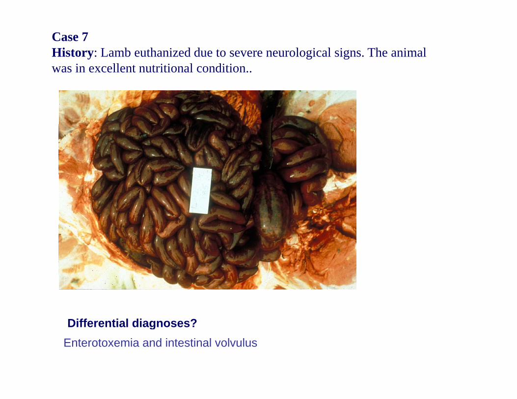
Case 7History: Lamb euthanized due to severe neurological signs. The animal was in excellent nutritional condition..
Differential diagnoses?Differential diagnoses?Enterotoxemia and intestinal volvulus


