36
Pathomechanism of fever. Fever of unknown origin. Fever and rash Dr. Szathmári Miklós Semmelweis University 1st Department of Medicine 11 April 2014.

Pathomechanism of fever. Fever
of unknown origin. Fever and rash
Dr. Szathmári Miklós
Semmelweis University
1st Department of Medicine
11 April 2014.
Regulation of body temperature
Controlled by the hypothalamus
Signals are received
from peripheral nerves (transmit information from warmth/cold
receptors)
from the temperature of the blood bathing the region
In a neutral temperature environment, the metabolic rate
of human produces more heat than necessary to
maintain the core temperature at 37C
Thermoregulatory center balances the heat production
derived from metabolic activity in muscle and the liver
with heat dissipation from the skin, and lungs
Harrison’s 17h Edition: Principles of Internal Medicine. McGraw Hill
Alterations in body temperature
Normal temperature:
Oral 36,8°C ± 0,5°C
Rectal 0.4°C higher than oral readings
Tympanic membrane (unadjusted mode) 0.8 °C lower than rectal
Fever: an a.m. temperature of > 37.2 °C or a p.m. temperature of
> 37.7 °C
Hyperpyrexia: > 41,5°C
Hypothermia: < 35°C rectal temperature
The normal daily temperature variation is typically 0.5 °C
- with lowest level at 6 a.m. and highest level at 4-6 p.m.
- During a febrile illness, the diurnal variation is usually
maintained a higher, febrile levels
Pathogenesis of fever
Fever is an elevation of body temperature that
occurs in conjunction with an increase in the
hypothalamic set point.
Neurons in vasomotor center are activated and
vasoconstriction occurs in the hands and feet (decreased
heat loss from the skin, and the patient feels cold)
Shivering, which increases heat production from the
muscles, starts
Non-shivering heat production from the liver
Behavioral adjustments (putting on more clothing) also
decreases heat loss
Pathogenesis of fever Pyrogen – substance that causes fever
Exogen pyrogens: microbial products, toxins, or whole microorganisms
Endotoxins produced by all gram-negative bacteria
Superantigens: pyrogenic products of gram-positive bacteria (enterotoxins of Staphylococcus aureus and the group A and B streptococcal toxins
Pyrogenic cytokines
Endogen pyrogens (IL-1, IL-6, TNF, interferon-alpha etc.)
Bacterial and fungal products, as well as viruses induce the synthesis and release of pyrogenic cytokines
Other inflammatory processes, trauma, tissue necrosis without microbial infection can also induce endogen pyrogen production.
Pyrogens induce PGE2 synthesis in the peripheral tissues (myalgias, arthralgias) – elevation of PGE2 in the brain – rapid release of cyclic AMP in the hypothalamus – activation of neuronal endings from the thermoregulatory center
Hyperthermia Uncontrolled increase in body temperature that exceeds the
body’s ability to lose heat. The setting of the hypothalamic thermoregulatory center is unchanged. Heat stroke
Exertional: in individuals exercising at elevated ambient temperature and/or humidity
Nonexertional: in very young or elderly individuals, particularly during heat waves.
Drug induced Result of the use of psychotropic and illicit drugs (MAOIs, ecstasy,
cocaine, amphetamines
Malignant neuroleptic syndrome Neuroleptic agent use (phenothiazines, haloperidol,
metoclopramide) . Muscle rigidity, extra pyramidal side effects, autonomic dysregulation, and hyperthermia
Serotonin syndrome SSRI. Clinical features: similar to the malignant neuroleptic syndrome,
but may be distinguished by the presence of diarrhea, tremor, and myoclonus
Treatment of fever and hyperthermia
Treatment of fever and its symptoms does no harm
and does not slow the resolution of common viral
and bacterial infections.
In bacterial infections, withholding antipyretic
therapy can be helpful in evaluating the
effectiveness of the antibiotic therapy.
Withholding antipyretics in some cases may
facilitate the diagnosis of an unusual febrile disease.
Hyperthermia does not respond to antipyretics.
Physical cooling (external and internal). Dantrolene
iv. or orally.

Continuous fever
The daily temperature variation is < 1°CTyphoid fever and lobar pneumonia
Types of fever
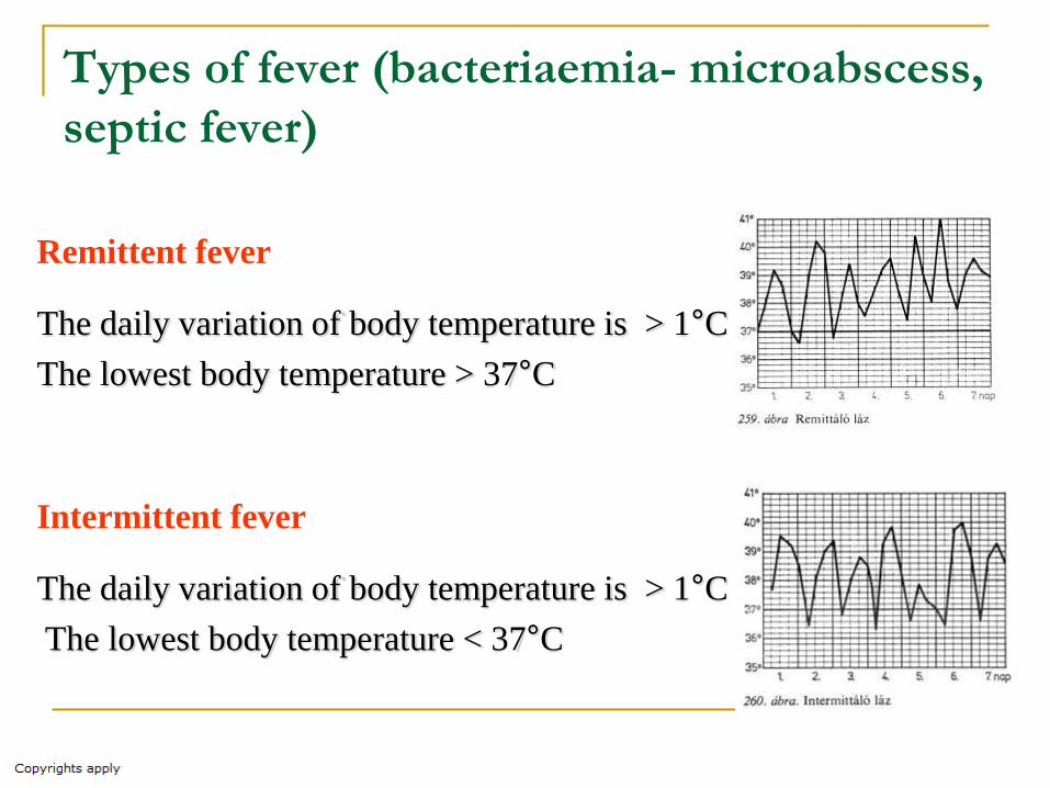
Remittent fever
The daily variation of body temperature is > 1°C
The lowest body temperature > 37°C
Intermittent fever
The daily variation of body temperature is > 1°C
The lowest body temperature < 37°C
Types of fever (bacteriaemia- microabscess,
septic fever)

Types of fever (relapsing fever)
Fever lasting 3-10 days is followed by afebrile periods of 3-10 days. (Pel-
Ebstein pattern). Hodgkin’s disease, other lymphomas.
Plasmodium vivax causes fever every third day. Plasmodium malariae
causes fever every fourth day.
Borellia infection
Definition and classification of
FUO
Classic Fever of Unknown Origin
Temperatures of > 38.3 oC (> 10 oF) on several occasions
Duration of fever is more than 3 weeks
Three outpatient visits or 3 days in the hospital without elucidation of a cause or 1 week of „ intelligent and invasive” ambulatory investigation
Definition and classification of FUO
Nosocomial FUO
Temperatures of > 38.3 oC (> 10 oF) on several occasions in a hospitalized patient
Three days of investigation, including 2 days’ incubation of cultures
Neutropenic FUO
Temperatures of > 38.3 oC (> 10 oF) on several occasions in a patient whose neutrophil count is < 500/µL
A specific cause is not identified after 3 days of investigation, including at least 2 days’ incubation of cultures
HIV-associated FUO
Temperatures of > 38.3 oC (> 10 oF) on several occasions over a period of > 4 weeks for outpatients or > 3 days for hospitalized patients with HIV infection
Appropriate investigation over 3 days, including 2 days’ incubation of cultures

Classic FUO in adult patients
Disease-groups Cases(%)
Infections 26
Neoplasms 13
Multisystem diseases (SLE,
RA, vasculitis,
granulomatous diaseases)
24
Others 8
Undiagnosed 25-30
Bleeker-Rovers et al. Medicine (Baltimore 2007;86:26.), de Kleijn et al.1997
The leading infectious causes of FUO
Extrapulmonary tuberculosis
Mononucleosis syndromes (EB, CMV) or HIV sometimes confounded by delayed antibody responses
Intraabdominal abscesses
Osteomyelitis (especially where prosthetic devices have been implanted)
Endocarditis
Fungal disease (histoplasmosis)
Noninfectious inflammatory diseases
in the background of FUO
Giant-cell arteritis (>50 years of age, this
disease accounts for 15-20% of FUO cases)
Still’s disease : leukocytosis, anemia, arthralgias,
polyserositis, lymphadenopathy, splenomegaly, rash
SLE
Granulomatous diseases
Sarcoidosis
Crohn’s disease
Granulomatous hepatitis
Miscellaneous causes of FUO in adults
Drug fever
β-lactam antibiotics
Cardiovascular drugs (quinidine)
Antineoplastic drugs
Drugs acting on the central nervous system (phenytoin)
Pulmonary embolism
Factitious fever
In young women in the health professions
Hereditary periodic fever syndromes
General considerations in adult FUO
The FUO is a rare condition.
As the duration of fever increases, the
likelihood of an infectious cause decreases.
In cases of infectious origin, the cause is not a
rare infectious disease, rather the unusual
manifestation of a common infectious disease.
In the elderly patients, multisystem disease is
the most frequent cause of FUO.

Approach to the patient with classic
FUO History (environmental, occupational, and professional)
Physical exam
Erythrocyte sedimentation rate and CRP should be determined
Routine blood chemistry (incl. complete blood count, liver function tests and serum bilirubin, serum creatinine, electrolytes, muscle enzymes, serum protein electrophoresis)
ANA, RF, VDRL, HIV, EBV, CMV, skin test to screen for tuberculosis
Hepatitis serology (if liver tests are positive)
Multiple blood samples (3-6), including samples for anaerobic culture
Urine analysis and cultures
Chest X-ray
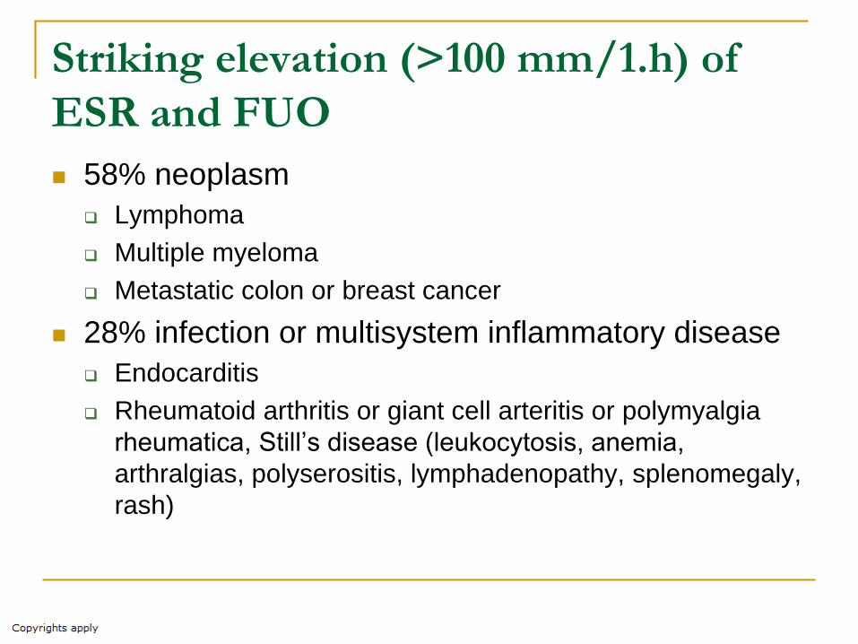
Striking elevation (>100 mm/1.h) of
ESR and FUO
58% neoplasm
Lymphoma
Multiple myeloma
Metastatic colon or breast cancer
28% infection or multisystem inflammatory disease
Endocarditis
Rheumatoid arthritis or giant cell arteritis or polymyalgia
rheumatica, Still’s disease (leukocytosis, anemia,
arthralgias, polyserositis, lymphadenopathy, splenomegaly,
rash)
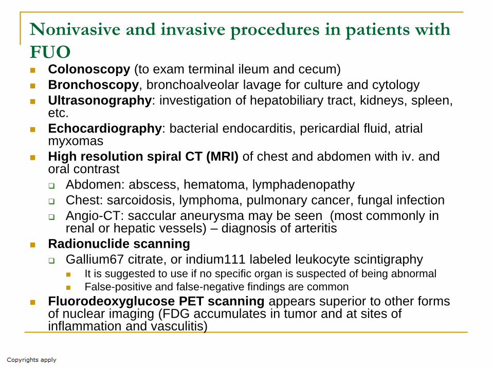
Nonivasive and invasive procedures in patients with
FUO Colonoscopy (to exam terminal ileum and cecum)
Bronchoscopy, bronchoalveolar lavage for culture and cytology
Ultrasonography: investigation of hepatobiliary tract, kidneys, spleen, etc.
Echocardiography: bacterial endocarditis, pericardial fluid, atrial myxomas
High resolution spiral CT (MRI) of chest and abdomen with iv. and oral contrast
Abdomen: abscess, hematoma, lymphadenopathy
Chest: sarcoidosis, lymphoma, pulmonary cancer, fungal infection
Angio-CT: saccular aneurysma may be seen (most commonly in renal or hepatic vessels) – diagnosis of arteritis
Radionuclide scanning
Gallium67 citrate, or indium111 labeled leukocyte scintigraphy It is suggested to use if no specific organ is suspected of being abnormal
False-positive and false-negative findings are common
Fluorodeoxyglucose PET scanning appears superior to other forms of nuclear imaging (FDG accumulates in tumor and at sites of inflammation and vasculitis)
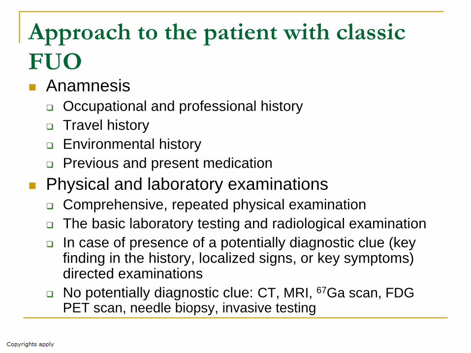
Approach to the patient with classic
FUO Anamnesis
Occupational and professional history
Travel history
Environmental history
Previous and present medication
Physical and laboratory examinations Comprehensive, repeated physical examination
The basic laboratory testing and radiological examination
In case of presence of a potentially diagnostic clue (key finding in the history, localized signs, or key symptoms) directed examinations
No potentially diagnostic clue: CT, MRI, 67Ga scan, FDG PET scan, needle biopsy, invasive testing
FUO in immuncompromised patients
HIV-associated FUO 80% infection (due to Mycobacterium avium or
Mycobacterium intracellulare, tuberculosis, Pneumocystis , cryptococcosis, CMV, etc.)
8% neoplasm, mostly non-Hodgkin lymphoma
Drug fever
Neutropenic FUO Neutropenic patients are susceptible to focal bacterial and
fungal, to bacteremic infection, to infections involving catheters and to perianal infections.
50-60% of febrile neutropenic patients are infected, and 20% are bacteremic.
Fever even after the recovery from neutropenia suggest systemic fungal infection (Candida, aspergillus)

Classification of rash that reflect systemic diasease
Not palpable, flat lesion defined by an area of changed color (blanchable erythema): macula
Elevated, solid skin lesions: Papula: < 5 mm
Plaque: > 5 mm with a flat, plateau-like surface
Nodulus: > 5 mm in diameter with a more rounded configuration
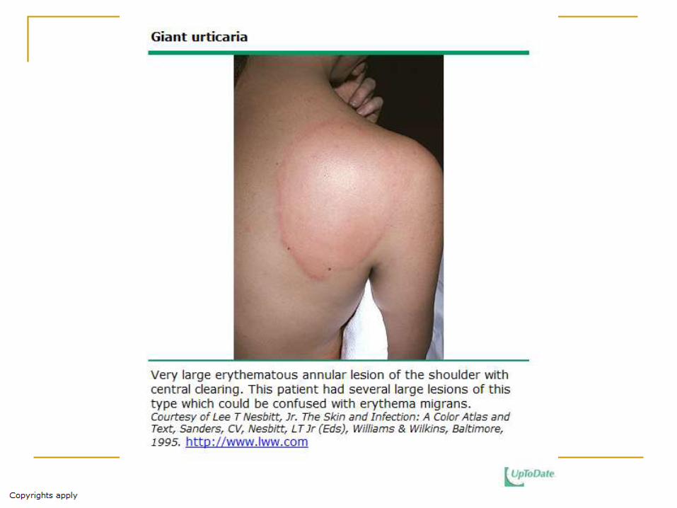
Urticaria: are papules or plaques that are pale pink and may appear annular(ringlike). Transient, lasting only 24-48 h in a defined area
Skin-bleedings Petechia: nonpalpable, flat lesion, < 3 mm in diameter
Ecchymosis: nonpalpable, flat lesion, > 3 mm
Palpable purpura: a raised lesion that is due to inflammation of the vessel wall with subsequent hemorrhage.
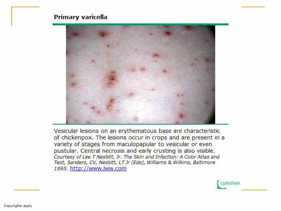
Elevated lesions containing fluid: Vesicula: < 5 mm, elevated, circumscribed lesion,
Bulla: > 5 mm
Pustula: raised lesion containing purulent exudate
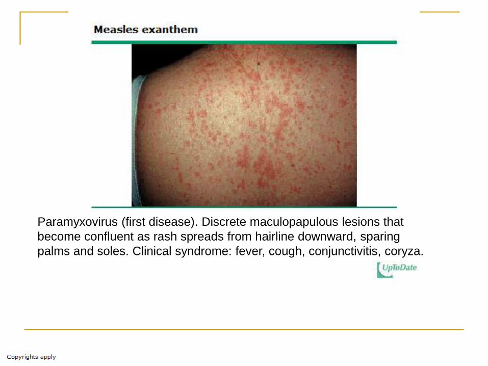
Paramyxovirus (first disease). Discrete maculopapulous lesions that
become confluent as rash spreads from hairline downward, sparing
palms and soles. Clinical syndrome: fever, cough, conjunctivitis, coryza.
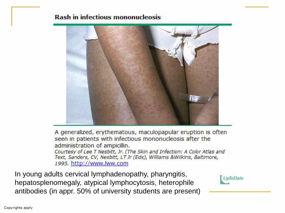
In young adults cervical lymphadenopathy, pharyngitis,
hepatosplenomegaly, atypical lymphocytosis, heterophile
antibodies (in appr. 50% of university students are present)
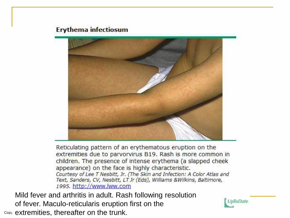
Mild fever and arthritis in adult. Rash following resolution
of fever. Maculo-reticularis eruption first on the
extremities, thereafter on the trunk.

Causes:
- Streptococcal,
mycobacterium or
Yersinia infections
-Drug-induced (penicillin,
contraceptives
-Sarcoidosis
Arthralgy in 50% of cases

Drug intake (sulfa,
phenytoin, penicillin;
Herpes simplex virus
Mycoplasma pneumoniae
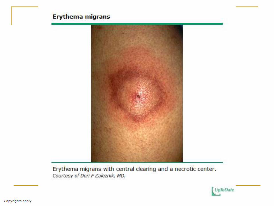
central erythema
area of clearing
another rim of erythema


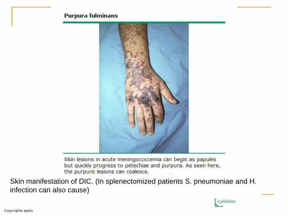
Skin manifestation of DIC. (In splenectomized patients S. pneumoniae and H.
infection can also cause)


![Fever of unknown origin (FUO): which are the factors influencing … · 2018. 3. 15. · Fever of unknown origin (FUO) was originally defined by Petersdorf and Beeson [1]asanillnessofmore](https://static.documents.pub/doc/80x56/60fe895bfa0f251e835ba0b5/fever-of-unknown-origin-fuo-which-are-the-factors-influencing-2018-3-15.jpg)


