Pathophysiology of Peripheral Nerve Lesions Part 3: Lower Extremity Entrapment Syndromes David A. Lake, PT, PhD Department of Physical Therapy Armstrong Atlantic State University Savannah, GA
Transcript
Pathophysiology of Peripheral Nerve LesionsPart 3: Lower Extremity Entrapment Syndromes
David A. Lake, PT, PhD
Department of Physical Therapy
Armstrong Atlantic State University
Savannah, GA
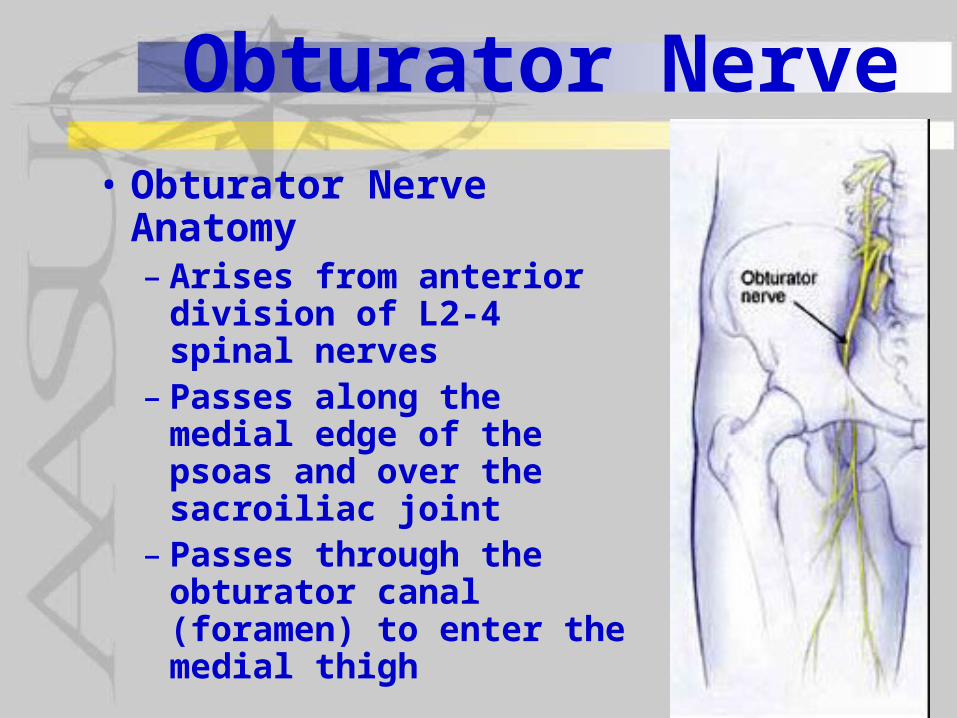
Obturator Nerve• Obturator Nerve
Anatomy– Arises from anterior
division of L2-4 spinal nerves
– Passes along the medial edge of the psoas and over the sacroiliac joint
– Passes through the obturator canal (foramen) to enter the medial thigh
neuropathy reported by patient include:– Pain along medial thigh - “obturator
neuralgia” common– Numbness along medial thigh also
common– Occassionally report gait
abnormalities – Rarely do patients report weakness
Obturator Nerve• Neurologic findings of Obturator
damage include: –Weakness in thigh adduction– Circumduction of thigh when walking– Occassionally wide stance – Positive EMG signs of denervation in
adductor muscles
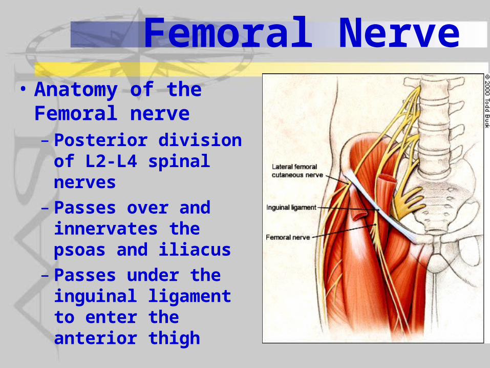
Femoral Nerve• Anatomy of the
Femoral nerve– Posterior division of
L2-L4 spinal nerves– Passes over and
innervates the psoas and iliacus
– Passes under the inguinal ligament to enter the anterior thigh
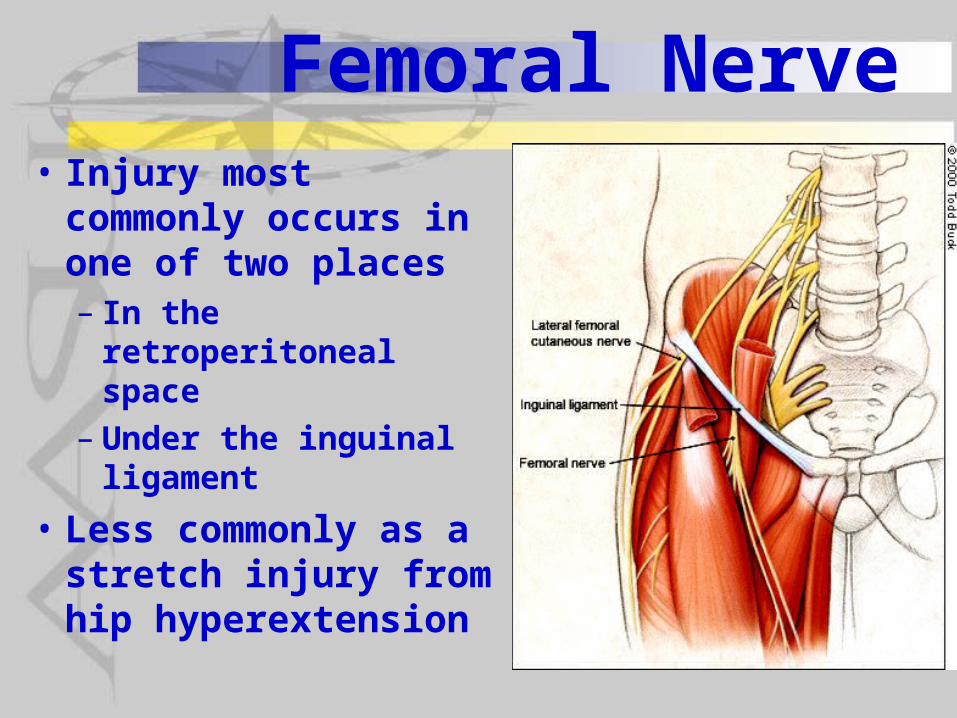
Femoral Nerve• Injury most
commonly occurs in one of two places– In the retroperitoneal
space– Under the inguinal
ligament
• Less commonly as a stretch injury from hip hyperextension
Femoral Nerve
• Injury in the retroperitoneal space–Most common secondary to
abdominal surgery and retroperitoneal hematomas
– Estimated that in up to 7.5% of hysterectomies there is femoral nerve damage
Femoral Nerve• Injury under the inguinal ligament–Most common secondary to nerve
compression during lithotomy positioning
– Estimated that femoral nerve damage occurs in up to 2.3% of total hip surgeries particularly in complicated revisions
– Less common from inguinal hematomas resulting from femoral vessel catheterization
Femoral Nerve• Symptoms of nerve injury reported by
patients–Most commonly unilateral but can be
bilateral after lithotomy–Weakness in quadriceps femoris muscles– Knee buckling on weightbearing– Easy loss of balance and falling– Numbness on anteromedial thigh & leg– Pain usually only with retroperitoneal
hematomas
Femoral Nerve• Diagnosis of femoral nerve injury–Weakness of quads with diminished or
eliminated patellar tendon reflex– Thigh adduction and ankle dorsiflexion
strength is normal–MR & CT for presumed space occupying
lesion– NCV studies of CMAP of femoral nerve
and SNAP of saphenous nerve show amplitudes and conduction velocities
– Spontaneous activity and recruitment of MUAPs of quadriceps femoris
Femoral Nerve• Terminology note:– Saphenous nerve is the sensory branch of
the femoral nerve– NCV - nerve conduction velocity but also
includes amplitude in of the compound action potentials from surface recordings
– CMAP - compound motor action potentials – SNAP - sensory nerve action potentials–MUAPs - motor unit action potentials
recorded with needle electrodes in the muscle
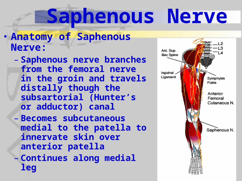
Saphenous Nerve• Anatomy of Saphenous
Nerve:– Saphenous nerve branches
from the femoral nerve in the groin and travels distally though the subsartorial (Hunter’s or adductor) canal
– Becomes subcutaneous medial to the patella to innervate skin over anterior patella
– Continues along medial leg
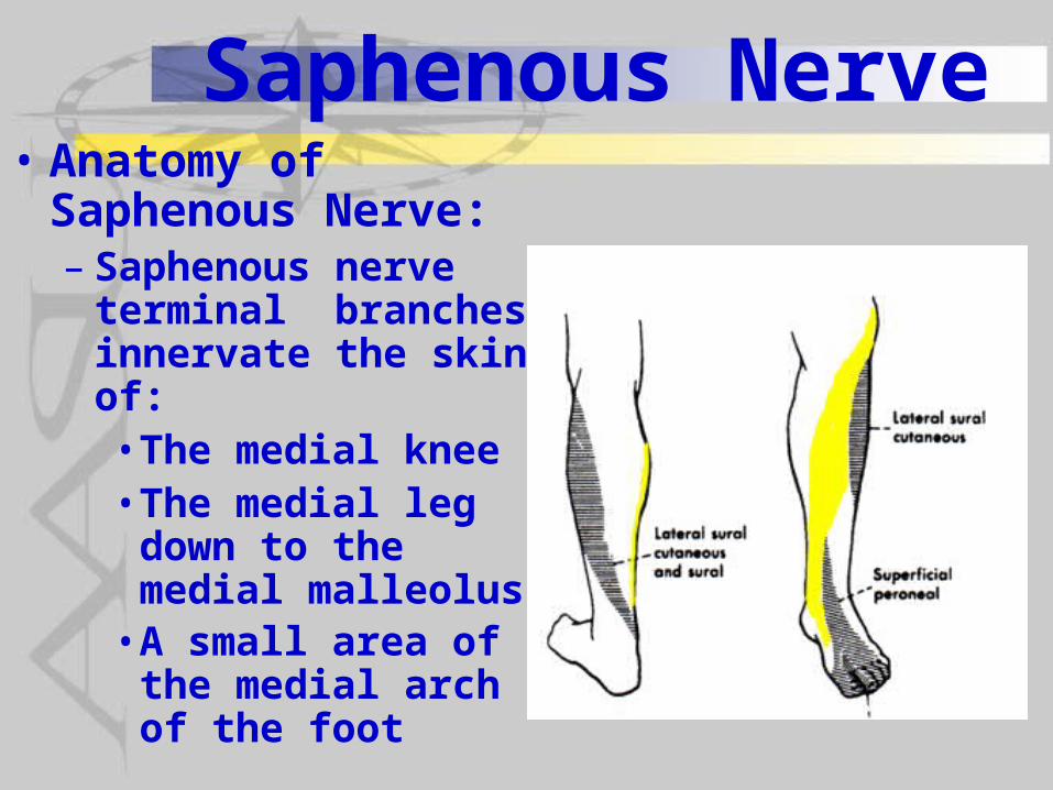
Saphenous Nerve• Anatomy of
Saphenous Nerve:– Saphenous nerve
terminal branches innervate the skin of: • The medial knee• The medial leg
down to the medial malleolus • A small area of the
medial arch of the foot
Saphenous Nerve• Neuropathies of Saphenous nerve
occur:– Occasionally through entrapment as it
exits the subsartorial canal next to the pes anserine bursa as a result of bursitis or other narrowing of the canal
–Most commonly the result of damage with:• Varicose vein surgery• Removal of the saphenous vein for
coronary artery bypass grafting • Arthroscopic surgery of the knee
Saphenous Nerve
• Primary symptoms of nerve damage reported by patients include:– Paresthesia, hyperthesias and pain
along the medial leg– Knee pain is also common and if
only the infrapatellar branch is damaged , there may only be anterior knee numbness
Saphenous Nerve
• Diagnosis is done with the following findings: SNAP of saphenous nerve– No weakness in quadriceps femoris
muscles– Normal EMG findings in quadriceps
femoris, hip adductors and iliacus– Occasionally + Tinel sign over
subsartorial canal
Lateral Femoral Cutaneous Nerve
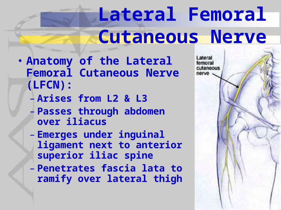
• Anatomy of the Lateral Femoral Cutaneous Nerve (LFCN):– Arises from L2 & L3– Passes through abdomen
over iliacus– Emerges under inguinal
ligament next to anterior superior iliac spine
– Penetrates fascia lata to ramify over lateral thigh
Lateral Femoral Cutaneous Nerve
• Neuropathy of the LFCN:– Termed Meralgia Paresthetica and most
commonly due to compression under the inguinal ligament
– Contributing factors can include:• Pregnancy• Obesity• Wearing a heavy tool belt or very tight belt• Automobile accident restrained by seatbelt• Chronic leaning against object such as
gymnastic bars
Lateral Femoral Cutaneous Nerve
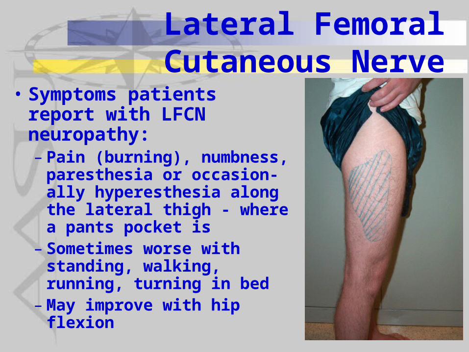
• Symptoms patients report with LFCN neuropathy:– Pain (burning), numbness,
paresthesia or occasion-ally hyperesthesia along the lateral thigh - where a pants pocket is
– Sometimes worse with standing, walking, running, turning in bed
–May improve with hip flexion
Lateral Femoral Cutaneous Nerve
• Diagnosis of LFCN neuropathy:– History of precipitating factor– Pattern of pain, numbness,
paresthesias along lateral thigh SNAP amplitude and conduction
velocity– Lack of quadriceps or adductor
weakness or sensory loss over femoral or obturator distributions
Lateral Femoral Cutaneous Nerve
• Some evidence for physical therapy intervention effectiveness from case study:– Thermal US & mobilization to
inguinal ligament followed by icepack– 3 treatments/week for 3 weeks
reduced pain from 6/10 to 2/10– Lasted until patient started running
again– Subsequent treatments reduced pain
again
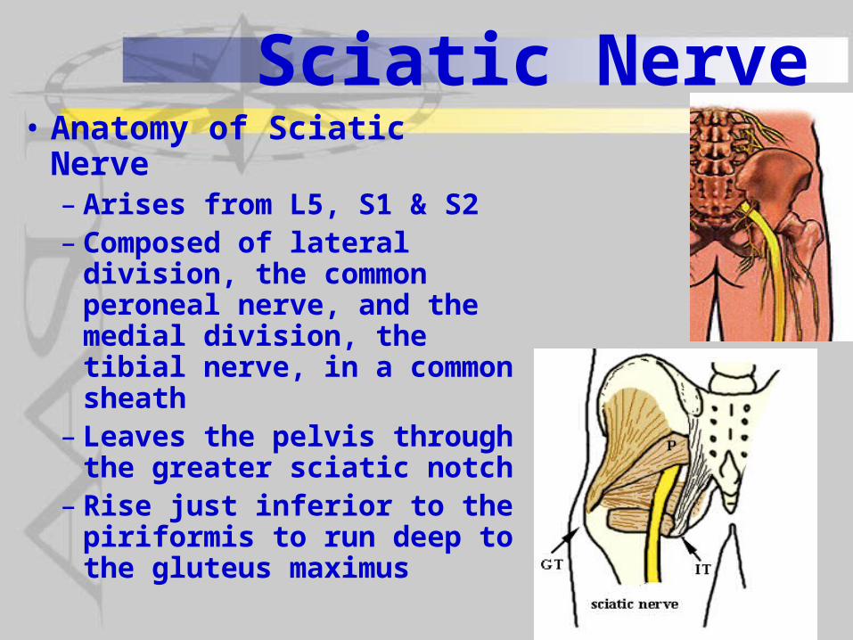
Sciatic Nerve• Anatomy of Sciatic Nerve– Arises from L5, S1 & S2– Composed of lateral
division, the common peroneal nerve, and the medial division, the tibial nerve, in a common sheath
– Leaves the pelvis through the greater sciatic notch
– Rise just inferior to the piriformis to run deep to the gluteus maximus
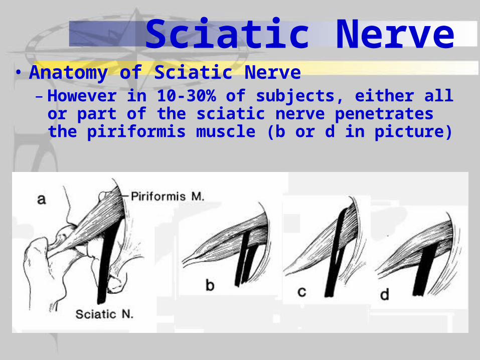
Sciatic Nerve• Anatomy of Sciatic Nerve– However in 10-30% of subjects, either all or
part of the sciatic nerve penetrates the piriformis muscle (b or d in picture)
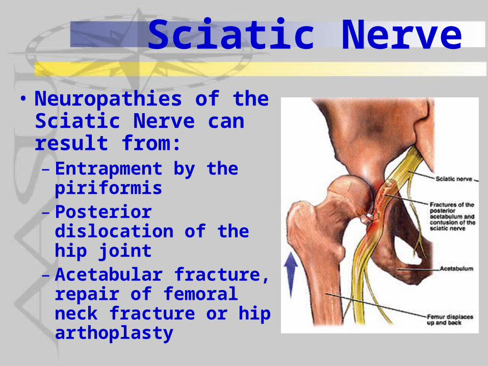
Sciatic Nerve
• Neuropathies of the Sciatic Nerve can result from:– Entrapment by the
piriformis– Posterior dislocation of
the hip joint– Acetabular fracture,
repair of femoral neck fracture or hip arthoplasty
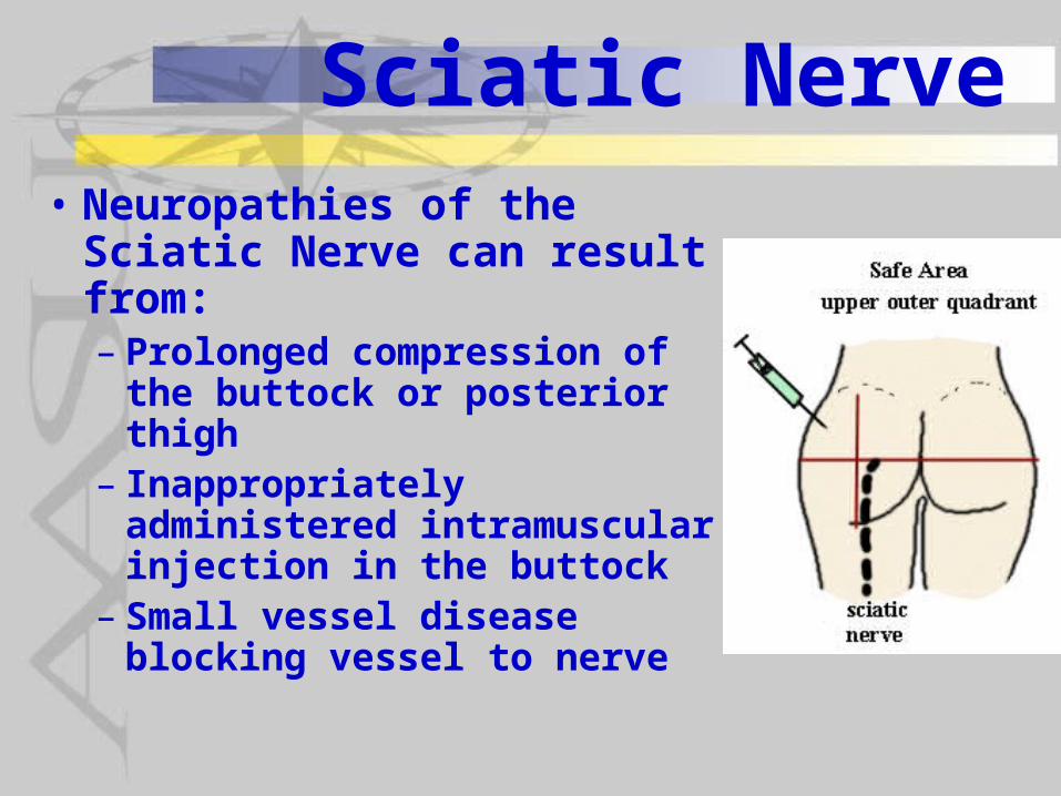
Sciatic Nerve• Neuropathies of the Sciatic
Nerve can result from:– Prolonged compression of
the buttock or posterior thigh– Inappropriately administered
intramuscular injection in the buttock
– Small vessel disease blocking vessel to nerve
Sciatic Nerve• Symptoms reported by patients with
Sciatic Neuropathies include: – Loss of muscle strength of all muscles
below the knee and the hamstrings and adductor magnus
– Paresthesias, numbness or pain in all areas below the knee except the medial leg area served by the saphenous nerve
Sciatic Nerve• In partial injury common peroneal
nerve more vulnerable because• fewer axons than tibial nerve• more exposed to traction injury being
tightly secured at fibular head and sciatic notch.
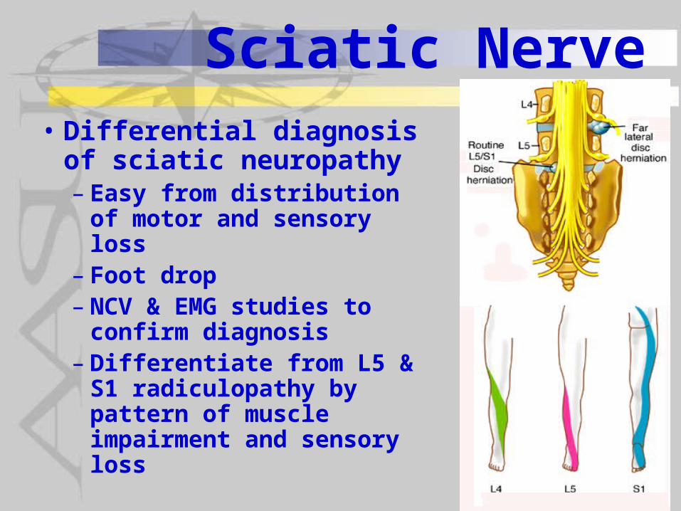
Sciatic Nerve• Differential diagnosis of
sciatic neuropathy – Easy from distribution of
motor and sensory loss– Foot drop– NCV & EMG studies to
confirm diagnosis– Differentiate from L5 & S1
radiculopathy by pattern of muscle impairment and sensory loss
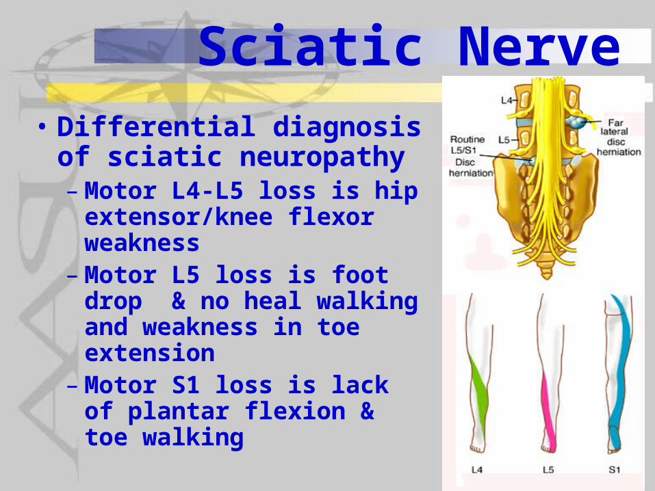
Sciatic Nerve• Differential diagnosis of
sciatic neuropathy –Motor L4-L5 loss is hip
extensor/knee flexor weakness
–Motor L5 loss is foot drop & no heal walking and weakness in toe extension
–Motor S1 loss is lack of plantar flexion & toe walking
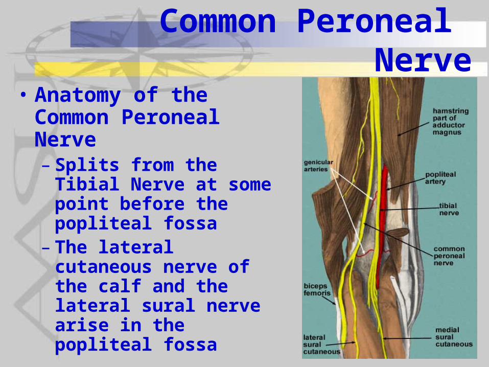
Common Peroneal Nerve
• Anatomy of the Common Peroneal Nerve – Splits from the Tibial
Nerve at some point before the popliteal fossa
– The lateral cutaneous nerve of the calf and the lateral sural nerve arise in the popliteal fossa
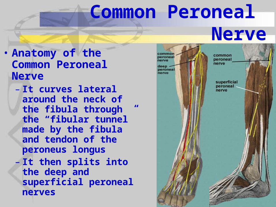
Common Peroneal Nerve
• Anatomy of the Common Peroneal Nerve – It curves lateral around
the neck of the fibula through the “fibular tunnel” made by the fibula and tendon of the peroneus longus
– It then splits into the deep and superficial peroneal nerves
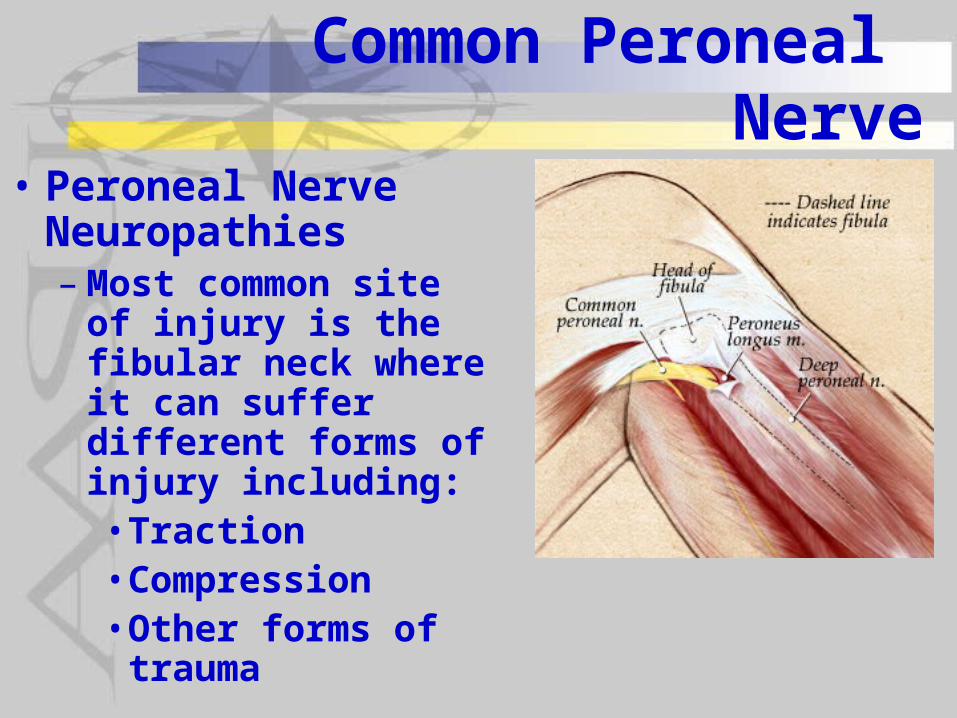
Common Peroneal Nerve
• Peroneal Nerve Neuropathies –Most common site of
injury is the fibular neck where it can suffer different forms of injury including:• Traction• Compression• Other forms of
trauma
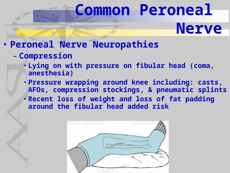
Common Peroneal Nerve
• Peroneal Nerve Neuropathies– Compression• Lying on with pressure on fibular head (coma,
anesthesia)• Pressure wrapping around knee including: casts,
AFOs, compression stockings, & pneumatic splints• Recent loss of weight and loss of fat padding around
the fibular head added risk
Common Peroneal Nerve
• Peroneal Nerve Neuropathies– Traction• Prolonged squating such as crop
harvesting, yoga meditation and exercises• Lithotomy positioning for prolonged
periods such as in childbirth• Ankle sprains
– Trauma• Blunt trauma as well as open wounds• Fibular fractures or dislocations• Surgical procedures such as arthroscopic
or open knee procedures
Common Peroneal Nerve
• Peroneal Nerve Neuropathies– Other factors• Diabetics and others with
polyneuropathies are particularly prone to injury at this point• Prolonged (> 30 min) cold applied to the
knee has been shown to produce irreversible injury to the common peroneal nerve at this point as well
Common Peroneal Nerve
• Symptoms of Peroneal Nerve Neuropathies include:– Complete or partial footdrop– Paresthesias or numbness on the
anterio-lateral leg & dorsum of the foot
–Mild, deep “boring” pain around the lateral leg and knee may be reported
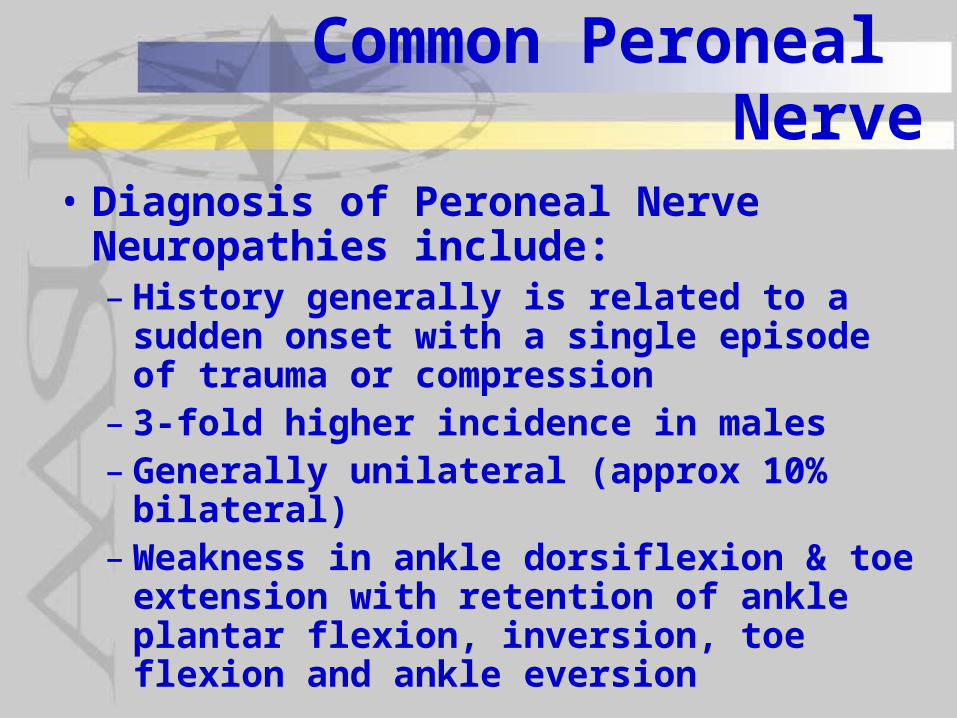
Common Peroneal Nerve
• Diagnosis of Peroneal Nerve Neuropathies include:– History generally is related to a sudden
onset with a single episode of trauma or compression
– 3-fold higher incidence in males– Generally unilateral (approx 10% bilateral)–Weakness in ankle dorsiflexion & toe
extension with retention of ankle plantar flexion, inversion, toe flexion and ankle eversion
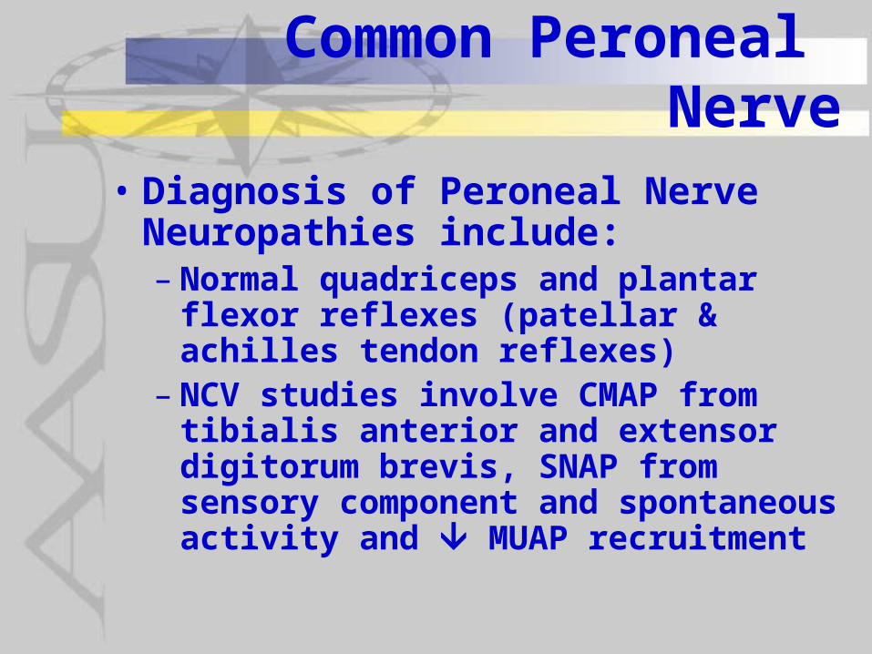
Common Peroneal Nerve
• Diagnosis of Peroneal Nerve Neuropathies include:– Normal quadriceps and plantar flexor
reflexes (patellar & achilles tendon reflexes)
– NCV studies involve CMAP from tibialis anterior and extensor digitorum brevis, SNAP from sensory component and spontaneous activity and MUAP recruitment
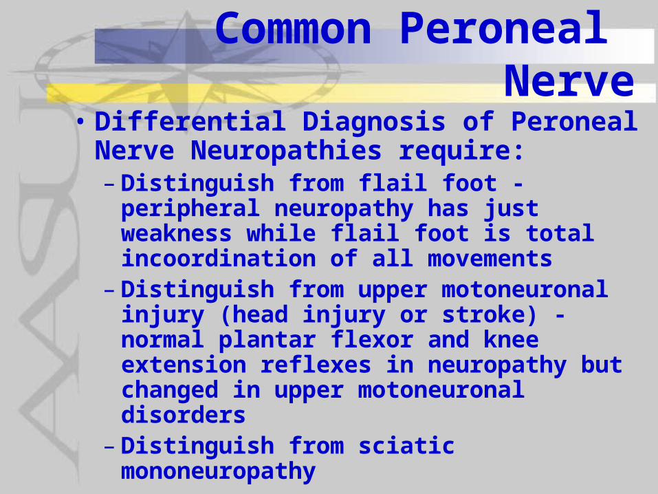
Common Peroneal Nerve
• Differential Diagnosis of Peroneal Nerve Neuropathies require:– Distinguish from flail foot - peripheral
neuropathy has just weakness while flail foot is total incoordination of all movements
– Distinguish from upper motoneuronal injury (head injury or stroke) - normal plantar flexor and knee extension reflexes in neuropathy but changed in upper motoneuronal disorders
– Distinguish from sciatic mononeuropathy
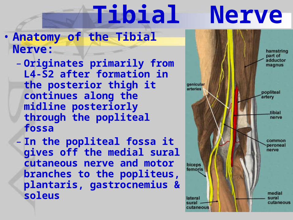
Tibial Nerve• Anatomy of the Tibial
Nerve:– Originates primarily from
L4-S2 after formation in the posterior thigh it continues along the midline posteriorly through the popliteal fossa
– In the popliteal fossa it gives off the medial sural cutaneous nerve and motor branches to the popliteus, plantaris, gastrocnemius & soleus
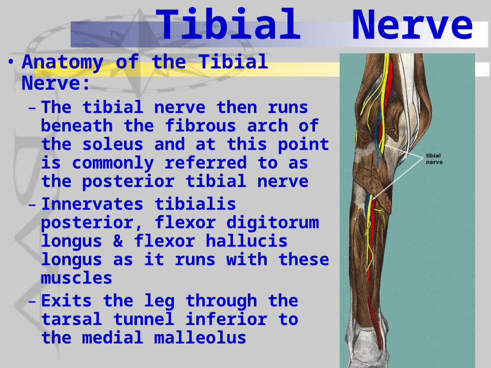
Tibial Nerve• Anatomy of the Tibial Nerve:– The tibial nerve then runs
beneath the fibrous arch of the soleus and at this point is commonly referred to as the posterior tibial nerve
– Innervates tibialis posterior, flexor digitorum longus & flexor hallucis longus as it runs with these muscles
– Exits the leg through the tarsal tunnel inferior to the medial malleolus
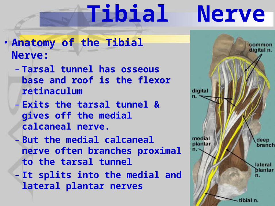
Tibial Nerve• Anatomy of the Tibial Nerve:– Tarsal tunnel has osseous
base and roof is the flexor retinaculum
– Exits the tarsal tunnel & gives off the medial calcaneal nerve.
– But the medial calcaneal nerve often branches proximal to the tarsal tunnel
– It splits into the medial and lateral plantar nerves
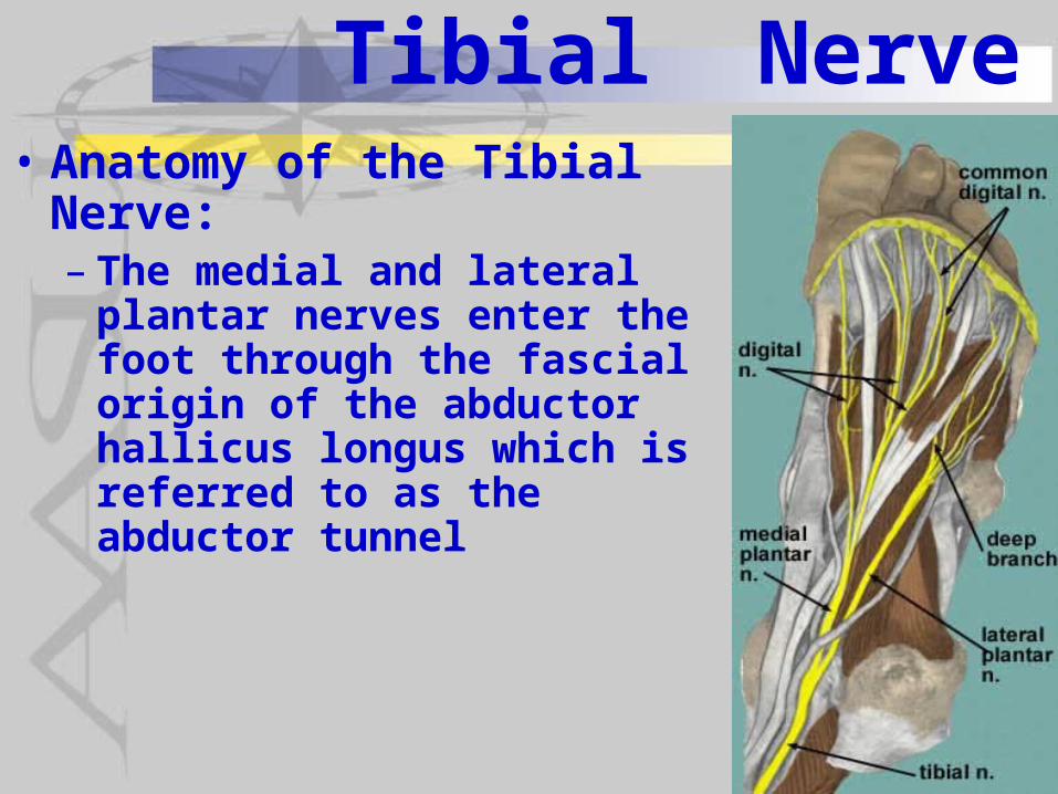
Tibial Nerve• Anatomy of the Tibial Nerve:– The medial and lateral plantar
nerves enter the foot through the fascial origin of the abductor hallicus longus which is referred to as the abductor tunnel
Tibial Nerve
• Tibial Neuropathies:– Damage in or around the popliteal fossa– Damage in the tarsal tunnel (tarsal
tunnel syndrome)
Tibial Nerve
• Tibial Neuropathies:– The popliteal fossa is the most common
site of tibial nerve injury (48% in a recent study) followed by distal to it - mostly in the tarsal tunnel (27%) and then proximal to it (25%)
–Most common etiology is trauma (56%) followed by ischemia (19%) & neoplasms (17%)
– Lesions proximal to the popliteal fossa most commonly from cast compression or blunt trauma
Tibial Nerve
• Tibial Neuropathies:– Popliteal lesions of the tibial nerve occur
mostly from penetrating and non-penetrating trauma, tibial dislocations during knee injury and only very rarely following surgical procedures
– Tibial nerve lesions distal to the popliteal fossa are primarily the result of tibial fractures, posterior compartment syndrome, and entrapment in the tendinous arch of the soleus or in fibrous bands between heads of gastrocnemius
Tibial Nerve
• Tibial Neuropathies:–Most common cause of tarsal tunnel
syndrome injury is secondary to trauma• Displaced fracture of distal tibia• Fracture of tarsal bones• Fracture of the calcaneous• Medial ankle sprains• Tenosynovitis of tendons in tarsal tunnel
• Tibial Neuropathies:– Other non-traumatic causes of tarsal
tunnel syndrome• Space occupying lesions such as
tumors, ganglia• Foot deformities such as varus heel
with pronated forefoot or valgus heel with abducted forefoot (pes planus)• Rarely but seen with patients with
diabetes and inflammatory arthritis
Tibial Nerve
• Symptoms:– Sensory disturbances in the distribution
of the sural, medial & lateral plantar and medial calcaneal nerves - posteromedial leg (calf), lateral ankle, on the lateral aspect, sole and heel of the foot
– If damage proximal to popliteal fossa weakness in ankle plantar flexion and inversion and toe flexion
–Weakness of knee flexion may be seen if denervation of gastrocnemius
Tibial Nerve
• Symptoms:– Baker’s cysts in the popliteal fossa may
also affect the common peroneal nerve– Entrapment as the tibial nerve passes
through the fibrous arch of the soleus produces severe pain and tenderness in the popliteal fossa and upper calf (soleus) made worse by weight-bearing & passive dorsiflexion of the ankle
– Entrapment in the tarsal tunnel foot paresthesias, pain and numbness are most prominent symptoms
Tibial Nerve
• Diagnosis:– History of tibial nerve symptoms with
symptoms most unique to tibial nerve being:• Hypersensitivity of the foot initially or after
nerve repair• Insensitivity of the foot with axonal loss and
foot ulcerations
– Imaging studies can show some obstructions and diagnosis fractures