74

Patient Positioning in Neurosurgery and Principles of Making a Craniotomy
Presenter: Dr. Shashank Ramdurg
Introduction
• Patient positioning critical and vital
• Control of bleeding and ventilation
• Sir Victor Horsley in 1906 used ‘ fork rest of professor Frazier’
• Head rest‐ an extension attached to the operating table
• Horsley and Krause also proposed the use of lateral position for posterior fossa surgeries

• Schede in 1905 used sitting position with patient leaning far forward
• de Martel in 1913 used sitting position and took credit for its routine use in posterior fossa surgeries
• He introduced a special chair and head fixation holder• Theoretical advantage of lowering ICP and venous
bleeding with risk of syncope and the inability to disarrange the draping from this position
‐ Bailey

• In early years‐ trial and error• Today though standardized, not absolute• Factors associated:
1. Age2. Site and nature of lesion3. Head position in relation to heart4. Position of anesthesiologist/ nurse5. Microscope and other imaging equipment
• Pediatric patients present a differentset of considerations
• Some operations have more than one acceptable position
Indications
• Most of the cranial procedures
• Anterior cervical spine
• Anterior approaches to the lumbar spine
• Carotid endarterctomies
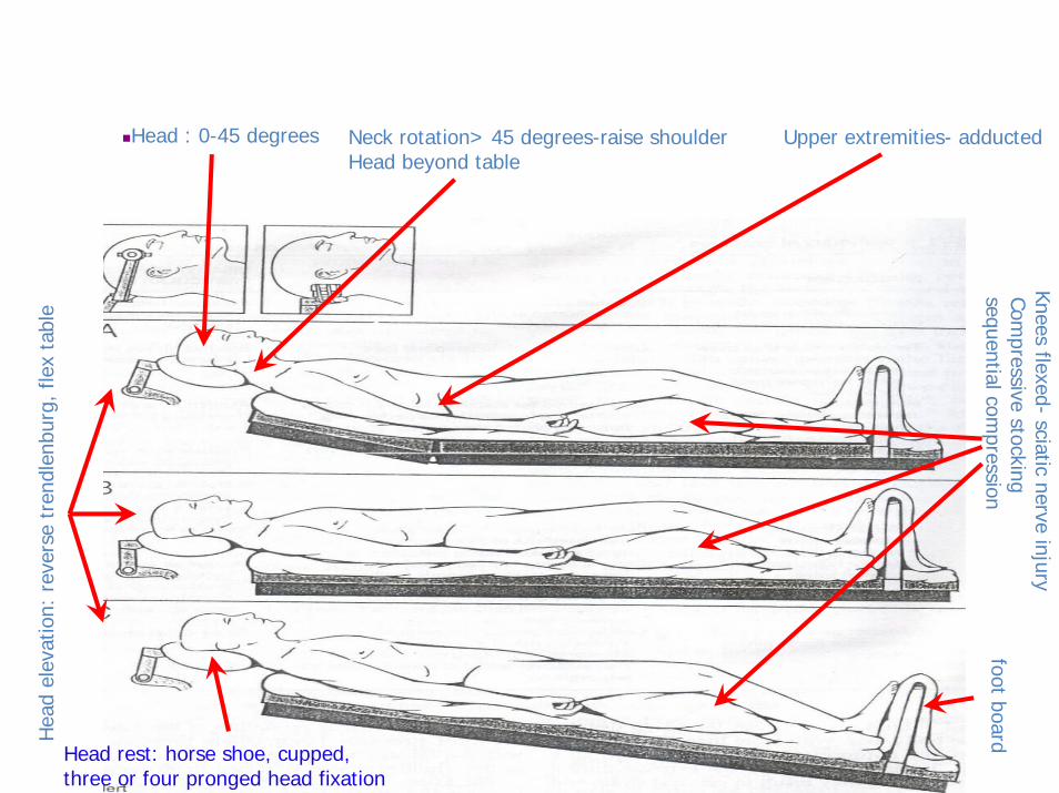
Head : 0-45 degrees Neck rotation> 45 degrees-raise shoulder Head beyond table
Hea
d el
evat
ion:
rev
erse
tre
ndle
nbur
g, f
lex
tabl
e
Upper extremities- adducted
Knees flexed-sciatic nerve injury
Compressive stocking
sequential compression
foot board
Head rest: horse shoe, cupped, three or four pronged head fixation

Positioning
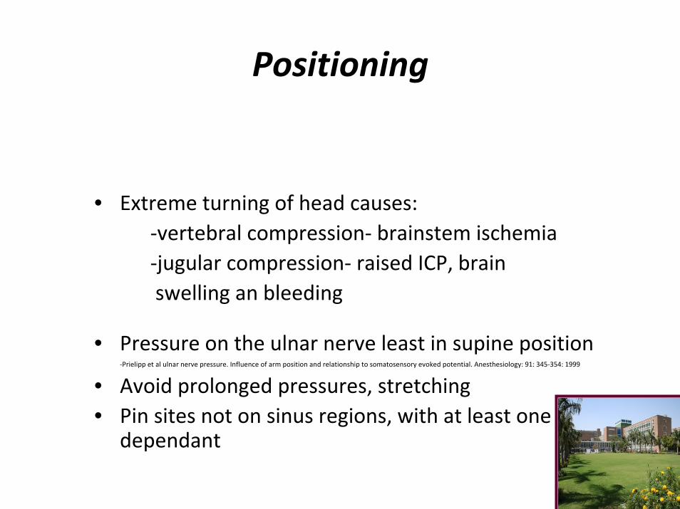
• Extreme turning of head causes:‐vertebral compression‐ brainstem ischemia‐jugular compression‐ raised ICP, brain swelling an bleeding
• Pressure on the ulnar nerve least in supine position‐Prielipp et al ulnar nerve pressure. Influence of arm position and relationship to somatosensory evoked potential. Anesthesiology: 91: 345‐354: 1999
• Avoid prolonged pressures, stretching• Pin sites not on sinus regions, with at least one
dependant

Lateral position
Indications
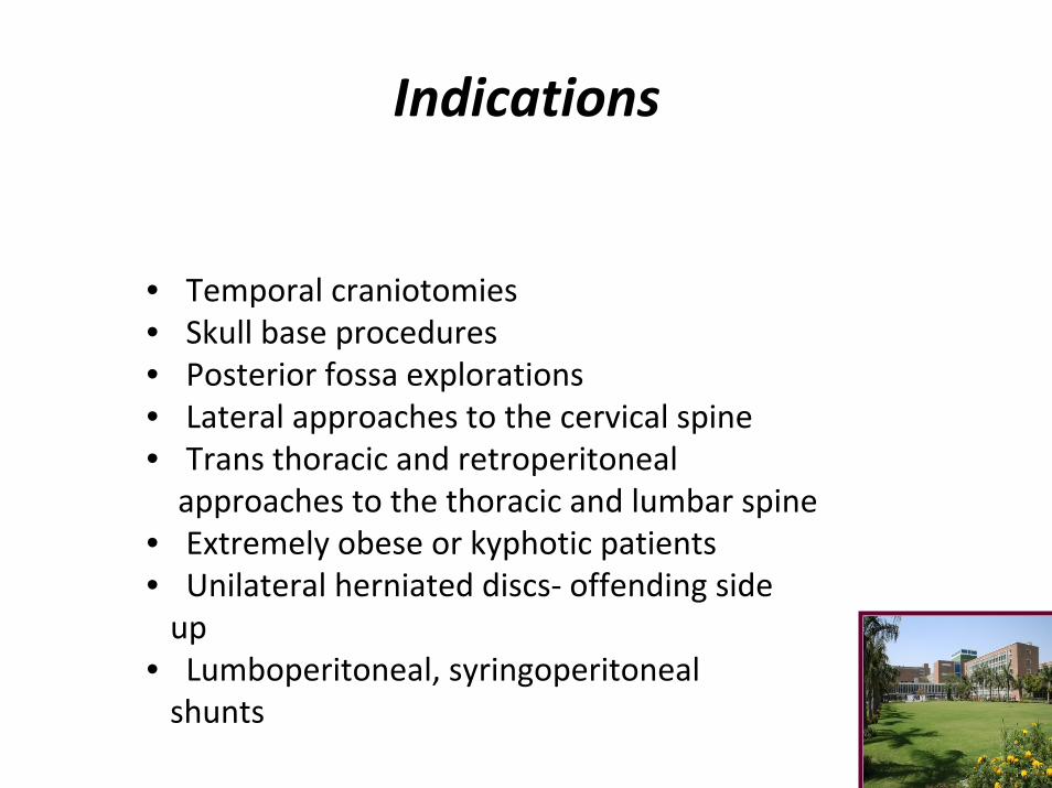
• Temporal craniotomies• Skull base procedures• Posterior fossa explorations• Lateral approaches to the cervical spine• Trans thoracic and retroperitonealapproaches to the thoracic and lumbar spine
• Extremely obese or kyphotic patients• Unilateral herniated discs‐ offending sideup
• Lumboperitoneal, syringoperitoneal shunts
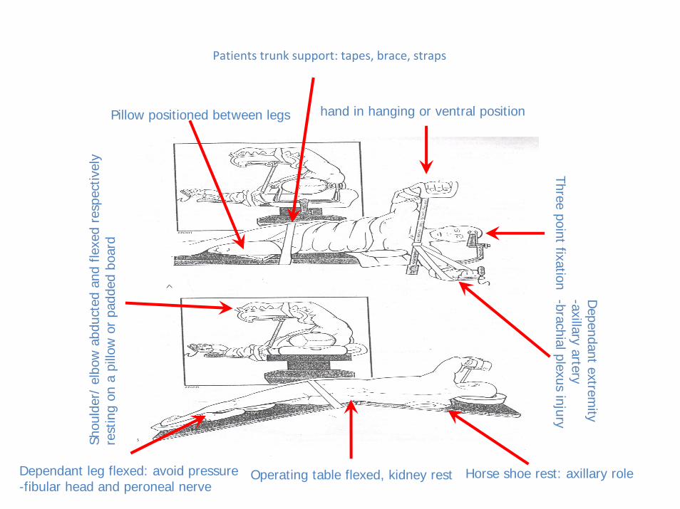
Patients trunk support: tapes, brace, straps
Operating table flexed, kidney rest
Three point fixationhand in hanging or ventral position
Shou
lder
/ el
bow
abd
ucte
d an
d fle
xed
resp
ectiv
ely
rest
ing
on a
pill
ow o
r pa
dded
boa
rd
Dependant extrem
ity-axillary artery-brachial plexus injury
Horse shoe rest: axillary role
Pillow positioned between legs
Dependant leg flexed: avoid pressure-fibular head and peroneal nerve
Positioning
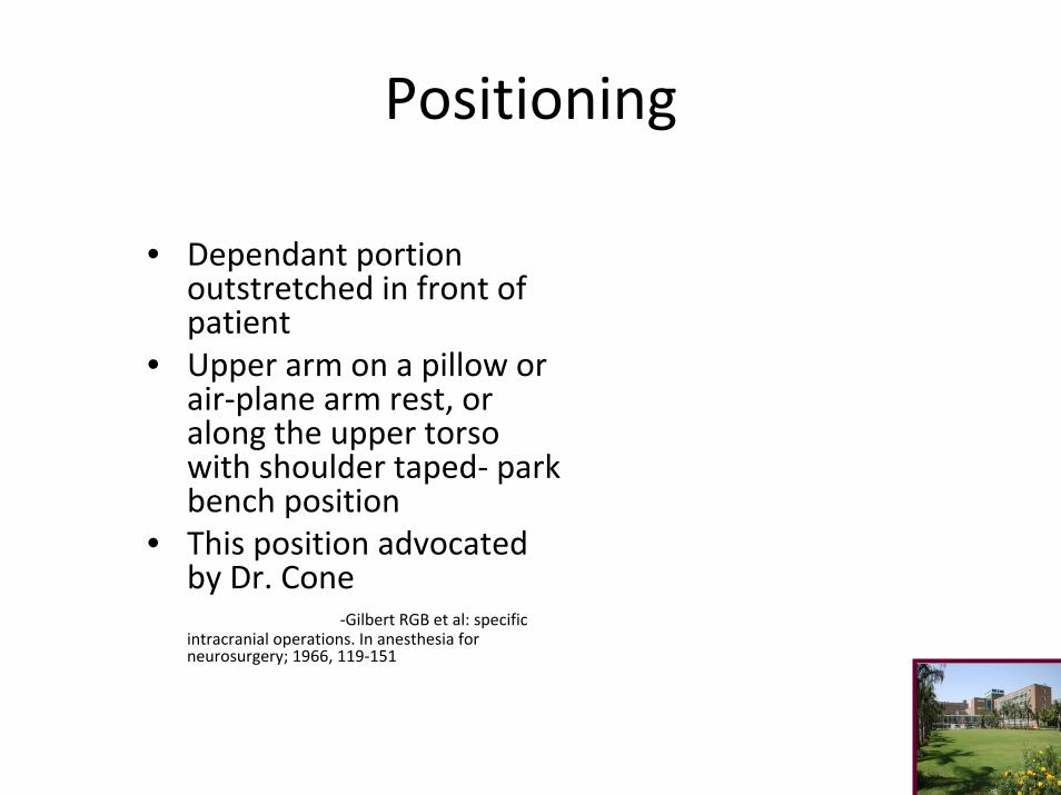
• Dependant portion outstretched in front of patient
• Upper arm on a pillow or air‐plane arm rest, or along the upper torso with shoulder taped‐ park bench position
• This position advocated by Dr. Cone
‐Gilbert RGB et al: specific intracranial operations. In anesthesia for neurosurgery; 1966, 119‐151

Indications
• Posterior fossa surgeries
• Sub‐ occipital regions
• Posterior approaches to the spine
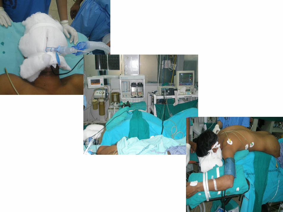

Technique
• Femoral, distal pulses checked• Genitalia should lie free• Extremes of head rotation and neck extension to be
avoided• In cases cord compromise‐ patient may be placed in halo
frame before turning him

Kneeling position
• Advantage:IVC pressure lowest in kneeling position
• Disadvantages:More time to positionMechanical injuriesDifficulty in changing curvatureHypotension
• Rarely: DVTPulmonary embolismRenal failurePost operative pain

Concorde position
Variant of prone position
Variant of prone position
Occipital trans-tentorial,supra cerebellar infra-tentorial approach
Less venous embolismfatigue
Hea
d hi
gher
tha
n he
art
Thre
e po
int
fixat
ion
Head flexed with extension of thoraco-lumbar region
Complications as in prone
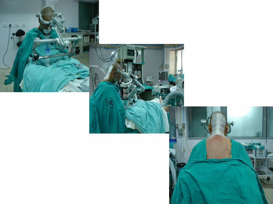
Indications
• Posterior fossa
• Cervical cord
• Sub temporal approaches
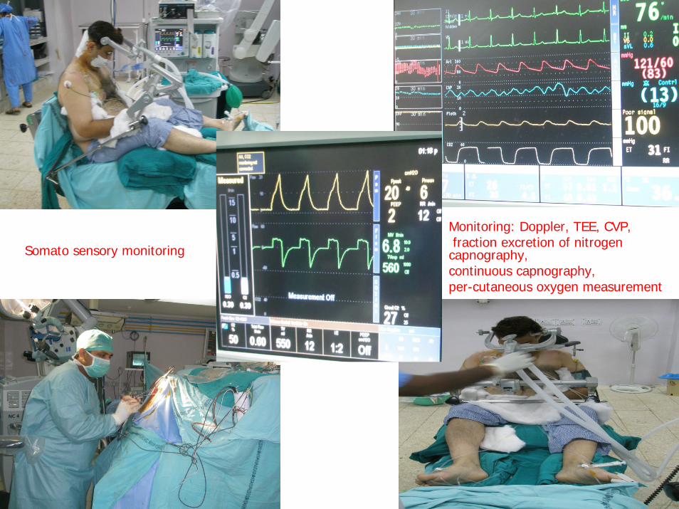
Monitoring: Doppler, TEE, CVP, fraction excretion of nitrogen
capnography, continuous capnography,per-cutaneous oxygen measurement
Somato sensory monitoring
Advantages and Disadvantages
• Advantages: Midline lesionsLow ICPImproved venous drainageDrainage of blood and CSFUnobstructed view of patients faceLess cerebellar retraction
• Complications: air embolism, hypotension, postoperative tension pneumocephalus, sub dural hematoma, quadriplegia and discomfort inupper extremities
Other complications
• Both brain and spinal cord at increased risk of cerebral
ischemia in the presence of mass lesions –Ernst PS et al intracranial and spinal cord hemodynamics in the sitting position in dogs in the presence and absence of ICP: Anesthesia analgesia: 1990
• Precautions: echocardiography, CVP, slow positioning, antigravity suit inflated with air
• Other rare complications: supra tentorial hematoma, cerebellar hemorrhages, peripheral nerve palsies, traumatic elbow dislocations
Indications
• AKA semi prone/ lateral oblique• Parieto occipital regions• Posterior fossa/ CP angle• Pineal and vermian region• Advantage: comfortable for the surgeon with less risk forembolism, Less retraction
Brachial plexus
• Supine in semi sitting/ semi fowler’s position
• Head turned to opposite side
• Ipsilateral arm‐ patients side, abducted 50 deg, arm rest
• Place roll beneath medial aspect of scapula

Trans‐ sphenoidal procedures
• Supine‐ horse shoe rest, c‐arm fluoroscopy, head tilt of 15‐20 degrees
• 10 degree head elevation with indwelling LP drain‐Banerji AK et al: trans nasal approach to pituitary adenomas. Neurol india: 34: 183: 1986

Complications
Position Complications Supine excessive head rotation, pressure sores, alopecia
Prone pressure sores, vascular compromise, brachial plexus injuries, stretch injuries, blindness, embolism, anesthetic problems
Concorde same
Three quarter prone same
Lateralbrachial plexus injuries, stretch injuries, pressurepalsies
Awake aspiration, asphyxiation, pressure palsies
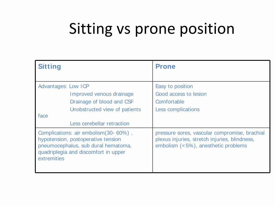
Sitting vs prone position
Sitting Prone
Advantages: Low ICPImproved venous drainageDrainage of blood and CSFUnobstructed view of patients
faceLess cerebellar retraction
Easy to positionGood access to lesionComfortableLess complications
Complications: air embolism(30- 60%) , hypotension, postoperative tension pneumocephalus, sub dural hematoma, quadriplegia and discomfort in upper extremities
pressure sores, vascular compromise, brachial plexus injuries, stretch injuries, blindness, embolism (<5%), anesthetic problems
Craniotomy principles
Skin flaps
• Neolithic period in 2000 B.C
• Trepanations made followed by scrapings of the skull till holes
• 19th century‐ trephines
• 1889 Wagner first osteoplastic bone flap
• Gigli saw for craniotomy‐ Obalinski in 1897
• Electric and gas powered high speed drills
Anatomic and neurovascular considerations
• 5 layers of scalp:
Skin
Subcutaneous tissue
Galea
loose areolar tissue
periosteum

Land marks
• Nasion• Bregma• Lambda• Inion• Pterion:Middle meningeal artery
• Asterion: Transverse sigmoidjunction
Nerves
• Fronto‐ temporal branch:
anterior branch
middle branch
posterior branch
• Middle division: 1 cm anterior to superficial temporal artery, subgaleal pad of fat
dissect between superficial and deep layers of superficial temporalis fascia
Blood supply
• Superficial temporal artery
• Occipital artery
• Posterior auricular artery
• Supra orbital and trochlear vessels
Planning
• Position of lesion
• Position of important structures
• Contingency plan for enlarging incision
• Obtain adequate closure
Principles
• General principals:
1. surgical exposure of the lesion
2. neuro vascular supply
3. cosmetic effect
• Types: Random pattern
Based on named vessel
• Length not > 1.5 times base
• Integrity of major vascular flap to be maintained
• Incision in hair containing region
• No crossed incisions

Principles
• Skin incised with galea• Pressure over the scalp• Periosteum raised with scalp or separately• Raney’s clips, bipolar, Dandy’s clamps• Adequate retraction• Inner surface protected with moistened gauze• Roller gauze• Dissect in interfascial fat which is encountered in 4 cm of orbital rim
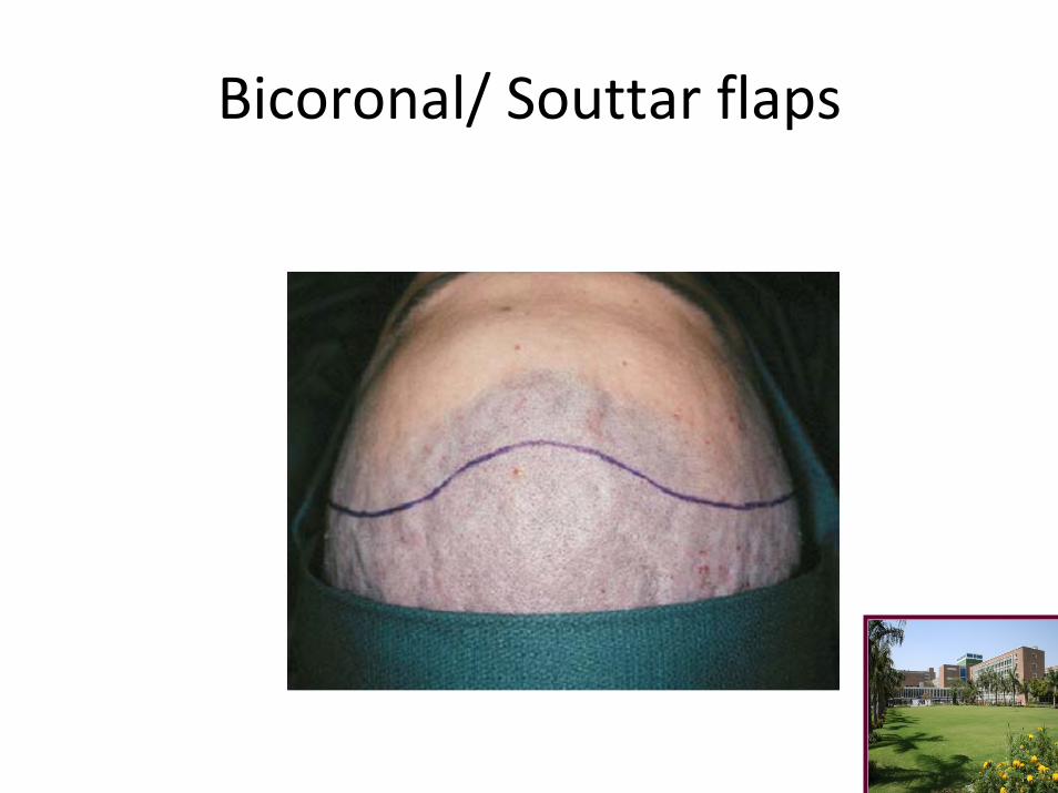
Bicoronal/ Souttar flaps
Bicoronal/ Souttar flaps
• Large exposures of anterior cranial fossa and sella• Fronto temporal lesions and cranial base • Superior to zygomatic arch, 1 cm anterior to tragus‐ extends
over the bregma to the corresponding site on the opposite side
• Reflect up to orbit rim• Supraorbital/ trochlear vessels

Frontal flap
•Exposes anterior frontal lobe
•Begins along coronal suture and curves anteriorly along the midline preferably ending at hair line
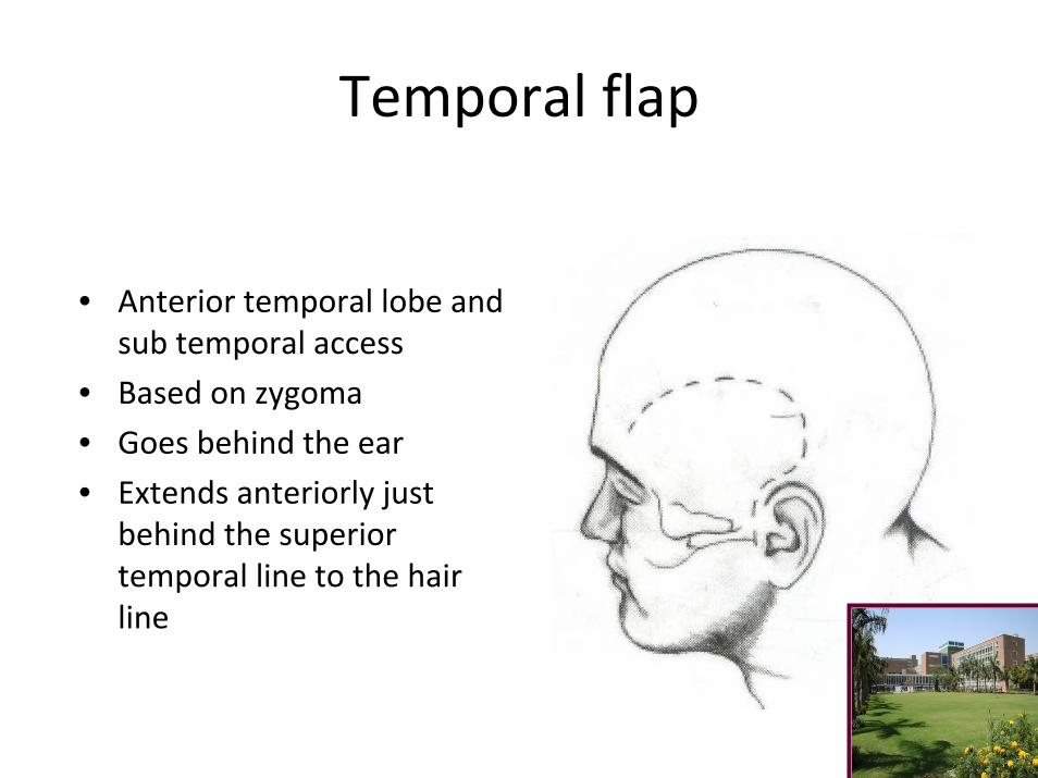
Temporal flap
• Anterior temporal lobe and sub temporal access
• Based on zygoma
• Goes behind the ear
• Extends anteriorly just behind the superior temporal line to the hair line
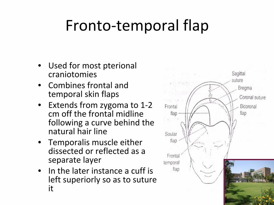
Fronto‐temporal flap
• Used for most pterional craniotomies
• Combines frontal and temporal skin flaps
• Extends from zygoma to 1‐2 cm off the frontal midline following a curve behind the natural hair line
• Temporalis muscle either dissected or reflected as a separate layer
• In the later instance a cuff is left superiorly so as to suture it
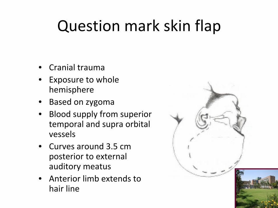
Question mark skin flap
• Cranial trauma• Exposure to whole
hemisphere• Based on zygoma• Blood supply from superior
temporal and supra orbital vessels
• Curves around 3.5 cm posterior to external auditory meatus
• Anterior limb extends to hair line
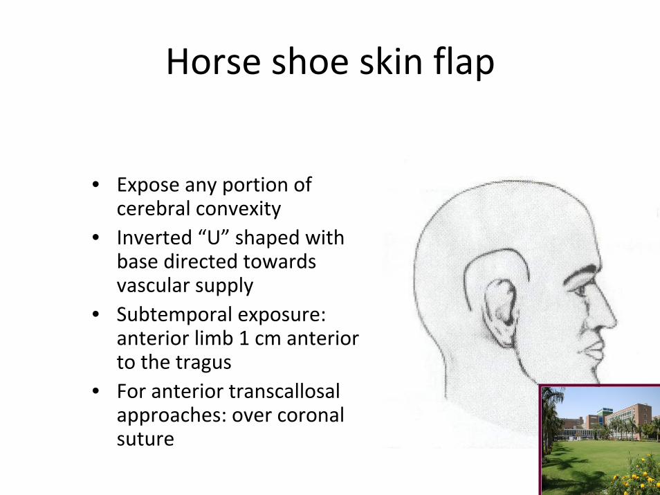
Horse shoe skin flap
• Expose any portion of cerebral convexity
• Inverted “U” shaped with base directed towards vascular supply
• Subtemporal exposure: anterior limb 1 cm anterior to the tragus
• For anterior transcallosal approaches: over coronal suture
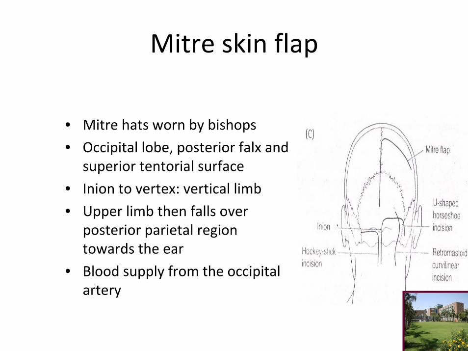
Mitre skin flap
• Mitre hats worn by bishops
• Occipital lobe, posterior falx and superior tentorial surface
• Inion to vertex: vertical limb
• Upper limb then falls over posterior parietal region towards the ear
• Blood supply from the occipital artery
Linear and curvilinear incisions
• Limited exposures
• Simplicity
• E.g..: MLSOC
RMSOC
Hockey stick incisions
Linear incisions for temporal lobe and sub temporalaccess
Principles of craniotomy
• Preoperative review of patient
• Preparation of scalp
• Positioning of patient on the table
• Scalp toilet
• Marking of the incision
• Draping
Types of craniotomies
• Flap craniotomy
• Trephine craniotomy
• Flap craniotomy: Osteoplastic
Free bone flap
Bone flaps
• Most direct access to target
• For cerebral convexity directly centered over the lesion
• Skull base lesions should be at the cranial base
• Number of burr holes varies
• Separation of underlying dura
• Beveling effect
• If dura is lacerated during cutting, saw should be turned of and removed backwards via entrance hole
• Air cells opened: remove the mucosapack with betadine soaked spongstanpack with bone waxcover it up with vascularized tissue
• Proposed bony cuts over the sinuses should be done last‐vascularityadherence
• Cut sinus can be sewn/ tamponade• Bony bleeds stopped with bone wax• Penfield’s retractors to separate dura• Epidural tacking sutures to control epidural bleeding
before opening dura• Others don’t in order to protect cortical blood vessels with
an intervening brain spoon• Tailor to avoid dural venous channels
Opening of Dura mater
• Manually palpate the dura• Dura opened as straight, curved or flap like incisions• Flaps based towards sinuses• Opened with sharp hook and knife• Incision further opened with dural scissors• Placement of cottonoid along the intended incision• Suitable cuff of dura around the bone for suturing later

Closure
• Closure in layers
• Check for BP‐ valsalva maneuver
• Hitch suture
• Water tight but not tension
• Bone flap replacement
• Skin closed in two layers

Frontal/ Bifrontal bone flaps
• Skin incisions: frontal, hockey stick, three quarters souttar• Suitable for frontal lobe, sub‐frontal approaches to anterior
skull base, and trans cortical access to ventricles• Burr holes: key point, anterior midline just above skull base,
multiple holes placed close together at midline• Avoid entering orbit• If orbit breached: bipolar cautery and close with bone wax• Last burr hole place posterior to key burr hole
• An extended frontal or bi‐frontal craniotomies for exposure of sella, anterior cranial base
• Supine with head extended for these• Holes placed on either sides of sagittal sinus and intervening bone is removed with roneguers or drill
• Either removed as single piece or conversion of frontal flap to bi‐frontal flap
• Combining a frontal flap with pterional flap
• Goals of surgery dictate the craniotomy• Bilateral orbital craniotomies may be added to minimize frontal lobe retraction
• Dural openings for a unilateral frontal craniotomy usually consist of flap reflected towards sagittal sinus
• For bi‐frontal access transverse incision will suffice
• Superior sagittal sinus will have to be ligated on both sides

Fronto‐ temporal (pterional) bone flap
• Popularized by Yasargil • Most useful for aneurysms of anterior circulation, basilar top, also tumors of retro orbital, parasellar and subfrontal areas
• Usually performed through right side• Supine position with head end elevated to 30 degrees and rotated by the same to opposite side
• Skin incision through standard fronto temporal ski incisions
• Temporalis muscle dissected or reflected
• Bone flap centered over the pterion
• Key burr hole, frontal burrhole, posterio burr hole, last burr hole just above the zygoma
• Further bone may be removed from the inferior temporal squama
• To improve vision, drill the sphenoid ridge
• Dural flap based on the orbit
• Addition of orbito‐zygomatic craniotomy will allow for a more lower and anterior approach
• Suited for para‐sellar, inter peduncular lesions
• Pterional+ anterior temporal craniotomy= upper basilar aneurysm

Krause
“ during all operations upon the brain, care must be exercised to avoid undue pressure on the thorax and abdomen which might interfere with respirations. Top of the table must be arranged to allow change of position”
“ operating room should be warm”
“ while operating on the cerebrum shoulders and thorax too be elevated to little less than 45 degrees. On operating on the side or posterior aspect of the head it is best to posture the patient on his side and allow his head to extend beyond the edge of the table. In all instances the assistant holds the head firmly with the fingers opposed to the jaws and cheeks.”
Surgery of the brain and spinal cord on personal experiences
Cushing
• In his report to chief surgeon:
“ ordinary pillows and sand bags are desirable. In order to get proper elevation of the head so that it can stand free of the surrounding, one or two sandbags, measuring 8*8*3 inches covered with rubber sheet, will be found convenient. A secure arrangement to prevent there slipping in the course of prolonged operation is essential”
• He also used a horseshoe head rest to allow for access to patients head and neck in the prone position

CP angle tumors
• Supine
• Lateral
• Three quarters prone
• Prone
• Sitting

Indications and Technique
• Mapping speech, motor sensory cortex
• Intractable epilepsy
• Tumors in eloquent areas
• Stereo‐tactic biopsies, DBS, chronic SDH, thermo‐coagulation of brain lesions
Technique
• Simple head rest or pin fixation may be used
• Maintenance of airway paramount importance
• Possibility of venous air embolism
• Monitoring of ETCO2 by nasal catheter
• Appropriate padding• Catheterization
Nerves
• Occipital branch of posterior auricular nerve‐ superior nuchal line‐ no deficits
• Supra orbital nerve‐ notch• In 8‐53% of patients foramen‐
open it up• Supra trochlear nerve• Temporal scalp supplied by
auricular temporal branch of mandibular nerve
• Greater occipital nerve supplies upto vertex
Thank youThank you