Page 1
1
SUPPLEMENTAL MATERIAL
Functional variants of POC5 identified in patients with idiopathic scoliosis
Shunmoogum A. Patten, Patricia Margaritte-Jeannin, Jean-Claude Bernard, Eudeline Alix,
Audrey Labalme, Alicia Besson, Simon L. Girard, Khaled Fendri, Nicolas Fraisse, Bernard Biot,
Coline Poizat, Amandine Campan-Fournier, Kariman Abelin-Genevois, Vincent Cunin, Charlotte
Zaouter, Meijiang Liao, Raphaelle Lamy, Gaetan Lesca, Rita Menassa, Charles Marcaillou,
Melanie Letexier, Damien Sanlaville, Jerome Berard, Guy A Rouleau, Françoise Clerget-
Darpoux, Pierre Drapeau, Florina Moldovan*, Patrick Edery*
* F.M. and P.E. jointly directed this work.
Page 2
2
Supplemental Results
High-Throughput Sequencing of POC5 in IS Families and Cases
Three additional POC5 SNVs, including 2 rare missense SNVs and a novel 5’UTR SNV were
identified (Supplemental Table 6). The pathogenicity and possible role of these mutations in IS
will be determined by further studies.
Supplemental Methods
Patients
The 41 multiplex idiopathic scoliosis (IS) families (F1-F41) include 135 affected individuals, 45
individuals of uncertain status and 150 unaffected individuals. 8/41 multiplex families included
5-11 affected individuals and in the remaining 33/41 multiplex families, 2-4 individuals were
affected (Supplemental Figure 11 A-C). Participants were seen at the Massues Center and at the
Hôpital Femme Mère Enfant, Lyon, France. Recruitment for this study started on March 1, 2000
and ended on July 31, 2012. The collection of 41 multiplex IS families was established between
March 1, 2000 and January 2, 2012. Multiplex IS families in which disease transmission
appeared to be consistent with an autosomal dominant trait were selected. Families where IS was
diagnosed in both parental branches were excluded from the study. An additional collection of
150 IS cases for whom familial information was not initially recorded was established between
January 2, 2012 and July 31, 2012. The control population was of similar ancestry (French,
French Canadian or European) and consisted of 1268 individuals. This control population was
not screened for the presence of IS.
Page 3
3
Phenotypic Characterization of Idiopathic Scoliosis
IS was diagnosed by combining clinical examination of the spine, including the forward bending
test (Adams test), with measurement of Cobb's angle on X-ray images. Both a Cobb's angle
greater than or equal to 15° (to minimize the risk of phenocopies) and vertebral rotation were
required for positive diagnosis of IS. Patients presenting spine curvature but no rotational
component were classed as “unknown status”, as were those with any other associated anomaly
of the spine or, more generally, with atypical IS. Individuals with normal clinical examination
and a strictly normal spine on radiograph were considered to be “unaffected”. All individuals
were clinically examined by at least one clinical geneticist and one orthopedist. Each radiograph
was carefully, and often repeatedly, checked by the geneticist (PE) and an orthopedist (BB, JCB,
NF, KAG, VC or JB). Medical records and spine radiographs from IS multiplex families and
cases harboring any of the c.G1336A (p.A446T), c.G1363C (p.A455P) or c.C1286T (pA429V)
POC5 functional SNVs (where “functional SNVs” indicates SNVs which, when over-expressed
in zebrafish, produce scoliosis-like traits) are presented in Supplemental Fig. 3A-E and
Supplemental Table 5.
Sample Collection and Legal Issues
EDTA blood samples were obtained from each participant, i.e., 150 IS cases and affected
members of families F1-F41 and their first-degree relatives (either affected or unaffected), as
often as possible. Similar blood samples were subsequently obtained also from relatives of IS
cases harboring the c.G1336A (p.A446T) POC5 SNV, i.e., one parent and daughter of case C39
and both parents of case C83. Blood samples were not available from relatives of case C58. DNA
was extracted from peripheral blood using a QIAmp DNA Blood Midi Kit (Qiagen), according to
the manufacturer’s instructions. Lithium-heparinate blood samples were collected from at least
one proband in each multiplex IS family. Lymphoblast cell lines were established and standard
Page 4
4
blood karyotyping was performed. The protocol for this study was approved by the local ethics
committee and was sponsored by the Hospices Civils de Lyon, France.
Genetic Refinement of the 5q13.3 Idiopathic Scoliosis Critical Region
A minimum common haplotype, shared by all affected members from family F2 was determined.
Towards this aim, DNA samples (500 ng) from all available individuals belonging to family F2
were hybridized on 700k Illumina HumanOmniExpress SNP arrays (Illumina) as described in the
manufacturer's protocol. Genotypes were analyzed and the locations of the recombination events
were refined using Merlin° software (1).
Whole-Exome Sequencing, Read Mapping and Variant Calling
After unsuccessful direct sequencing of a number of candidate genes from the refined 5q13.3 and
3q12.3 IS critical intervals, whole-exome sequencing was performed in 3 patients from family
F2, as previously described (2, 3). Exome capture and high-throughput sequencing were
performed at McGill University and Génome Québec Innovation Center (Montréal, Canada).
Exomes were enriched using an Agilent SureSelect all-exome kit (V4 optimized for Illumina
HiSEQ sequencing), with 2 µg of subjects' genomic DNA. This enrichment is designed to cover
approximately 50 Mb of genomic sequences, mainly protein coding sequences. Exon-enriched
DNA libraries were sequenced (paired-end, 2x100 bp) using an Illumina HiSEQ 2000 platform in
accordance with the manufacturer’s instructions.
The total number of reads was on average 6,845,663,492 per patient. The Burrows-Wheeler
Aligner (BWA, (4) was used as the main aligner for mapping against the human genome (hg19),
which was indexed using the bwtsw algorithm included with BWA. Alignment was performed
using a maximum mismatch penalty of three. All other parameters from BWA were left at their
Page 5
5
default values. The alignment was generated in paired-end mode, and SAMTOOLS (5) was used
to store the alignment. Duplicate reads were marked using Picard (http://picard.sourceforge.net)
and were excluded. Average coverage of consensus coding sequence (CCDS) was calculated for
each sample using GATK (6). After duplicate read removal, mean coverage was 134X, with a
range of 130X to 141X for the different DNA samples (Supplemental Table 1, Supplemental
Figure 2). The average transition to transversion ratio was 2.81 after applying the PASS only
filter value. The “Best Practice Variant Detection with GATK v2” was used to generate SNP and
call indels (6, 7). SNP and indels in samples sequenced on the Illumina HiSEQ system were
called using a set of 88 samples sequenced in the same conditions. Variant frequencies were
established by comparison to the 1000 Genomes database. Sequences were annotated using
ANNOVAR (8) and the RefSeq and dbSNP132 databases. To identify putative IS mutations, a
filter was applied to retain novel variants or variants with very low-frequency missense alleles
(minor allele frequency (MAF) < 5%) in the databases (1000 Genomes Project, dbSNP and our
in-house control exomes (n=1165)).
Sanger Sequencing and Statistical Analyses
Sanger sequencing
A DNA fragment containing the c.G1336A (p.A446T) POC5 rare SNV identified by whole-
exome sequencing was PCR-amplified for classic Sanger sequencing in all individuals from IS
family F2, one proband from each of the remaining 40 IS families (F1 and F3-F41) and 150 IS
cases (C1-C150). This amplicon was then sequenced in all available individuals from IS families
F19, F31, F35 and F41, in which the c.G1336A (p.A446T) or the c.G1363C (p.A455P) POC5
SNVs were detected. The PCR primers used were as follows: Forward
5’CTTTTCATAAGGTGGGACCT3’; Reverse 5’TCCGATGCCCTTACCAG3’. PCR was
Page 6
6
performed on a FlexCycler (AnalytikJena). PCR products were purified using commonly applied
methods before analysis of amplicons on an ABI 3730xl DNA Analyzer (Applied Biosystems).
Statistical analysis
The allelic frequencies of the c.G1336A (p.A446T) and c.C1286T (p.A429V) POC5 SNVs were
each compared in 191 IS cases (41 familial cases and 150 isolated cases from this study) to the
control population (1268 individuals). A one-tailed Fischer’s exact test was performed to test the
hypothesis that the POC5 variant are more frequent in the IS population than in controls.
Founder Effect Studies
DNA samples from all available individuals of IS families F2, F19, F35 and F41, case C39, her
husband and daughter, case C83 and her parents, and case C58 (no samples were available from
relatives of case C58) were genotyped using 700k Illumina HumanOmniExpress SNP arrays
(Illumina), as described above. Haplotypes were reconstructed when possible and the length of
the shared haplotype was determined (Supplemental Figure 4). Haplotypes were reconstructed
when possible, using Merlin software and figures were drawn using Haplopainter° software (9).
High-Throughput Sequencing of POC5 in IS Families and Cases
Whole exonic, flanking intronic and regulatory POC5 sequences were studied using a Fluidigm
Access Array° device (IntegraGen). A proband was analyzed from each of 40 multiplex IS
families (F1/ F3-F41) and 150 IS cases (C1-150). Primer pairs were designed using an in-house
pipeline based on Primer3° (Supplemental Table 7). 5.5 Kb of POC5 sequences were covered by
42 overlapping amplicons, average length 277 bp. Each sample was quantified with Picogreen°,
and 50 ng of DNA were used to prepare the library, according to the Fluidigm recommendations.
Universal tags, Rd1 and Rd2, were added to the 5’ end of the forward and reverse primers,
Page 7
7
respectively, for the first round of PCR, which was performed on the Access Array°. Illumina
adapters, P5 and P7, as well as barcodes were added to the pooled PCR products for the second
round of PCR, performed in microplates. Paired-end sequencing was performed on the Illumina°
MiSeq system after quality control (Fragment Analyser, AATI°) and quantification.
Zebrafish Maintenance
Zebrafish (Danio rerio; wild type AB strain) embryos were raised at 28.5 °C, collected and
staged using standard methods. All procedures described here were carried out in accordance
with the guidelines set out by the Canadian Council for Animal Care (CCAC), the CHU Sainte-
Justine Research Center, and the Comité de Déontologie de l'Expérimentation sur les Animaux
(CDEA), which is the local animal care committee at the University of Montreal. This study was
approved by the ethics committee for CHU Sainte-Justine Research Center, University of
Montreal (ZF-09-60/Category B). Fish were anaesthetized in 0.02% tricaine (MS-222; Sigma
Chemical, St. Louis, MO) in phosphate-buffered saline (PBS) prior to all procedures.
Poc5 Knockdown in Zebrafish
poc5 expression was knocked down using a morpholino antisense oligomer targeting the ATG of
the zebrafish poc5 ortholog (XM_685988). The translation-blocking morpholino,
5′-GTTCATTTGAAGGTCTATTACATCT-3′, was supplied by Gene Tools (Philomath, OR).
The morpholino was injected into single-cell stage zebrafish embryos at doses of 2 ng/embryo, 4
ng/embryo and 6 ng/embryo.
A splice-blocking morpholino (5′-ACCGCAAGTGCAATACAAACCTTAA-3′) was also used
to knocdown poc5 expression in zebrafish. The splice blocking morpholino was designed to bind
poc5 mRNA at the junction across the 3′ end of exon 5 and the 5′ end of intron 5. To test for loss
of proper poc5 mRNA processing, PCR primers (forward- 5′-CATGTCAGCCAGGTCTGTGT -
Page 8
8
3′, reverse- 5′-TCCATCTCAGCATTCACAGC-3′) were designed to bind cDNA at sites
corresponding to the 5′ end of exon 5 and the 3′ end of exon 6 of the poc5 mRNA. Amplified
cDNA was visualized using gel electrophoresis.
Expression of Wild-Type and Mutated Human POC5 SNVs in Zebrafish
In vitro mRNA synthesis and microinjection into embryos
Wild-type and mutated versions of human POC5 were produced from a myc-tagged ORF clone
of human POC5 (Origene) and injected into zebrafish embryos. Site-directed mutagenesis was
performed on this vector using a QuikChange® XL Site-Directed Mutagenesis Kit (Agilent). The
sequences of primers used for this assay are listed in Supplemental Table 8. Messenger RNAs
were obtained from linearized constructs, using the T7 RNA polymerase and the mMESSAGE
mMACHINE kit (Ambion). Transcription products were extracted by phenol:chloroform,
precipitated in isopropanol, and diluted in nuclease-free water (Ambion) with 0.05% Fast Green
vital dye (Sigma-Aldrich). mRNAs were injected into one- or two-cell stage embryos using a
Picospritzer III pressure ejector. The final injection volume was ~1.5 nl, at a concentrations of 25
ng/µl, 50 ng/µl, 100 ng/µl and 150 ng/µl mRNA. Injected and non-injected embryos were then
incubated in appropriate media at 28.5 °C for 24 h, and assessed for viability. Morphological
differences between mutant injected, wild-type injected and non-injected embryos were assessed
under an Olympus SZX12 stereoscope.
Western blot
Total protein extracts (40 µg) were obtained from 3 dpf wt-POC5 and mut-POC5 zebrafish.
Proteins were resolved on a 12% polyacrylamide gel and transferred onto a PVDF membrane.
Membranes were blocked in 0.1% PBS-Tween, 5% Skim Milk for one hour followed by
overnight incubation at 4 °C with primary antibodies: rabbit anti-myc (rabbit polyclonal; at a
Page 9
9
dilution of 1:2000; Sigma; catalogue# AV38156) or mouse anti- γ tubulin (Sigma; catalogue#
T6557; 1:5000) in 0.1% PBS-Tween with 5% BSA. After washing, membranes were incubated
for 1 h at room temperature with secondary antibodies: donkey anti-rabbit-HRP or donkey anti-
mouse HRP, as appropriate (both polyclonal antibodies from Jackson ImmunoResearch;
catalogue# 715-035-151 and 711-036-152 respectively; dilution 1:10,000) in 0.1% PBS-Tween
with 5% milk. Blots were revealed by ECL after a 10-second exposure.
Statistical analysis
Statistical analyses were performed and data were plotted using SigmaPlot 11.0 (Systat Software
Inc., CA). A Chi-squared (χ2) test was used to analyze the statistical significance of differences
in the zebrafish phenotype distributions between experimental groups.
Three-dimensional Imaging and Reconstruction of Zebrafish Bone
Juvenile zebrafish underwent a micro-CT scan (SkyScan 1072 High Resolution Desktop Micro-
CT System, Microtomograph, SkyScan) for three-dimensional (3D) visualization of the skeleton
after 3D imaging and subsequent reconstruction. Acquisition parameters for the scan were as
follows: 35 kV, 215 µA, step rotation of 0.9°, pixel size 4-7 microns; images were reconstructed
using NRecon (Version: 1.6.1.3).
Poc5 in situ Hybridization in Zebrafish
Total RNA was extracted from 48 hours post-fertilization zebrafish embryos using Trizol
(Invitrogen). This RNA was reverse transcribed using a Reverse Transcription Kit (Qiagen,
Valencia, CA). The cDNA produced was PCR-amplified using Poc5 primers, and the PCR
Page 10
10
products served as templates for in vitro transcription to produce Poc5 RNA probes, as previously
described (10, 11).
Poc5 primers were as follows: Poc5F primer: CAGATCTCTAACCAGAGGAAAGATG and
T7_Poc5R primer
TAATACGACTCACTATAGGGAGAGTATTGGACTCTCCATGACTATTGG (T7 promoter
sequence is underlined). These primers were used to generate an antisense probe. T7_Poc5F:
TAATACGACTCACTATAGGGAGACAGATCTCTAACCAGAGGAAAGATG (T7 promoter
sequence is underlined) and Poc5R: GTATTGGACTCTCCATGACTATTGG were used to
generate the sense (control) probe. PCR products were then transcribed in vitro, using T7 RNA
polymerase, to produce RNA probes. RNA probes were labeled with DIG using a DIG RNA
labeling kit (Roche). DIG-labeled RNA probes were precipitated in 0.2 M EDTA, 4 M LiCl, and
100% ethanol overnight at −20 °C, and suspended in DEPC-treated water. The purified probe
was visualized on Agarose gel. Probes were stored at −80 °C.
Whole-mount in situ hybridizations were performed on staged zebrafish embryos using both
sense and antisense poc5 riboprobes. Briefly, staged embryos (15-72 hpf) were fixed overnight in
4% paraformaldehyde before dehydrating in methanol. For use, embryos were rehydrated in
phosphate-buffered saline with 0.1% Tween-20 (PBSt). Embryos were permeabilized with
proteinase K and hybridized with riboprobes overnight at 70 °C. The next day, embryos were
prehybridized in graded solutions of 75%, 50%, and 25% 2X saline-sodium citrate (SSC)
solutions, then washed in 0.2X SSC for 30 minutes at 68 °C. Embryos were placed in blocking
solution for several hours, before incubating with α-DIG antibody overnight. Finally, embryos
were washed again and incubated in NBT/BCIP staining solution in the dark, until staining on the
embryos was sufficiently visible. Younger embryos (15-24 hpf) were automatically processed for
hybridization, SSC washes and incubation with α-DIG antibody in the in situ hybridization
Page 11
11
system, Flogentec (www.flogentec.com), prior to staining as previously described (12). Embryos
were stored in glycerol and visualized using an Olympus Stereomicroscope.
Page 12
12
Supplemental Figure 1
Supplemental Figure 1. Genetic Refinement of the 5q13.3 Idiopathic Scoliosis Interval in
Family F2
Whole-genome genotyping was performed in IS family F2 using an IlluminaOmniExpress chip,
revealing a minimum IS critical interval of 5.582 Mb on chromosome 5q. Haplotypes are
illustrated for some family members and with only some SNPs for clarity. The grey haplotype
harbors the IS-causing gene. Arrowheads show centromeric and telomeric recombination events.
The refined 5q13.3 critical IS interval is proximally bounded by rs300263 and distally bounded
by rs4704627.
Page 13
13
Supplemental Figure 2
Supplemental Figure 2. Exome Capture Efficiency
Exome capture efficiency is shown for each individual sequenced in this study. The x-axis
presents the coverage in total number of reads, while the y-axis shows the percentage of the total
targeted region, on a per-base calculation.
Page 14
14
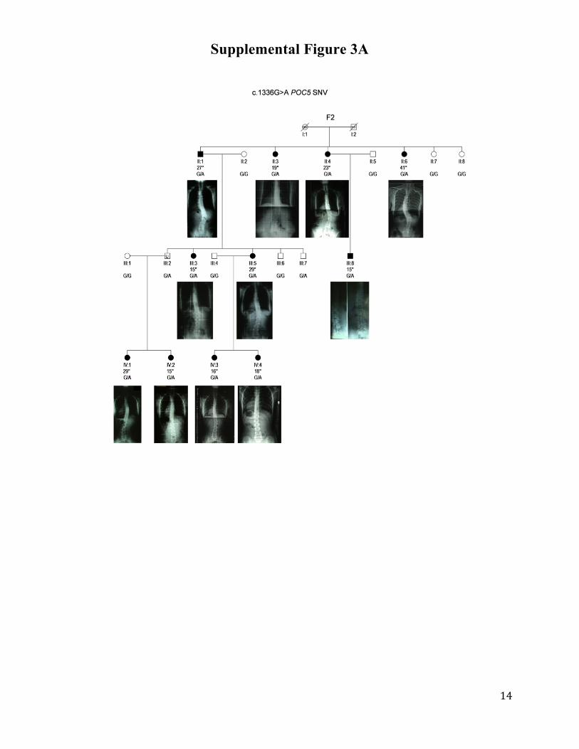
Supplemental Figure 3A
Page 15
15
Supplemental Figure 3B
Page 16
16
Supplemental Figure 3C
Page 17
17
Supplemental Figure 3D
Page 18
18
Supplemental Figure 3E
Supplemental Figures 3A-E. Pedigrees and Spine Radiographs of Idiopathic Scoliosis Families
and Cases Carrying either c.G1336A, c.G1363C or c.C1286T POC5 SNVs.
Page 19
19
Supplemental Figure 4
Supplemental Figure 4. Haplotype Analysis of Families and Cases harboring the c.G1336A
POC5 SNV
Haplotypes at the POC5 locus were reconstructed. The most likely haplotype was determined
using Merlin° software. When different haplotypes had a similar likelihood, reconstruction was
considered impossible (IS patients C39 and C58). All IS patients in whom haplotypes could be
reconstructed carried the c.G1336A POC5 SNV on the same ancestral haplotype, denoted H
(boxed). Genotypes of C39 and C58 (not shown) were compatible with haplotype H.
Page 20
20
Supplemental Figure 5
Supplemental Figure 5. Morphological phenotype of Poc5 knockdown zebrafish
Knockdown embryos (MO-poc5) show abnormal axial phenotypes compared to non-injected
wild-type (WT) embryos. The morphological phenotype of poc5 knockdown zebrafish can be
rescued by over-expression of human POC5.
Page 21
21
Supplemental Figure 6
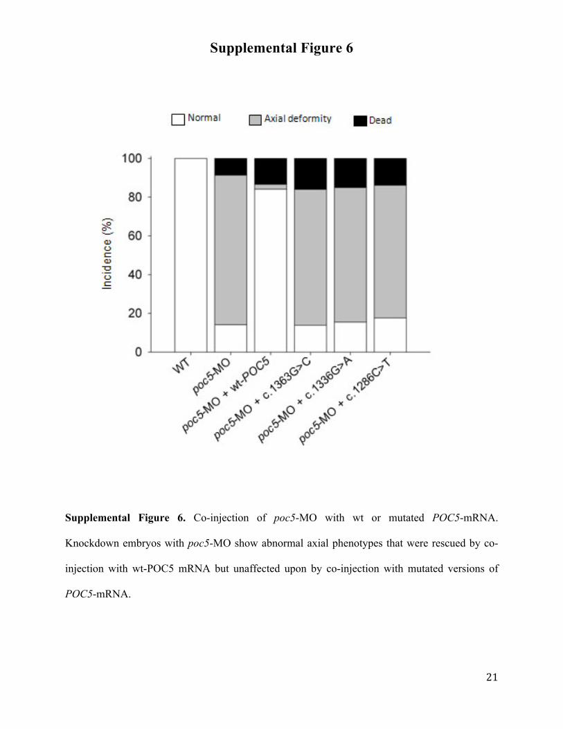
Supplemental Figure 6. Co-injection of poc5-MO with wt or mutated POC5-mRNA.
Knockdown embryos with poc5-MO show abnormal axial phenotypes that were rescued by co-
injection with wt-POC5 mRNA but unaffected upon by co-injection with mutated versions of
POC5-mRNA.
Page 22
22
Supplemental Figure 7
Supplemental Figure 7. Poc5 splice-blocking morpholino (SBMO) injection results in axial
deformities in zebrafish. Reverse transcriptase-polymerase chain reaction (RT-PCR) exhibiting
the loss of proper splicing of poc5 mRNA in poc5-splice blocking morphants (A). The increased
band size is an indicator of the retention of poc5 intron 5 following mRNA processing.
Knockdown embryos (48 hpf) with poc5-SMBO show abnormal axial phenotypes (mild to
severe) compared to non-injected wild-type (WT) embryos (B). SMBO, splice blocking
morpholino; WT, wild-type.
Page 23
23
Supplemental Figure 8 A
B
Supplemental Figure 8. Dose-reponse of POC5 mRNA overexpression in Zebrafish. (A) POC5
mRNAs overexpression led to mild to severe axial phenotypes. Mild axial phenotype is
highlighted by a black arrow. (B) Wild-type POC5 (wt-POC5) or mutated POC5 (mut-POC5)
mRNAs were injected at concentrations of 25 ng/µl, 50 ng/µl, 100 ng/µl and 150 ng/µl.
Page 24
24
Supplemental Figure 9
Supplemental Figure 9. Expression of Human Poc5 in Zebrafish
Human POC5 is expressed in zebrafish injected with myc-tagged wild-type POC5 (wt-POC5) or
mutated POC5 (mut-POC5) mRNAs, but not in non-injected wild-type (WT) fish.
Page 25
25
Supplemental Figure 10
Supplemental Figure 10. In situ Poc5 Expression Pattern in Zebrafish
In situ hybridization with specific zebrafish poc5 antisense at 15 hpf (A), 24 hpf (B), 48 hpf (C),
72 hpf (D) and sense probes at 3dpf (E). Poc5 was expressed ubiquitously during early
somitogenesis. Its expression became restricted to the head and bud region by 24 hpf. By 48 hpf
and 72 hpf, its expression became even more confined to the brain.
Page 26
26
Supplemental Figure 11A
Page 27
27
Supplemental Figure 11B
Page 28
28
Supplemental Figure 11C
Supplemental Figures 11A-C. Pedigrees of Idiopathic Scoliosis Families F1-F41
Filled symbols indicate affected individuals (i.e., idiopathic scoliosis with Cobb’s angle of at
least 15° and rotation of vertebrae). U: uncertain status (e.g. idiopathic scoliosis with Cobb’s
angle below 15° or no rotation). +: DNA sample or blood lymphocytes available. ND: Status not
determined.
Page 29
29
Supplemental Table 1- Whole-Exome Coverage
Table S2- Whole-Exome Coverage
Mean depth coverage
Coverage > 10X
Coverage > 20X
Patient 1(II:6)
Patient 2(III:8)
Patient 3(IV:4)
131X
98.5%
89.9%
130X
98.5%
87.6%
141X
98.8%
90.0%
Page 30
30
Supplemental Table 2- Complete List of the 172 Candidate Variants (SNVs (A) +Indels (B)) (A) Single Nucleotide Variants (SNVs)
Chromosome 3 Variant (NCBI:hg 19) RS_ID Variant (NCBI:hg 19) RS_ID
g.96069538T>A rs13096522 g.98252027G>A rs1529047 g.97517118G>C rs4857294 g.98281078C>T rs6797035 g.97541018C>T rs974572 g.98281349G>T rs9850648 g.97591153C>T rs17301717 g.98299365T>G rs1051712 g.97594261G>A rs6782766 g.98307630C>T rs75450904 g.97660106A>C rs4857302 g.98312581G>C rs4857406 g.97664725C>T rs2172257 g.98512825T>A rs14310 g.97726747T>A rs832032 g.98518072A>G rs17270986 g.97805954T>C rs13082722 g.99643176C>T rs793440 g.97806616G>A rs4518168 g.99886662G>A rs11537816 g.97806944T>C rs80220955 g.100354524A>G rs1144122 g.97806999T>C rs6439602 g.100368546A>G rs61730367 g.97851998A>C rs79920650 g.100374740T>C rs9866111 g.97852083C>T rs75045884 g.100712249T>C rs3732895 g.97852229T>A rs9849637 g.100944932A>G rs75852013 g.97868795A>G rs4857076 g.100963154G>A rs571391 g.97887865G>A rs4133320 g.101066717T>A rs2433031 g.97887985T>A rs4133321 g.101232048A>G g.97888042A>T rs4133322 g.101232093C>A rs55749605 g.97926625A>G rs9837684 g.101283792C>G rs3762735 g.97927329C>T rs28411367 g.101370529T>A g.97958054T>C rs9851509 g.101383562G>A rs11712748 g.97958253A>G rs9847708 g.101443461T>C rs994573 g.97958280C>T rs9828347 g.101445570G>A rs111912421 g.97983561G>C rs9289564 g.105588069G>A rs11711088 g.97983942A>G rs9853906 g.107096547G>A rs709564 g.97983981A>G rs9871143 g.97984280C>T rs17195192 g.98001777G>C rs72487753 g.98002419A>G rs16839214 g.98002587A>G rs16839611 g.98217178T>A rs55639376 g.98220243C>A rs73140298 g.98241847G>C rs6807441 g.98250862C>A rs3749260 g.98250986C>T rs2230344
Page 31
31
Chromosome 5 Variant (NCBI:hg 19) RS_ID Variant (NCBI:hg 19) RS_ID
g.73932315T>C rs9176 g.77656300G>C rs4072852 g.73980960C>T rs71627068 g.77784542C>T rs11740697 g.73981270T>C g.77784643C>T g.74324437G>A rs3811986 g.78076160C>T rs2173012 g.74324548G>A rs3811987 g.78111674A>G rs34152768 g.74324902C>T rs4704166 g.78135241C>T rs25414 g.74364300G>A rs10942729 g.78181423C>T rs17220759 g.74400386G>C rs961098 g.78181477C>T rs1065757 g.74443132C>T rs1422698 g.78324352A>G rs1805074 g.74921686G>A rs9332464 g.78326750G>C rs1805073 g.74962768C>T rs6453139 g.78340286A>G rs532964 g.74981103C>T rs34678567 g.78379537T>G g.75001582A>G rs17672542 g.78421959G>A rs3733890 g.75003678T>C rs2307111 g.78532658C>T rs3733893 g.75427518C>T rs1423099 g.78573790A>T rs13182512 g.75858215C>T rs58087114 g.78671747G>T rs80274918 g.75913301A>G rs2069702 g.79024734A>G rs1541813 g.75913305T>C rs2069685 g.79028327G>A g.75923294T>G rs2431352 g.79028472C>T rs4704585 g.75923307A>G rs2909888 g.79028726A>G rs13158477 g.75932965G>C rs2455230 g.79029594T>C rs1019762 g.75948650A>G rs2431363 g.79086883G>A rs1129770 g.76003254A>T rs463188 g.79095417C>T rs10043986 g.76003258C>T rs464494 g.79172136A>G rs265005 g.76114859C>G rs2242991 g.79172189C>G rs74916729 g.76114963C>T rs1529505 g.79282798G>C rs9293796 g.76115069C>T rs2243072 g.79331434A>C g.76128521G>A rs616235 g.79331450T>G g.76359024C>A rs34400049 g.79351859G>A rs405482 g.76373240A>G rs2303713 g.79351860G>A rs447875 g.76373241G>C rs2303714 g.79361265G>C rs1866389 g.76722443G>A rs40594 g.79375724G>C rs2288395 g.76728837T>C rs335631 g.76734084C>T rs33204 g.76878139T>C rs13176191 g.77298619A>T rs11552314 g.77425028A>T rs6453373
Page 32
32
(B) Indels
Chromosome 3 Variant
(NCBI:hg19) Reference Allele
Mutant Allele
Variant class RS_ID
96069450 CAT C deletion rs34039875 96152499 AT A deletion rs112236687 97367230 C CA insertion rs3214668 97926079 AT A deletion rs5851109 97984691 G GAA insertion rs34155016 98073591 TA T deletion rs11288615 98110406 G GA insertion 98220492 AAG A deletion 98225846 TAGA T deletion rs10603022 98518160 T TAA insertion rs113737993 98518160 T TA insertion rs113737993 99833338 CTG C deletion
100170600 A ATCCTAGAAGGCATTCTCATGAGGACCAGG
AATTCCGATGCCGATCGTC insertion 100175184 TC T deletion rs11338136 100295909 A ATTGTCT insertion rs5851214 100570787 TA TAAA insertion 100570787 TA T deletion 100945069 T(TA)20 TTA deletion 101177901 GA G deletion 101232055 AGG AG deletion 101232056 GGAA G deletion 101370529 TAA TAAA insertion 101370529 TAA T deletion 101370529 TAA TA deletion 101399910 GA G deletion 101443145 AT A deletion rs75043935 101576029 T TACTTTTAGAAAGCTTTAATAACC insertion rs3217713 105377236 CT C deletion rs55698856
Chromosome 5 Variant
(NCBI:hg19) Reference Allele
Mutant Allele
Variant class RS_ID
73980963 GC G deletion rs70976124 74491715 TTCA T deletion rs10563854 75648938 TA T deletion 75648940 AT A deletion rs112425421 76011613 A ACGGCCGCGGGAAG insertion 76359090 G GA insertion rs34239222 76916335 G GC insertion rs5868876 77524068 T TA insertion rs5868908 77745853 C CA insertion rs113934564 78671727 A ATT insertion 78981369 TAACTG T deletion 78981381 TAAAA T deletion 79279310 T TTGA insertion rs3841613
Page 33
33
Supplemental Table 3- Data on the c.G1336A (p.A446T), c.C1286T (p.A429V) and c.G1363C (p.A455P) POC5 SNVs
!
!
Gene!Full'name'of'
protein! Chromosome! Rs'number! Genomic!Coding'DNA'
Sequence!Protein!
POC5% POC5!centriolar!protein!homolog! 5! rs34678567! g.74981103! c.G1336A! p.A446T!
! ! ! rs146984380! g.74981153! c.C1286T! p.A429V!
! ! !B! g.74981076! !c.G1363C! p.A455P!
Supplemental Table 4– Summary of POC5 Sequencing Data in 41 IS families, 150 Cases and 1268 Controls
Data
Families (n=41, including 330 individuals and 135
patients)
Cases with unknown pedigree data
(n=150)
Controls matched for ethnicity with families and cases
(n=1268)
Comparison of
allelic frequency of the rare variant in IS cases vs controls (Fischer’s exact
test)
Sequencing Method
Exome + Sanger Sanger Sanger (n=103)
Exome (n=1165)
c.G1336A (p.A446T)
4/41 (9.8%) MAF= 4.88%
3/150 (2%) MAF= 1.00%
0/103
19/1165 (1.6%) MAF=0.82%
p=0.0445
c.G1363C (p.A455P)
1/41
0/150 (0%)
0/103 0/1165 (0%)
N/A
c.C1286T (p.A429V)
0/41
5/150 (3.3%) MAF= 1.67%
0/103
9/1165 (0.8%) MAF= 0.39%
p=0.0273
N/A-‐ Not applicable (novel mutation)
Page 34
34
Supplemental Table 5- Clinical Data for Idiopathic Scoliosis Patients with c.G1336A, c.G1363C or c.C1286T POC5 SNV *NA : Not available
IS Family Patient Age at
diagnosis (years old)
Cobb's angle on radiograph
(age: years old) Spine deformity
Apical vertebrae Therapy
F2 II-‐1 Fortuitous 27° (73) Right thoracolumbar D12-‐L1 None
II-‐3 Fortuitous 19° (74) Right lumbar L1 None
II-‐4 Fortuitous 23° (71) Right lumbar L2 None
II-‐6 Adolescence 41° (67) Right thoracolumbar L1 None
III-‐3 Adolescence 15° (49) Right thoracolumbar L1 Bracing
III-‐5 Fortuitous 29° (46) Right thoracolumbar D12 None
III-‐8 Fortuitous 15° (42) Right thoracolumbar L1 None
IV-‐1 Adolescence 29°/29° (20) Right thoracic/Left lumbar D8/L2 Physiotherapy
IV-‐2 Fortuitous 15° (16) Right thoracic D7 None
IV-‐3 Fortuitous 14°/16° (19) Right thoracic/Left lumbar D8/L1 None
IV-‐4 12 18° (15) Left thoracic/Left lumbar L1 Physiotherapy
F19 II-‐3 <18 98° (77) Left lumbar L1 None
III-‐2 Fortuitous 15° (47) Right lumbar L3 None
III-‐4 15 38° (41) Right thoracic T9 None
IV-‐1 10 29°/21° (13) Right thoracic/Left lumbar T10/L2 Bracing
F35 I-‐1 12 76°/80° (55) Right thoracic/Left lumbar T9/L2 Bracing
II-‐2 14 45° (43) Right thoracic T7 Bracing
F41 II-‐1 12 52°/39° (14) Right thoracic/Left lumbar T9/L3 Bracing
II-‐2 12 15°/18°/17° (14) Left thoracic/Right thoracic/Left lumbar
T3/T9/L2 Physiotherapy
F31 II-‐1 Fortuitous 18°/15° (44) Right thoracic/Left lumbar T9/L2 None
II-‐2 10 24° (11) Right thoracic T9 Bracing
C150 II-‐1 10 33° (13) Right thoracolumbar T11 Bracing
C39 II-‐1 66 19° (69) Left lumbar L3 Bracing
C58 II-‐1 10 28°/25° (13) Right thoracic/Left lumbar T9/L2 Physiotherapy+bracing
C83 II-‐1 11 27°/25° (12) Right thoracic/Left lumbar T9/L3 Bracing
C1 II-‐1 15 18° Right thoracic/Left lumbar T8/L2 None
C77 II-‐1 12 37°/45° (12) Left thoracic/Right thoracic T3/T9 Bracing
C137 II-‐1 10 64°/60° (13) Right thoracic/Left lumbar T7/L2 Surgery
C149 II-‐1 13 30° (13) NA* NA* Physiotherapy
Page 35
35
Supplemental Table 6- Additional POC5 SNVs Identified Using High-Throughput POC5 Sequencing
IS Patients
Position (Mb)
GRCh37/hg19
dbSNP138 DNA Change
Mutation AA change
Transcript ID
Ensembl
Protein ID
Ensembl
AA Position
Allele frequency
(dbSNP, 1000 Genomes)
Effect Prediction (SNPnexus)
C10 74990497 rs190991771
T>C Missense I/V ENST00000428202
ENSP00000410216 225
No allele frequency Benign
ENST00000380475
ENSP00000369842 108 Benign
ENST00000446329
ENSP00000399481
200 Benign
C39, F41 (II.1, II.2)
74998501 rs200926172 C>T Missense D/N ENST00000428202
ENSP00000410216 148
No allele frequency Benign
ENST00000380475
ENSP00000369842
31 Possibly damaging
ENST00000446329
ENSP00000399481 123 Benign
C128 75013289 Novel C>A 5'UTR -‐
CpG:41 Island Change
Page 36
36
Supplemental Table 7- Primers Used for High-Throughput POC5 Sequencing
ID#PCR Left#primer#sequence Right#primer#sequence PJ1210142_0001 AGGTTCCCTCTCAACACTTTGA ACATCATGGAGACATCATGTTCA PJ1210142_0002 TATTTTGATGCTGTAATCAGCAAC GTGGGGTCTTTTAATCCCTCTG PJ1210142_0003 GTTACAAAGCATGGTAGAGCTTGAA CGGACCATTCATCCTGAAAGTA PJ1210142_0004 ATGGTAGAGCTTGAAAAAGCCTCT CTCAAGCAACTGCAGCAAAATA PJ1210142_0005 CAGAGGGATTAAAAGACCCCACT GATCTTAACAAATGTTTATTGGGTTAA PJ1210142_0006 TCTGGAAGCTGAGGTACTACTTTCA CACTGTGGGTGTTGAACATGTC PJ1210142_0007 AATTAATTTCCCAACAGCAGAAA CTCCCATGAGCTCAGTTGTTGT PJ1210142_0008 AGTCTATGAACTCTCAGGAAAAAGACTT GATTTTGCTGTGGATTTTCTGC PJ1210142_0009 ACTCACCACTGTGACTGGATGA ATTTTAAGTGCCTGTGTATTCTTCA PJ1210142_0010 CCTGAGTAGCTGGGACTACAGG GCATCTTCTGTTCACGTTCCTG PJ1210142_0011 CTCCCAAAGTGCTGGGATTAC CTCCGATGCCCTTACCAGTTAC PJ1210142_0012 ACCATTTCTTCTGATGCAGCAG TTTGTGATTTATAGGGATAGACTCCA PJ1210142_0013 CGGCTGGTGGGGATG TGTAAACATCTAAATTTTTGTTAGGACCA PJ1210142_0014 TTCTTTTCCTTGAACACCAGGA CACATGTGGAAGGAAGTAGTCTGA PJ1210142_0015 ACCAATCACCAAAATCTCCTCA TCTTCTTTGTATCTCAAATTGTTTTGC PJ1210142_0016 ACCAAAATCTCCTCAAATCTTTTT TTCTTCTTTGTATCTCAAATTGTTTTG PJ1210142_0017 ACCGCGCCCAACTAATAATTT TTGTCAAGCAAGAGCTGAAGAA PJ1210142_0018 TTGTCTGCATTCTTGATTAAAGACC TGACCAGTACTACCAGAGAACTTTACTG PJ1210142_0019 AGATGCTTACCATAGCAACTTTGG TGTACACTTACCACCATGTTATGTTT PJ1210142_0020 CACTACGGAACGCCAGACTTT TGTGAACTACGTAGCTTGCTTAACC PJ1210142_0021 AAAACAACTATAATAAGTGATCCTGAGCA TGAGAAAATCACTTTACCAATTGC PJ1210142_0022 TGAATTCAGAAGTCTAACATCCATCA TGATCCATACCTATCCTCTCTAGCA PJ1210142_0023 TTATCTAGCAAGACATTTACTGAATCTCA TTTTAAGAAGTGATATTAACGAACAAACT PJ1210142_0024 TCTAGCAAGACATTTACTGAATCTCAGT GTTTTAAGAAGTGATATTAACGAACAAAC PJ1210142_0025 TGCTGAAGACCTACTGCATATGAA TGATGAAAACCTTCAGAAGATGG PJ1210142_0026 CTCAGGAAACAAAAGATTTTTAGAAA CTTCTCACCCAGTCATGGATTT PJ1210142_0027 CCTGAACTCCAAAGATCAAGCA CGTCTTTTAGCCTTCCAGTATGG PJ1210142_0028 GCTGGTGAGGAAGAGTCAGCTA GGTATCTCATGCCAGTCTGTGC PJ1210142_0029 AATATGCTGAAATTATTCCTCTTACTACA ACTGCTACCTTCTTAAATTATGTGTGA PJ1210142_0030 ATTCATCAAACCACAAAATGTTG GTGTTCCTATATCCCCAGCATG PJ1210142_0031 TGAAGTTCAAAGAAAAATCAAGCTG TCATCCTAAGGGAGAATTGGTG PJ1210142_0032 ATCACTGATCTGGACAGGCATT TTGATTATTGGCCTCTCTTAGGAAG PJ1210142_0033 AACCTTGACTATGAAGAATATCATGAA AATAAGCATGGGACTCTATTATGGTAA PJ1210142_0034 ATGACTGTGAAGCACAGGGTTC CCAGCCCTCTTCAAACTGTTAA PJ1210142_0035 CTCCAGAAATCTAAATCCATATTTTTG CATTTTAGCTACCTCTCATGAATGC PJ1210142_0036 GAAATAAGAGAAATTTAAAACATTTCATG GAGGTGAAACAAATGTTCAAGAAA PJ1210142_0037 TTGAAAAACTCCCTTGTAAATGG GGAAAGGATTTTATCTTAAATATCAAGG PJ1210142_0038 AAAAATCCTAGTTTTCCCTTACATTCA TGCAGATTTGGATACTGTTGCA PJ1210142_0039 CATCCCTCACTCCTGCTCACT CACTTGCTGACACTGCAGCT PJ1210142_0040 GCGCCAAGGAACTTTAAATCTC GCAGATTGCTGAAACAAAGGAC PJ1210142_0041 CGACCAAATCCCGACTCCT CGCCCCCTACCAACCTG PJ1210142_0042 TTCAAACTGCAGGGAGGAATTA CTCCCGGAGCCGCTTAG
Page 37
37
Supplementary Table 8- Primers (5’-3’) Used for Site-Directed Mutagenesis of Human POC5 Open Reading Frame
c.G1336A c.G1363C c.C1286T
Forward GAGGAGCCAGCGTGACTGCCGTTCC Reverse GGAACGGCAGTCACGCTGGCTCCTC
Forward ACCAGGGCTGCTTCCACATCTTCTGTTCACG Reverse CGTGAACAGAAGATGTGGAAGCAGCCCTGGT
Forward TCACGTTCCTGTTTCTCCTCTTGGTGCAGGATC Reverse GATCCTGCACCAAGAGGAGAAACAGGAACGTGA
All substitutions are noted in bold, underlined. All constructs were verified by sequencing.
Page 38
38
References
1. Abecasis, G.R., Cherny, S.S., Cookson, W.O., and Cardon, L.R. 2002. Merlin-‐-‐rapid analysis of dense genetic maps using sparse gene flow trees. Nat Genet 30:97-‐101.
2. Merner, N.D., Girard, S.L., Catoire, H., Bourassa, C.V., Belzil, V.V., Riviere, J.B., Hince, P., Levert, A., Dionne-‐Laporte, A., Spiegelman, D., et al. 2012. Exome sequencing identifies FUS mutations as a cause of essential tremor. Am J Hum Genet 91:313-‐319.
3. Girard, S.L., Gauthier, J., Noreau, A., Xiong, L., Zhou, S., Jouan, L., Dionne-‐Laporte, A., Spiegelman, D., Henrion, E., Diallo, O., et al. 2011. Increased exonic de novo mutation rate in individuals with schizophrenia. Nat Genet 43:860-‐863.
4. Li, H., and Durbin, R. 2009. Fast and accurate short read alignment with Burrows-‐Wheeler transform. Bioinformatics 25:1754-‐1760.
5. Li, H., Handsaker, B., Wysoker, A., Fennell, T., Ruan, J., Homer, N., Marth, G., Abecasis, G., and Durbin, R. 2009. The Sequence Alignment/Map format and SAMtools. Bioinformatics 25:2078-‐2079.
6. DePristo, M.A., Banks, E., Poplin, R., Garimella, K.V., Maguire, J.R., Hartl, C., Philippakis, A.A., del Angel, G., Rivas, M.A., Hanna, M., et al. 2011. A framework for variation discovery and genotyping using next-‐generation DNA sequencing data. Nat Genet 43:491-‐498.
7. McKenna, A., Hanna, M., Banks, E., Sivachenko, A., Cibulskis, K., Kernytsky, A., Garimella, K., Altshuler, D., Gabriel, S., Daly, M., et al. 2010. The Genome Analysis Toolkit: a MapReduce framework for analyzing next-‐generation DNA sequencing data. Genome Res 20:1297-‐1303.
8. Wang, K., Li, M., and Hakonarson, H. 2010. ANNOVAR: functional annotation of genetic variants from high-‐throughput sequencing data. Nucleic Acids Res 38:e164.
9. Thiele, H., and Nurnberg, P. 2005. HaploPainter: a tool for drawing pedigrees with complex haplotypes. Bioinformatics 21:1730-‐1732.
10. Cha, Y.R., and Weinstein, B.M. 2012. Use of PCR template-‐derived probes prevents off-‐target whole mount in situ hybridization in transgenic zebrafish. Zebrafish 9:85-‐89.
11. Thisse, C., and Thisse, B. 2008. High-‐resolution in situ hybridization to whole-‐mount zebrafish embryos. Nat Protoc 3:59-‐69.
12. Bekri, A., Billaud, M., and Thelu, J. 2014. Analysis of NUAK1 and NUAK2 expression during early chick development reveals specific patterns in the developing head. Int J Dev Biol 58:379-‐384.