P04163_05/SK00521-5 p. 1/25 PD-L1 IHC 28-8 pharmDx SK005 50 tests for use with Autostainer Link 48 Table of Contents 1. Intended Use ....................................................................................................................................................................2 2. Summary and Explanation .............................................................................................................................................2 2.1 Non-squamous non-small cell lung cancer (NSCLC) ..........................................................................................2 2.2 Squamous cell carcinoma of the head neck (SCCHN) ........................................................................................2 2.3 Urothelial carcinoma ..............................................................................................................................................2 2.4 Melanoma ................................................................................................................................................................2 3. Principle of Procedure ....................................................................................................................................................3 4. Materials Provided ..........................................................................................................................................................3 5. Materials Required, but not Supplied ............................................................................................................................4 6. Precautions ......................................................................................................................................................................4 7. Storage .............................................................................................................................................................................5 8. Specimen Preparation ....................................................................................................................................................5 8.1 Paraffin-Embedded Sections .................................................................................................................................5 8.2 Use of Decalcified Tissues ....................................................................................................................................5 8.3 Cut Section Storage Recommendation ................................................................................................................5 9. Reagent Preparation .......................................................................................................................................................5 10. Staining Procedure on the Autostainer Link 48............................................................................................................6 11. Quality Control ................................................................................................................................................................6 12. Assay Verification ...........................................................................................................................................................7 13. Staining Interpretation ....................................................................................................................................................7 13.1 Staining Interpretation: Non-squamous NSCLC ..................................................................................................7 13.2 Staining Interpretation: Squamous cell carcinoma of the head and neck (SCCHN).........................................8 13.3 Staining Interpretation: Urothelial carcinoma (UC) .............................................................................................8 13.4 Staining Interpretation: Melanoma ........................................................................................................................8 14. Slide Evaluation...............................................................................................................................................................8 15. Limitations .......................................................................................................................................................................9 15.1 General Limitations ................................................................................................................................................9 15.2 Product-Specific Limitations ...............................................................................................................................10 16. Non-Clinical Performance Evaluation .........................................................................................................................10 16.1 Analytical Specificity for PD-L1 IHC 28-8 pharmDx ...........................................................................................10 16.2 Normal and Neoplastic Tissues...........................................................................................................................10 17. Performance Evaluation ...............................................................................................................................................12 17.1 Performance Evaluation: Non-squamous NSCLC .............................................................................................12 17.2 Performance Evaluation: SCCHN ........................................................................................................................17 17.3 Performance Evaluation: UC ...............................................................................................................................19 17.4 Performance Evaluation: Melanoma ...................................................................................................................20 18. Troubleshooting ............................................................................................................................................................23 19. References .....................................................................................................................................................................24
Transcript
P04163_05/SK00521-5 p. 1/25
PD-L1 IHC 28-8 pharmDx SK005 50 tests for use with Autostainer Link 48
Table of Contents
1. Intended Use .................................................................................................................................................................... 2
2. Summary and Explanation ............................................................................................................................................. 2
2.1 Non-squamous non-small cell lung cancer (NSCLC) .......................................................................................... 2
2.2 Squamous cell carcinoma of the head neck (SCCHN) ........................................................................................ 2
5. Materials Required, but not Supplied ............................................................................................................................ 4
8.2 Use of Decalcified Tissues .................................................................................................................................... 5
10. Staining Procedure on the Autostainer Link 48 ............................................................................................................ 6
11. Quality Control ................................................................................................................................................................ 6
15.1 General Limitations ................................................................................................................................................ 9
16.1 Analytical Specificity for PD-L1 IHC 28-8 pharmDx ........................................................................................... 10
16.2 Normal and Neoplastic Tissues........................................................................................................................... 10
PD-L1 IHC 28-8 pharmDx is a qualitative immunohistochemical assay using Monoclonal Rabbit Anti-PD-L1, Clone 28-8 intended for use in the detection of PD-L1 protein in formalin-fixed, paraffin-embedded (FFPE) non-squamous non-small cell lung cancer (NSCLC), squamous cell carcinoma of the head and neck (SCCHN), urothelial carcinoma (UC), and melanoma tissues using EnVision FLEX visualization system on Autostainer Link 48. PD-L1 protein expression is defined as the percentage of evaluable tumor cells exhibiting partial or complete membrane staining at any intensity. Tumor PD-L1 status is defined by indication specific staining interpretation.
Tumor Indication* Intended Use PD-L1 Expression Clinical
Cut Off
nsNSCLC PD-L1 expression as detected by PD-L1 IHC 28-8 pharmDx in non-squamous NSCLC and SCCHN may be associated with enhanced survival from OPDIVO® (nivolumab).
≥1%, ≥5%, ≥10%
SCCHN ≥1%
UC PD-L1 expression as detected by PD-L1 IHC 28-8 pharmDx in UC may be associated with enhanced response rate from OPDIVO®.
≥1%
Melanoma Positive PD-L1 status as determined by PD-L1 IHC 28-8 pharmDx in melanoma is correlated with the magnitude of the treatment effect on progression-free survival from OPDIVO®.
≥1%
*For details on staining interpretation, refer to section 13 of the product insert and indication specific PD-L1 IHC 28-8 pharmDx Interpretation Manuals.
2. Summary and Explanation
Binding of the PD-1 ligands, PD-L1 and PD-L2, to the PD-1 receptor found on T cells, inhibits T-cell proliferation and cytokine production. Up-regulation of PD-1 ligands occurs in some tumors and signaling through this pathway can contribute to inhibition of active T-cell immune surveillance of tumors (1). OPDIVO® (nivolumab) is a human immunoglobulin G4 (IgG4) monoclonal antibody that binds to the PD-1 receptor and blocks its interaction with PD-L1 and PD-L2, releasing PD-1 pathway-mediated inhibition of the immune response, including the antitumor immune response (2). In syngeneic mouse tumor models, blocking PD-1 activity resulted in decreased tumor growth (3). Clinical benefit from OPDIVO® and clinical utility of PD-L1 IHC 28-8 pharmDx has been investigated in NSCLC, SCCHN, UC, and melanoma.
2.1 Non-squamous non-small cell lung cancer (NSCLC): Detection of PD-L1 expressing tumor cells in a non-squamous non-small cell lung cancer patient specimen may indicate an enhanced survival benefit to OPDIVO® (nivolumab) treatment for the patient. Specimens from patients in OPDIVO® clinical studies sponsored by Bristol-Myers Squibb were tested using PD-L1 IHC 28-8 pharmDx. Clinical study CHECKMATE-057 investigated the clinical validity of PD-L1 IHC 28-8 pharmDx for the assessment of PD-L1 status in non-squamous NSCLC patients treated with OPDIVO® (5,11). The anti-PD-L1 immunotherapeutic OPDIVO® treatment effect has been correlated with PD-L1 expression in patients with advanced NSCLC.
2.2 Squamous cell carcinoma of the head and neck (SCCHN): Detection of PD-L1 expressing tumor cells in SCCHN patient specimens may indicate an enhanced survival benefit to OPDIVO® (nivolumab) treatment for the patient. Specimens from patients in OPDIVO® clinical studies sponsored by Bristol-Myers Squibb were tested using PD-L1 IHC 28-8 pharmDx. Clinical study CHECKMATE-141 investigated the clinical validity of PD-L1 IHC 28-8 pharmDx for the assessment of PD-L1 status in SCCHN patients treated with OPDIVO® (16). 2.3 Urothelial carcinoma (UC): Detection of PD-L1 expressing tumor cells in UC patient specimens may indicate an enhanced response rate benefit to OPDIVO® (nivolumab) treatment for the patient. Specimens from patients in OPDIVO® clinical studies sponsored by Bristol-Myers Squibb were tested using PD-L1 IHC 28-8 pharmDx. Clinical study CHECKMATE-275 investigated the clinical validity of PD-L1 IHC 28-8 pharmDx for the assessment of PD L1 status in UC patients treated with OPDIVO® (18).
2.4 Melanoma: Cytotoxic T-lymphocyte-associated antigen-4 (CTLA-4) is a negative regulator of T-cell activity. YERVOY® (ipilimumab) is a monoclonal antibody that binds to CTLA-4 and blocks the interaction of CTLA-4 with its ligands, CD80/CD86. Blockade of CTLA-4 has been shown to augment T-cell activation and proliferation, which contributes to a general increase in antitumor immune response (4).
PD-1 and CTLA-4 inhibit antitumor immunity through complementary and non-redundant mechanisms. OPDIVO® monotherapy and combination of OPDIVO® and YERVOY® has demonstrated significantly greater improvement in progression-free survival over YERVOY®
alone among patients with advanced melanoma who had not previously received treatment (8-10).
Melanoma tumor tissue specimens from patients treated with OPDIVO® in clinical studies sponsored by Bristol-Myers Squibb were tested using PD-L1 IHC 28-8 pharmDx. Clinical study CHECKMATE-067 investigated the clinical validity of PD-L1 IHC 28-8 pharmDx for the assessment of PD-L1 expression in melanoma patients treated with OPDIVO®, OPDIVO® in combination with YERVOY®, and YERVOY®
alone.
OPDIVO® and YERVOY® are trademarks owned by Bristol-Myers Squibb.
P04163_05/SK00521-5 p. 3/25
3. Principle of Procedure
PD-L1 IHC 28-8 pharmDx contains optimized reagents and protocol required to complete an IHC staining procedure of FFPE specimens using PT Link Pre-treatment Module and Autostainer Link 48 (6). Following incubation with the primary monoclonal antibody to PD-L1 or the Negative Control Reagent (NCR), specimens are incubated with a linker antibody specific to the host species of the primary antibody, and then are incubated with a ready-to-use visualization reagent consisting of secondary antibody molecules and horseradish peroxidase molecules coupled to a dextran polymer backbone. The enzymatic conversion of the subsequently added chromogen results in precipitation of a visible reaction product at the site of the antigen. The color of the chromogenic reaction is modified by a chromogen enhancement reagent. The specimen may then be counterstained and coverslipped. Results are interpreted using a light microscope. Control Slides containing two formalin-fixed, paraffin-embedded human cell lines are provided to validate staining runs.
4. Materials Provided
PD-L1 IHC 28-8 pharmDx (Code SK005) Each kit includes 19.5 mL of Primary Antibody and contains the reagents necessary to perform 50 tests in up to 15 individual runs. The materials listed below are sufficient for 50 tests (50 slides incubated with Primary Antibody to PD-L1 and 50 slides incubated with the corresponding Negative Control Reagent; 100 test slides in total). Additional Primary Antibody to PD-L1 is provided in the kit to stain 15 cell line control slides. The number of tests is based on the use of 2 x 150 µL per slide of each reagent except DAB+ and Target Retrieval Solution.
The kit provides materials sufficient for a maximum of 15 individual staining runs.
Monoclonal rabbit anti-PD-L1 in a buffered solution, containing stabilizing protein, and 0.015 mol/L sodium azide.
1 x 15 mL
Negative Control Reagent*
Monoclonal rabbit control IgG antibody in a buffered solution, containing stabilizing protein, and 0.015 mol/L sodium azide. *Monoclonal rabbit control IgG sold under license from Cell Signaling Technology.
1 x 34.5 mL
LINKER, Anti-Rabbit
Mouse secondary antibody against rabbit immunoglobulins in a buffered solution containing stabilizing protein and 0.015 mol/L sodium azide.
1 x 34.5 mL
Visualization Reagent-HRP
Dextran coupled with peroxidase molecules and goat secondary antibody molecules against rabbit and mouse immunoglobulins in a buffered solution containing stabilizing protein and an antimicrobial agent.
15 x 7.2 mL
DAB+ Substrate Buffer
Buffered solution containing hydrogen peroxide and an antimicrobial agent.
1 x 5 mL DAB+ Chromogen
3,3’-diaminobenzidine tetrahydrochloride in an organic solvent.
Buffered solution, pH 6.1, containing detergent and an antimicrobial agent.
15 slides PD-L1 IHC 28-8 pharmDx Control Slides
Each slide contains sections of two pelleted, formalin-fixed, paraffin-embedded cell lines: NCI-H226** with positive PD-L1 protein expression (originating from human lung squamous cell carcinoma with positive PD-L1 protein expression) and MCF-7 with negative PD-L1 protein expression (originating from human breast adenocarcinoma with negative PD-L1 protein expression).
PD
-L1,
S
K00
5
ph
arm
Dx
XX
XX
X
**Dr. AF Gazdar and Dr. JD Minna at NIH are acknowledged for their contribution in developing NCI-H226 (ATCC Number: CRL-5826™) (7). Note: All reagents included are formulated specifically for use with this kit. In order for the test to perform as specified, no substitutions, other than EnVision FLEX Target Retrieval Solution, Low pH (50x) (Code K8005) can be made. PD-L1 IHC 28-8 pharmDx has been tailored for use with Autostainer Link 48. Please refer to the User Guides for your Autostainer Link 48 and PT Link for further information.
5. Materials Required, but not Supplied
PT Link Pre-treatment Module (Code PT100/PT101/PT200) PT Link rinse station (Code PT109) Autostainer Link 48 (Code AS480) EnVision FLEX Wash Buffer (20x) (Code K8007) EnVision FLEX Hematoxylin (Link) (Code K8008) Distilled or deionized water (reagent-quality water) Timer Positive and negative tissues to use as process controls (see Quality control section and Table 2) Microscope slides: Dako FLEX IHC Microscope Slides (Code K8020) or Fisherbrand Superfrost Plus charged slides. Coverslips Permanent mounting medium and ancillary reagents required for mounting coverslips Light microscope (4x–40x objective magnification)
6. Precautions
1. For in vitro diagnostic use. 2. For professional users. 3. This product contains sodium azide (NaN3), a chemical highly toxic in pure form. At product concentrations, though not classified as
hazardous, NaN3 may react with lead and copper plumbing to form highly explosive build-ups of metal azides. Upon disposal, flush with large volumes of water to prevent metal azide build-up in plumbing (12).
4. Primary Antibody, Negative Control Reagent, Linker, and Visualization Reagent contain material of animal origin. 5. Specimens, before and after fixation, and all materials exposed to them, should be handled as if capable of transmitting infection, and
disposed of with proper precautions (13). 6. Incubation times, temperatures, or methods other than those specified may give erroneous results. 7. Reagents have been optimally diluted. Further dilution may result in loss of antigen staining. 8. The Visualization Reagent, Liquid DAB+ chromogen and prepared DAB+ Substrate-Chromogen solution may be affected adversely if
exposed to excessive light levels. Do not store system components or perform staining in strong light, such as direct sunlight. 9. Paraffin residue may lead to false negative results.
10. Use of reagent volumes other than recommended may result in loss of visible PD-L1 immunoreactivity. 11. Large tissue sections may require 3x150µl of reagent. 12. As a general rule, persons under 18 years of age are not allowed to work with this product. Users must be carefully instructed in the
proper work procedures, the dangerous properties of the product and the necessary safety instructions. Please refer to Safety Data Sheet (SDS) for additional information.
13. Wear appropriate Personal Protective Equipment to avoid contact with eyes and skin. 14. Unused solution should be disposed of according to local, regional, national and international regulations. 15. Safety Data Sheet available for professional users on request.
Danger DAB+ Chromogen: 1–5% biphenyl-3,3',4,4'-tetrayltetraammonium tetrachloride H350 May cause cancer.
H341 Suspected of causing genetic defects. P201 Obtain special instructions before use. P202 Do not handle until all safety precautions have been read and understood. P280 Wear protective gloves. Wear eye or face protection. Wear protective clothing. P308 + P313 IF exposed or concerned: Get medical attention. P405 Store locked up. P501 Dispose of contents and container in accordance with all local, regional, national and international
regulations.
Warning EnVision FLEX Target Retrieval Solution, Low pH (50x): 1-5% citric acid H319 Causes serious eye irritation. H411 Toxic to aquatic life with long lasting effects. P280 Wear eye or face protection. P273 Avoid release to the environment. P264 Wash hands thoroughly after handling. P305 + P351 + P338 IF IN EYES: Rinse cautiously with water for several minutes. Remove contact lenses, if present and easy
to do. Continue rinsing. P337 + P313 If eye irritation persists: Get medical attention. P501 Dispose of contents and container in accordance with all local, regional, national and international
regulations. 7. Storage
Store all components of PD-L1 IHC 28-8 pharmDx, including Control Slides, in original container in the dark at 2-8 °C when not in use on Autostainer Link 48. Do not use the kit after the expiration date printed on the outside of the kit box. If reagents are stored under any conditions other than those specified in this package insert, they must be validated by the user. There are no obvious signs to indicate instability of this product; therefore, positive and negative controls should be run simultaneously with patient specimens.
8. Specimen Preparation
Specimens must be handled to preserve the tissue for IHC staining. Standard methods of tissue processing should be used for all specimens.
8.1 Paraffin-Embedded Sections Formalin-fixed, paraffin-embedded tissues are suitable for use. Recommended handling and processing conditions are: <30 minutes ischemia time prior to immersion in fixative, and 24-48 hours fixation time in 10% neutral buffered formalin. Alternative fixatives have not been validated and may give erroneous results. Specimens should be blocked into a thickness of 3 or 4 mm, fixed in 10% neutral buffered formalin, and dehydrated and cleared in a series of alcohols and xylene, followed by infiltration with melted paraffin. The paraffin temperature should not exceed 60 °C.
8.2 Use of Decalcified Tissues The use of PD-L1 IHC 28-8 pharmDx on decalcified tissues has not been validated and is not recommended.
8.3 Cut Section Storage Recommendation Tissue specimens should be cut into sections of 4-5 µm. After sectioning, tissues should be mounted on Fisherbrand Superfrost Plus charged slides or Dako FLEX IHC Microscope Slides (Code K8020) and then placed in a 58 ± 2 °C oven for 1 hour. To preserve antigenicity, tissue sections, once mounted on slides, should be stored in the dark at 2-8 °C, or room temperature up to 25 °C, and stained within 4 months of sectioning. Slide storage and handling conditions should not exceed 25 °C at any point post-mounting to ensure tissue integrity and antigenicity.
9. Reagent Preparation
The following reagents must be prepared prior to staining: EnVision FLEX Target Retrieval Solution, Low pH (50x) Prepare a sufficient quantity of 1x Target Retrieval Solution, Low pH by diluting Target Retrieval Solution, Low pH (50x) 1:50 using distilled or deionized water (reagent-quality water); the pH of 1x Target Retrieval Solution should be 6.1 ± 0.2. One 30 mL bottle of Target Retrieval Solution, Low pH (50x), diluted 1:50 will provide 1.5 L of 1x reagent, sufficient to fill one PT Link tank which will treat up to 24 slides per use. Discard 1x Target Retrieval Solution after three uses and do not use after 5 days following dilution. Note the FLEX Target Retrieval Solution, low pH (50x) is a red colored solution. Additional EnVision FLEX Target Retrieval Solution, Low pH (50x), if required, is available as Code K8005. EnVision FLEX Wash Buffer (20x) Prepare a sufficient quantity of Wash Buffer by diluting Wash Buffer (20x) 1:20 using distilled or deionized water (reagent-quality water) for the wash steps. Store unused 1x solution at 2-8 ºC for no more than one month. Discard buffer if cloudy in appearance. Refer to the User Guide for your Autostainer Link 48 for further information.
P04163_05/SK00521-5 p. 6/25
EnVision FLEX Wash Buffer (20x) is available as Code K8007.
DAB+ Substrate-Chromogen Solution This solution should be mixed thoroughly prior to use. Any precipitate developing in the solution does not affect staining quality. To prepare DAB+ Substrate-Chromogen Solution, add 1 drop of Liquid DAB+ Chromogen per 1 mL of DAB+ Substrate Buffer and mix. Prepared Substrate-Chromogen is stable for 5 days if stored in the dark at 2-8 C. Important Notes: If using an entire bottle of DAB+ Substrate Buffer, add 9 drops of DAB+ chromogen. Although the label states 7.2 mL, this is the useable volume and does not account for the “dead volume” in the bottle. The color of the Liquid DAB+ Chromogen in the bottle may vary from clear to lavender-brown. This will not affect the performance of this product. Dilute per the guidelines above. Addition of excess Liquid DAB+ Chromogen to the DAB+ Substrate Buffer will result in deterioration of the positive signal.
10. Staining Procedure on the Autostainer Link 48
Procedural Notes The user should read these instructions carefully and become familiar with all components and instrumentation prior to use (see Precautions). All reagents should be equilibrated to room temperature (20-25 C) prior to immunostaining. Likewise, all incubations should be performed at room temperature. Do not allow tissue sections to dry during the staining procedure. Dried tissue sections may display increased nonspecific staining. All of the required steps and incubation times for staining are preprogrammed in the Dako Link software. Please refer to the User Guides for Autostainer Link 48 and PT Link for further information on programming protocols and loading slides and reagents. Note: The reagents and instructions supplied in this system have been designed for optimal performance when used with the recommended reagents and materials. Further dilution of the reagents or alteration of incubation times or temperatures may give erroneous or discordant results. Staining Protocol Please select the PD-L1 IHC 28-8 pharmDx staining protocol from the options in the DakoLink drop down menu. All of the required steps and incubation times for staining are preprogrammed in the DakoLink. If the appropriate PD-L1 IHC 28-8 pharmDx protocols are not on your server please contact your local Technical Service Representative to obtain/install the protocols. Step 1: Deparaffinization, Rehydration and Target Retrieval (3-in-1) Procedure Recommended procedure:
For details, please refer to the PT Link User Guide.
Set PT Link (Code PT100/PT101/PT200) Preheat and Cool to 65 C. Set Heat to 97 °C for 20 minutes. ►Fill PT Link tanks with 1.5 L per tank of Target Retrieval Solution, Low pH, 1x working solution to cover the tissue sections. ►Preheat the Target Retrieval Solution to 65 C. ►Immerse Autostainer racks containing mounted, FFPE tissue sections into the pre-heated Target Retrieval Solution, Low pH (1x working
solution) in PT Link tank. Incubate for 20 minutes at 97 C. ►As soon as target retrieval incubation time has been completed and the temperature has cooled to 65 °C, remove each Autostainer
slide rack with the slides from the PT Link tank and immediately place the Autostainer rack with slides into a tank (e.g., PT Link Rinse Station, Code PT109) containing diluted, room temperature Wash Buffer (Code K8007).
►Immerse slides in diluted, room temperature Wash Buffer for 5 minutes. Step 2: Staining procedure After deparaffinization, rehydration and target retrieval (3-in-1) procedure, the Autostainer racks with slides are placed on Autostainer Link 48. Ensure slides remain wet with buffer while loading and prior to initiating run. The instrument will perform the staining process by applying the appropriate reagent, monitoring the incubation time and rinsing slides between reagents. The reagent times are preprogrammed in the DakoLink software. Step 3: Counterstain Slides should be counterstained for 7 minutes with EnVision FLEX Hematoxylin (Link) (Code K8008). The Hematoxylin incubation time is preprogrammed in the protocol. Step 4: Mounting Non-aqueous, permanent mounting media is required. Note: Some fading of stained slides over time may occur, depending on several factors including, but not limited to, counterstaining, mounting materials and methods, and slide storage conditions. To minimize fading, store slides in the dark at room temperature (20-25 C).
11. Quality Control
Reagents in PD-L1 IHC 28-8 pharmDx have been quality controlled by immunohistochemistry using the target retrieval and staining procedures outlined above. Deviations in the recommended procedures for tissue fixation, processing and embedding in the user’s laboratory may produce significant variability in results. Quality controls should be included in each staining run. These quality controls are
P04163_05/SK00521-5 p. 7/25
specified in Table 2 and include: a H&E stained patient tissue specimen; lab-supplied positive and negative control tissues; and a Dako supplied Control Slide (15). In the USA, consult the quality control guidelines of the College of American Pathologists (CAP) Certification Program for Immunohistochemistry; see also CLSI Quality Assurance for Immunocytochemistry, Approved Guideline (14) for additional information.
12. Assay Verification
Prior to initial use of a staining system in a diagnostic procedure, the user should verify the assay’s performance by testing it on a series of lab-supplied tissues with known IHC performance characteristics representing known positive and negative tissues. Refer to the quality control procedures outlined in the quality control section above. These quality control procedures should be repeated for each new antibody lot, or whenever there is a change in assay parameters. Troubleshooting options for potential problems, their causes and suggested corrective actions are outlined in Table 20.
13. Staining Interpretation
For each staining run, slides should be examined in the order presented in Table 2 to determine the validity of the staining run and enable assessment of the staining of the sample tissue. Staining interpretation for the PD-L1 IHC 28-8 pharmDx in various indications is summarized in Table 1. Table 1: Staining Interpretation
PD-L1 IHC 28-8 pharmDx Staining Interpretation*
Slide evaluation should be performed by a pathologist using a light microscope. For evaluation of the PD-L1 immunohistochemical staining and scoring, 4x objective magnification can be used for initial assessment of the entire specimen followed by the 10-20x objectives for scoring (40x can be utilized for confirmation if needed). PD-L1 staining is indicated with a brown (3,3’-diaminobenzidine, DAB) reaction product.
PD-L1-positive staining is defined as complete circumferential and/or partial linear plasma membrane staining of tumor cells at any intensity. The entire specimen must be evaluated. All viable tumor cells on the entire PD-L1 stained patient slide must be evaluated and included in the PD-L1 scoring assessment. A minimum of 100 viable tumor cells should be present in the PD-L1 stained patient slide to determine the percentage of stained cells. Refer to indication specific staining interpretation sections in this product insert for detailed guidelines.
Cytoplasmic staining, if present, is not considered for scoring purposes. Non-malignant cells and immune cells (e.g., infiltrating lymphocytes or macrophages) may also stain with PD-L1; however, these should not be included in the scoring for the determination of PD-L1 positivity.
SCCHN Cells within foci of dysplasia and carcinoma in situ are excluded from scoring. Accompanying H&E slides allow for the proper assessment of invasive carcinoma, carcinoma in situ, and adjacent normal epithelium.
≥1%
UC Cells within foci of dysplasia and carcinoma in situ are excluded from scoring. Accompanying H&E slides allow for the proper assessment of invasive carcinoma, carcinoma in situ, and adjacent normal epithelium.
≥1%
Melanoma Melanin should be excluded when scoring plasma membrane staining. Comparison to a sequential slide stained with NCR may be useful for identifying and excluding melanin content.
≥1%
*For additional guidance on PD-L1 scoring, please refer to the indication specific staining interpretation sections below and to the relevant PD-L1 IHC 28-8 pharmDx Interpretation Manual for each respective indication.
13.1 Staining Interpretation: Non-squamous NSCLC The entire specimen must be evaluated. All viable tumor cells on the entire PD-L1 stained patient slide must be evaluated and included in the PD-L1 scoring assessment. A minimum of 100 viable tumor cells should be present in the PD-L1 stained patient slide to determine the percentage of stained cells. For non-squamous NSCLC specimens, record the percentage of viable tumor cells exhibiting circumferential and/or partial linear plasma membrane PD-L1 staining at any intensity.
P04163_05/SK00521-5 p. 8/25
13.2 Staining Interpretation: Squamous cell carcinoma of the head and neck (SCCHN) This assay was validated for invasive SCCHN tissue samples and not for lesions with foci of dysplasia or carcinoma in situ. H&E stained slides should accompany each PD-L1 stained sample to allow proper assessment of invasive carcinoma, carcinoma in situ, and adjacent normal epithelium. The entire specimen must be evaluated. All viable tumor cells on the entire PD-L1 stained patient slide must be included in the PD-L1 scoring assessment. A minimum of 100 viable tumor cells should be present in the PD-L1 stained patient slide to determine the percentage of stained cells. For SCCHN specimens, record the percentage of viable tumor cells exhibiting circumferential and/or partial linear plasma membrane PD-L1 staining at any intensity.
13.3 Staining Interpretation: Urothelial carcinoma (UC) This assay was validated for invasive UC tissue samples and not for lesions with foci of dysplasia or carcinoma in situ. H&E stained slides should accompany each PD-L1 stained sample to allow proper assessment of invasive carcinoma, carcinoma in situ, and adjacent normal epithelium. The entire specimen must be evaluated. All viable tumor cells on the entire PD-L1 stained patient slide must be included in the PD-L1 scoring assessment. A minimum of 100 viable tumor cells should be present in the PD-L1 stained patient slide to determine the percentage of stained cells. For UC specimens, record the percentage of viable tumor cells exhibiting circumferential and/or partial linear plasma membrane PD-L1 staining at any intensity.
13.4 Staining Interpretation: Melanoma The entire specimen must be evaluated. All viable tumor cells on the entire PD-L1 stained patient slide must be evaluated and included in the PD-L1 scoring assessment. A minimum of 100 viable tumor cells should be present in the PD-L1 stained patient slide to determine the percentage of stained cells. Specimens are considered PD-L1 positive if ≥ 1% of melanoma cells exhibit circumferential and/or partial linear plasma membrane PD-L1 staining of tumor cells at any intensity. Specimens -are considered PD-L1 negative if < 1% of melanoma cells exhibit circumferential and/or partial linear plasma membrane PD-L1 staining of tumor cells at any intensity. NOTE: When interpreting melanoma patient specimens, brown melanin pigmentation may be present. Melanin should be excluded when scoring plasma membrane staining; comparison to a sequential slide stained with NCR may be useful for identifying and excluding melanin content. If highly elevated melanin precludes scoring of plasma membrane staining of tumor cells, the specimen may be excluded from interpretation and deemed to be indeterminate.
A hematoxylin and eosin (H&E) stain of the tissue specimen is evaluated first to assess tissue histology and preservation quality. Note: The H&E may be reviewed again in the context of the patient tissue slides stained with the Negative Control Reagent and Primary Antibody (steps 5 and 6).
The PD-L1 IHC 28-8 pharmDx and H&E stain should be performed on serial sections from the same paraffin block of the specimen. Tissue specimens should be intact, well-preserved, and should confirm tumor indication.
2. Control Slide (Dako-supplied)
The Control Cell Line Slide stained with the PD-L1 Primary Antibody from PD-L1 IHC 28-8 pharmDx should be examined to ascertain that all reagents are functioning properly. The Control Slide contains the PD-L1-positive cell line pellet and PD-L1-negative cell line pellet.
One Control Slide should be stained with the Primary Antibody to PD-L1 in each staining run. NCI-H226 (PD-L1-positive control cell line originating from human lung squamous cell carcinoma with positive PD-L1 protein expression) acceptance criteria: Plasma membrane staining of ≥80% of cells at ≥2+ average staining
intensity. Non-specific staining <1+ intensity. MCF-7 (PD-L1-negative control cell line originating from human breast adenocarcinoma with negative PD-L1 protein expression) acceptance criteria: No specific staining. Non-specific staining <1+ intensity. Note that staining of a few cells in the
MCF-7 cell pellet may occasionally be observed. The following acceptance criteria are applicable: the presence of ≤10 total cells with distinct plasma membrane staining, or cytoplasmic staining with ≥1+ intensity within the boundaries of the MCF-7 cell pellet are acceptable.
If either of the Control Cell Lines does not meet these criteria, all results with the patient specimens should be considered invalid.
P04163_05/SK00521-5 p. 9/25
3. Positive Control Tissue Slides (Lab-supplied)
The Positive Control Tissue Slides stained with both PD-L1 Primary Antibody and Negative Control Reagent should be examined next. These slides verify that the fixation method and target retrieval process are effective. Known Positive Tissue Controls should only be utilized for monitoring the correct performance of processed tissues and test reagents, NOT as an aid in formulating a specific diagnosis of patient samples.
Controls should be biopsy/surgical specimens of the same tumor indication as the patient specimen, fixed, processed and embedded as soon as possible in the same manner as the patient sample(s). Use intact specimens for interpretation of staining results as necrotic or degenerated cells often stain nonspecifically. The tissues selected for use as the Positive Tissue Controls should give weak to moderate positive staining when stained with PD-L1 to aid in detection of subtle changes in assay sensitivity. Two positive tissue control slides should be included in each staining run. Slide stained with PD-L1: Presence of brown plasma membrane staining should be observed. Non-specific staining should be ≤1+. Slide stained with Negative Control Reagent: No membrane staining. Non-specific staining should be ≤1+. If the positive tissue controls fail to demonstrate appropriate positive staining, results with the test specimens should be considered invalid.
4. Negative Control Tissue Slides (Lab-supplied)
The Negative Control Tissue Slides (known to be PD-L1 negative) stained with both PD-L1 Primary Antibody and Negative Control Reagent should be examined next to verify the specificity of the labeling of the target antigen by the Primary Antibody. Alternatively, negative portions of the Positive Control Tissue may serve as the Negative Control Tissue, but this should be verified by the user.
Controls should be biopsy/surgical specimens of the same tumor indication as the patient specimen, fixed, processed and embedded as soon as possible in the same manner as the patient sample(s). Two Negative Tissue Control Slides should be included in each staining run. Slide stained with PD-L1: No membrane staining in tumor cells. Non-specific staining should be ≤1+. Slide stained with Negative Control Reagent: No membrane staining. Non-specific staining should be ≤1+. If specific plasma membrane staining occurs in the Negative Control Tissue Slides, results with the patient specimen should be considered invalid.
5. Patient tissue slide stained using the Negative Control Reagent
Examine patient specimens stained with the Negative Control Reagent from PD-L1 IHC 28-8 pharmDx. Negative Control Reagent is used in place of the Primary Antibody and aids in interpretation of specific staining at the antigen site.
Absence of plasma membrane staining verifies the specific labeling of the target antigen by the PD-L1 Primary Antibody. Non-specific staining should be ≤1+.
6. Patient tissue slide stained using the PD-L1 primary antibody
Examine the entire slide of the patient specimens stained with the PD-L1 Primary Antibody from PD-L1 IHC 28-8 pharmDx last. Refer to Summary and Explanation, Limitations, and Non-Clinical Performance Evaluation for specific information regarding PD-L1 IHC 28-8 pharmDx immunoreactivity.
Positive staining intensity should be assessed within the context of any non-specific background staining observed on the Negative Control Reagent slide in the same run. As with any immunohistochemical test, a negative result means that the antigen was not detected, not necessarily that the antigen was absent in the cells/tissue assayed. For staining interpretation guidelines, refer to section 13.
15. Limitations
15.1 General Limitations
1. Immunohistochemistry is a multi-step diagnostic process that requires specialized training in the selection of the appropriate reagents; tissue selection, fixation, and processing; preparation of the immunohistochemistry slide, and interpretation of the staining results.
2. Tissue staining is dependent on the handling and processing of the tissue prior to staining. Improper fixation, freezing, thawing, washing, drying, heating, sectioning, or contamination with other tissues or fluids may produce artifacts, antibody trapping, or false-negative results. Inconsistent results may be due to variations in fixation and embedding methods, or to inherent irregularities within the tissue.
3. Excessive or incomplete counterstaining may compromise proper interpretation of results. 4. The clinical interpretation of any positive staining or its absence must be evaluated within the context of clinical presentation,
morphology and other histopathological criteria. The clinical interpretation of any staining, or its absence, must be complemented by morphological studies and proper controls as well as other diagnostic tests. It is the responsibility of a qualified pathologist, who is familiar with the antibodies, reagents and methods used, to interpret the stained slide. Staining must be performed in a certified, licensed laboratory under the supervision of a pathologist who is responsible for reviewing the stained slides and assuring the adequacy of positive and negative controls.
5. Tissues from persons infected with hepatitis B virus and containing hepatitis B surface antigen (HBsAg) may exhibit non-specific staining with horseradish peroxidase (17).
P04163_05/SK00521-5 p. 10/25
6. Reagents may demonstrate unexpected reactions in previously untested tissue types. The possibility of unexpected reactions in tested tissue types cannot be completely eliminated due to biological variability of antigen expression in neoplasms, or other pathological tissues. Contact Dako Technical Support with documented unexpected reactions.
7. False-positive results may be seen due to non-Immunological binding of proteins or substrate reaction products. They may also be caused by pseudo peroxidase activity (erythrocytes) and endogenous peroxidase activity (cytochrome c) (14).
8. The reagents and instructions supplied in this system have been designed for optimal performance. Further dilution of the reagents or alteration of incubation times or temperatures may give erroneous or discordant results.
15.2 Product-Specific Limitations
1. False-negative results could be caused by degradation of the antigen in the tissues over time. Tissue sections, once mounted on slides, should be stored in the dark at 2-8 °C, or room temperature up to 25 °C, and stained within 4 months of sectioning. Slide storage and handling conditions should not exceed 25 °C at any point post-mounting to ensure tissue integrity and antigenicity.
2. For optimal and reproducible results, the PD-L1 protein requires target retrieval pre-treatment when tissues are fixed in neutral buffered formalin and paraffin embedded.
3. Do not substitute reagents from different lot numbers of this product, or from kits of other manufacturers. The only exception is the EnVision FLEX Target Retrieval Solution, Low pH (50x), which, if required, is available as Code K8005.
4. Stained control cell lines should be used only for validation of the staining run and should not be used as a guide to score the staining reaction in patient tissue sections.
5. Use of PD-L1 IHC 28-8 pharmDx on tissues with fixatives other than 10% neutral buffered formalin has not been validated. 6. Excisional, incisional, punch or core needle biopsies were considered acceptable sample types for the clinical trials using PD-L1 IHC
28-8 pharmDx. Fine needle aspirates or other cytology specimens were insufficient for biomarker analysis and were excluded from the clinical trials using PD-L1 IHC 28-8 pharmDx.
16. Non-Clinical Performance Evaluation 16.1 Analytical Specificity for PD-L1 IHC 28-8 pharmDx The primary antibody for PD-L1 IHC 28-8 pharmDx is a rabbit monoclonal anti-human PD-L1 antibody, clone 28-8. The immunogen used for the antibody generation is a purified recombinant human PD-L1 containing the extracellular domain (Phe19-Thr239) of human PD-L1. IHC staining with the PD-L1 Primary Antibody showed no cross-reactivity for PD-L2 exogenously expressed in Chinese hamster ovary (CHO) cells. PD-L1 IHC 28-8 pharmDx specifically detects PD-L1 membrane protein expressed in tumor cells in FFPE tissues, which can be completely abolished by the addition of PD-L1 antigen. PD-L1 IHC 28-8 pharmDx does not detect PD-L1 membrane protein in PD-L1 knock-out tumor cells in which the PD-L1 gene is genetically deleted. 16.2 Normal and Neoplastic Tissues Table 3 summarizes Monoclonal Rabbit Anti-Human PD-L1 antibody immunoreactivity on the recommended panel of normal tissues. Table 4 summarizes Monoclonal Rabbit Anti-Human PD-L1 antibody immunoreactivity on neoplastic tissues in multi-tumor tissue microarrays. All tissues were formalin-fixed, paraffin-embedded and stained with PD-L1 IHC 28-8 pharmDx according to the instructions in this package insert. PD-L1 IHC 28-8 pharmDx detected PD-L1 protein localized in the plasma membrane of cell types known to express the PD-L1 antigen such as immune cells and cells of epithelial origin mainly tumor cells.
Table 3: Summary of PD-L1 IHC 28-8 pharmDx Normal Tissue Reactivity
Tissue Type (# tested)
Positive Plasma Membrane Staining: Tissue Elements
17.1.1 Analytical Sensitivity: Non-squamous NSCLC Analytical sensitivity of PD-L1 IHC 28-8 pharmDx was tested on 112 unique cases of non-squamous NSCLC FFPE specimens staged I to IV using a manufactured production lot. Assessment of PD-L1 expression demonstrated staining across a range of 0-100% positive tumor cells and 0-3 staining intensity. 17.1.2 Repeatability/External Reproducibility: Non-squamous NSCLC The Repeatability and External Reproducibility of PD-L1 IHC 28-8 pharmDx was evaluated at Dako and three external testing sites respectively. The performance data are provided in Table 5 and Table 6. Negative percent agreement (NPA), positive percent agreement (PPA) and overall percent agreement (OA) of independent pair-wise comparison of the tests were determined for each PD-L1 expression level evaluated. The most frequently occurring observation was applied as a reference to calculate the NPA, PPA, OA and the corresponding 95% Wilson Score confidence intervals.
Inter-instrument Each of 34 non-squamous NSCLC specimens with a range of PD-L1 IHC expression was tested on each of three Autostainer Link 48 instruments, repeated twice. The slides were blinded and randomized prior to scoring. A total of 204 independent pair-wise comparisons were performed.
Inter-analyst Each of 34 non-squamous NSCLC specimens with a range of PD-L1 IHC expression was tested by three analysts, repeated twice on one Autostainer Link 48 instrument. The slides were blinded and randomized prior to scoring. A total of 204 independent pair-wise comparisons were performed.
Inter-day Each of 34 non-squamous NSCLC specimens with a range of PD-L1 IHC expression was tested over five non-consecutive days on the Autostainer Link 48 instrument. The slides were blinded and randomized prior to scoring. A total of 170 independent pair-wise comparisons were performed.
Inter-lot Each of 20 non-squamous NSCLC specimens with a range of PD-L1 IHC expression was tested with two replicates with each of five reagent lots on the Autostainer Link 48 instrument. A total of 160 independent pair-wise comparisons were performed.
Intra-run Each of 34 non-squamous NSCLC specimens with a range of PD-L1 IHC expression was tested with five replicates within a run on the Autostainer Link 48 instrument. The slides were blinded and randomized prior to scoring. A total of 167 (10%) and 168 (1% and 5%) independent pair-wise comparisons were performed.
Each of 24 non-squamous NSCLC with a range of PD-L1 IHC expression was tested on five non-consecutive days. Inter-site analysis was performed between three sites on a total of 360 independent pair-wise comparisons.
Intra-site assay Each of 24 non-squamous NSCLC with a range of PD-L1 IHC expression was tested on five non-consecutive days at each of three study sites. Intra-site analysis was performed for three sites on a total of 360 independent pair-wise comparisons.
Scoring of 30 non-squamous NSCLC specimens with a range of PD-L1 IHC expression, stained with PD-L1 IHC 28-8 pharmDx, was performed by three pathologists, one at each of three study sites, on three non-consecutive days. Inter-observer analysis was performed between three sites on a total of 270 independent pair-wise comparisons.
Scoring of 30 non-squamous NSCLC specimens with a range of PD-L1 IHC expression, stained with PD-L1 IHC 28-8 pharmDx, was performed by three pathologists, one at each of three study sites, on three non-consecutive days. Intra-observer analysis was performed for three sites on a total of 270 independent pair-wise comparisons.
17.1.3 Clinical Performance Evaluation: Non-squamous NSCLC Clinical utility of PD-L1 IHC 28-8 pharmDx was evaluated in CHECKMATE-057, a Phase 3, randomized, open-label study of nivolumab vs docetaxel in adult ( 18 years) subjects with advanced or metastatic non-squamous cell NSCLC after failure of prior platinum doublet -based chemotherapy. A total of 582 subjects were randomized at 112 sites in 22 countries (Argentina, Australia, Austria, Brazil, Canada, Chile, Czech Republic, France, Germany, Hong Kong, Hungary, Italy, Mexico, Norway, Peru, Poland, Romania, Russian Federation, Singapore, Spain, Switzerland, and United States). Subjects were randomized 1:1 and stratified according to 1) prior use of maintenance therapy vs. no use of maintenance therapy and 2) second-line vs. third-line therapy. Pre-study (baseline) tumor tissue specimens were collected prior to randomization and prior to first treatment to conduct pre-planned analyses of efficacy according to predefined baseline PD-L1 expression levels (secondary objective). The primary endpoint was overall survival (OS). Other secondary endpoints were objective response rate (ORR), progression-free survival (PFS), and disease-related symptom improvement by 12 weeks, as measured by the Lung Cancer Symptom Scale (LCSS). The baseline demographic and disease characteristics were generally balanced between randomized subjects in the nivolumab and docetaxel groups. The mean age was 62 years (range: 21 to 85) with 34% ≥65 years of age and 7% ≥75 years of age. The majority of patients were white (92%) and male (55%); baseline ECOG performance status was 0 (31%) or 1 (69%). Seventy-nine percent of patients were former/current smokers. Tumor specimens were collected from non-squamous NSCLC tumors, consistent with the inclusion requirements for the study. Frequencies of PD-L1 expression at each of the predefined baseline expression levels in all randomized subjects in CHECKMATE-057 are presented in Table 7.
P04163_05/SK00521-5 p. 15/25
Table 7: Frequency of Pre-Study PD-L1 Expression in All Randomized Subjects with non-squamous NSCLC – CHECKMATE-057
Population PD-L1 Expression Category Nivolumab 3 mg/kg
Without PD-L1 Expression at Baseline (N(%)) 61 (20.9) 66 (22.8) 127 (21.8)
Patients with PD-L1 expression by all predefined expression levels in the OPDIVO® group were associated with enhanced survival compared to docetaxel, whereas survival was similar to docetaxel in patients with no PD-L1 expression. Meaningful differences in median OS were observed in nivolumab over docetaxel subgroups when analyzed by PD-L1 expression level. Median OS was 17.1, 18.2, and 19.4 months for nivolumab subjects compared to 9.0, 8.1, and 8.0 months for docetaxel subjects with 1%, 5%, and 10% PD-L1 expression levels, respectively. There were no differences in OS between the treatment groups in subjects with <1%, <5%, and <10% expression levels, with ranges of median OS of 9.7 to 10.4 months for nivolumab and 10.1 to 10.3 months for docetaxel. The unstratified hazard ratios (HR) and median overall survival (OS) are presented in Figure 1. The Kaplan-Meier plot for subgroups by PD-L1 expression level is shown in Figure 2 and Figure 3.
Figure 1: Forest Plot - OS Based on PD-L1 Expression in non-squamous NSCLC Patients – CHECKMATE-057
Note: The unstratified hazard ratio and the corresponding 95% CI were estimated in a Cox proportional hazards model using the randomized arm as a single covariate
17.2.1 Analytical Sensitivity: SCCHN Analytical sensitivity of PD-L1 IHC 28-8 pharmDx was tested on 236 unique cases of SCCHN FFPE specimens staged I to IV using a manufactured production lot. Assessment of PD-L1 expression demonstrated staining across a range of 0-95% positive tumor cells and 0-3 staining intensity. 17.2.2 Repeatability/External Reproducibility: SCCHN The Repeatability and External Reproducibility of PD-L1 IHC 28-8 pharmDx was evaluated at Dako and three external testing sites, respectively. Average negative agreement (ANA), average positive agreement (APA), overall agreement (OA), and corresponding 95% confidence intervals were performed and are reported in Table 8 and Table 9.
Table 8: Repeatability of PD-L1 IHC 28-8 pharmDx – SCCHN
Repeatability Method % Agreement (95% CI)
≥1% Expression Level
Inter-Lot Each of 39 SCCHN specimens with a range of PD-L1 IHC expression was tested with each of three assay build lots on the Autostainer Link 48 instrument. A total of 115 pair-wise comparisons were performed.
ANA 100 (96.9, 100) APA 100 (96.6, 100) OA 100 (98.4, 100)
Table 9: Full Reproducibility of the PD-L1 IHC 28-8 pharmDx - SCCHN, tested at three external sites
Reproducibility Method % Agreement (95% CI)
≥1% Expression Level
Inter-site assay (three sites) A set of 32 SCCHN specimens, with a range of PD-L1 IHC expression, was
tested on five non-consecutive days at each of three sites. Inter-site analysis was performed between three sites on a total of 2400 pair-wise comparisons.
ANA 96.0 (91.5, 99.2) APA 96.3 (92.9, 99.2) OA 96.2 (92.2, 99.2)
Intra-site assay A set of 32 SCCHN specimens, with a range of PD-L1 IHC expression, was tested on five non-consecutive days at each of three sites. Intra-site analysis was performed for three sites on a total of 960 pair-wise comparisons.
ANA 97.2 (94.7, 99.1) APA 97.4 (95.3, 99.2) OA 97.3 (95.0, 99.2)
Inter-observer (three observers)
Three separate scoring evaluations of a set of 38 SCCHN specimens, demonstrating a range of PD-L1 IHC expression, stained with PD-L1 IHC 28-8 pharmDx, were performed by three pathologists, with a minimum of a 14-day washout period between reads. Inter-observer analysis was performed between three pathologists on a total of 1026 pair-wise comparisons.
ANA 97.1 (94.6, 99.4) APA 97.1 (94.7, 99.4) OA 97.1 (94.7, 99.4)
Intra-observer Three separate scoring evaluations of a set of 38 SCCHN specimens, demonstrating a range of PD-L1 IHC expression, stained with PD-L1 IHC 28-8 pharmDx, were performed by three pathologists, with a minimum of a 14-day washout period between reads. Intra-observer analysis was performed for three pathologists on a total of 342 pair-wise comparisons.
ANA 97.1 (94.2, 99.4) APA 97.1 (94.3, 99.4) OA 97.1 (94.2, 99.4)
17.2.3 Clinical Performance Evaluation: SCCHN Clinical utility of PD-L1 IHC 28-8 pharmDx was evaluated in CHECKMATE-141, an open label, randomized Phase 3 clinical trial of nivolumab vs therapy of investigator's choice in recurrent or metastatic platinum-refractory squamous cell carcinoma of the head and neck (SCCHN). Patients were randomized at 55 sites in 15 countries (Argentina, Brazil, Canada, France, Germany, Hong Kong, Italy, Japan, Korea, Netherlands, Spain, Switzerland, Taiwan, United Kingdom, and United States of America). Subjects were randomized 2:1 (nivolumab: investigator’s choice) and stratified according to prior cetuximab treatment (yes/no). Pre-study (baseline) tumor tissue specimens were collected prior to randomization and prior to first treatment to conduct pre-planned analyses of efficacy according to predefined baseline PD-L1 expression levels (exploratory objective). The major efficacy outcome measure was OS. Additional efficacy outcome measures were PFS and ORR. In this trial, a total of 361 patients were randomized; 240 patients to OPDIVO and 121 patients to investigator’s choice (45% received docetaxel, 43% received methotrexate, and 12% received cetuximab). The median age was 60 years (range: 28 to 83) with 31% ≥65 years of age, 83% were White, 12% Asian, and 4% were Black, and 83% male. Baseline ECOG performance status was 0 (20%) or 1 (78%), 76% were former/current smokers, 90% had Stage IV disease, 45% of patients received only one prior line of systemic therapy, the remaining 55% received two or more prior lines of systemic therapy, and 25% had HPV p16-positive tumors, 24% had HPV p16-negative tumors, and 51% had unknown status. Tumor specimens were collected from SCCHN tumors from either a primary or metastatic site, consistent with the inclusion requirements for the study. 327 subjects (out of 361 total subjects) had tumor tissue collected at baseline with the following site proportion: 29.7% primary, 52.0% metastasis, and 18.3% not reported.
P04163_05/SK00521-5 p. 18/25
Frequencies of PD-L1 expression at each of the predefined baseline expression levels in all randomized subjects in CHECKMATE-141 are presented in Table 10.
Table 10: Frequency of Pre-Study PD-L1 Expression in All Randomized Subjects with SCCHN – CHECKMATE-141
Population PD-L1 Expression Category Nivolumab 3 mg/kg
Without PD-L1 Expression at Baseline (N (%)) 79 (32.9) 22 (18.2) 101 (28.0)
The Phase 3 CHECKMATE-141 trial demonstrated a statistically significant improvement in OS for subjects randomized to nivolumab as compared with investigator’s choice at a pre-specified interim analysis (78% of the planned number of events for final analysis). The median OS was 7.5 months for nivolumab subjects compared to 5.1 months for investigator’s choice subjects with a hazard ratio of 0.70 (95% CI: 0.53, 0.92). In pre-specified exploratory subgroup analyses, using the PD-L1 IHC 28-8 pharmDx assay, the hazard ratio for survival was 0.89 (95% CI: 0.54, 1.45) with median survivals of 5.7 months and 5.8 months for the nivolumab and chemotherapy arms, respectively, in patients with <1% PD-L1 expression. The HR for survival was 0.55 (95% CI: 0.36, 0.83) with median survivals of 8.7 months and 4.6 months for the nivolumab and chemotherapy arms, respectively, in subjects with ≥1% PD-L1 expression (16). The unstratified hazard ratios (HR) and median overall survival (OS) are presented in Table 11. Hazard ratios and median overall survival by PD-L1 expression level subgroup are shown in Table 12.
Table 11: Overall Survival in CHECKMATE-141
OPDIVO (n=240)
Investigator’s Choice (n=121)
Overall Survival
Deaths (%) 133 (55%) 85 (70%)
Median (months) (95% CI)
7.5 (5.5, 9.1)
5.1 (4.0, 6.0)
Hazard ratio (95% CI)a 0.70 (0.53, 0.92)
p-valueb,c 0.0101
a Based on stratified proportional hazards model. b Based on stratified log-rank test. c p-value is compared with 0.0227 of the allocated alpha for this interim analysis.
P04163_05/SK00521-5 p. 19/25
Table 12: Median Overall Survival by PD-L1 Expression Level Subgroup
17.3.1 Analytical Sensitivity: UC Analytical sensitivity of PD-L1 IHC 28-8 pharmDx was tested on 138 unique cases of human UC FFPE specimens staged III to IV using a manufactured production lot. Assessment of PD-L1 expression demonstrated staining across a range of 0-90% positive tumor cells and 0-3 staining intensity.
17.3.2 Repeatability/External Reproducibility: UC The Repeatability and External Reproducibility of PD-L1 IHC 28-8 pharmDx was evaluated at Dako and three external testing sites respectively. Average negative agreement (ANA), average positive agreement (APA), overall agreement (OA), and corresponding 95% confidence intervals were performed and are reported in Table 13 and Table 14.
Table 13: Repeatability of PD-L1 IHC 28-8 pharmDx - UC
Repeatability Method % Agreement (95% CI)
≥1% Expression Level
Combined Precision Each of 29* urothelial carcinoma specimens with a range of PD-L1 IHC expression was tested in the following conditions: 5 operators/instruments/days using one assay lot. A total of 286 independent pair-wise comparisons were performed.
ANA 96.6 (91.7, 100.0) APA 96.4 (90.3, 100.0) OA 96.5 (91.0, 100.0)
Inter-observer Each of 72 urothelial carcinoma specimens with a range of PD-L1 IHC expression were read 3 times by 3 different pathologists with a 2-week washout period in between reads. A total of 1944 independent pair-wise comparisons were performed.
ANA 90.3 (85.2, 94.8) APA 93.1 (89.2, 96.4) OA 91.9 (87.6, 95.7)
Intra-observer Each of 72 urothelial carcinoma specimens with a range of PD-L1 IHC expression were read 3 times by the same pathologist with a 2-week washout period in between reads. A total of 3 pathologists were used and 648 independent pair-wise comparisons were performed.
ANA 95.2 (92.6, 97.4) APA 96.6 (94.6, 98.2) OA 96.0 (93.8, 97.8)
Inter-lot Each of 29* urothelial carcinoma specimens with a range of PD-L1 IHC expression was tested with 3 different assay build lots on the Autostainer Link 48 instrument. A total of 87 independent pair-wise comparisons were performed.
ANA 97.8 (93.8, 100.0) APA 97.6 (92.3, 100.0) OA 97.7 (93.1, 100.0)
Intra-run Each of 29* urothelial carcinoma specimens with a range of PD-L1 IHC expression was tested with five replicates within the same run by the same operator on the same Autostainer Link 48 instrument. A total of 290 independent pair-wise comparisons were performed.
ANA 96.3 (90.3, 100.0) APA 96.8 (92.7, 100.0) OA 96.6 (91.7, 100.0)
*A total of 30 urothelial carcinoma specimens were tested in combined precision, inter-lot, and intra-run studies, one specimen was found to be not evaluable.
P04163_05/SK00521-5 p. 20/25
Table 14: Reproducibility of the PD-L1 IHC 28-8 pharmDx - UC, tested at three external sites
Reproducibility Method % Agreement (95% CI)
≥1% Expression Level
Inter-site assay (three sites)
Each of 46 urothelial carcinoma specimens with a range of PD-L1 IHC expression was tested on five non-consecutive days. Inter-site analysis was performed between three sites on a total of 3440 pair-wise comparisons.
ANA 88.1 (82.2, 93.7) APA 85.8 (77.7, 92.7) OA 87.0 (80.5, 93.2)
Intra-site assay
Each of 46 urothelial carcinoma specimens with a range of PD-L1 IHC expression was tested on five non-consecutive days. Intra-site analysis was performed for three sites on a total of 1376 pair-wise comparisons.
ANA 93.2 (89.5, 96.5) APA 91.9 (87.2, 96.0) OA 92.6 (88.6, 96.2)
Inter-observer (one observer at each of three sites)
Scoring of 78 urothelial carcinoma specimens with a range of PD-L1 IHC expression was performed by three pathologists, one at each of three study sites, on three non-consecutive days. Inter-observer analysis was performed between three sites on a total of 2106 pair-wise comparisons.
ANA 91.9 (87.8, 95.7) APA 92.5 (88.5, 96.1) OA 92.2 (88.1, 95.9)
Intra-observer Scoring of 78 urothelial carcinoma specimens with a range of PD-L1 IHC expression was performed by three pathologists, one at each of three study sites, on three non-consecutive days. Intra-observer analysis was performed for three sites on a total of 702 pair-wise comparisons.
ANA 96.1 (94.0, 98.0) APA 96.4 (94.4, 98.2) OA 96.3 (94.3, 98.0)
17.3.3 Clinical Performance Evaluation: UC Clinical utility of PD-L1 IHC 28-8 pharmDx was evaluated in CHECKMATE-275, A phase II single arm clinical trial of nivolumab in subjects with metastatic or unresectable urothelial cancer who have progressed or recurred following treatment with a platinum agent. A total of 270 subjects were randomized to nivolumab at 63 sites in 11 countries (Australia, Belgium, Czech Republic, Finland, Germany, Italy, Japan, Poland, Spain, Sweden, and United States of America). Pre-study (baseline) tumor tissue specimens were systematically collected and PD-L1 status determined prospectively to define PD-L1 sub groups for pre-specified efficacy analysis. Major efficacy outcome measures included confirmed objective response rate (ORR) as assessed by independent radiographic review committee (IRRC) using Response Evaluation Criteria in Solid Tumors (RECIST v1.1) and duration of response (DOR). The median age was 66 years (range 38 to 90), 78% were male, 86% of patients were white. Twenty-seven percent had non-bladder urothelial carcinoma and 84% had visceral metastases. Thirty-four percent of patients had disease progression following prior platinum-containing neoadjuvant or adjuvant therapy. Twenty-nine percent of patients had received ≥2 prior systemic regimens in the metastatic setting. Thirty-six percent of patients received prior cisplatin only, 23% received prior carboplatin only, and 7% were treated with both cisplatin and carboplatin in the metastatic setting. Forty-six percent of patients had an ECOG performance status of 1. Eighteen percent of patients had a hemoglobin <10 g/dL, and twenty-eight percent of patients had liver metastases at baseline. Patients were included regardless of their PD-L1 status. In study CHECKMATE-275, of the 270 patients, 46% were defined as having PD-L1 expression of ≥1% (defined as ≥1% of tumor cells expressing PD-L1). The remaining 54% of patients, were classified as having PD-L1 expression of <1% (defined as <1% of tumor cells expressing PD-L1). Confirmed ORR in all patients and the two PD-L1 subgroups are summarized in Table 15. Median time to response was 1.9 months (range; 1.6-7.2).
10.3 (1.9+, 12.0+) 7.6 (3.7+,12.0+) NE (1.9+, 12.0+)
*Estimated from the Kaplan-Meier Curve
17.4 Performance Evaluation: Melanoma
17.4.1 Analytical Sensitivity: Melanoma Analytical sensitivity of PD-L1 IHC 28-8 pharmDx was tested on 104 unique cases of melanoma FFPE specimens staged I to IV. Assessment of PD-L1 expression demonstrated staining across a range of 0-100% positive tumor cells and 0-3 staining intensity. 17.4.2 Repeatability/External Reproducibility: Melanoma The Repeatability and External Reproducibility of PD-L1 IHC 28-8 pharmDx was evaluated at Dako and three external testing sites respectively. Average negative agreement (ANA), average positive agreement (APA), overall agreement (OA), and corresponding 95% confidence intervals were performed and are reported in Table 16 and Table 17.
P04163_05/SK00521-5 p. 21/25
Table 16: Repeatability of PD-L1 IHC 28-8 pharmDx - Melanoma
Each of 16 melanoma specimens with a range of PD-L1 IHC expression was tested on each of five Autostainer Link 48 instruments. The slides were blinded and randomized prior to scoring. A total of 160 pair-wise comparisons were performed.
ANA 89.5% (83.2, 93.6%) APA 90.5% (84.8, 94.2%) OA 90.0% (86.0, 92.9%)
Inter-analyst
Each of 16 melanoma specimens with a range of PD-L1 IHC expression was tested by six analysts on one Autostainer Link 48 instrument. The slides were blinded and randomized prior to scoring. A total of 235 pair-wise comparisons were performed.
ANA 96.4% (92.9, 98.2%) APA 96.8% (93.8, 98.4%) OA 96.6% (94.5, 97.9%)
Inter-day
Each of 16 melanoma specimens with a range of PD-L1 IHC expression was tested over five non-consecutive days on the Autostainer Link 48 instrument. The slides were blinded and randomized prior to scoring. A total of 160 pair-wise comparisons were performed.
ANA 95.5% (90.3, 97.9%) APA 96.8% (93.1, 98.6%) OA 96.3% (93.5, 97.9%)
Inter-lot
Each of 16 melanoma specimens with a range of PD-L1 IHC expression was tested with each of three reagent lots on the Autostainer Link 48 instrument. The slides were blinded and randomized prior to scoring. A total of 702 pair-wise comparisons were performed.
ANA 98.6% (97.3, 99.3%) APA 98.8% (97.8, 99.4%) OA 98.7% (98.0, 99.2%)
Intra-run
Each of 16 melanoma specimens with a range of PD-L1 IHC expression was tested with five replicates within a run on the Autostainer Link 48 instrument. The slides were blinded and randomized prior to scoring. A total of 153 pair-wise comparisons were performed.
ANA 97.1% (92.6, 98.9%) APA 97.7% (94.1, 99.1%) OA 97.4% (94.9, 98.7%)
Table 17: Reproducibility of the PD-L1 IHC 28-8 pharmDx - Melanoma, tested at three external sites
Each of 24 melanoma specimens with a range of PD-L1 IHC expression was tested on five non-consecutive days. Inter-site analysis was performed between three sites on a total of 1800 pair-wise comparisons.
ANA 91.7% (89.0, 94.5%) APA 89.6% (86.0, 93.2%) OA 90.8% (87.9, 93.9%)
Intra-site assay
Each of 24 melanoma specimens with a range of PD-L1 IHC expression was tested on five non-consecutive days at each of three study sites. Intra-site analysis was performed for three sites on a total of 720 pair-wise comparisons.
ANA 94.5% (91.6, 97.3%) APA 93.1% (89.2, 96.6%) OA 93.9% (90.7, 96.9%)
Inter-observer (one observer at
each of three sites)
Scoring of 30 melanoma specimens with a range of PD-L1 IHC expression, stained with PD-L1 IHC 28-8 pharmDx, was performed by three pathologists, one at each of three study sites, on three non-consecutive days. Inter-observer analysis was performed between three sites on a total of 810 pair-wise comparisons.
ANA 87.5% (83.8, 91.2%) APA 88.7% (85.4, 92.0%)
OA 88.2% (85.0, 91.5 %)
Intra-observer (one observer at
each of three sites)
Scoring of 30 melanoma specimens with a range of PD-L1 IHC expression, stained with PD-L1 IHC 28-8 pharmDx, was performed by three pathologists, one at each of three study sites, on three non-consecutive days. Intra-observer analysis was performed for three sites on a total of 270 pair-wise comparisons.
ANA 93.0% (88.2, 97.2%) APA 93.7% (89.4, 97.4%) OA 93.3% (89.2, 97.4%)
17.4.3 Clinical Performance Evaluation: Melanoma PD-L1 IHC 28-8 pharmDx was evaluated using specimen from patients enrolled in clinical trial CHECKMATE-067, a Phase 3, randomized, double-blind study of nivolumab monotherapy or nivolumab in combination with ipilimumab versus ipilimumab monotherapy in patients with previously untreated metastatic melanoma. A total of 1296 patients were enrolled at 137 sites in 21 countries (Australia, Austria, Belgium, Canada, Czech Republic, Denmark, Finland, France, Germany, Ireland, Israel, Italy, Netherlands, New Zealand, Norway, Poland, Spain, Sweden, Switzerland, United Kingdom, and United States). Of the 1296 patients enrolled, 945 patients were randomized to one of the three treatment arms in a 1:1:1 ratio and stratified by PD-L1 status (≥5% by a clinical trial assay), BRAF status, and AJCC M stage. The baseline demographic and disease characteristics were generally balanced between randomized patients in the treatment groups. The median age was 60 years (range: 18 to 90) with 40% ≥65 years of age and 13% ≥75 years of age. The majority of patients were white (97%) and male (65%). Pre-study (baseline) tumor specimens were collected from melanoma patients with 86% collected from metastatic sites. Of the 945 randomized study patients, retrospective testing of archival tumor tissue with PD-L1 IHC 28-8 pharmDx assay was performed for 915 (97%) patients. For 55 (6%) study patients, melanin precluded evaluation of PD-L1 expression status and PD-L1 expression status was unknown for 47 (5%) patients due to consent withdrawal or missing samples. Thus PD-L1 expression status was ascertained for 843 (89%) study patients. The proportion of patients with tumor PD-L1 expression at 1% and <1% levels were balanced between the treatment groups. PD-L1 expression status for study patients with PD-L1 IHC pharmDx test results in CHECKMATE-067 are presented in Table 18.
P04163_05/SK00521-5 p. 22/25
Table 18: Frequency of PD-L1 Expression in All Randomized Subjects with Melanoma – CHECKMATE-067
Number of subjects, n (%) Nivolumab Nivolumab + ipilimumab Ipilimumab
PD-L1 quantifiable subjects a 288 278 277 PD-L1 expression level: 1% 171 (59.4) 155 (55.8) 164 (59.2) <1% 117 (40.6) 123 (44.2) 113 (30.8) a Number of quantifiable PD-L1 results only; does not include the number indeterminate PD-L1 results
A pre-planned retrospective analysis of efficacy based on PD-L1 expression (secondary objective) was performed. The co-primary endpoint of progression free survival (PFS) was evaluated across PD-L1 subgroups defined as <1 and 1% in all three arms of the study. The PFS Hazard Ratios (HRs) and median PFS by PD-L1 expression level are presented in Table 19. The Kaplan-Meier plot for subgroups by PD-L1 expression at 1% and <1% levels is shown in Figure 4 and Figure 5. Table 19: Summary of Progression-free Survival by PD-L1 Level and Treatment Group - All Randomized Subjects with Melanoma – CHECKMATE-067
a Hazard ratio for treatment effect based on Cox proportional hazard model with treatment, PD-L1 status, and treatment by PD-L1 status interaction
Abbreviations: CI = confidence interval, NR = not reached, PFS = progression-free survival
Figure 4: Kaplan-Meier Plot of Progression Free Survival of Melanoma Patients - All Randomized Subjects with ≥1% PD-L1 Expression at Baseline – CHECKMATE-067
P04163_05/SK00521-5 p. 23/25
Figure 5: Kaplan-Meier Plot of Progression Free Survival of Melanoma Patients - All Randomized Subjects with <1% PD-L1 Expression at Baseline CHECKMATE-067
Use of the 1% cut off was further supported by a retrospective analysis of outcomes on the basis of PD-L1 expression in 2 additional sub groups of 1- 5% and >5% PD-L1 expression levels. Outcomes in the 1-5% PD-L1 and >5% PD-L1 subgroups was comparable to >1% cut off. See Figure 6 below. PD-L1 IHC 28-8 pharmDx has not been validated to discriminate PD-L1 expression levels between 1 and 5%.
Figure 6. Forest Plot: PFS Based on PD-L1 Expression Comparing OPDIVO-Containing Arms
18. Troubleshooting
Table 20: Troubleshooting
Problem Probable Cause Suggested Action 1. No staining of control or specimen slides
1a. Programming error. 1a. Verify that the SK005 PD-L1 IHC 28-8 pharmDx program was selected for programming of slides.
1b. Lack of reaction with DAB+ Substrate-Chromogen Solution (DAB)
1b. Verify that DAB+ Substrate-Chromogen Solution was prepared properly.
1c. Sodium azide in wash buffer. 1c. Use only Dako Wash Buffer, Code K8007. 1d. Degradation of Control Slide 1d. Check kit expiration date and kit storage
conditions on outside of package. 2. Weak staining of specimen slides. 2a. Inappropriate fixation method used. 2a. Ensure that only neutral buffered formalin fixative
and approved fixation methods are used. 2b. Insufficient reagent volume applied. 2b. Check size of tissue section and reagent volume
applied. 2c. Inappropriate wash buffer used. 2c. Use only Dako Wash Buffer, Code K8007.
P04163_05/SK00521-5 p. 24/25
3. Weak staining of specimen slides or the positive cell line on the Dako-supplied Control Slide.
3a. Inadequate target retrieval. 3a. Verify that the 3-in-1 pre-treatment procedure was correctly performed.
3b. Inappropriate wash buffer used. 3b. Use only Dako Wash Buffer, Code K8007
4. Excessive background staining of slides.
4a. Paraffin incompletely removed. 4a. Verify that the 3-in-1 pre-treatment procedure was correctly performed.
4b. Slides dried while loading onto the Autostainer Link 48.
4b. Ensure slides remain wet with buffer while loading and prior to initiating run.
4c. Nonspecific binding of reagents to tissue section.
4c. Check for proper fixation of the specimen and/or the presence of necrosis.
4d. Inappropriate fixation method used. 4d. Ensure that only neutral buffered formalin fixative and approved fixation methods are used.
5. Tissue detached from slides. 5a. Use of incorrect microscope slides. 5a. Use Dako FLEX IHC Microscope Slides, (Code K8020), or Fisherbrand Superfrost Plus slides.
5b. Inadequate preparation of specimens
5b. Cut sections should be placed in a 58 ± 2 °C oven for 1 hour prior to staining.
6. Excessively strong specific staining. 6a. Inappropriate fixation method used. 6a. Ensure that only approved fixatives and fixation methods are used.
6b. Inappropriate wash buffer used. 6b. Use only Dako Wash Buffer, Code K8007. 7. Target Retrieval Solution is cloudy in appearance when heated.
7. When heated the Target Retrieval Solution turns cloudy in appearance.
7. This is normal and does not influence staining.
NOTE: If the problem cannot be attributed to any of the above causes, or if the suggested corrective action fails to resolve the problem, please call Dako Technical Support for further assistance. Additional information on staining techniques and specimen preparation can be found in Dako Education Guide: Immunohistochemical Staining Methods (15) (available from Dako).
19. References
1. Topalian SL, Drake CG, Pardoll DM. Targeting the PD-1/B7-H1(PD-L1) pathway to activate anti-tumor immunity. Curr Opin Immunol 2012;24(2):207-212.
2. Wang C, Thudium KB, Han M, et al. In vitro characterization of the anti-PD-1 antibody nivolumab, BMS-936558, and in vivo toxicology in non-human primates. Cancer Immunol Res 2014;2(9):846-56.
3. OPDIVO® package insert. 4. YERVOY® package insert. 5. Borghaei H, Paz-Ares L, Horn L, et al. Nivolumab versus Docetaxel in advanced nonsquamous non-small-cell lung cancer. N Engl J
Med 2015; 10.1056/NEJMoa1507643. 6. Phillips T, Simmons P, Inzunza HD, et al. Development of an automated PD-L1 immunohistochemistry (IHC) assay for non-small cell
lung cancer. Appl Immuno Molec Morph 2015; 23(8):541-9. 7. R. Phelps, et al., Journal of Cellular Biochemistry Supplement (1996) 24:32-91. 8. Weber JS, D’Angelo SP, Minor D, et al. Nivolumab versus chemotherapy in patients with advanced melanoma who progressed after
anti-CTLA-4 treatment (CheckMate 037): a randomised, controlled, open-label, phase 3 trial. Lancet Oncol 2015; 16: 375–84. 9. Postow M, Chesney J, Pavlick A, et al. Nivolumab and ipilimumab versus ipilimumab in untreated melanoma. N Engl J Med.
2015;372(21):2006-17. 10. Larkin J, Chiarion-Sileni V, Gonzalez R et al. Combined Nivolumab and Ipilimumab or Monotherapy in Untreated Melanoma. N Engl J
Med. 2015;373(1):23-34. 11. Topalian SL, Hodi FS, Brahmer JR, et al. Safety, Activity, and Immune Correlates of Anti-PD-1 Antibody in Cancer. New Eng. J. Med.
2012; 366(26):2455-2465. 12. Department of Health, Education and Welfare, National Institutes for Occupational Safety and Health, Rockville, MD. “Procedures for
the decontamination of plumbing systems containing copper and/or lead azides.” DHHS (NIOSH) Publ. No. 78-127, Current 13. August 16, 1976.
13. Clinical and Laboratory Standards Institute (formerly NCCLS). Protection of Laboratory Workers From Occupationally Acquired Infections; Approved Guideline – Fourth Edition. CLSI document M29-A4 [ISBN 1-56238-962-9]. Clinical and Laboratory Standards Institute, 950 West Valley Road, Suite 2500, Wayne, Pennsylvania 19087 – 1898 USA, 2014.
14. Clinical and Laboratory Standards Institute (formerly NCCCLS). Quality assurance for Design Control and Implementation of Immunohistochemistry Assays; Approved guideline. CLSI document I/LA28-A2;Vol. 31 No. 4 (ISBN 1-56238-745-6) CLSI, 940 West Valley Road, Suite 1400, Wayne, Pennsylvania 19087 USA; 2011.
15. Taylor CR and Rudbeck L. Education Guide: Immunohistochemical Staining Methods. Sixth Edition. Dako, Carpinteria, California; 2013.
16. Ferris RL, Blumenschein G, Fayette J, et. al. Nivolumab for Recurrent Squamous-Cell Carcinoma of the Head and Neck, N Engl J Med. 2016: DOI: 10.1056/NEJMoa1602252.
17. Omata M, Liew C-T, Ashcavai M, Peters RL. Nonimmunologic binding of horseradish peroxidase to hepatitis B surface antigen: a possible source of error in immunohistochemistry. Am J Clin Path 1980; 73:626.
18. Sharma P, Retz M, et. al. Nivolumab in metastatic urothelial carcinoma after platinum therapy (CheckMate 275): a multicentre, single-arm, phase 2 trial, Lancet Oncology 2017; (18): 312–322.


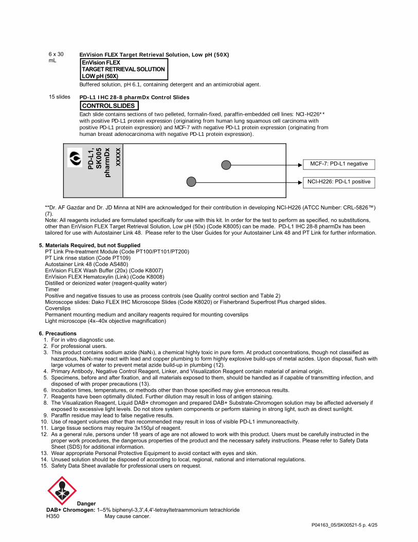


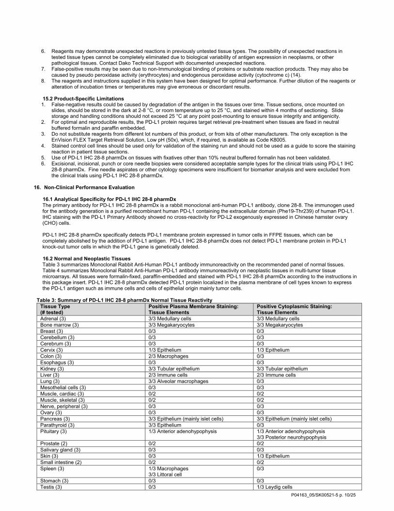

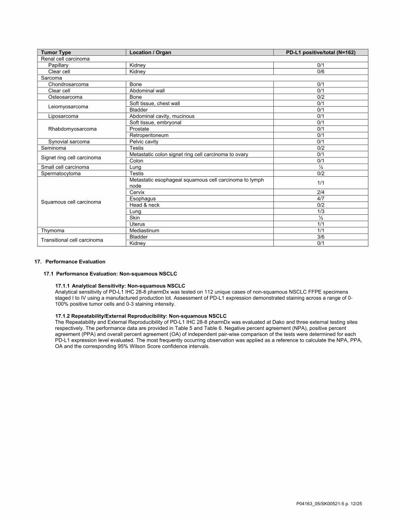


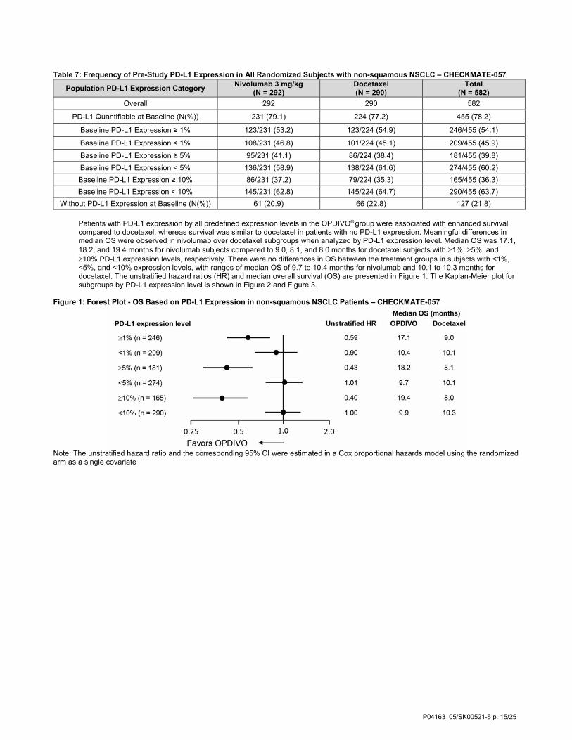


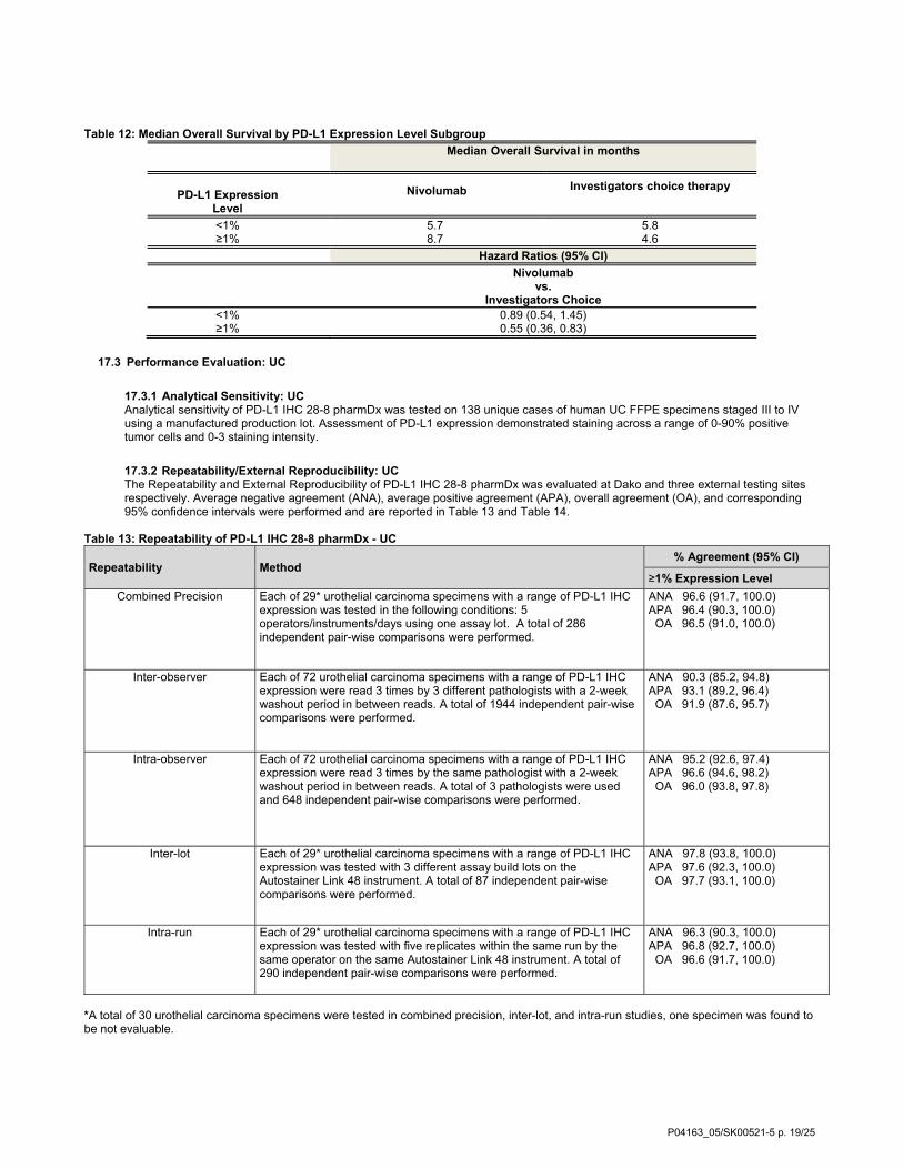
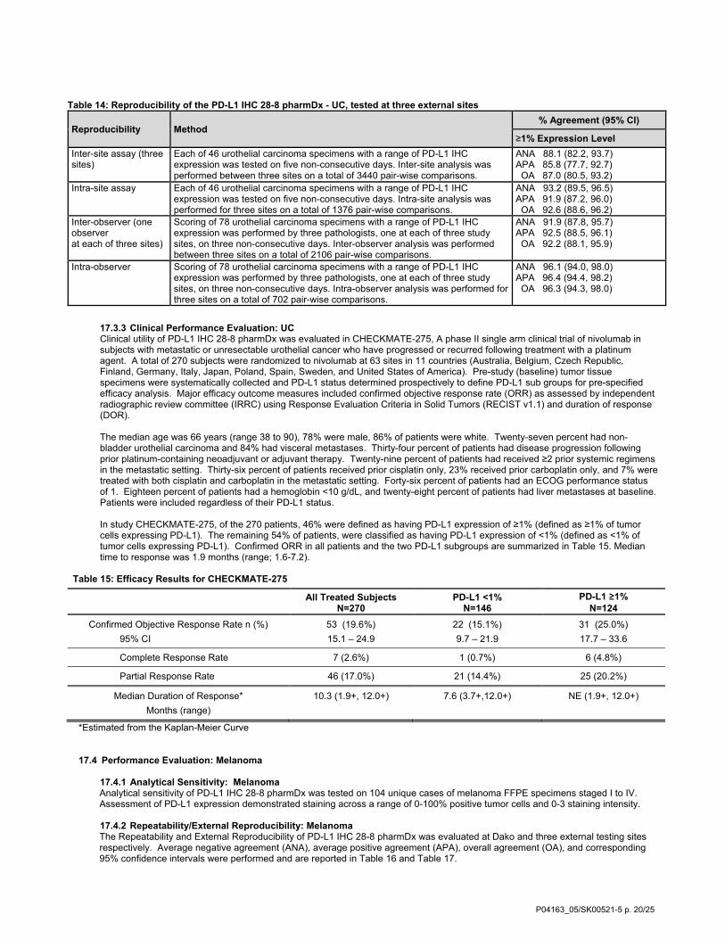

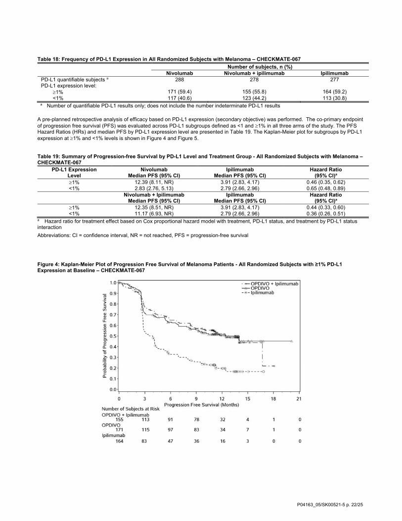
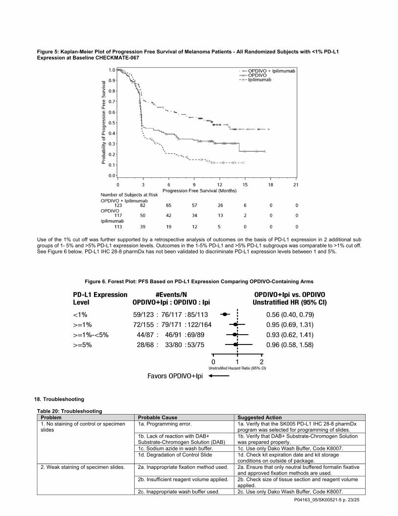


![Anti-PD-L1 antibody [28-8] · For IHC detection kit, Rabbit specific IHC polymer detection kit HRP/DAB (ab209101) is recommended. Immunohistochemistry (Formalin/PFA-fixed paraffin-embedded](https://static.documents.pub/doc/80x56/5f5a978f55ad5f6039367e63/anti-pd-l1-antibody-28-8-for-ihc-detection-kit-rabbit-specific-ihc-polymer-detection.jpg)


