Page 1
S1
Electronic Supporting Information
Triflyloxy-Substituted Carboranes as Useful Weakly
Coordinating Anions
Loren P. Press, Billy J. McCulloch, Weixing Gu, Chun-Hsing Chen, Bruce M. Foxman, and Oleg
V. Ozerov*
1Department of Chemistry, Texas A&M University, 3255 TAMU, College Station, TX 77842.
2Department of Chemistry, Brandeis University, MS 015, 415 South Street, Waltham, MA
02454.
*Email: [email protected] ; Phone: +1-979-845-5870
Electronic Supplementary Material (ESI) for ChemComm.This journal is © The Royal Society of Chemistry 2015
Page 2
S2
Table of Contents
I. General Considerations S3
II. Synthesis of Carborane and Palladium Compounds S5
III. Catalytic Hydrodefluorination Studies S21
IV. Graphical NMR Spectra and MALDI Mass Spectra S28
V. X-ray Crystallography Studies S102
VI. Supporting Information References S121
Page 3
S3
I. General Considerations
Unless specified otherwise, all manipulations were performed under an argon atmosphere
using standard Schlenk or glovebox techniques. Pentane, diethyl ether, tetrahydrofuran,
mesitylene and benzene were dried over sodium benzophenone ketyl, distilled or vacuum
transferred and stored over molecular sieves in an Ar-filled glovebox. Methyl tert-butyl ether
(MTBE) was used as received. 5-fluororesorcinol was prepared according to published
procedure. 1 (cod)PdCl2 was prepared according to published procedure. 2
Trifluoromethanesulfonic acid (HOTf) was vacuum transferred and stored under an inert
atmosphere. Trimethylsilyl trifluoromethanesulfonate (Me3SiOTf) was vacuum transferred and
stored in an argon glovebox free of donor solvents. Trityl chloride (Ph3CCl) was recrystallized
from toluene and pentane at –30 °C in a glovebox free of donor solvents. Triethyl silane (Et3SiH)
and triisopropylsilane (iPr3SiH) were stirred over calcium hydride then vacuum transferred and
stored in a glovebox free of donor solvents. Antimony pentachloride (SbCl5) was vacuum
transferred and used immediately. 4-fluorobenzotrifluoride (4-F-C6H4CF3) and perfluorotoluene
(C6F5CF3) were stirred over calcium hydride then vacuum transferred and stored in a glovebox
free of donor solvents. [Me3NH][HCB11Cl11] was prepared according to published procedures.3
All other chemicals were used as received from commercial vendors. All NMR spectra were
recorded on a Varian iNova 300 spectrometer (1H NMR 299.951 MHz, 31P NMR 121.425 MHz,
13C NMR 75.413 MHz), Varian Mercury 300 spectrometer (13C NMR 75.426 MHz), Varian
iNova 400 spectrometer (1H NMR, 399.755 MHz; 13C NMR, 100.518 MHz; 31P NMR 161.92
MHz; 11B NMR 128 MHz, 29Si NMR 79.458 MHz), or a Varian iNova NMR 500 (1H NMR,
499.425 MHz/ 499.683 MHz; 13C NMR, 75.424 MHz/ 125.580 MHz; 31P NMR, 202.171 MHz)
spectrometer. Chemical shifts are reported in δ/ppm. For 1H and 13C{1H} NMR spectra, the
Page 4
S4
residual solvent peak was used as an internal reference. 1H NMR spectra in C6D5Br were
referenced 7.30 ppm. For 1H and 13C{1H} NMR spectra in 2:1 o-difluorobenzene:C6D6 the
residual benzene solvent peak was used as an internal reference. 13C{1H} NMR spectra in
C6D5Br were referenced by setting the most downfield signal to 130.9 ppm. Et3SiF and Et2SiF2
were identified by 19F NMR spectroscopy by comparing to literature values.4 31P NMR spectra
were referenced externally using 85% H3PO4 at δ 0 ppm. 19F NMR spectra were referenced
externally using 1.0 M CF3CO2H in CDCl3 at δ –78.5 ppm. 29Si NMR spectra were referenced
externally to δ = 0 ppm by using Me4Si. 11B NMR spectra were referenced externally to δ = 0
ppm by using BF3・Et2O. Elemental analyses were performed by CALI Labs, Inc. (Parsippany,
NJ). All simulated MALDI(–) spectra were generated using a publicly available isotope
distribution calculator and mass spectrometry plotter.5
Page 5
S5
II. Synthesis of Carborane and Palladium Compounds
Synthesis of Cs[HCB11H10OTf] – A 100 mL Schlenk flask was loaded with
Cs[HCB11H11] (1.58 g, 5.75 mmol) and a PTFE coated stir bar. The flask was evacuated and
charged with argon. HOTf (17.4 g, 116 mmol) was added under argon flow. Cs[HCB11H11] did
not immediately dissolve. The flask was placed into a 65 °C oil bath for 24 h under argon flow.
All HOTf was then removed in vacuo using a short path distillation apparatus and a 60 °C oil
bath affording a clear residue. To this residue, 3 mL of 1.0 M Cs2CO3 (aq) was added until
slightly basic. An additional 20 mL of distilled water was added. All solvent was removed in
vacuo giving white solid. The product was extracted with MTBE (3 × 75 mL) and filtered
through Celite. All solvent was removed in vacuo giving a clear resin which was >95% pure
Cs[HCB11H10OTf] via 1H, 11B and 19F NMR spectroscopy. Isolation of Cs[HCB11H10OTf] as
described was successful on multiple attempts but the protocol was sensitive to minor variations
in conditions, particularly temperature. Several impurities were have been observed in varying
amounts (5-20%) via MALDI(–) MS and 19F NMR; of those impurities, only
Cs[HCB11H9(OTf)2] was identified: 19F NMR (470 MHz, 25 °C, CD3CN): δ –77.7 (s, 3F), –78.0
(s, 3F)). Yield of Cs[HCB11H10OTf]: 2.39 g (98%). 1H NMR (400 MHz, 25 °C, CD3CN): δ
2.47-0.79 (br m, 10H, B-H), 2.32 (br s, 1H, C-H). 11B NMR (128 MHz, 25 °C, CD3CN): δ 8.5 (s,
1B), –14.1 (d, 5B, JB-H = 145 Hz), –17.8 (d, 5B, JB-H = 155 Hz). 13C{1H} NMR (101 MHz, 25
°C, CD3CN): δ 119.3 (q, JC-F = 316 Hz, CF3), 42.6 (br s, 1C, C-H). 19F NMR (470 MHz, 25 °C,
CD3CN): δ –78.1 (s, 3F).
Synthesis of Cs[HCB11Cl10OTf] – A 250 mL Schlenk flask containing
Cs[HCB11H10OTf] (2.39 g, 5.62 mmol) and a PTFE coated stir bar was charged with freshly
distilled SbCl5 (50 mL, 391 mmol) via glass volumetric pipette under argon flow. The flask was
Page 6
S6
fitted with a reflux condenser and placed into a 180 °C oil bath for 60 h under argon flow. The
reaction turned dark brown after several hours of heating. The SbCl5 was then removed in vacuo
using a short path distillation apparatus giving a brown residue. To this residue, 3 mL of 1.0 M
Cs2CO3 (aq) was added until slightly basic. An additional 20 mL of distilled water was added.
The mixture was refluxed for 4 h giving cream colored slurry. All solvent was removed in vacuo
giving a cream colored solid. The product was extracted with MTBE (3 × 150 mL) and filtered
through Celite. All solvent was removed in vacuo giving a white yellow solid. 1H and 19F NMR
analysis corroborated by MALDI(–) MS revealed the white solid to be 87% pure
Cs[HCB11Cl10OTf]. The product was purified via recrystallization from boiling water as
described below. The solids were loaded into a Schlenk flask with 30 mL of distilled water. The
water was brought to a boil and filtered hot (frit and receiving flask were taken out of a 180 °C
oven and used immediately). The solvent became yellow and upon cooling a white crystalline
solid precipitated out of solution. The solvent was decanted and the solids were washed with 20
mL of distilled water. The recrystallization process was repeated a second time. Residual solvent
was removed in vacuo at 170 °C for 24 h giving a white crystalline solid which was >95% pure
Cs[HCB11Cl10OTf] via 1H, 11B and 19F NMR spectroscopy. Yield 1.98 g (46%). 1H NMR (500
MHz, 25 °C, CD3CN): δ 4.16 (br s, 1H, C-H). 11B NMR (128 MHz, 25 °C, CD3CN): δ –0.40 (s,
1B, para-B), –10.9 (s, 5B, meta-B), –13.0 (s, 5B, ortho-B). 13C{1H} NMR (101 MHz, 25 °C,
CD3CN): δ 119.2 (q, JC-F = 316 Hz, 1C, CF3), 46.4 (br s, 1C, C-H). 19F NMR (470 MHz, 25 °C,
CD3CN): δ –77.0 (s, 3F). Anal. Calcd. for Cs[HCB11Cl10OTf]: C, 3.13; H, 0.13, B, 15.48.
Found: C, 3.40 ; H, 0.20, B, 15.06.
Synthesis of Cs[HCB11Cl9(OTf)2] - A 50 mL three-neck flask was loaded with
Cs[HCB11H11] (0.45 g, 1.63 mmol), a PTFE coated stir bar and HOTf (8 mL , 90.4 mmol). The
Page 7
S7
flask was equipped with a water-cooled condenser and hose adapter connected via Tygon tubing
to an inverted filter funnel submerged in a solution of NaOH and Na2SO3. Chlorine gas was
delivered to the reaction flask from a lecture bottle of chlorine gas with a Monel valve through
Tygon tubing and a bubbler with concentrated HCl solution. The third neck was equipped with a
glass stopper. Chlorine gas was slowly added into the flask and the reaction mixture was heated
to 160 °C for 50 h. The flow of chlorine gas was ceased and reaction mixture was allowed to
cool down to room temperature. All volatiles were removed in vacuo and the resulting white
powder was washed with hexanes then dried in vacuo for 3 h. The products were dissolved in
acetonitrile and identified as Cs[HCB11Cl9(OTf)2] on the basis of 19F NMR spectroscopy, and
MALDI(–) MS. 19F NMR (470 MHz, 25 °C, CD3CN): δ –76.6 (s, 3F), –76.7 (s, 3F).
Synthesis of Cs[HCB11H5Br5OTf] - A 50 mL Schlenk flask was loaded with
Cs[HCB11H11] (0.283 g, 1.03 mmol) and a PTFE coated stir bar. The flask was evacuated and
charged with argon. HOTf (3.40 g, 22.6 mmol) was added under argon flow. The flask was
placed into a 65 °C oil bath for 24 hours under argon flow. The HOTf was then removed in
vacuo using a short path distillation apparatus and a 60 °C oil bath affording a clear residue. To
this residue, 3 mL of 1.0 M Cs2CO3 (aq) was added until slightly basic. All solvent was removed
in vacuo giving white solid. The product was extracted with MTBE (3 × 25 mL) and filtered
through Celite. All solvent was removed in vacuo giving Cs[HCB11H10OTf] as a clear resin.
Under argon flow the flask was charged with 20 mL of dichloromethane followed by Br2 (9 mL,
175 mmol). The flask was fitted with a reflux condenser cooled to 0 °C and placed into a 60 °C
oil bath for 48 h under argon flow. All volatiles were removed in vacuo giving tan powder. This
powder was dissolved in a minimum of boiling water and filtered hot. Upon cooling tan solid
precipitated from solution. This was washed with water and dried in vacuo at 170 °C. An X-ray
Page 8
S8
quality crystal was obtained by slow evaporation of a dichloromethane solution of
Cs[HCB11H5Br5OTf]. MALDI(-) MS and 19F NMR revealed approximately 5% hexa-
brominated product Cs[HCB11H4Br6OTf]. The compound was purified further by dissolving
Cs[HCB11H5Br5OTf] in MTBE/toluene and layering with hexane affording colorless crystals.
Yield: 521 mg (62%). 1H NMR (400 MHz, 25 °C, CD3CN): δ 2.82 (br s, 1H, C-H), 2.43-1.60 (br
m, 5H, B-H). 11B NMR (128 MHz, 25 °C, CD3CN): δ 3.26 (s, 1B), –11.4 (s, 5B), –20.6 (d, JB-H =
166 Hz, 5B). 13C{1H} NMR (101 MHz, 25 °C, CD3CN): δ 119.4 (q, JC-F = 320 Hz, 1C, CF3),
40.2 (br s, 1C, C-H). 19F NMR (376 MHz, 25 °C, CD3CN): –77.1 (s, 3F). Anal. Calcd. for
Cs[HCB11H5Br5OTf]: C, 2.94; H, 0.74, B, 14.53. Found: C, 3.16 ; H, 0.99, B, 14.28.
Synthesis of Cs[HCB11H8(OTf)3] - A 50 mL Schlenk flask was loaded with
Cs[HCB11H11] (0.326 g, 1.19 mmol) and a PTFE coated stir bar. The flask was evacuated and
charged with argon. HOTf (5.88 g, 39.2 mmol) was added under argon flow. The flask was fitted
with a reflux condenser and placed into a 170 °C oil bath for 60 hours under argon flow. The
HOTf was removed in vacuo using a short path distillation apparatus affording a tan residue. To
this residue, 3 mL of 1.0 M Cs2CO3 (aq) was added until slightly basic. The tan residue did not
fully dissolve. An additional 20 mL of distilled water was added and the mixture was brought to
a boil at which point the tan residue dissolved. Upon cooling a white solid precipitated from
solution. The solvent was decanted and the white solid was washed with 20 mL of water. All
solvent was removed in vacuo giving a white solid. 1H 11B, and 19F NMR corroborated by
MALDI(-) MS revealed the white solid to be >95% pure Cs[HCB11H8(OTf)3]. The compound
was further purified via recrystallization from acetone and water. Yield: 772 mg (90 %). 1H
NMR (400 MHz, 25 °C, CD3CN): δ 2.57 (br s, 1H, C-H), 2.72-0.83 (br m, 8H, B-H). 11B{1H}
NMR (128 MHz, 25 °C, CD3CN): δ 4.0 (s, 1B), –0.28 (s, 2B), –16.05 (s, 1B), –16.6 (s, 2B), –
Page 9
S9
21.3 (s, 2B), –23.1 (s, 2B), –23.8 (s, 1B). 13C{1H} NMR (101 MHz, 25 °C, CD3CN): δ 119.4 (q,
JC-F = 318 Hz, 2C, CF3), 119.3 (q, JC-F = 320 Hz, 1C, CF3), 39.5 (s, 1C, C-H). 19F NMR (376
MHz, 25 °C, CD3CN): –77.43 (s, 6F), –77.75 (s, 3F). Anal. Calcd. for Cs[HCB11H8(OTf)3]: C,
6.67; H, 1.26, B, 16.51. Found: C, 6.93 ; H, 0.91, B, 16.29.
Synthesis of [Ph3C][OTf] –A 50 mL Schlenk flask was loaded with Ph3CCl (0.524 g,
1.88 mmol) and dissolved in 6 mL of benzene. Via syringe, Me3SiOTf (1.22 mL, 6.76 mmol)
was added giving a bright orange solution. The reaction was stirred for 2 h at room temperature
and solvent was removed in vacuo giving orange powder. The orange powder was washed with
pentane (2 × 15 mL). All solvent was decanted and the solids were dried in vacuo. Yield: 715 mg
(97 %). 1H NMR (500 MHz, 25 °C, CDCl3): δ 8.24 (apparent t, JH-H = 7.3 Hz, 3H, [Ph3C]+), 7.89
(apparent t, JH-H = 7.4 Hz, 6H, [Ph3C]+), 7.70 (d, JH-H = 7.7 Hz, 6H, [Ph3C]+). 13C{1H} NMR
(101 MHz, 25 °C, CDCl3): δ 210.7 (s, 1C, [Ph3C]+), 143.4 (s, [Ph3C]+), 142.7 (s, [Ph3C]+), 139.9
(s, [Ph3C]+), 130.7 (s, [Ph3C]+), 120.7 (q, JC-F = 321 Hz, 1C, CF3), . 19F NMR (376 MHz, 25 °C,
CDCl3): δ –79.0 (s, 3F).
Previously, [Ph3C][OTf] was synthesized by Martin and co-workers6,7 from Ph3CCl and
trifluoracetyl triflate and by Bosnich and co-workers8 from Ph3CCl and AgOTf. In the latter
work, no yield was reported and the purity of [Ph3C][OTf] was only ~80% due to Ph3COH
formation. Literature NMR data follow: 1H NMR (CF3CO2H)6 δ 8.26 (tt, J = 7.5 and 1 Hz, 3H),
7.90 (tt, J = 7.5 and 1 Hz, 6H), 7.78 (tt, J = 7.5 and 1 Hz, 6H); 1H NMR (CH3NO2)7 δ 7.87 (m,
12), 8.33 (m, 3H); 19F NMR (CH3NO2)7 δ –78.1 (s); 1H NMR (CD2Cl2)8 δ 8.30 (br s, 3H), 7.92
(br s, 6H), 7.70 (br s, 6H).
Synthesis of [Ph3C][HCB11Cl10OTf] – A 50 mL Schlenk flask was loaded with
Cs[HCB11Cl10OTf] (0.195 g, 0.254 mmol) and [Ph3C][OTf] (0.0998 g, 0.254 mmol). 8 mL of
Page 10
S10
fluorobenzene was added and the mixture was stirred for 12 h. The mixture was filtered and
solvent removed in vacuo giving yellow residue. Washing with pentane (3 × 10 mL), decanting
and removing all volatiles in vacuo gave a bright yellow powder. Surprisingly
Ph3C[HCB11Cl10OTf] was poorly soluble in common solvents such as CD3CN and CDCl3 giving
broad indiscernible resonances in the 1H NMR spectrum. The compound had excellent solubility
in C6D5Br. Yield: 212 mg (95 %). 1H NMR (500 MHz, 25 °C, C6D5Br): δ 7.76 (apparent t, JH-H =
7.2 Hz, 3H, [Ph3C]+), 7.43 (apparent t, JH-H = 7.8 Hz, 6H, [Ph3C]+), 7.15 (d, JH-H = 7.5 Hz, 6H,
[Ph3C]+), 2.78 (br s, 1H, C-H) . 11B NMR (128 MHz, 25 °C, C6D5Br): δ –0.98 (s, 1B), –11.43 (s,
5B), –13.77 (s, 5B). 13C{1H} NMR (101 MHz, 25 °C, C6D5Br): δ 209.2 (s, 1C, [Ph3C]+), 143.0
(s, [Ph3C]+), 142.2 (s, [Ph3C]+), 139.1 (s, [Ph3C]+), 130.2 (s, [Ph3C]+), 118.5 (q, JC-F = 318 Hz,
1C, CF3), 45.8 (br s, 1C, [HCB11Cl10OTf]–). 19F NMR (470 MHz, 25 °C, C6D5Br): δ –76.38.
Anal. Calcd. for [Ph3C][HCB11Cl10OTf]: C, 28.70; H, 1.84, B, 13.53. Found: C, 28.67 ; H, 1.72,
B, 13.27.
Synthesis of Na[HCB11Cl11] – A 250 mL Schlenk flask was loaded with
[Me3NH][HCB11Cl11] (1.0927 g, 1.877 mmol), Na2CO3 (0.263 g, 1.877 mmol), 100 mL of
MeOH, 20 mL of H2O and a PTFE coated stirbar. The mixture was heated to reflux for 2 h
followed by removal of all solvent in vacuo. The resultant off white solid was dried in vacuo
over night at 200 °C then brought into a glovebox. The solid was suspended in fluorobenzene
and filtered. The frit was washed with additional fluorobenzene until the entire product was
washed through. All solvent was removed in vacuo and the resultant white solid was dried for 12
h at 200 °C. The product was used without further purification and found to be >95% pure
Na[HCB11Cl11] via 1H and 11B NMR spectroscopy. Yield: 0.620 g (61%). 1H NMR (300 MHz,
25 °C, CD3CN): δ 4.10 (br s, 1H, C–H).
Page 11
S11
Synthesis of [Ph3C][HCB11Cl11] 9 –A 50 mL culture tube was loaded with
Na[HCB11Cl11] (0.540 g, 0.991 mmol), Ph3CCl (0.278 g, 0.997 mmol), and 15 mL of
fluorobenzene. The mixture was stirred for 6 h then filtered and all solvent was removed in
vacuo giving a bright yellow solid. This was washed with pentane (2 × 20 mL) and dried in
vacuo. The product was used without further purification and found to be >95% pure
[Ph3C][HCB11Cl11] via 1H and 11B NMR spectroscopy. Yield: 0.654 g (86%). 1H NMR (400
MHz, 25 °C, C6D5Br): δ 7.72 (br s, 3H, [Ph3C]+), 7.41 (br s, 6H, [Ph3C]+), 7.13 (br s, 6H,
[Ph3C]+), 2.84 (br s, 1H, [HCB11Cl11]–). 11B NMR (128 MHz, 25 °C, C6D5Br): δ –2.58 (br s, 1B),
–10.18 (br s, 5B), –13.39 (br s, 5B). 13C{1H} NMR (101 MHz, 25 °C, C6D5Br): δ 209.1 (br s, 1C,
[Ph3C]+), 142.9 (s, [Ph3C]+), 142.3 (s, [Ph3C]+), 139.2 (s, [Ph3C]+), 130.2 (s, [Ph3C]+), 46.7 (br s,
1C, [HCB11Cl11]–).
Comparison of [Ph3C][HCB11Cl10OTf] with other [Ph3C][WCA] NMR signals –
Three J. Young NMR tubes were loaded with [Ph3C][HCB11Cl10OTf] (0.062 g, 0.071 mmol),
[Ph3C][HCB11Cl11] (0.058 g, 0.076 mmol), and [Ph3C][B(C6F5)4] (0.079 g, 0.086 mmol),
respectively. 800 μL of C6D5Br was added to each NMR tube giving intense yellow orange
solutions. Each sample was analyzed by 1H, 13C{1H} and 19F NMR spectroscopy (See S11,
Figure 2.1, Tables 2.1 and 2.2). Surprisingly, compound [Ph3C][OTf] was poorly soluble in
C6D5Br and no useful NMR signals could be obtained.
Page 12
S12
Table S2.1 - Comparison of 1H NMR resonances of various [Ph3C][WCA] salts in C6D5Br
[Ph3C][WCA] ortho C-H
(δ, m)
meta C-H
(δ, m)a
para C-H
(δ, m)a
carborane C-H
(δ, m)
[Ph3C][HCB11Cl10OTf] 7.14, br d 7.43, br at 7.76, br at 2.78, br s
[Ph3C][HCB11Cl11] 7.13, br s 7.41. br s 7.72, br s 2.84, br s
[Ph3C][ [B(C6F5)4]] 7.11, br s 7.34, br at 7.71, br s n/a
a. at = apparent triplet
0123456789ppm
[Ph3C][HCB11Cl10OTf] – 1H NMR (C6D5Br, 25 °C)
[Ph3C][HCB11Cl11] – 1H NMR (C6D5Br, 25 °C)
[Ph3C][B(C6F5)4] – 1H NMR (C6D5Br, 25 °C)
Figure S2.1 – Comparison of 1H NMR spectra of various [trityl][WCA] in C6D5Br
Page 13
S13
Synthesis of [Et3Si][HCB11Cl10OTf] – A 10 mL PTFE capped glass vial was charged
with [Ph3C][HCB11Cl10OTf] (0.216 g, 0.246 mmol), fluorobenzene (1 mL) and triethylsilane
(1.5 g, 9.3 mmol) resulting in a colorless solution. All solvent was removed in vacuo and the
white solid was washed with pentane, dried in vacuo, collected and stored in a –30 °C glovebox
freezer. The white solid was determined to be >95% pure [Et3Si][HCB11Cl10OTf] via 1H, 11B,
13C{1H}, 19F and 29Si{1H} NMR. Yield: 143 mg (77%). An X-Ray quality crystal was grown by
charging a 10 mL PTFE capped glass vial with [Ph3C][HCB11Cl10OTf] and triethylsilane in an
analogous fashion as described above then adding fluorobenzene until all solids dissolved.
Aliquots of the solution were layered with additional fluorobenzene placed into a pentane
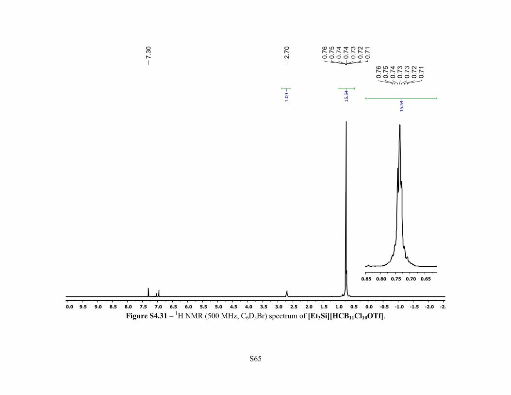
chamber affording clear colorless block crystals. 1H NMR (500 MHz, 25 °C, C6D5Br): δ 2.70 (br
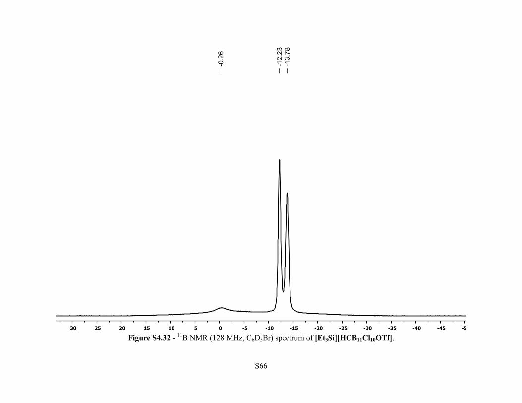
s, 1H, C-H), 0.76-0.71 (m, 15H, Et3Si). 11B NMR (128 MHz, 25 °C, C6D5Br): δ –0.26 (br s, 1B),
–12.23 (s, 5B), –13.78 (s, 5B). 13C{1H} NMR (126 MHz, 25 °C, C6D5Br): δ 117.6 (q, JC-F = 324
Hz, 1C, CF3), 48.2 (br s, 1C, [HCB11Cl10(OSO2CF3)]–), 5.21 (s, 3C, Et3Si), 5.18 (s, 3C, Et3Si).
19F NMR (470 MHz, 25 °C, C6D5Br): δ –71.9 (s, 3F). 29Si NMR (79.5 MHz, 25 °C, C6D5Br): δ
77.7 (s, 1Si).
Synthesis of [iPr3Si][HCB11Cl10OTf] - A 10 mL PTFE capped glass vial was charged
with [Ph3C][HCB11Cl10OTf] (0.141 g, 0.160 mmol), fluorobenzene (1 mL) and
Table S2.2 - Comparison of 13C{1H} NMR resonances of various [Ph3C][WCA] salts in C6D5Br
[Ph3C][WCA] Ph3C+ (δ, m)
[Ph3C][HCB11Cl10OTf] 209.2, s
[Ph3C][HCB11Cl11] 209.1, br s
[Ph3C][B(C6F5)4] 209.4, s
Page 14
S14
triisopropylsilane (1.6 g, 10.1 mmol) resulting in a colorless solution. All solvent was removed in
vacuo and the white solid was washed with pentane, dried in vacuo, collected and stored in a –30
°C glovebox freezer. The white solid was determined to be >95% pure [iPr3Si][HCB11Cl10OTf]
via 1H, 11B 13C{1H}, 19F and 29Si{1H} NMR. Yield: 0.088 g (69%). An X-Ray quality crystal
was grown by charging a 10 mL PTFE capped glass vial with [Ph3C][HCB11Cl10OTf] and
triisopropylsilane as described above then adding fluorobenzene until all solids dissolved.
Aliquots of the solution were then layered with additional fluorobenzene and placed into a
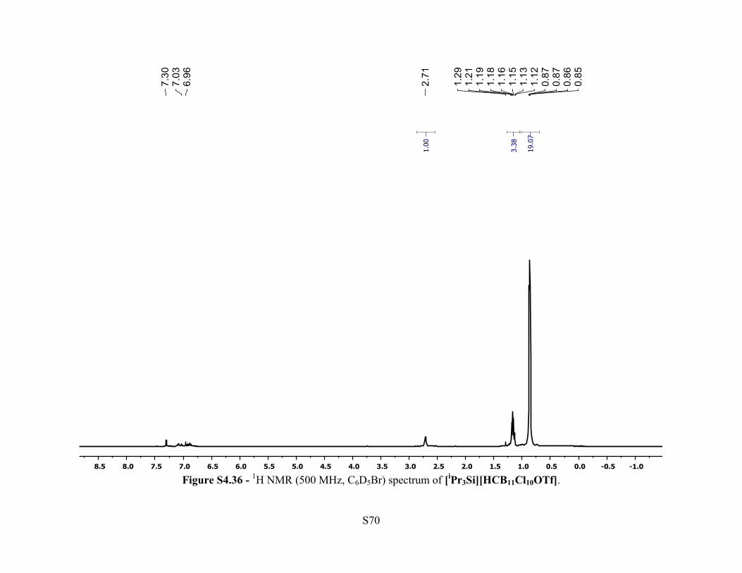
pentane chamber affording colorless needle crystals. 1H NMR (500 MHz, 25 °C, C6D5Br): δ 2.71
(br s, 1H, [HCB11Cl10OTf]–), 1.21-1.12 (m, 3H, Si(CH(CH3)2)3), 0.87-0.85 (m, 18H,
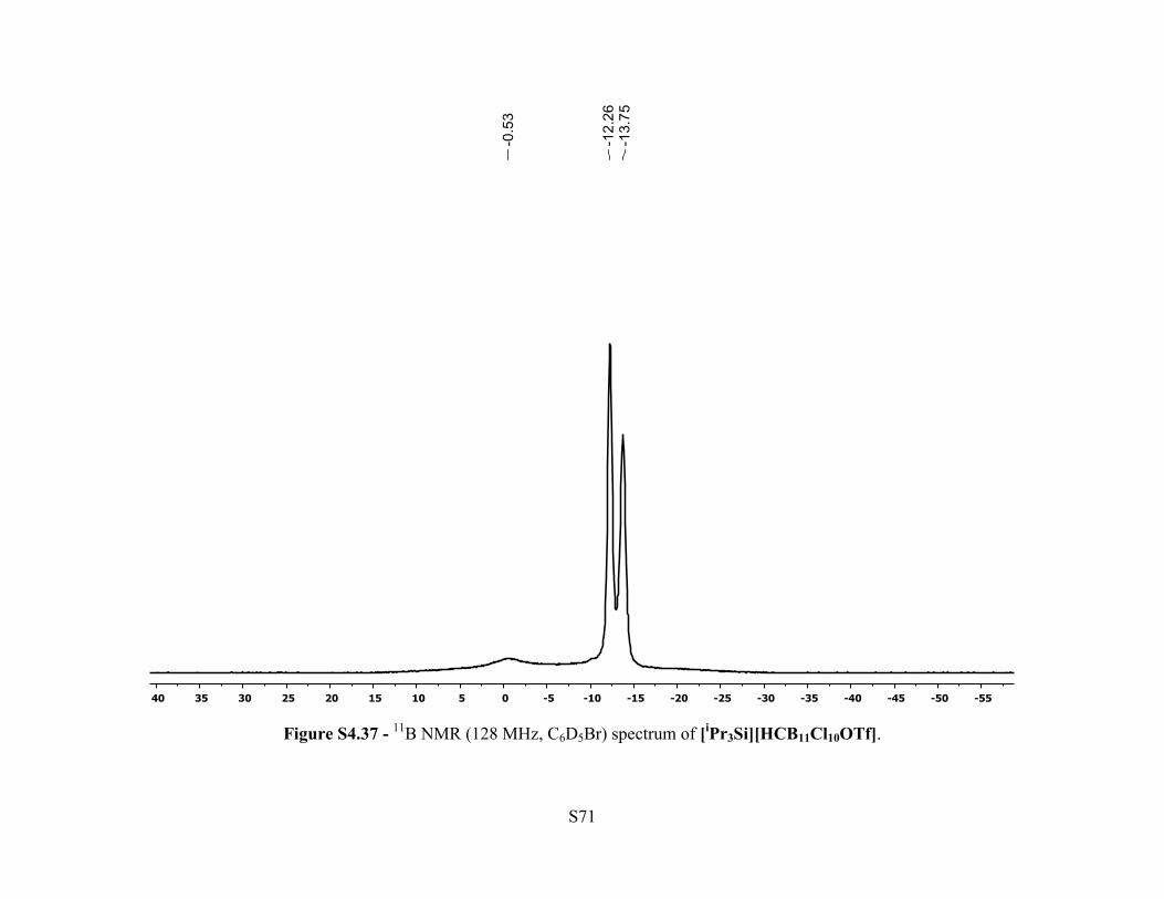
Si(CH(CH3)2)3). 11B NMR (128 MHz, 25 °C, C6D5Br): δ –0.53 (br s, 1B), –12.26 (s, 5H), –13.75
(s, 5H). 13C{1H} NMR (126 MHz, 25 °C, C6D5Br): δ -OTf carbon not observed, 48.2 (br s, 1C,
[HCB11Cl10OTf]–), 16.3 (s, Si(CH(CH3)2)3), 16.2 (s, Si(CH(CH3)2)3), 12.9 (s, Si(CH(CH3)2)3).
19F NMR (470 MHz, 25 °C, C6D5Br): δ –70.4 (s, 3F). 29Si NMR (79.5 MHz, 25 °C, C6D5Br): δ
74.8 (s).
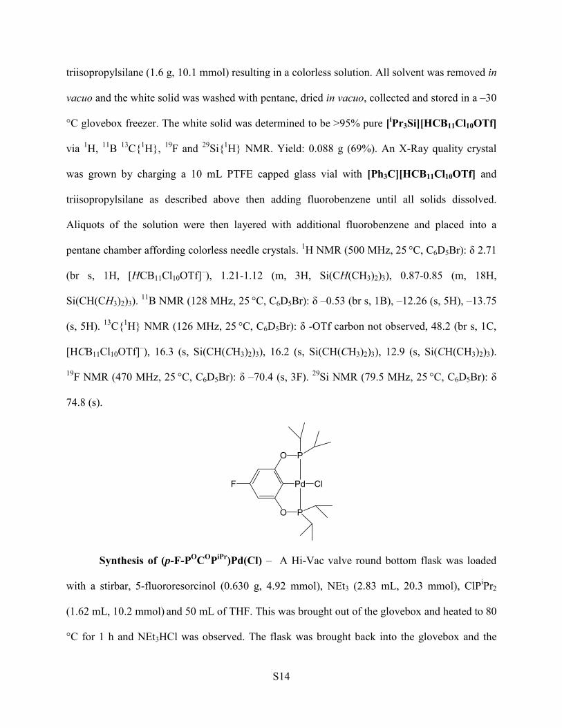
Synthesis of (p-F-POCOPiPr)Pd(Cl) – A Hi-Vac valve round bottom flask was loaded
with a stirbar, 5-fluororesorcinol (0.630 g, 4.92 mmol), NEt3 (2.83 mL, 20.3 mmol), ClPiPr2
(1.62 mL, 10.2 mmol) and 50 mL of THF. This was brought out of the glovebox and heated to 80
°C for 1 h and NEt3HCl was observed. The flask was brought back into the glovebox and the
Page 15
S15
flask was loaded with (cod)PdCl2 (1.48 g, 5.18 mmol). The reaction mixture was heated to 80 °C
for 24 h and then brought back in a glovebox. The mixture was filtered through a plug of silica
and the resultant light yellow solution was concentrated, layered with pentane and placed in a –
30 °C freezer overnight. Solvent was decanted, the white-yellow precipitate was washed with
pentane and the solids were dried in vacuo. Yield: 1.28 g (52%). 1H NMR (500 MHz, 25 °C,
C6D6): δ 6.43 (d, JH-F = 9.7 Hz 2H, Ar-H), 2.13-2.04 (m, 4H, P-CH(CH3)2), 1.24 (dvt, JH-H = 9.5
Hz, JP-H = 8.5 Hz, 12H, P-CH(CH3)2), 1.03 (dvt, JH-H = 8.5 Hz, JP-H = 7.0 Hz, 12H, P-CH(CH3)2).
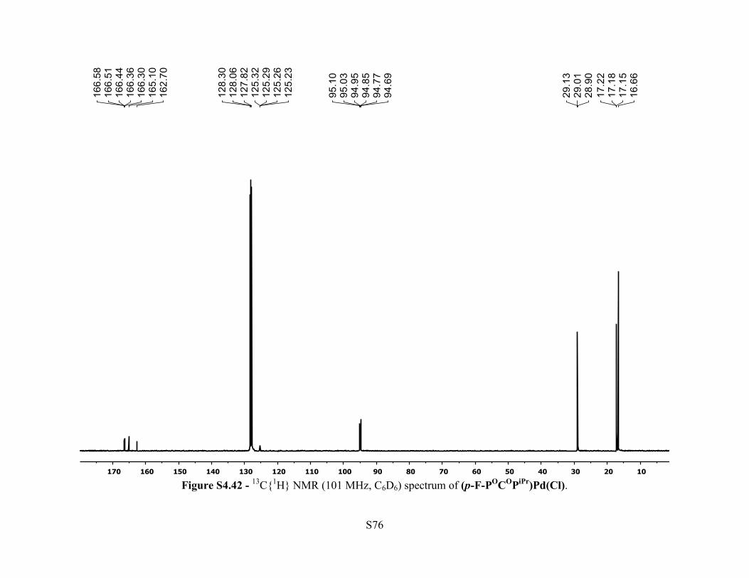
13C{1H} NMR (101 MHz, 25 °C, C6D6): δ 166.4 (dt, JC-F = 14.4 Hz, JC-P = 7.0 Hz, 2C, Ar C–
OPR2), 163.9 (d, JC-F = 241 Hz, 1C, Ar p-C–F ), 125.3 (m, J = 3.1 Hz, 1C, Ar C–Pd), 94.9 (dt,
JC-F = 26.1 Hz, JC-P = 7.6 Hz, 2C, Ar C–H), 29.0 (vt, JC-P = 11.3 Hz, P–CH(CH3)2, 4C), 17.2 (vt,
JC-P = 3.7 Hz, 4C, P–CH(CH3)2), 16.6 (s, 4C, P–CH(CH3)2). 19F NMR (376 MHz, 25 °C, C6D6):
δ –114.8 (t, JH-F = 9.7 Hz, 1F, Ar p-F). 31P{1H} NMR (202 MHz, 25 °C, C6D6): δ 190.2 (s). Anal.
Calcd. for (p-F-POCOPiPr)Pd(Cl): C, 43.13; H, 6.03. Found: C, 43.15 ; H, 6.15.
Synthesis of [(p-F-POCOPiPr)2Pd2(Cl)][HCB11Cl10OTf] in situ – A J. Young tube was
loaded with (p-F-POCOPiPr)Pd(Cl) (0.0133 g, 0.0265 mmol) and C6D6. [Et3Si][HCB11Cl10OTf]
(0.020 g, 0.0266 mmol) was added. The J. Young tube was sealed and shaken. Immediately
white precipitate began to form and via 1H NMR analysis, Et3SiCl was observed. The white
precipitate was collected on a frit, washed with benzene and pentane, then allowed to dry. The
white precipitate was dissolved in fluorobenzene and aliquots of the solution were placed into
vials then layered with fluorobenzene and put into a pentane chamber. X-Ray quality crystals of
[(p-F-POCOPiPr)2Pd2(Cl)][HCB11Cl10OTf] formed after several days.
Page 16
S16
Synthesis of (p-F-POCOPiPr)Pd(OTf) – A 50 mL Schlenk flask was loaded with (p-F-
POCOPiPr)Pd(Cl) (0.758 g, 1.51 mmol) and 10 mL of benzene. Via syringe, Me3SiOTf (2.75
mL, 15.2 mmol) was added and the reaction was stirred at room temperature for 20 minutes. The
solvent was removed in vacuo giving a white solid. The product was recrystallized by dissolving
in a minimum of toluene, layering with pentane and placed in a –30 °C glovebox freezer
overnight. The white solid recovered was dried in vacuo. Yield: 0.742 g (80%). 1H NMR (500
MHz, 25 °C, C6D6): δ 6.28 (d, JH-F = 9.7 Hz, 2H, Ar-H), 2.24-2.15 (m, J = 7.0 Hz, 4H, P–
CH(CH3)2), 1.19 (dvt, JH-H = 10.0 Hz, JP-H = 8.5 Hz, 12H, P–CH(CH3)2), 0.94 (dvt, JH-H = 8.3
Hz, JP-H = 7.2 Hz, 12H, P–CH(CH3)2). 13C{1H} NMR (126 MHz, 25 °C, C6D6): δ 166.8 (dt, JC-F
= 14.5 Hz, JC-P = 6.3 Hz, 2C, Ar C–OPR2), 164.3 (d, JC-F = 243 Hz, 1C, Ar p-C–F), 120.8 (q, JC-F
= 318 Hz, 1C, -CF3). 116.5 (m, 1C, Ar C–Pd), 95.5 (dt, JC-F = 26.2 Hz, JC-P = 7.6 Hz, 2C, Ar C–
H), 29.1 (vt, JC-P = 11.3 Hz, 4C, P–CH(CH3)2), 17.1(vt, JC-P = 3.8 Hz, 4C, P–CH(CH3)2), 16.4 (s,
4C, P–CH(CH3)2) . 19F NMR (470 MHz, 25 °C, C6D6): δ –77.7 (s, 3F, -CF3), –112.8 (t, JF-H = 9.7
Hz, 1F, Ar-F). 31P{1H} NMR (202 MHz, 25 °C, C6D6): δ 192.8 (s). Anal. Calcd. for (p-F-
POCOPiPr)Pd(OTf): C, 37.12; H, 4.92. Found: C, 37.22; H, 5.02.
Page 17
S17
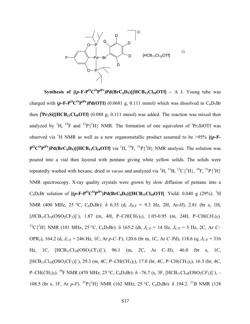
Synthesis of [(p-F-POCOPiPr)Pd(BrC6D5)][HCB11Cl10OTf] – A J. Young tube was
charged with (p-F-POCOPiPr)Pd(OTf) (0.0681 g, 0.111 mmol) which was dissolved in C6D5Br
then [iPr3Si][HCB11Cl10OTf] (0.088 g, 0.111 mmol) was added. The reaction was mixed then
analyzed by 1H, 19F and 31P{1H} NMR. The formation of one equivalent of iPr3SiOTf was
observed via 1H NMR as well as a new organometallic product assumed to be >95% [(p-F-
POCOPiPr)Pd(BrC6D5)][HCB11Cl10OTf] via 1H, 19F, 31P{1H} NMR analysis. The solution was
poured into a vial then layered with pentane giving white yellow solids. The solids were
repeatedly washed with hexane, dried in vacuo and analyzed via 1H, 11B, 13C{1H}, 19F, 31P{1H}
NMR spectroscopy. X-ray quality crystals were grown by slow diffusion of pentane into a
C6D5Br solution of [(p-F-POCOPiPr)Pd(BrC6D5)][HCB11Cl10OTf]. Yield: 0.040 g (29%). 1H
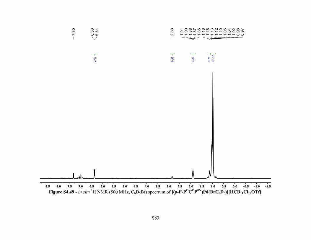
NMR (400 MHz, 25 °C, C6D5Br): δ 6.35 (d, JH-F = 9.3 Hz, 2H, Ar-H), 2.81 (br s, 1H,
[HCB11Cl10(OSO2CF3)]–), 1.87 (m, 4H, P–CH(CH3)2), 1.05-0.95 (m, 24H, P–CH(CH3)2).
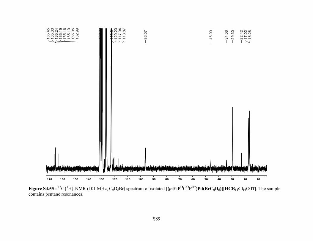
13C{1H} NMR (101 MHz, 25 °C, C6D5Br): δ 165.2 (dt, JC-F = 14 Hz, JC-P = 5 Hz, 2C, Ar C–
OPR2), 164.2 (d, JC-F = 246 Hz, 1C, Ar p-C–F), 120.6 (br m, 1C, Ar C–Pd), 118.6 (q, JC-F = 316
Hz, 1C, [HCB11Cl10(OSO2CF3)]–), 96.1 (m, 2C, Ar C–H), 46.0 (br s, 1C,
[HCB11Cl10(OSO2CF3)]–), 29.3 (m, 4C, P–CH(CH3)2), 17.0 (br, 4C, P–CH(CH3)2), 16.3 (br, 4C,
P–CH(CH3)2). 19F NMR (470 MHz, 25 °C, C6D5Br): δ –76.7 (s, 3F, [HCB11Cl10(OSO2CF3)]–), –
108.5 (br s, 1F, Ar p-F). 31P{1H} NMR (162 MHz, 25 °C, C6D5Br): δ 194.2. 11B NMR (128
Page 18
S18
MHz, 25 °C, C6D5Br): δ –1.07 (br s, 1B), –11.6 (br s, 5B), –13.9 (br s, 5B). Anal. Calcd. for [(p-
F-POCOPiPr)Pd(BrC6D5)][HCB11Cl10OTf]: C, 24.72; H, 3.27; B, 9.41. Found: C, 24.55; H,
2.99; B, 9.24.
Synthesis of [(p-F-POCOPiPr)Pd][HCB11Cl10OTf] in situ – A J. Young tube was
charged with (p-F-POCOPiPr)Pd(OTf) (0.0232 g, 0.0377 mmol), 10 μL of C6F6, 10 μL of
mesitylene, C6D6, then [Et3Si][HCB11Cl10OTf] (0.0285 g, 0.0380 mmol) was added. The
reaction was mixed then analyzed by 1H, 11B, 19F and 31P{1H} NMR. Quantitative formation of
[(p-F-POCOPiPr)Pd][HCB11Cl10OTf] and concomitant formation of Et3SiOTf was observed by
1H and 19F NMR analysis.
Synthesis of [(p-F-POCOPiPr)Pd][HCB11Cl10OTf] - A 10 mL PTFE capped glass vial
was charged (p-F-POCOPiPr)Pd(OTf) (0.0413 g, 0.0672 mmol) and fluorobenzene (1 mL).
[Et3Si][HCB11Cl10OTf] (0.0505 g, 0.0673 mmol) was added and the solution was stirred for 5
minutes then all solvent was removed in vacuo giving white solid. Recrystallization from toluene
and pentane gave white colorless crystals. X-ray quality crystals of [(p-F-
POCOPiPr)Pd][HCB11Cl10OTf] were grown from an o-difluorobenzene solution layered with
pentane at room temperature. Yield: 0.029 g (39%).). 1H NMR (500 MHz, 25 °C, C6D6): δ 6.12
(d, JH-F = 9.4 Hz, 2H, Ar-H), 2.33 (br s, 1H, [HCB11Cl10(OSO2CF3)]–), 1.94 (m, 4H, P–
CH(CH3)2), 0.98 (br dvt, JH-H = 9.5 Hz, JP-H = 9.2 Hz, 12H, P–CH(CH3)2), 0.80 (br dvt, JH-H =
7.7 Hz, JP-H = 7.5 Hz, 12H, P–CH(CH3)2). 11B NMR (128 MHz, 25 °C, C6D6): δ –0.46 (br s, 1B),
–11.31 (s, 5B), –13.20 (s 5B). 13C{1H} NMR (126 MHz, 25 °C, C6D6): δ 166.2 (dt, JC-F = 14 Hz,
JC-P = 6.3 Hz, 2C, Ar C–OPR2), 164.6 (d, JC-F = 247 Hz, 1C, Ar p-C–F), 118.6 (q, JC-F = 320 Hz,
1C, [HCB11Cl10(OSO2CF3)]–), 112.9 (m, 1C, Ar C-Pd), 96.1 (dt, JC-F = 26.5 Hz, JC-P = 7.6 Hz,
2C, Ar C–H), 47.1 (br s, 1C, [HCB11Cl10(OSO2CF3)]–), 29.5 (vt, JC-P = 11.5 Hz, 4C, P–
Page 19
S19
CH(CH3)2), 17.3 (vt, JC-P = 3.3 Hz, 4C, P–CH(CH3)2), 16.3 (s, 4C, P–CH(CH3)2). 19F NMR (470
MHz, 25 °C, C6D6): δ –74.6 (s, 3F, [HCB11Cl10(OSO2CF3)]–), –110.5 (br m, 1F, Ar-p-F).
31P{1H} NMR (202 MHz, 25 °C, C6D6): δ 193.9. Anal. Calcd. for [(p-F-
POCOPiPr)Pd][HCB11Cl10OTf]: C, 21.81; H, 2.84; B, 10.80. Found: C, 21.94; H, 2.73; B, 10.54.
Synthesis of [(p-F-POCOPiPr)Pd][HCB11Cl11] – A J. Young NMR tube was charged with
(p-F-POCOPiPr)Pd(OTf) (0.0721 g, 0.060 mmol), Na[HCB11Cl11] (0.064 g, 0.0609 mmol) and a
2:1 o-difluorobenzene:C6D6 solvent mixture. The mixture was agitated for 24 h at room
temperature then filtered through a plug of celite. All solvent was removed in vacuo resulting in
a white solid. X-ray quality crystals of [(p-F-POCOPiPr)Pd][HCB11Cl11] were grown from
fluorobenzene solution layered with hexanes at room temperature. Yield: 0.105 g (91%). 1H
NMR (400 MHz, 25 °C, 2:1 ODFB:C6D6): δ 6.17 (d, JH-F = 9.5 Hz, 2H, Ar-H), 2.63 (br s, 1H,
[HCB11Cl11]–), 1.97 (br m, 4H, P–CH(CH3)2), 1.05-0.99 (br m, 24H, P–CH(CH3)2). 11B NMR
(128 MHz, 25 °C, 2:1 ODFB:C6D6): δ –2.22 (br s, 1B), –9.69 (br s, 5B), –12.83 (br s, 5B).
13C{1H} NMR (101 MHz, 25 °C, 2:1 ODFB:C6D6): δ 165.7 (m, 2C, Ar C–OPR2), 164.8 (d, JC-F
= 247 Hz, 1C, Ar p-C–F), Ar C–Pd signal could not be identified, 95.9 (dt, JC-F = 26.5 Hz, JC-P =
7.8 Hz, 2C, Ar C–H), 47.7 (br s, 1C, [HCB11Cl11]–), 30.1 (br vt, JC-P = 10.4 Hz, 4C, P–
CH(CH3)2), 17.5 (br s, 4C, P–CH(CH3)2), 16.2 (s, 4C, P–CH(CH3)2). δ 19F NMR (376 MHz, 25
°C, 2:1 ODFB:C6D6): δ –110.2 (br s, Ar-p-F). 31P{1H} NMR (162 MHz, 25 °C, 2:1
ODFB:C6D6): δ 193.9. Anal. Calcd. for [(p-F-POCOPiPr)Pd][HCB11Cl11]: C, 23.11; H, 3.16; B,
12.04. Found: C, 23.20; H, 3.06; B, 11.96.
Synthesis of [(Et3Si)2OTf][HCB11Cl11] – A vial was loaded with [Ph3C][HCB11Cl11]
(0.116 g, 0.152 mmol), a PTFE stir bar and 2 mL of PhF. Me3SiOTf (136 mg, 0.612 mmol) and
Et3SiH (80 mg, 0.688 mmol) were added and the solution went colorless. The reaction was
Page 20
S20
stirred for 1 h at room temperature then all solvent was removed in vacuo giving yellow oil
determined to be >95% pure [(Et3Si)2OTf][HCB11Cl11] via 1H, 11B, 13C{1H}, 19F and 29Si{1H}
NMR. The use of Et3SiOTf in place of Me3SiOTf gave the same product,
[(Et3Si)2OTf][HCB11Cl11]. Yield: 0.109 g (80%). 1H NMR (400 MHz, 25 °C, C6D5Br): δ 2.88
(br s, 1H, [HCB11Cl11]–), 0.85–0.71 (m, 30H, [(Et3Si)2OTf]. 11B NMR (128 MHz, 25 °C,
C6D5Br): δ –2.54 (s, 1B), –10.09 (s, 5B), –13.23 (s, 5B). 13C{1H} NMR (126 MHz, 25 °C,
C6D5Br): δ 117.4 (q, JC-F = 323 Hz, 1C, [(Et3Si)2OTf]+), 47.0 (s, 1C, [HCB11Cl11]–), 5.52-5.04
(m, 3H, [(Et3Si)2OTf]+), 5.12 (s, 3H, [(Et3Si)2OTf]+). 19F NMR (376 MHz, 25 °C, C6D5Br): δ –
73.7. 29Si NMR (79.5 MHz, 25 °C, C6D5Br): δ 75.5 (s).
Page 21
S21
III. Catalytic Hydrodefluorination Studies
Entries 1-5 refer to Table 1 in the main text.
Description of experiments in Table 1, by entry.
CAUTION: In certain cases, C-F activation reactions may proceed very rapidly, self-
accelerating and releasing dangerous amounts of heat. In addition, these reactions may
generate hydrogen and possibly even other gases. Great care and preliminary testing of
safe conditions are necessary for performing reactions in closed vessels. All reactions were done
in 20 mL polypropylene vials pierced with a 20 gauge 1.5” needle to allow gas to escape.
Entry 1 – A 20 mL polypropylene vial was loaded with [Ph3C][HCB11Cl10OTf] (2.9 mg, 3.3
μmol), C6F6 (20 μL, 0.17 mmol), o-dichlorobenzene (0.3 mL), C6F5CF3 (0.30 mL, 2.1 mmol),
and a PTFE coated stirbar. Et3SiH (1.1 mL, 6.9 mmol) was added slowly and the mixture was
allowed to stir for 24 h at room temperature. The mixture was taken up into a J. Young tube,
sealed, taken out of the glovebox and analyzed via 19F NMR spectroscopy. A trace amount
(<1%) of Et3SiF (δ = –177.5 ppm) was observed.
Entry 2 – A 20 mL polypropylene vial was loaded with [Ph3C][HCB11Cl10OTf] (29 mg, 33
μmol), C6F6 (20 μL, 0.17 mmol), o-dichlorobenzene (0.3 mL), C6F5CF3 (0.30 mL, 2.1 mmol),
and a PTFE coated stirbar. Et3SiH (1.1 mL, 6.9 mmol) was added slowly and the mixture was
allowed to stir for 24 h at room temperature. The mixture was taken up into a J. Young tube,
sealed, taken out of the glovebox and analyzed via 19F NMR spectroscopy. After 24 h, 20% of
the C6F5CF3 had been consumed. Et3SiF (s, δ = –177.5 ppm) was observed in a 20% yield.
Entry 3 – A 20 mL polypropylene vial was loaded with [Ph3C][HCB11Cl11] (2.5 mg, 3.3 μmol),
C6F6 (20 μL, 0.17 mmol), o-dichlorobenzene (0.3 mL), C6F5CF3 (0.30 mL, 2.1 mmol), and a
Page 22
S22
PTFE coated stirbar. Et3SiH (1.1 mL, 6.9 mmol) was added slowly and the mixture was allowed
to stir for 24 h at room temperature. The mixture was taken up into a J. Young tube, sealed, taken
out of the glovebox and analyzed via 19F NMR spectroscopy. After 24 h, all C6F5CF3 had been
consumed. Et3SiF (s, δ = –177.5 ppm) and Et2SiF2 (br s, δ = –145.3 ppm) were observed in a
59% and 10% yield, respectively.
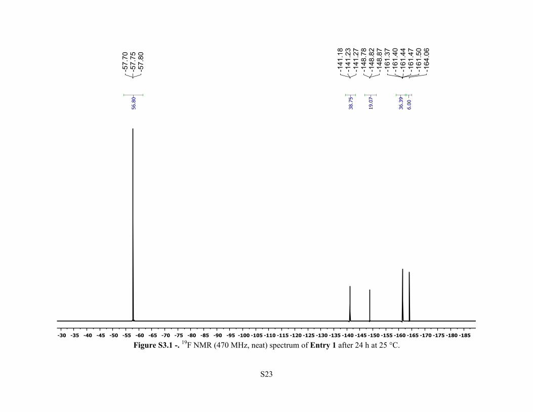
Entry 4 – A 20 mL polypropylene vial was loaded with [Ph3C][HCB11Cl10OTf] (2.9 mg, 3.3
μmol), C6F6 (20 μL, 0.17 mmol), o-dichlorobenzene (0.3 mL), 4-F-C6H4CF3 (0.30 mL, 2.4
mmol), and a PTFE coated stirbar. Et3SiH (1.1 mL, 6.9 mmol) was added slowly and the mixture
was allowed to stir for 24 h at room temperature. The mixture was taken up into a J. Young tube,
sealed, taken out of the glovebox and analyzed via 19F NMR spectroscopy. After 24 h, 85% of
the 4-F-C6H4CF3 had been consumed. Et3SiF (s, δ = –177.5 ppm) was observed in an 87% yield.
Entry 5 – A 20 mL polypropylene vial was loaded with [Ph3C][HCB11Cl11] (2.5 mg, 3.3 μmol),
C6F6 (20 μL, 0.17 mmol), o-dichlorobenzene (0.3 mL), 4-fluorobenzotrifluoride (0.30 mL, 2.4
mmol), and a PTFE coated stirbar. Et3SiH (1.1 mL, 6.9 mmol) was added slowly and gas
evolved violently. The solution turned dark brown. The mixture was allowed to stir for 1 h at
room temperature. The mixture was taken up into a J. Young tube, sealed, taken out of the
glovebox and analyzed via 19F NMR spectroscopy. After 1 h, all 4-F-C6H4CF3 had been
consumed. Et3SiF (s, δ = –177.5 ppm) was observed in a 95% yield.
Page 23
S23
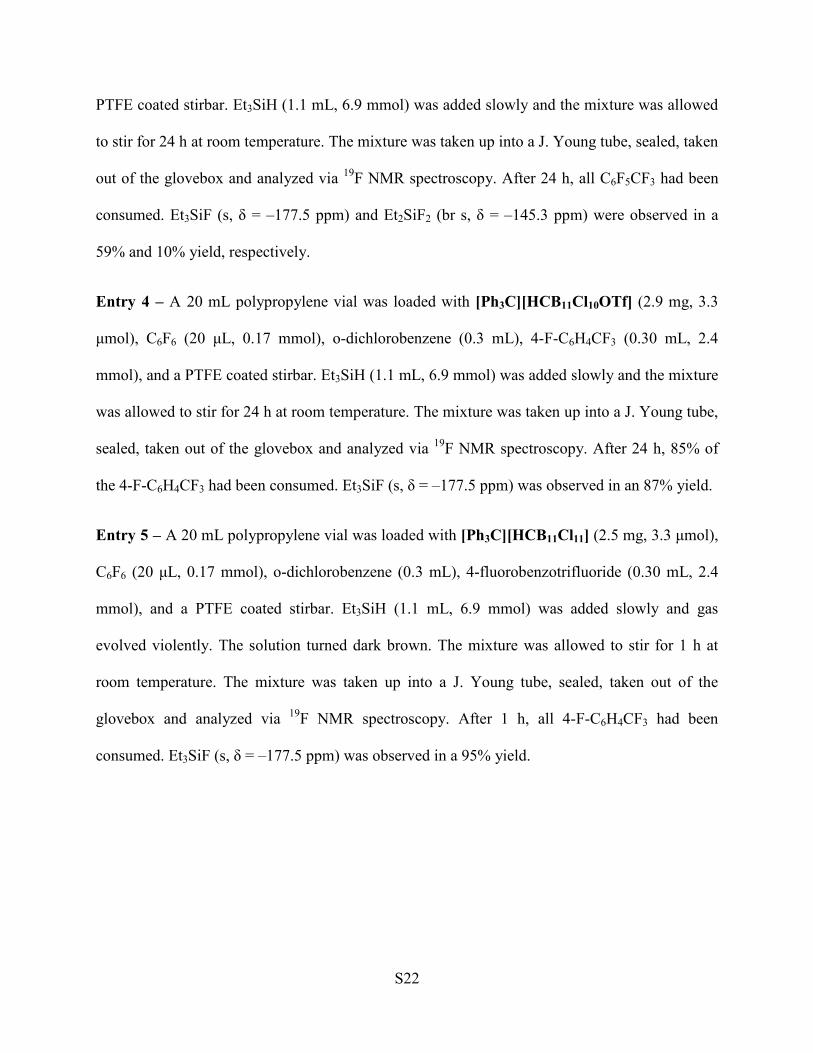
Figure S3.1 -. 19F NMR (470 MHz, neat) spectrum of Entry 1 after 24 h at 25 °C.
Page 24
S24
Figure S3.2 - 19F NMR (376 MHz, neat) spectrum of Entry 2 after 24 h at 25 °C.
Page 25
S25
Figure S3.3 - 19F NMR (470 MHz, neat) spectrum of Entry 3 after 24 h at 25 °C.
Page 26
S26
Figure S3.4 - 19F NMR (376 MHz, neat) spectrum of Entry 4 after 24 h at 25 °C.
Page 27
S27
Figure S3.5 - 19F NMR (376 MHz, neat) spectrum of Entry 5 after 1 h at 25 °C.
Page 28
S28
Figure S4.1a - MALDI (–) Mass Spectrum of Cs[HCB11H10OTf].
IV. Graphical NMR Spectra and MALDI Mass Spectra
[HCB11H10OTf]–
[HCB11H10OTf]–
Page 29
S29
Figure S4.1b – Simulated MALDI (–) mass spectrum of Cs[HCB11H10OTf].
Page 30
S30
Figure S4.2 – 1H NMR (400 MHz, CD3CN) spectrum of Cs[HCB11H10OTf].
Page 31
S31
Figure S4.3 – 11B NMR (128 MHz, CD3CN) spectrum of Cs[HCB11H10OTf].
Page 32
S32
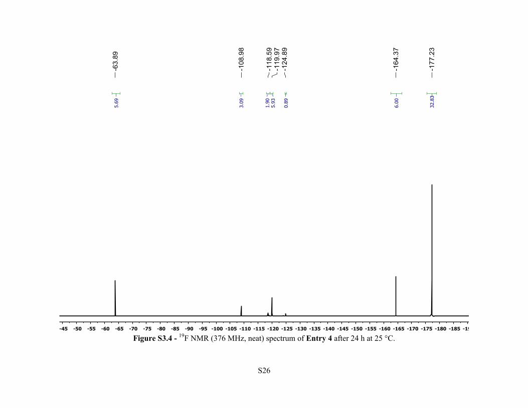
Figure S4.4 13C{1H} NMR (101 MHz, CD3CN) spectrum of Cs[HCB11H10OTf].
Page 33
S33
Figure S4.5 – 19F NMR (470 MHz, CD3CN) spectrum of Cs[HCB11H10OTf]. Cs[HCB11H9(OTf)2] is the major impurity followed by CsOTf.The other resonances were not identified.
Page 34
S34
Figure S4.6a - MALDI (–) mass spectrum of Cs[HCB11H10OTf],where Cs[HCB11H9(OTf)2] is observed. The other signals were not identified.
[HCB11H10OTf]–
[HCB11H9(OTf)2]– ??
??
Page 35
S35
Figure S4.6b – Simulated MALDI (–) mass spectrum of Cs[HCB11H9(OTf)2].
Page 36
S36
Figure S4.7 – 1H NMR (500 MHz, CD3CN) spectrum of Cs[HCB11Cl10OTf]. Spectrum contains resonances of toluene and pentane.
Page 37
S37
Figure S4.8 – 19F NMR (470 MHz, CD3CN) of Cs[HCB11Cl10OTf]. The sample contains Cs[HCB11Cl9(OTf)2].
Page 38
S38
Figure S4.9 – 11B NMR (128 MHz, CD3CN) spectrum of Cs[HCB11Cl10(OTf)].
Page 39
S39
Figure S4.10a - MALDI (–) mass spectrum of Cs[HCB11Cl10OTf]. Cs[HCB11Cl10(OH)] is observed.
[HCB11Cl10(OH)]– [HCB11Cl10OTf]–
[HCB11Cl10OTf]–
Page 40
S40
Figure S4.10b – Simulated MALDI (–) mass spectrum of Cs[HCB11Cl10OTf].
Page 41
S41
Figure S4.10c – Simulated MALDI (–) mass spectrum of Cs[HCB11Cl10(OH)].
Page 42
S42
Figure S4.11 – 13C{1H} NMR (101 MHz, CD3CN) spectrum of Cs[HCB11Cl10OTf].
Page 43
S43
Figure S4.12 – 19F NMR (470 MHz, 25 °C, CD3CN) spectrum of Cs[HCB11Cl9(OTf)2] after reacting Cs[HCB11H11] with Cl2 in triflic acid. Resonance at –79.3 ppm is the residual triflic acid.
Page 44
S44
Figure S4.13a – MALDI (–) mass spectrum of Cs[HCB11Cl9(OTf)2]. (Top: from 100-1500; Bottom: from 700-800).
[HCB11Cl9(OTf)2]–
Page 45
S45
Figure S4.13b – Simulated MALDI (–) mass spectrum of Cs[HCB11Cl9(OTf)2].
Page 46
S46
Figure S4.14 – 1H NMR (400 MHz, CD3CN) spectrum of Cs[HCB11H5Br5OTf]. The spectrum contains an H2O resonance.
Page 47
S47
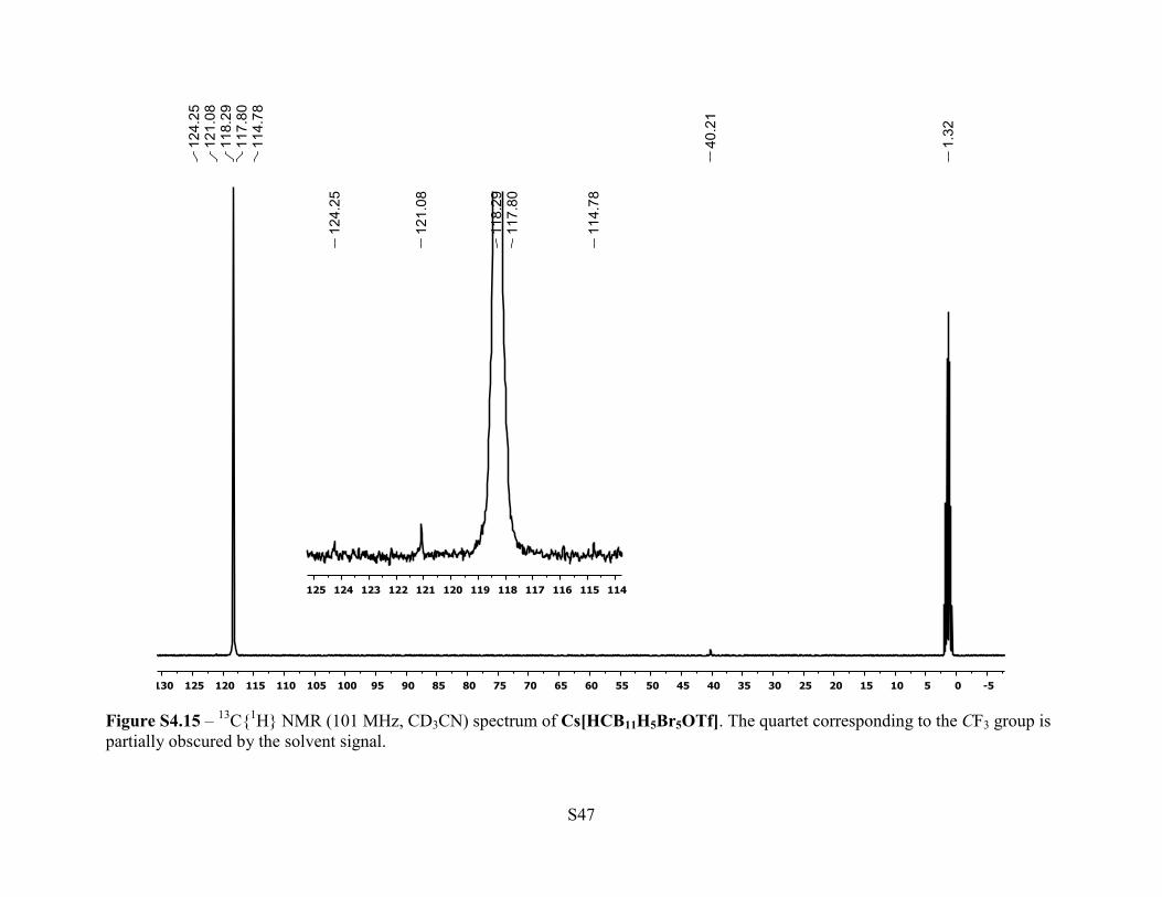
Figure S4.15 – 13C{1H} NMR (101 MHz, CD3CN) spectrum of Cs[HCB11H5Br5OTf]. The quartet corresponding to the CF3 group is partially obscured by the solvent signal.
Page 48
S48
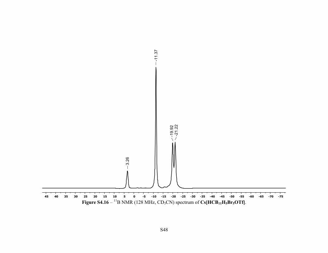
Figure S4.16 – 11B NMR (128 MHz, CD3CN) spectrum of Cs[HCB11H5Br5OTf].
Page 49
S49
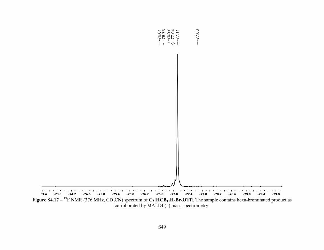
Figure S4.17 – 19F NMR (376 MHz, CD3CN) spectrum of Cs[HCB11H5Br5OTf]. The sample contains hexa-brominated product as
corroborated by MALDI (–) mass spectrometry.
Page 50
S50
Figure S4.18a - MALDI (–) mass spectrum of Cs[HCB11H5Br5OTf]. Cs[HCB11H4Br6OTf] is observed.
[HCB11H5Br5OTf]–
[HCB11H5Br5OTf]– [HCB11H4Br6OTf]–
Page 51
S51
Figure S4.18b - Simulated MALDI (–) mass spectrum of Cs[HCB11H5Br5OTf].
Page 52
S52
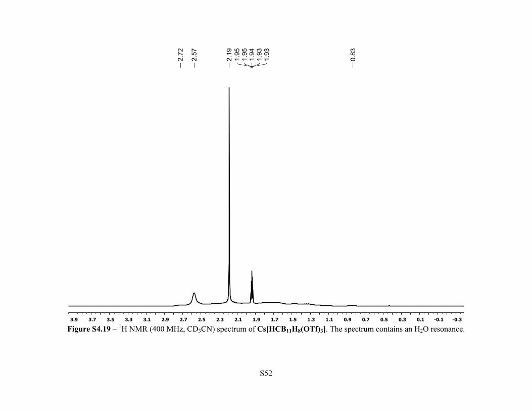
Figure S4.19 – 1H NMR (400 MHz, CD3CN) spectrum of Cs[HCB11H8(OTf)3]. The spectrum contains an H2O resonance.
Page 53
S53
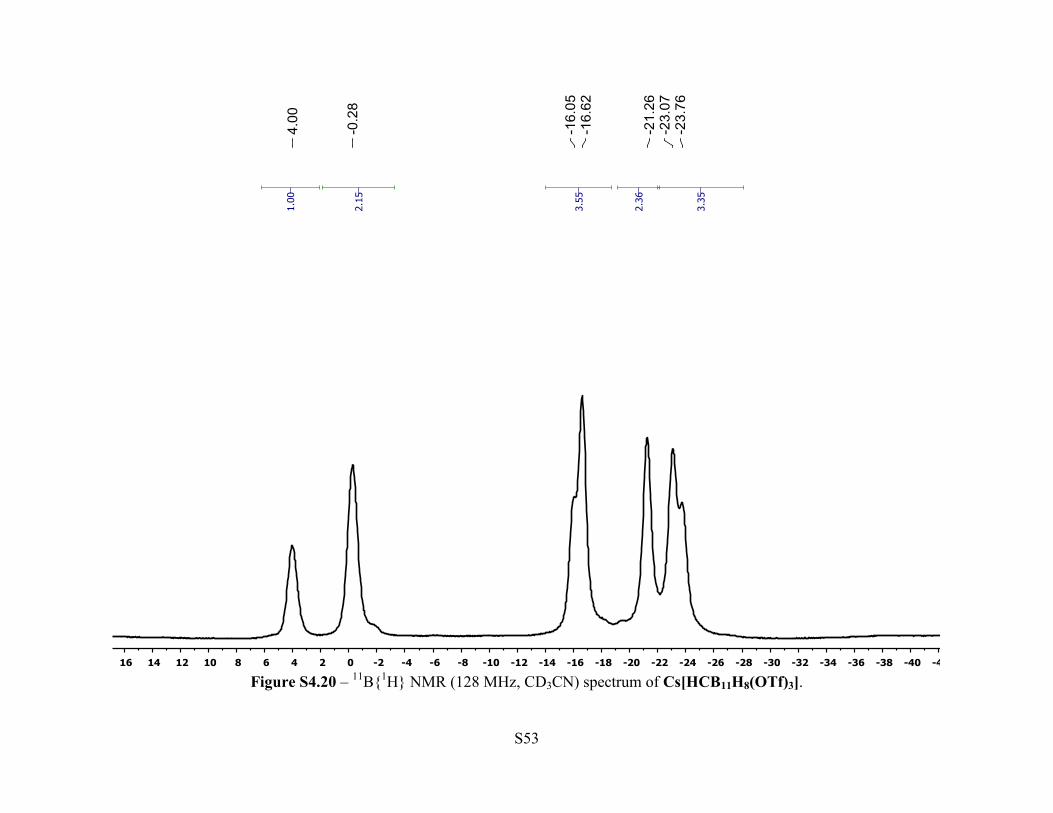
Figure S4.20 – 11B{1H} NMR (128 MHz, CD3CN) spectrum of Cs[HCB11H8(OTf)3].
Page 54
S54
Figure S4.21 – 13C{1H} NMR (101 MHz, CD3CN) spectrum of Cs[HCB11H8(OTf)3].
Page 55
S55
Figure S4.22 – 19F NMR (376 MHz, CD3CN) spectrum of Cs[HCB11H8(OTf)3]. The sample contains a CsOTf resonance.
Page 56
S56
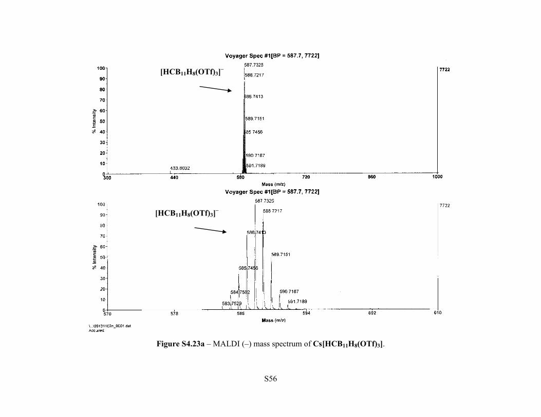
Figure S4.23a – MALDI (–) mass spectrum of Cs[HCB11H8(OTf)3].
[HCB11H8(OTf)3]–
[HCB11H8(OTf)3]–
Page 57
S57
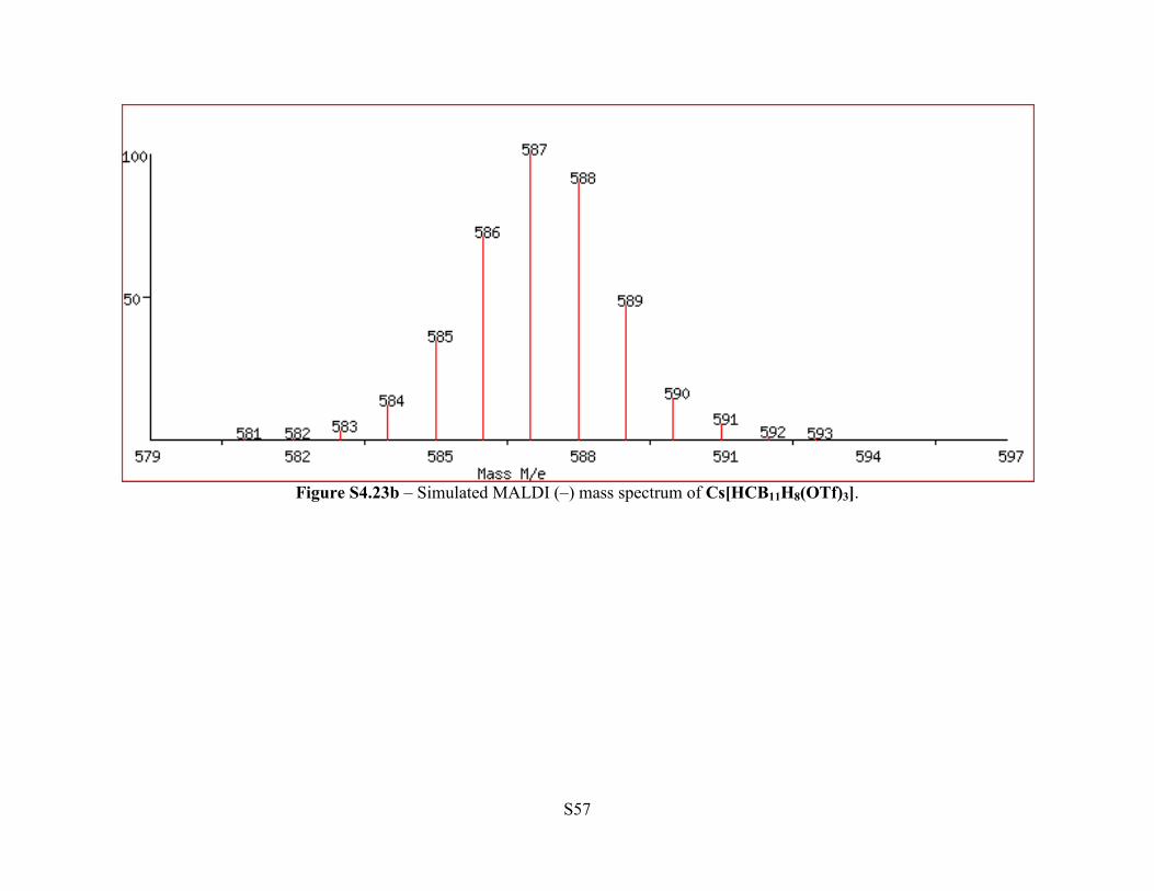
Figure S4.23b – Simulated MALDI (–) mass spectrum of Cs[HCB11H8(OTf)3].
Page 58
S58
Figure S4.24 - 1H NMR (500 MHz, CDCl3) of [Ph3C][OTf]. The sample contains pentane resonances.
Page 59
S59
Figure S4.25 -. 13C{1H} NMR (101 MHz, CDCl3) of [Ph3C][OTf].
Page 60
S60
Figure S4.26 - 19F NMR (376 MHz, CDCl3) of [Ph3C][OTf].
Page 61
S61
Figure S4.27 - 1H NMR (500 MHz, C6D5Br) of [Ph3C][HCB11Cl10OTf]. The sample contains pentane resonances.
Page 62
S62
Figure S4.28 11B NMR (128 MHz, C6D5Br) of [Ph3C][HCB11Cl10OTf].
Page 63
S63
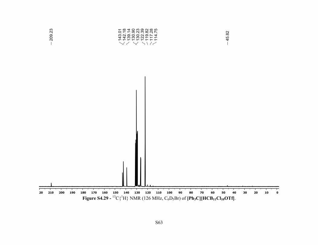
Figure S4.29 - 13C{1H} NMR (126 MHz, C6D5Br) of [Ph3C][HCB11Cl10OTf].
Page 64
S64
Figure S4.30 – 19F NMR (470 MHz, C6D5Br) of [Ph3C][HCB11Cl10OTf]. The sample contains [Ph3C][HCB11Cl9(OTf)2] resonances.
Page 65
S65
Figure S4.31 – 1H NMR (500 MHz, C6D5Br) spectrum of [Et3Si][HCB11Cl10OTf].
Page 66
S66
Figure S4.32 - 11B NMR (128 MHz, C6D5Br) spectrum of [Et3Si][HCB11Cl10OTf].
Page 67
S67
Figure S4.33 - 13C{1H} NMR (126 MHz, C6D5Br) spectrum of [Et3Si][HCB11Cl10OTf].
Page 68
S68
Figure S4.34 - 19F NMR (470 MHz, C6D5Br) spectrum of [Et3Si][HCB11Cl10OTf].
Page 69
S69
Figure S4.35 - 29Si{1H} NMR (79.5 MHz, C6D5Br) spectrum of [Et3Si][HCB11Cl10OTf].
Page 70
S70
Figure S4.36 - 1H NMR (500 MHz, C6D5Br) spectrum of [iPr3Si][HCB11Cl10OTf].
Page 71
S71
Figure S4.37 - 11B NMR (128 MHz, C6D5Br) spectrum of [iPr3Si][HCB11Cl10OTf].
Page 72
S72
Figure S4.38 - 13C{1H} NMR (126 MHz, C6D5Br) spectrum of [iPr3Si][HCB11Cl10OTf].
Page 73
S73
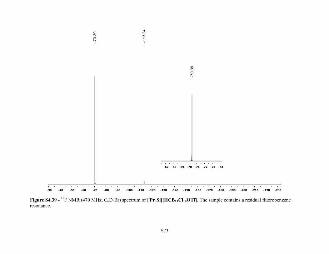
Figure S4.39 - 19F NMR (470 MHz, C6D5Br) spectrum of [iPr3Si][HCB11Cl10OTf]. The sample contains a residual fluorobenzene resonance.
Page 74
S74
Figure S4.40 - 29Si{1H} NMR (79.5 MHz, C6D5Br) spectrum of [iPr3Si][HCB11Cl10OTf].
Page 75
S75
Figure S4.41 - 1H NMR (500 MHz, C6D6) spectrum of (p-F-POCOPiPr)Pd(Cl).
Page 76
S76
Figure S4.42 - 13C{1H} NMR (101 MHz, C6D6) spectrum of (p-F-POCOPiPr)Pd(Cl).
Page 77
S77
Figure S4.43 - 31P{1H} NMR (202 MHz, C6D6) spectrum of (p-F-POCOPiPr)Pd(Cl).
Page 78
S78
Figure S4.44 19F NMR (470 MHz, C6D6) spectrum of (p-F-POCOPiPr)Pd(Cl).
Page 79
S79
Figure S4.45 1H NMR (500 MHz, C6D6) spectrum of (p-F-POCOPiPr)Pd(OTf).
Page 80
S80
Figure S4.46 - 13C{1H} NMR (126 MHz, C6D6) spectrum of (p-F-POCOPiPr)Pd(OTf).
Page 81
S81
Figure S4.47 - 19F NMR (470 MHz, C6D6) spectrum of (p-F-POCOPiPr)Pd(OTf).
Page 82
S82
Figure S4.48 31P{1H} NMR (202 MHz, C6D6) spectrum of (p-F-POCOPiPr)Pd(OTf).
Page 83
S83
Figure S4.49 - in situ 1H NMR (500 MHz, C6D5Br) spectrum of [(p-F-POCOPiPr)Pd(BrC6D5)][HCB11Cl10OTf].
Page 84
S84
Figure S4.50 - in situ 11B NMR (128 MHz, C6D5Br) spectrum of [(p-F-POCOPiPr)Pd(BrC6D5)][HCB11Cl10OTf].
Page 85
S85
Figure S4.51 - in situ 19F NMR (470 MHz, C6D5Br) spectrum of [(p-F-POCOPiPr)Pd(BrC6D5)][HCB11Cl10OTf].
Page 86
S86
Figure S4.52 - in situ 31P{1H} NMR (202 MHz, C6D5Br) spectrum of [(p-F-POCOPiPr)Pd(BrC6D5)][HCB11Cl10OTf].
Page 87
S87
Figure S4.53 - 1H NMR (400 MHz, C6D5Br) spectrum of isolated [(p-F-POCOPiPr)Pd(BrC6D5)][HCB11Cl10OTf]. The sample contains pentane resonances.
Page 88
S88
Figure S4.54 - 11B NMR (128 MHz, C6D5Br) spectrum of isolated [(p-F-POCOPiPr)Pd(BrC6D5)][HCB11Cl10OTf].
Page 89
S89
Figure S4.55 - 13C{1H} NMR (101 MHz, C6D5Br) spectrum of isolated [(p-F-POCOPiPr)Pd(BrC6D5)][HCB11Cl10OTf]. The sample contains pentane resonances.
Page 90
S90
Figure S4.56 - 19F NMR (376 MHz, C6D5Br) spectrum of isolated [(p-F-POCOPiPr)Pd(BrC6D5)][HCB11Cl10OTf].
Page 91
S91
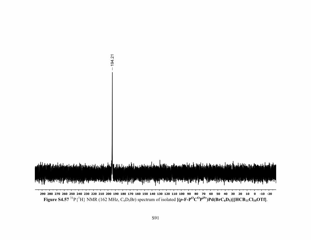
Figure S4.57 31P{1H} NMR (162 MHz, C6D5Br) spectrum of isolated [(p-F-POCOPiPr)Pd(BrC6D5)][HCB11Cl10OTf].
Page 92
S92
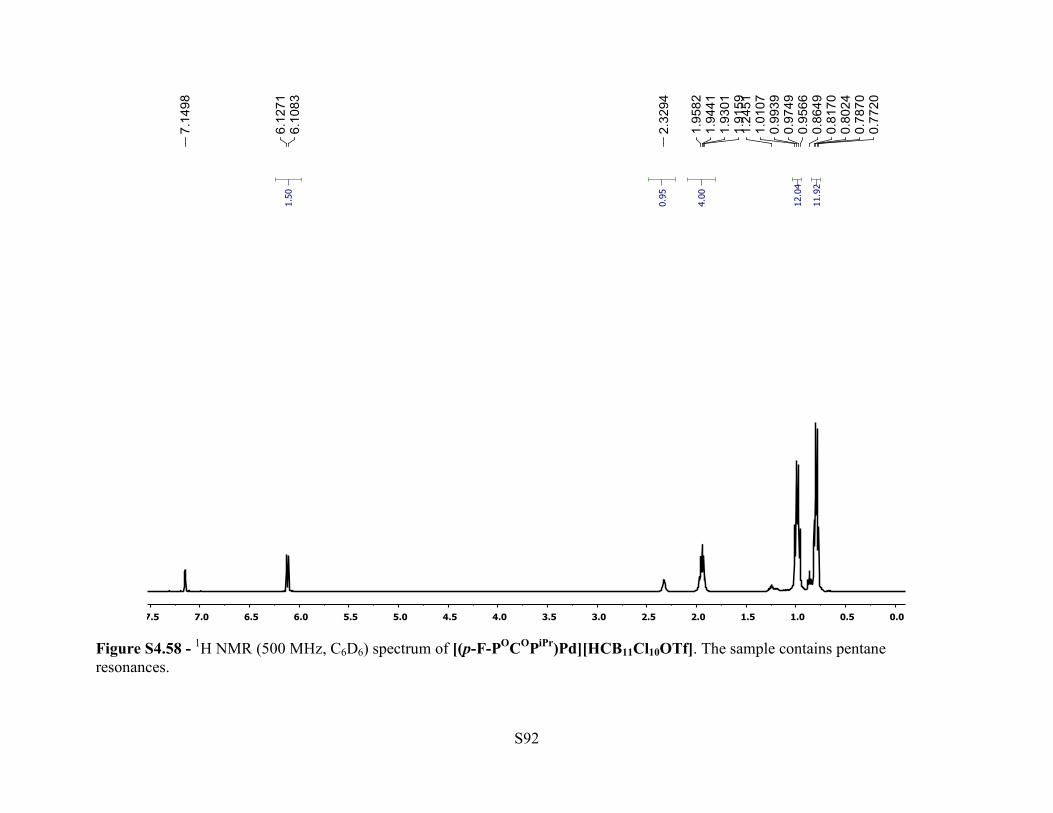
Figure S4.58 - 1H NMR (500 MHz, C6D6) spectrum of [(p-F-POCOPiPr)Pd][HCB11Cl10OTf]. The sample contains pentane resonances.
Page 93
S93
Figure S4.59 - 11B NMR (128 MHz, C6D6) spectrum of [(p-F-POCOPiPr)Pd][HCB11Cl10OTf].
Page 94
S94
Figure S4.60 - 13C{1H} NMR (126 MHz, C6D6) spectrum of [(p-F-POCOPiPr)Pd][HCB11Cl10OTf]. The sample contains pentane resonances.
Page 95
S95
Figure S4.61 - 19F NMR (470 MHz, C6D6) spectrum of [(p-F-POCOPiPr)Pd][HCB11Cl10OTf].
Page 96
S96
Figure S4.62 - 31P{1H} NMR (202 MHz, C6D6) spectrum of [(p-F-POCOPiPr)Pd][HCB11Cl10OTf].
Page 97
S97
Figure S4.63 - 1H NMR (400 MHz, C6D5Br) spectrum of [(Et3Si)2OTf][HCB11Cl11]. The sample contains pentane resonances.
Page 98
S98
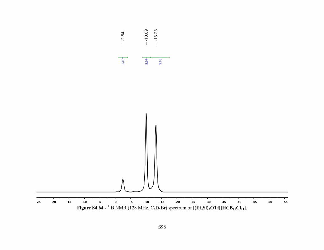
Figure S4.64 - 11B NMR (128 MHz, C6D5Br) spectrum of [(Et3Si)2OTf][HCB11Cl11].
Page 99
S99
Figure S4.65 - 13C{1H} NMR (101 MHz, C6D5Br) spectrum of [(Et3Si)2OTf][HCB11Cl11].
Page 100
S100
Figure S4.66 19F NMR (376 MHz, C6D5Br) spectrum of [(Et3Si)2OTf][HCB11Cl11].
Page 101
S101
Figure S4.67 - 29Si{1H} NMR (79.5 MHz, C6D5Br) spectrum of [(Et3Si)2OTf][HCB11Cl11].
Page 102
S102
V. X-Ray Crystallography Studies
Crystallographic information in the form of CIF files is available for free from the Cambridge
Crystallographic Data Centre: CCDC 1060341 is Cs[HCB11H8(OTf)3], 1060342 is
Cs[HCB11H5Br5OTf3], 1060343 is Et3Si[HCB11Cl10OTf], 1060344 is [(FPOCOP)Pd-Cl-
Pd(POCOPF)][HCB11Cl10OTf], 1060345 is iPr3Si[HCB11Cl10OTf], 1060346 is
Ph3C[HCB11Cl10OTf], 1060347 is [(FPOCOP)Pd(C6D5Br)][HCB11Cl10OTf], 1060348 is
[(FPOCOP)Pd][HCB11Cl10OTf], 1060349 is [(FPOCOP)Pd][HCB11Cl11], 1061042 is
Cs[HCB11Cl9(OTf)2].
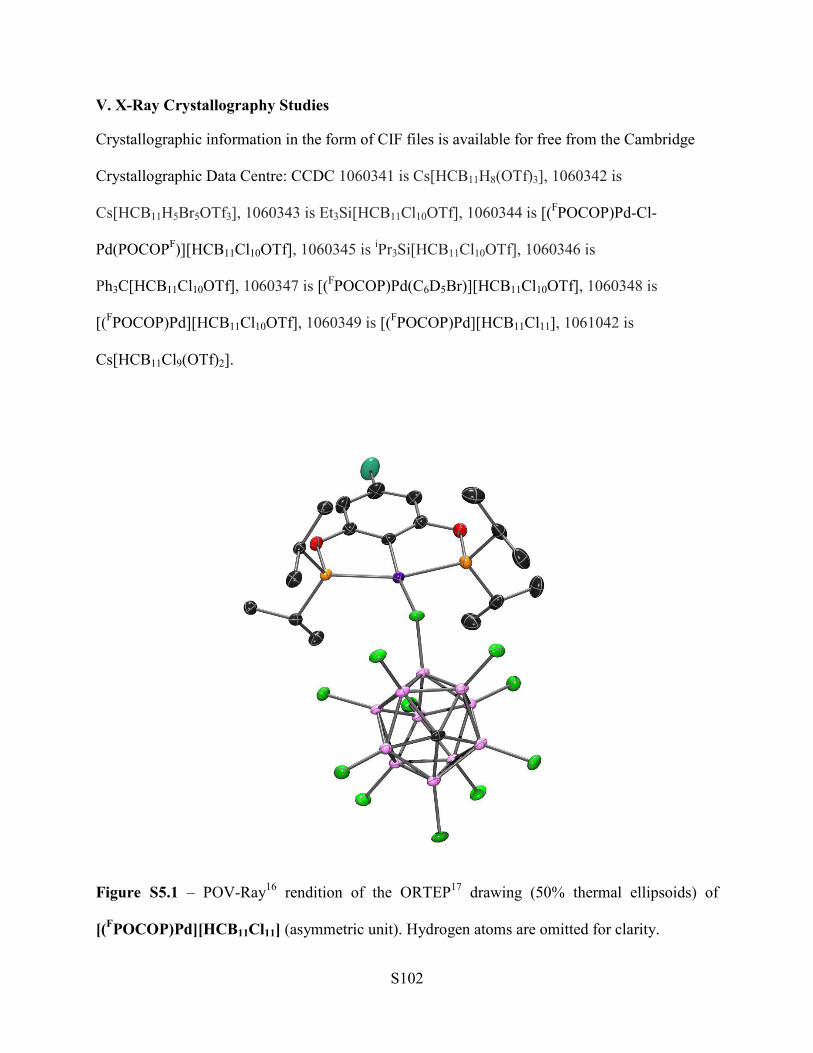
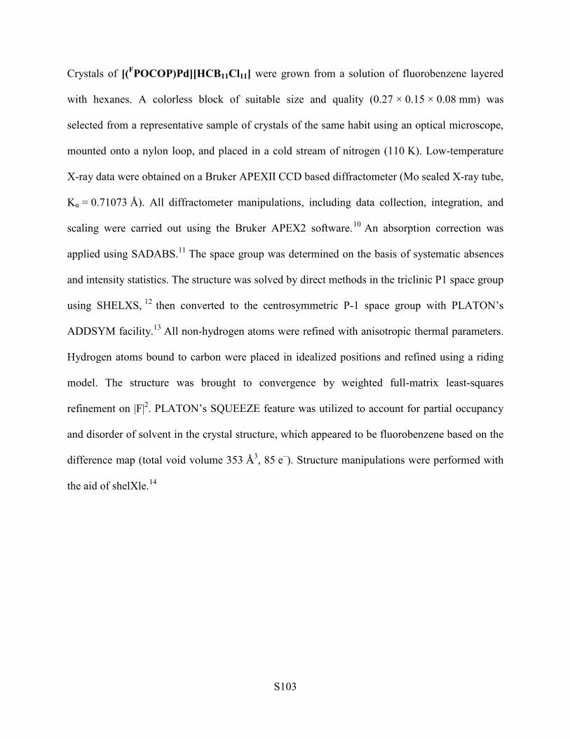
Figure S5.1 – POV-Ray16 rendition of the ORTEP17 drawing (50% thermal ellipsoids) of
[(FPOCOP)Pd][HCB11Cl11] (asymmetric unit). Hydrogen atoms are omitted for clarity.
Page 103
S103
Crystals of [(FPOCOP)Pd][HCB11Cl11] were grown from a solution of fluorobenzene layered
with hexanes. A colorless block of suitable size and quality (0.27 × 0.15 × 0.08 mm) was
selected from a representative sample of crystals of the same habit using an optical microscope,
mounted onto a nylon loop, and placed in a cold stream of nitrogen (110 K). Low-temperature
X-ray data were obtained on a Bruker APEXII CCD based diffractometer (Mo sealed X-ray tube,
Kα = 0.71073 Å). All diffractometer manipulations, including data collection, integration, and
scaling were carried out using the Bruker APEX2 software.10 An absorption correction was
applied using SADABS.11 The space group was determined on the basis of systematic absences
and intensity statistics. The structure was solved by direct methods in the triclinic P1 space group
using SHELXS, 12 then converted to the centrosymmetric P-1 space group with PLATON’s
ADDSYM facility.13 All non-hydrogen atoms were refined with anisotropic thermal parameters.
Hydrogen atoms bound to carbon were placed in idealized positions and refined using a riding
model. The structure was brought to convergence by weighted full-matrix least-squares
refinement on |F|2. PLATON’s SQUEEZE feature was utilized to account for partial occupancy
and disorder of solvent in the crystal structure, which appeared to be fluorobenzene based on the
difference map (total void volume 353 Å3, 85 e–). Structure manipulations were performed with
the aid of shelXle.14
Page 104
S104
Figure S5.2 – POV-Ray16 rendition of the ORTEP17 drawing (50% thermal ellipsoids) of
[(FPOCOP)Pd][HCB11Cl10OTf] (asymmetric unit). Hydrogen atoms are omitted for clarity.
Crystals of [(FPOCOP)Pd][HCB11Cl10OTf] were grown from a solution of ortho-
difluorobenzene layered with pentane. A clear colorless block of suitable size and quality
(0.34 × 0.32 × 0.08 mm) was selected from a representative sample of crystals of the same habit
using an optical microscope, mounted onto a nylon loop, and placed in a cold stream of nitrogen
(110 K). Low-temperature X-ray data were obtained on a Bruker APEXII CCD based
diffractometer (Mo sealed X-ray tube, Kα = 0.71073 Å). All diffractometer manipulations,
including data collection, integration, and scaling were carried out using the Bruker APEX2
software.10 An absorption correction was applied using SADABS.11 The space group was
Page 105
S105
determined on the basis of systematic absences and intensity statistics. The structure was solved
by direct methods in the triclinic P-1 space group using SHELXS.12 All non-hydrogen atoms
were refined with anisotropic thermal parameters. Hydrogen atoms bound to carbon were placed
in idealized positions and refined using a riding model. The structure was brought to
convergence by weighted full-matrix least-squares refinement on |F|2. A check for missed
symmetry was performed with PLATON’s ADDSYM facility, finding no apparent higher
symmetry.13 Structure manipulations were performed with the aid of shelXle.14
Figure S5.3 – POV-Ray16 rendition of the ORTEP17 drawing (50% thermal ellipsoids) of
[(FPOCOPiPr)Pd(BrC6D5)][HCB11Cl10OTf] (asymmetric unit). Hydrogen atoms are omitted for
clarity.
Page 106
S106
Crystals of [(FPOCOPiPr)Pd(BrC6D5)][HCB11Cl10OTf] were grown from a solution of
bromobenzene-d5 by vapor diffusion of pentane. A colorless block of suitable size and quality
(0.33 × 0.21 × 0.06 mm) was selected from a representative sample of crystals of the same habit
using an optical microscope, mounted onto a nylon loop, and placed in a cold stream of nitrogen
(150 K). Low-temperature X-ray data were obtained on a Bruker APEXII CCD based
diffractometer (Mo sealed X-ray tube, Kα = 0.71073 Å). All diffractometer manipulations,
including data collection, integration, and scaling were carried out using the Bruker APEX2
software.10 An absorption correction was applied using SADABS.11 The space group was
determined on the basis of systematic absences and intensity statistics. The structure was solved
by direct methods in the monoclinic P21/n space group using SHELXS.12 All non-hydrogen
atoms were refined with anisotropic thermal parameters. Hydrogen atoms bound to carbon were
placed in idealized positions and refined using a riding model. The structure was brought to
convergence by weighted full-matrix least-squares refinement on |F|2. Structure manipulations
were performed with the aid of shelXle.14 A check for missed symmetry was run using the
ADDSYM program within PLATON,14 revealing no apparent higher symmetry.
Page 107
S107
Figure S5.4 – POV-Ray16 rendition of the ORTEP17 drawing (50% thermal ellipsoids) of
[Ph3C][HCB11Cl10OTf] • C6H5F (asymmetric unit). Hydrogen atoms are omitted for clarity.
Crystals of [Ph3C][HCB11Cl10OTf] • C6H5F were grown from a fluorobenzene solution layered
with pentane. A clear yellow block of suitable size and quality (0.27 x 0.19 x 0.11 mm) was
selected from a representative sample of crystals of the same habit using an optical microscope,
mounted onto a nylon loop, and placed in a cold stream of nitrogen (150 K). Low-temperature X-
ray data were obtained on a Bruker APEXII CCD based diffractometer (Mo sealed X-ray tube,
Kα = 0.71073 Å). All diffractometer manipulations, including data collection, integration, and
scaling were carried out using the Bruker APEX2 software.10 An absorption correction was
applied using SADABS.11 The space group was determined on the basis of systematic absences
Page 108
S108
and intensity statistics. The structure was solved by direct methods in the orthorhombic Pbca
space group using SHELXS.12 All non-hydrogen atoms were refined with anisotropic thermal
parameters. Hydrogen atoms bound to carbon were placed in idealized positions and refined
using a riding model. The structure was brought to convergence by weighted full-matrix least-
squares refinement on |F|2. Structure manipulations were performed with the aid of shelXle.14 A
check for missed symmetry was run using the ADDSYM program within PLATON, revealing no
apparent higher symmetry.13
A solvent molecule consistent with fluorobenzene was identified in the electron difference map,
but was unable to be satisfactorily modeled, due to an apparent 6-fold rotational disorder. The
solvent electron density was therefore accounted for with SQUEEZE, which revealed four voids
in the unit cell (292 Å3, 104 e-), consistent with one fluorobenzene solvent molecule (50 e-) per
asymmetric unit. Our model is therefore inconsistent with the checkCIF calculated values of
moiety/sum formulae, formula weight (Mr), density (Dx), absorption coefficient (mu), and F000,
which accounts for all checkCIF alerts level A–C.
Page 109
S109
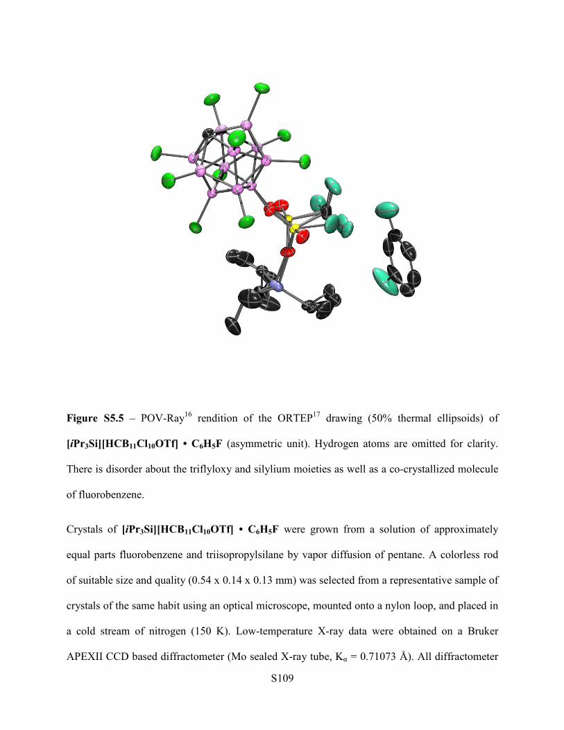
Figure S5.5 – POV-Ray16 rendition of the ORTEP17 drawing (50% thermal ellipsoids) of
[iPr3Si][HCB11Cl10OTf] • C6H5F (asymmetric unit). Hydrogen atoms are omitted for clarity.
There is disorder about the triflyloxy and silylium moieties as well as a co-crystallized molecule
of fluorobenzene.
Crystals of [iPr3Si][HCB11Cl10OTf] • C6H5F were grown from a solution of approximately
equal parts fluorobenzene and triisopropylsilane by vapor diffusion of pentane. A colorless rod
of suitable size and quality (0.54 x 0.14 x 0.13 mm) was selected from a representative sample of
crystals of the same habit using an optical microscope, mounted onto a nylon loop, and placed in
a cold stream of nitrogen (150 K). Low-temperature X-ray data were obtained on a Bruker
APEXII CCD based diffractometer (Mo sealed X-ray tube, Kα = 0.71073 Å). All diffractometer
Page 110
S110
manipulations, including data collection, integration, and scaling were carried out using the
Bruker APEX2 software.10 An absorption correction was applied using SADABS.11 The space
group was determined on the basis of systematic absences and intensity statistics. The structure
was solved by direct methods in the monoclinic P21/c space group using SHELXS.12 All non-
hydrogen atoms were refined with anisotropic thermal parameters. Hydrogen atoms bound to
carbon were placed in idealized positions and refined using a riding model. The structure was
brought to convergence by weighted full-matrix least-squares refinement on |F|2. Structure
manipulations were performed with the aid of shelXle.14 A check for missed symmetry was run
using the ADDSYM program within PLATON, revealing no apparent higher symmetry.13
Similarity restraints were applied to the triflyloxy and triisopropylsilyl moieties to handle the
refinement of the disorder.
Page 111
S111
Figure S5.6 – POV-Ray16 rendition of the ORTEP17 drawing (50% thermal ellipsoids) of
[Et3Si][HCB11Cl10OTf] (asymmetric unit). Hydrogen atoms are omitted for clarity. There are
two distinct molecules within the asymmetric unit. There is disorder about the triflyloxy and
silylium moieties of one of the molecules.
Crystals of [Et3Si][HCB11Cl10OTf] were grown from a fluorobenzene solution by vapor
diffusion of pentane. A colorless block of suitable size and quality (0.28 x 0.17 x 0.16 mm) was
selected from a representative sample of crystals of the same habit using an optical microscope,
mounted onto a nylon loop, and placed in a cold stream of nitrogen (150 K). Low-temperature X-
ray data were obtained on a Bruker APEXII CCD based diffractometer (Mo sealed X-ray tube,
Kα = 0.710 73 Å). All diffractometer manipulations, including data collection, integration, and
Page 112
S112
scaling were carried out using the Bruker APEX2 software.10 An absorption correction was
applied using SADABS.11 The space group was determined on the basis of systematic absences
and intensity statistics. The structure was solved by direct methods in the monoclinic P21/n space
group using XS12 (incorporated in SHELXTL). All non-hydrogen atoms were refined with
anisotropic thermal parameters. Hydrogen atoms bound to carbon were placed in idealized
positions and refined using a riding model. The structure was brought to convergence by
weighted full-matrix least-squares refinement on |F|2. A check for missed symmetry was run
using the ADDSYM program within PLATON,13 revealing no apparent higher symmetry.
Similarity restraints were applied to the triflyloxy and silylium moieties to handle the refinement
of the disorder. The checkCIF report yielded three moderate-level alerts attributed to the
disordered alkyl chains of the triethylsilyl moieties, specifically identifying the prolate nature of
the disorded carbons, and the resultant imprecision of C–C bond lengths. High residual density
(1:49 e− Å−3) was found near one of the disordered silicon centers (Si1_5 – 0.04 Å), and a hole in
the residual density (−0.99 e− Å−3) was also observed near the same atom (Si1_5 – 0.65 Å); this
is attributed to imperfect modeling of the disorder. Large X–O–Y angles (>140°) were observed,
as expected in light of previously reported triflate-bridged trimethylsilylium cations.
Page 113
S113
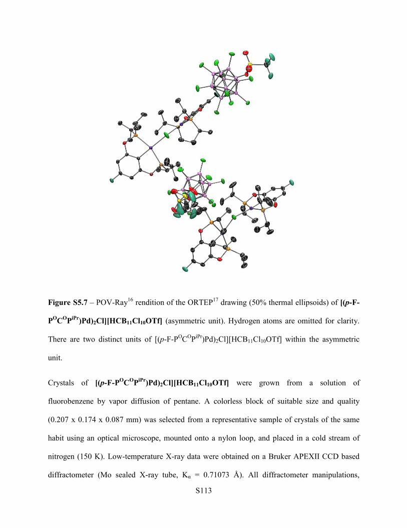
Figure S5.7 – POV-Ray16 rendition of the ORTEP17 drawing (50% thermal ellipsoids) of [(p-F-
POCOPiPr)Pd)2Cl][HCB11Cl10OTf] (asymmetric unit). Hydrogen atoms are omitted for clarity.
There are two distinct units of [(p-F-POCOPiPr)Pd)2Cl][HCB11Cl10OTf] within the asymmetric
unit.
Crystals of [(p-F-POCOPiPr)Pd)2Cl][HCB11Cl10OTf] were grown from a solution of
fluorobenzene by vapor diffusion of pentane. A colorless block of suitable size and quality
(0.207 x 0.174 x 0.087 mm) was selected from a representative sample of crystals of the same
habit using an optical microscope, mounted onto a nylon loop, and placed in a cold stream of
nitrogen (150 K). Low-temperature X-ray data were obtained on a Bruker APEXII CCD based
diffractometer (Mo sealed X-ray tube, Kα = 0.71073 Å). All diffractometer manipulations,
Page 114
S114
including data collection, integration, and scaling were carried out using the Bruker APEX2
software.10 An absorption correction was applied using SADABS.11 The space group was
determined on the basis of systematic absences and intensity statistics. The structure was solved
by direct methods in the monoclinic P21/n space group using SHELXS.12 All non-hydrogen
atoms were refined with anisotropic thermal parameters. Hydrogen atoms bound to carbon were
placed in idealized positions and refined using a riding model. The structure was brought to
convergence by weighted full-matrix least-squares refinement on |F|2. Structure manipulations
were performed with the aid of shelXle.14 A check for missed symmetry was run using the
ADDSYM program within PLATON, revealing no apparent higher symmetry.13 Similarity
restraints were applied to the triflyloxy moieties to handle the refinement of the disorder.
Figure S5.8a – POV-Ray16 rendition of the ORTEP17 drawing (50% thermal ellipsoids) of
Cs[HCB11H5Br5OTf] (asymmetric unit). The asymmetric unit contains one cesium atom (in
blue, located on two special positions at 50% occupancy) and one carborane anion. Hydrogen
atoms are omitted for clarity. Cs[HCB11H4Br6OTf] co-crystallizes as a minor component
(~6%). This is agreement with the ~5% of Cs[HCB11H4Br6OTf] observed by MALDI MS.
Page 115
S115
Figure S5.8b – POV-Ray16 rendition of the ORTEP17 drawing (50% thermal ellipsoids) of
Cs[HCB11H5Br5OTf] showing the coordination about cesium atom (in blue). Hydrogen atoms
are omitted for clarity. Cs[HCB11H4Br6OTf] co-crystallizes as a minor component (~6%). This
is in agreement with the ~5% of [HCB11H4Br6OTf]- observed by MALDI MS. The asymmetric
unit contains one cesium atom (in blue, located on two special positions at 50% occupancy) and
one carborane anion (well represented by a 94:6 mixture of [HCB11H5Br5OTf] and
[HCB11H4Br6OTf]).
Crystals of Cs[HCB11H5Br5OTf] were grown from a solution of dichloromethane by slow
evaporation of solvent. A colorless sheet of suitable size and quality (0.20 x 0.12 x 0.08 mm)
was selected from a representative sample of crystals of the same habit using an optical
microscope, mounted onto a nylon loop, and placed in a cold stream of nitrogen (150 K). Low-
temperature X-ray data were obtained on a Bruker APEXII CCD based diffractometer (Mo
sealed X-ray tube, Kα = 0.71073 Å). All diffractometer manipulations, including data collection,
Page 116
S116
integration, and scaling were carried out using the Bruker APEX2 software.10 An absorption
correction was applied using SADABS.11 The space group was determined on the basis of
systematic absences and intensity statistics. The structure was solved by the direct methods in the
orthorhombic Pbcn space group using SHELXS.12 All non-hydrogen atoms were refined with
anisotropic thermal parameters. Hydrogen atoms bound to carbon and boron were placed in
idealized positions and refined using a riding model. The structure was brought to convergence
by weighted full-matrix least-squares refinement on |F|2. Structure manipulations were performed
with the aid of shelXle.14 A check for missed symmetry was run using the ADDSYM program
within PLATON, revealing no apparent higher symmetry.13 Cs[HCB11H4Br6OTf] co-
crystallizes as a minor component (~6%), and the asymmetric unit is satisfactorily modeled as a
94:6 mixture of Cs[HCB11H5Br5OTf] and Cs[HCB11H4Br6OTf]. This is agreement with the
~5% of [HCB11H4Br6OTf]- observed by MALDI MS.
Figure S5.9a – POV-Ray16 rendition of the ORTEP17 drawing (50% thermal ellipsoids) of
Cs[HCB11H8(OTf)3] (asymmetric unit). Hydrogen atoms are omitted for clarity.
Page 117
S117
Figure S5.9b – POV-Ray16 rendition of the ORTEP17 drawing (50% thermal ellipsoids) of
Cs[HCB11H8(OTf)3] showing coordination about cesium (in blue). Hydrogen atoms are omitted
for clarity.
Crystals of Cs[HCB11H8(OTf)3] were grown from a solution of dichloromethane by slow
evaporation of solvent. A colorless block of suitable size and quality (0.55 x 0.20 x 0.10 mm)
was selected from a representative sample of crystals of the same habit using an optical
microscope, mounted onto a nylon loop, and placed in a cold stream of nitrogen (150 K). Low-
temperature X-ray data were obtained on a Bruker APEXII CCD based diffractometer (Mo
sealed X-ray tube, Kα = 0.71073 Å). All diffractometer manipulations, including data collection,
integration, and scaling were carried out using the Bruker APEX2 software.10 An absorption
correction was applied using SADABS.11 The space group was determined on the basis of
Page 118
S118
systematic absences and intensity statistics. The structure was solved by the Patterson method in
the monoclinic P21/c space group using SHELXS.12 All non-hydrogen atoms were refined with
anisotropic thermal parameters. Hydrogen atoms bound to carbon and boron were placed in
idealized positions and refined using a riding model. The structure was brought to convergence
by weighted full-matrix least-squares refinement on |F|2. Structure manipulations were performed
with the aid of shelXle.14 A check for missed symmetry was run using the ADDSYM program
within PLATON, revealing no apparent higher symmetry.13
Figure S5.10a – POV-Ray16 rendition of the ORTEP17 drawing (50% thermal ellipsoids) of
Cs[HCB11Cl9(OTf)2] (asymmetric unit). Hydrogen atoms are omitted for clarity. There is
disorder about the 7-position triflyloxy group.
Page 119
S119
Figure S5.10b – POV-Ray16 rendition of the ORTEP17 drawing (50% thermal ellipsoids) of
Cs[HCB11Cl9(OTf)2] ] showing coordination about cesium (in blue). Disorder about the 7-
position triflyloxy group omitted for clarity. Hydrogen atoms are omitted for clarity.
Crystals of Cs[HCB11Cl9(OTf)2] were grown from a solution of acetonitrile by slow evaporation
of solvent. A colorless plate of suitable size and quality (0.42 x 0.14 x 0.05 mm) was selected
from a representative sample of crystals of the same habit using an optical microscope. All
operations were performed on a Bruker-Nonius Kappa Apex2 diffractometer, using graphite-
monochromated MoKα radiation. All diffractometer manipulations, including data collection,
integration, scaling, and absorption corrections were carried out using the Bruker Apex2
software. 15 Preliminary cell constants were obtained from three sets of 12 frames. Data
collection was carried out at 120 K, using a frame time of 30 sec and a detector distance of 60
Page 120
S120
mm. The optimized strategy used for data collection consisted of two phi and seven omega scan
sets, with 0.5° steps in phi or omega; completeness was 99.8%. A total of 2234 frames were
collected. Final cell constants were obtained from the xyz centroids of 9002 reflections after
integration.
From the systematic absences, the observed metric constants and intensity statistics, space group
Pbca was chosen initially; subsequent solution and refinement confirmed the correctness of the
initial choice. The structures were solved using SIR-92,16 and refined (full-matrix-least squares)
using the Oxford University Crystals for Windows program.17 All ordered non-hydrogen atoms
were refined using anisotropic displacement parameters; the hydrogen atoms attached to the
carborane C atom was fixed at a calculated geometric position 0.95 Å from C(1) and refined as a
riding atom.
Compound Cs[HCB11Cl9(OTf)2] contained significant disorder, which was resolved (in part)
successfully. The resolvable disorder involved the sulfonate oxygen atoms of the triflyloxy
moiety attached to B(7); modeling of the disorder of the CF3 group was not successful. The two-
component disorder (major: O4/O5/O6; minor: O41/O51/O61) was described with a constraint
such that the occupancies of the major (anisotropic displacement parameters, occupancy
0.777(9)) and minor components (isotropic displacement parameters) sum to 1.0. It appears that
the disorder is caused by two positions for the CB11 cage, related by a small rotation
approximately about the B5-B8 axis. It was not possible to model the lower level of disorder in
the other triflyloxy moiety. The final least-squares refinement converged to R1 = 0.0326 (I >
2σ(I), 5328 data) and wR2 = 0.0870 (F2, 7782 data, 356 parameters).
Page 121
S121
VI. Supporting Information References
1. S. Wei-Chuan, K.R. Gee, D. H. Klaubert and R. P. Haugland, J. Org. Chem. 1997, 62, 6469.
2. J. R. Doyle and D. Drew, Inorg. Synth., 1990, 28, 346.
3. W. X. Gu, B. J. McCulloch, J. H. Reibenspies and O. V. Ozerov, Chem. Commun., 2010, 46,
820.
4. C. Douvris and O. V. Ozerov, Science, 2008, 321, 1188.
5. Isotope Distribution Calculator and Mass Spec Plotter,
http://www.sisweb.com/mstools/isotope.htm, (accessed July 2015).
6. T. R. Forbus and J. C. Martin, J. Org. Chem., 1979, 44, 313-314.
7. T. R. Forbus and J. C. Martin, J. Org. Chem., 1987, 52, 4156–4159.
8. T. K. Hollis and B. Bosnich, J. Am. Chem. Soc., 1995, 117, 4570-4581.
9. Synthesis of [Ph3C][HCB11Cl11] from Ag[HCB11Cl11] with Ph3CBr in a toluene/acetonitrile
was previously described by Reed and co-workers. C. A. Reed, Acc. Chem. Res., 2009, 43, 121.
10. APEX2, Version 2013.2-0; Bruker AXS Inc., Madison, WI, 2013.
11. SADABS, Version 2008/1; Bruker AXS Inc.: Madison, WI, 2008.
12. G. M. Sheldrick, Acta Crystallogr. A, 2008, 64, 112–122.
13. A. L. Spek, J. Appl. Crystallogr., 2003, 36, 7–13.
14. C. B. Hübschle and G. M. Sheldrick, J. Appl. Crystallogr., 2011, 44, 1281–1284.
15 . Apex2, Version 2 User Manual, M86-E01078, Bruker Analytical X-ray Systems, Madison,
WI, June 2006.
16. A. Altomare, G. Cascarano, G. Giacovazzo, A. Guagliardi, M.C. Burla, G. Polidori and M. J.
Camalli, J. Appl. Cryst., 1994, 27, 435.
Page 122
S122
17. P. W. Betteridge, J. R. Carruthers, R. I. Cooper, K. Prout and D. J. Watkin, J. Appl. Cryst.,
2003, 36, 1487.


![, Helpdesk CCTV Kit - iget.eu · CCTV Kit Helpdesk. ... 6\QFKURQL]DFH 6H]QDP ýtVOR NDQiOX 6H]QDP XGiORVWt ýDVRYi RVD](https://static.documents.pub/doc/80x56/5b8106b47f8b9a2b678b6853/-helpdesk-cctv-kit-igeteu-cctv-kit-helpdesk-6qfkurqldfh-6hqdp-ytvor.jpg)

