Available on line www.jocpr.com Journal of Chemical and Pharmaceutical Research __________________________________________________ ISSN No: 0975-7384 CODEN(USA): JCPRC5 J. Chem. Pharm. Res., 2010, 2(4):835-850 ______________________________________________________________________________ 835 Comparative conformational, structural and vibrational study on the molecular structure of tyrosine and L-DOPA using density functional theory Shamoon Ahmad Siddiqui 1 , Anoop Kumar Pandey 2 , Apoorva Dwivedi 2 , Sudha Jain 3 , Neeraj Misra 2* 1 Shri Ramswaroop Memorial College of Engg. and Management, Lucknow, India 2 Department of Physics, Lucknow University, Lucknow, India 3 Department of Chemistry, Lucknow University, Lucknow, India __________________________________________________________ ABSTRACT A brief conformational, structural and vibrational study has been performed on the molecular structure of two well known amino acids tyrosine and L-DOPA. The equilibrium geometry, harmonic vibrational frequencies, infrared intensities and Raman scattering activities were calculated by the Density Functional B3LYP method employing 6-311G(d,p) as the basis set and the vibrational studies were interpreted in terms of potential energy distribution (P.E.D.). The internal coordinates were optimized repeatedly to maximize the P.E.D. contributions. A detailed interpretation of the infrared and Raman spectra of tyrosine and L-DOPA is reported in the present work. The similarities and differences between the vibrational spectra of the two molecules studied have been highlighted. The calculations are in agreement with experiment. The thermodynamic calculations related to the title compounds were also performed at B3LYP/6-311G(d,p) level of theory. The FT-Raman and FT-IR spectra of tyrosine and L-DOPA have been taken from literature. Keywords: FT-IR and FT-Raman Spectra; Density functional theory; P.E.D.; Tyrosine and L- DOPA; Vibrational study. ______________________________________________________________________________ INTRODUCTION Tyrosine (4-hydroxyphenylalanine) also known as L-tyrosine is a non essential amino acid that is a building block of protein. It is a precursor of the neurotransmitter dopamine, as well as a
Transcript
Available on line www.jocpr.com
Journal of Chemical and Pharmaceutical Research __________________________________________________
Comparative conformational, structural and vibrational study on
the molecular structure of tyrosine and L-DOPA using density functional theory
Shamoon Ahmad Siddiqui1, Anoop Kumar Pandey2, Apoorva Dwivedi2, Sudha Jain3,
Neeraj Misra2*
1Shri Ramswaroop Memorial College of Engg. and Management, Lucknow, India
2Department of Physics, Lucknow University, Lucknow, India 3Department of Chemistry, Lucknow University, Lucknow, India
__________________________________________________________ ABSTRACT A brief conformational, structural and vibrational study has been performed on the molecular structure of two well known amino acids tyrosine and L-DOPA. The equilibrium geometry, harmonic vibrational frequencies, infrared intensities and Raman scattering activities were calculated by the Density Functional B3LYP method employing 6-311G(d,p) as the basis set and the vibrational studies were interpreted in terms of potential energy distribution (P.E.D.). The internal coordinates were optimized repeatedly to maximize the P.E.D. contributions. A detailed interpretation of the infrared and Raman spectra of tyrosine and L-DOPA is reported in the present work. The similarities and differences between the vibrational spectra of the two molecules studied have been highlighted. The calculations are in agreement with experiment. The thermodynamic calculations related to the title compounds were also performed at B3LYP/6-311G(d,p) level of theory. The FT-Raman and FT-IR spectra of tyrosine and L-DOPA have been taken from literature.
Keywords: FT-IR and FT-Raman Spectra; Density functional theory; P.E.D.; Tyrosine and L-DOPA; Vibrational study. ______________________________________________________________________________
INTRODUCTION
Tyrosine (4-hydroxyphenylalanine) also known as L-tyrosine is a non essential amino acid that is a building block of protein. It is a precursor of the neurotransmitter dopamine, as well as a
Neeraj Misra et al J. Chem. Pharm. Res., 2010, 2(4): 835-850 _____________________________________________________________________________
836
precursor to the adrenal harmones norepinephrine and epinephrine. The body can make tyrosine from the amino acid phenylalanine [1]. We get tyrosine in our body by eating protein-rich foods like meat, fish, eggs, dairy products and beans. Tyrosine is used by the body to make the neurotransmitter noradrenaline. Noradrenaline is believed to be in short supply in the brains of people who are depressed [2]. Tyrosine may help athletes avoid overtraining, due to its ability to offset fatigue [3]. Because tyrosine is a precursor of dopamine, supplementing with tyrosine may heighten mental alertness, increase feeling of well being, decrease feelings of depression, and offset physical and mental fatigue [4,5]. Tyrosine also serves to protect the integrity of the skin. Melanin, a substance which acts to protect the skin when the epidermis has been exposed to ultraviolet light, is derived from tyrosine. If a shortage of melanin is present within the body, skin defenses will be compromised. Melanin, which is derived from tyrosine, chemically reacts with sunlight to form a protective shield that protects the deeper layers of skin tissue [6]. People suffering from neurological degeneracy may also benefit from supplemental tyrosine [7]. The conversion of tyrosine to L-DOPA is catalyzes by tyrosine hydroxylase (TyrOH). TyrOH is a non-heme iron enzyme which uses molecular oxygen to hydroxylate tyrosine to form L-dihydroxyphenylalanine (L-DOPA) [8,9]. Levodopa or L-DOPA (3,4-dihydroxy-L-phenylalanine) is an intermediate in dopamine biosynthesis. L-DOPA is used as a prodrug to increase dopamine levels for the treatment of Parkinson’s disease, since it is able to cross the blood-brain barrier whereas dopamine itself cannot. Once L-DOPA has entered the central nervous system, it is metabolized to dopamine by aromatic L-amino acid decarboxylase [10,11]. The initial enzymatic reaction in the biosynthesis of brain catecholamines involves the formation of the catechol amino acid L-dihydroxyphenylalanine (L-DOPA) from tyrosine. Once formed, the L-DOPA is immediately decarboxylated to form dopamine, which in some neurons, is further transformed to norepinephrine [12]. L-DOPA can not be detected normally in the brain or in the blood [13], and thus it is unlikely that circulating L-DOPA is a physiological precursor for brain catecholamines. However, when exogenous L-DOPA is administered to experimental animals, the concentration of dopamine in the brain increases [14,15]. This observation, coupled with the finding that dopamine concentrations measured at autopsy in brains of patients with Parkinson’s disease are low [16], suggested that exogenous L-DOPA might be useful in the treatment of Parkinsonism. That hypothesis has now been confirmed in numerous clinical studies [17-19]. As a part of our ongoing research work [20-24], here in the present communication we report the vibrational study carried out on tyrosine and L-DOPA. To the best of our knowledge neither the complete Raman and IR spectra nor the comparative quantum chemical calculations for tyrosine and L-DOPA have been reported so far in the literature. Therefore, the aim of this paper is to interpret theoretically calculated vibrational spectra of the two well known amino acids tyrosine and L-DOPA by means of the potential energy distribution (P.E.D.) analysis of all fundamental vibrational modes. In this regard, with the help of VEDA program both the P.E.D. analysis and its optimization were carried out [25]. Based on the DFT calculations we also interpreted the experimental IR and Raman spectra of these title compounds. FT-IR and FT-Raman spectra The FT-IR and FT-Raman spectra of tyrosine and L-DOPA were taken from the Sigma-Aldrich chemical company (USA) with a stated purity of greater than 99% in condensed phase [26-29]. The observed frequencies from FT-IR and FT-Raman spectra of tyrosine and L-DOPA are given in Table 2 and 3 respectively.
Neeraj Misra et al J. Chem. Pharm. Res., 2010, 2(4): 835-850 _____________________________________________________________________________
837
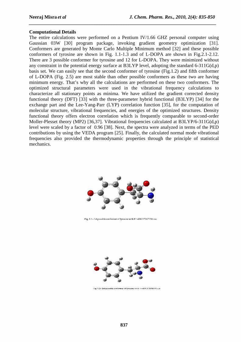
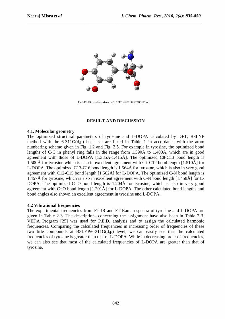
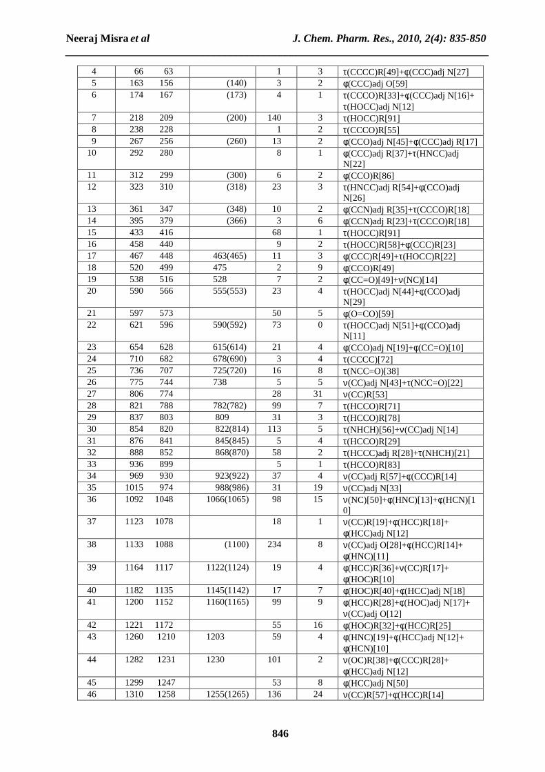
Computational Details The entire calculations were performed on a Pentium IV/1.66 GHZ personal computer using Gaussian 03W [30] program package, invoking gradient geometry optimization [31]. Conformers are generated by Monte Carlo Multiple Minimum method [32] and these possible conformers of tyrosine are shown in Fig. 1.1-1.3 and of L-DOPA are shown in Fig.2.1-2.12. There are 3 possible conformer for tyrosine and 12 for L-DOPA. They were minimized without any constraint in the potential energy surface at B3LYP level, adopting the standard 6-311G(d,p) basis set. We can easily see that the second conformer of tyrosine (Fig.1.2) and fifth conformer of L-DOPA (Fig. 2.5) are most stable than other possible conformers as these two are having minimum energy. That’s why all the calculations are performed on these two conformers. The optimized structural parameters were used in the vibrational frequency calculations to characterize all stationary points as minima. We have utilized the gradient corrected density functional theory (DFT) [33] with the three-parameter hybrid functional (B3LYP) [34] for the exchange part and the Lee-Yang-Parr (LYP) correlation function [35], for the computation of molecular structure, vibrational frequencies, and energies of the optimized structures. Density functional theory offers electron correlation which is frequently comparable to second-order Moller-Plesset theory (MP2) [36,37]. Vibrational frequencies calculated at B3LYP/6-311G(d,p) level were scaled by a factor of 0.96 [38]. Next, the spectra were analyzed in terms of the PED contributions by using the VEDA program [25]. Finally, the calculated normal mode vibrational frequencies also provided the thermodynamic properties through the principle of statistical mechanics.
Neeraj Misra et al J. Chem. Pharm. Res., 2010, 2(4): 835-850 _____________________________________________________________________________
838
Neeraj Misra et al J. Chem. Pharm. Res., 2010, 2(4): 835-850 _____________________________________________________________________________
839
Neeraj Misra et al J. Chem. Pharm. Res., 2010, 2(4): 835-850 _____________________________________________________________________________
840
Neeraj Misra et al J. Chem. Pharm. Res., 2010, 2(4): 835-850 _____________________________________________________________________________
841
Neeraj Misra et al J. Chem. Pharm. Res., 2010, 2(4): 835-850 _____________________________________________________________________________
842
RESULT AND DISCUSSION 4.1. Molecular geometry The optimized structural parameters of tyrosine and L-DOPA calculated by DFT, B3LYP method with the 6-311G(d,p) basis set are listed in Table 1 in accordance with the atom numbering scheme given in Fig. 1.2 and Fig. 2.5. For example in tyrosine, the optimized bond lengths of C-C in phenyl ring falls in the range from 1.390Å to 1.400Å, which are in good agreement with those of L-DOPA [1.385Å-1.415Å]. The optimized C8-C13 bond length is 1.500Å for tyrosine which is also in excellent agreement with C7-C12 bond length [1.510Å] for L-DOPA. The optimized C13-C16 bond length is 1.564Å for tyrosine, which is also in very good agreement with C12-C15 bond length [1.562Å] for L-DOPA. The optimized C-N bond length is 1.457Å for tyrosine, which is also in excellent agreement with C-N bond length [1.458Å] for L-DOPA. The optimized C=O bond length is 1.204Å for tyrosine, which is also in very good agreement with C=O bond length [1.201Å] for L-DOPA. The other calculated bond lengths and bond angles also shown an excellent agreement in tyrosine and L-DOPA. 4.2 Vibrational frequencies The experimental frequencies from FT-IR and FT-Raman spectra of tyrosine and L-DOPA are given in Table 2-3. The descriptions concerning the assignment have also been in Table 2-3. VEDA Program [25] was used for P.E.D. analysis and to assign the calculated harmonic frequencies. Comparing the calculated frequencies in increasing order of frequencies of these two title compounds at B3LYP/6-311G(d,p) level, we can easily see that the calculated frequencies of tyrosine is greater than that of L-DOPA. While in decreasing order of frequencies, we can also see that most of the calculated frequencies of L-DOPA are greater than that of tyrosine.
Neeraj Misra et al J. Chem. Pharm. Res., 2010, 2(4): 835-850 _____________________________________________________________________________
843
Table 1: Optimized geometrical parameters of tyrosine and L-DOPA
Table 2: Vibrational wave numbers obtained for tyrosine at B3LYP/6-311G(d,p) in cm-1, Experimental frequencies from FT-IR and FT-Raman spectra in cm-1, IR intensities (Km mol-1), Raman scattering activities
(Ǻ4 amu-1) and assignment with P.E.D. percentage in square brackets.
Table 3: Vibrational wave numbers obtained for L-DOPA at B3LYP/6-311G(d,p) in cm-1, Experimental
frequencies from FT-IR and FT-Raman spectra in cm-1, IR intensities (Km mol-1), Raman scattering activities (Ǻ4 amu-1) and assignment with P.E.D. percentage in square brackets.
Table 4: Theoretically computed energies (a.u), zero-point vibrational energies (kcal mol-1), rotational constants (GHz), entropies (Cal mol-1 K-1) and dipole moment (D) for Tyrosine and L-Dopa.
Parameters Tyrosine L-Dopa Total energy -630.17618551 -705.41091503 Zero-point energy 121.22195 123.89533 Rotational Constants
2.37059 0.34807 0.31036
1.74391 0.30353 0.24353
Entropy Total Translational Rotational Vibrational
111.395 41.404 30.508 39.483
119.330 42.850 31.077 45.403
Dipole moment 3.348 2.289 4.3 Vibrational assignment Tyrosine has 24 atoms and 66 normal modes of fundamental vibration. All of the 66 fundamental vibrations are IR and Raman active. L-DOPA has 25 atoms and 69 normal modes of fundamental vibration. Here also all the 69 fundamental vibrations are IR and Raman active. Detailed description of vibrational modes can be given by means of normal coordinate analysis. The detailed vibrational assignments are achieved by comparing the band positions and intensities observed in FT-IR and FT-Raman spectra with wave numbers and intensities from molecular modeling calculations given in Table 2 and 3.
Neeraj Misra et al J. Chem. Pharm. Res., 2010, 2(4): 835-850 _____________________________________________________________________________
848
4.3.1 C-C Vibrations For tyrosine, the C-C aromatic stretch known as semi-circle stretching predicted at 762, 857, 969, 1146, 1175, 1287, 1411, 1566 and 1590 cm-1 are in excellent agreement with experimental observations of both FT-IR and FT-Raman spectra with significant P.E.D. For L-DOPA, the C-C aromatic stretch known as semi-circle stretching predicted at 744, 930, 974, 1088, 1258, 1355, 1415, 1496, 1581 and 1595 cm-1 are also in good agreement with experimental observations of both FT-IR and FT-Raman spectra with significant P.E.D. The other C-C vibrations, like bending and torsional vibrations are also found in excellent agreement with experimental data with appropriate P.E.D., for both title compounds. 4.3.2 C-H Vibrations The hetero aromatic structure shows the presence of C-H stretching vibration in the region 3000-3100 cm-1, which is the characteristic region for the ready identification of C-H stretching vibration [39]. Accordingly in the present study for tyrosine, the C-H vibrations are calculated at 3019, 3039 and 3070 cm-1 respectively with significant P.E.D. and are supported by experimental data. While for L-DOPA, the C-H vibrations are calculated at 3046 and 3062 cm-1 respectively with appropriate P.E.D. and are supported excellently by experimental data. 4.3.3 O-H Vibrations In a vibrational spectra, the strength of hydrogen bond determines the position of O-H band. Usually the O-H stretching appears at 3600-3400 cm-1 [40]. In this study tyrosine showed a very strong absorption peak at 3608 and 3680 cm-1 with P.E.D. [100], which are due to the O-H stretching vibration. While L-DOPA also showed very strong absorption peak at 3608, 3637 and 3694 cm-1 with same P.E.D., which are also due to the O-H stretching vibration. The different bending vibrations of the hydroxyl groups are also identified and they are listed in Table 2 and 3. 4.3.4 NH2 group Vibrations The scaled NH2 asymmetric stretch is calculated at 3433 cm-1 for tyrosine and also at 3433 cm-1 for L-DOPA with P.E.D.[100]. Whereas NH2 symmetric stretch is calculated at 3355 cm-1 for tyrosine and at 3354 cm-1 for L-DOPA with P.E.D.[100]. The various bending vibrations of NH2 group for both title compounds are also found to be in good agreement with the experimental data with appropriate P.E.D. 4.3.5 Methylene group Vibrations The asymmetric CH2 stretching vibrations are generally observed in the region 3100-3000 cm-1, while the symmetric stretch will appear between 3000-2900 cm-1 [41]. For tyrosine, the symmetric CH2 stretching vibrations is calculated at 2904 cm-1 and asymmetric CH2 stretching vibrations are calculated at 2941 and 2960 cm-1 with significant P.E.D. and are also supported by experimental data. For L-DOPA, the symmetric CH2 stretching vibrations is calculated at 2908 cm-1 and the asymmetric CH2 stretching vibrations are calculated at 2941 and 2957 cm-1 with appropriate P.E.D. and are also supported by experimental data. The various bending CH2 vibrations are also found to be in excellent agreement with experimental data for both title compounds. 4.3.6 Carbonyl Absorption Carbonyl absorptions are sensitive and both the carbon and oxygen atoms of the carbonyl group move during the vibration and they have nearly equal amplitude. In the present study the C=O stretching vibration is observed at 1761 cm-1 for tyrosine and at 1757 cm-1 for L-DOPA with significant P.E.D. and is also supported by experimental data and literature.
Neeraj Misra et al J. Chem. Pharm. Res., 2010, 2(4): 835-850 _____________________________________________________________________________
849
4.4 Other Molecular Properties Several calculated thermodynamic parameters are presented in Table 4. The zero point vibrational energy (ZPVE) of tyrosine is less than the L-DOPA, but the difference seems to be insignificant. The total energies are found less in L-DOPA, in comparison to the tyrosine. The total, translational, rotational and vibrational entropy of tyrosine is less than that of L-DOPA at room temperature, but the difference are only marginal. Values of rotational constants and dipole moment are greater in tyrosine, than that of L-DOPA.
CONCLUSION
Attempts have been made in the present work for the proper frequency assignments for the compound tyrosine and L-DOPA from the FT-IR and FT-Raman spectra. The equilibrium geometries and harmonic frequencies of tyrosine and L-DOPA were determined and analysed at DFT level of theory utilizing 6-311G(d,p) basis set, giving allowance for the lone pairs through diffused functions. The vibrational frequency calculation proved that both structures are stable (no imaginary frequency). The difference between the observed and scaled wave numbers values of most of the fundamentals is very small. Any discrepancy noted between the observed and the calculated frequencies may be due to the fact that the calculations have been actually done on a single molecule in the gaseous state contrary to the experimental values recorded in the presence of intermolecular interactions. The potential energy distribution contribution to each of the observed frequency shows the reliability and accuracy of the normal mode analysis. Therefore, the assignments made at higher levels of theory with only reasonable deviations from the experimental values, seems to be correct. Acknowledgement The corresponding author (Neeraj Misra) is grateful to UGC (new Delhi) for providing the financial assistance.
REFERENCES
[1] RJ Wurtman; MC Lewis. Karger, 1991, 32, 94-109. [2] AJ Gelenberg et al. J. Affec. Disord., 1990, 19, 125-132. [3] W Romanowski; S Grabiec. Acta Physiol. Pol., 1974, 25, 127-134. [4] HR Lieberman; S Corkin; BJ Spring; RJ Wurtman; JH Growden. Am. J. Clin. Nutr., 1985, 42, 366-370. [5] LE Banderet; HR Lieberman. Brain Res. Bull., 1989, 22, 759-762. [6] AJ Gelenberg; CJ Gibson; JD Wojcik. Psychopharmacol. Bull., 1982, 18, 7-18. [7] JS Meyer et al. J. Am. Geriatr Soc., 1977, 7, 289-298. [8] KE Goodwill; C Sabatier; C Marks; R Raaq; PF Fitzpatrick; RC Stevens. Nat. Struct. Biol., 1997, 4(7), 578-585. [9] KE Goodwill; C Sabatier; RC Stevens. Biochem., 1998, 37(39), 13437-13445. [10] JH Waite et al. J. Adhesion, 2005, 81, 1-21. [11] PB Messersmith et al. Macromol., 2006, 39, 1740-1748. [12] J Glowinski; RJ Baldessarini. Pharmacol. Rev., 1966, 18, 1201-1238. [13] AH Anton; DF Sayre. J. Pharmacol. Exp. Ther., 1964, 145, 326-336. [14] LJ Poirier; P Singh; TL Sourkes; R Boucher. Brain Res., 1967, 6, 654-666. [15] J Constantinidis; G Bartholini; R Tissot; A Pletscher. Experientia, 1968, 24(2), 130-131. [16] H Ehringer; O Hornykiewicz. Klin Wochenschr., 1960, 38, 1236-1239. [17] GC Cotzias; PS Papavasiliou; R Gellene. New Eng. J. Med., 1969, 280(7), 337-345.
Neeraj Misra et al J. Chem. Pharm. Res., 2010, 2(4): 835-850 _____________________________________________________________________________
850
[18] MD Yahr; RC Duvoisin; MM Hoehn; MJ Schear; RE Barrett. Trans. Amer. Neurol. Ass., 1968, 93, 56-63. [19] DB Calne; GM Stern; AS Spiers; DR Laurence. Lancet., 1969, 8, 973-976. [20] SA Siddiqui; A Dwivedi; N Misra; N Sundaraganesan. Journal of Molecular Structure: THEOCHEM, 2007, 847, 101-102. [21] SA Siddiqui; A Dwivedi; PK Singh; T Hasan; S Jain; N Sundaraganesan; H Saleem; N Misra. J. Theo. Comput. Chem., 2009, 8(3), 433-450. [22] SA Siddiqui; A Dwivedi; A Pandey; PK Singh; T Hasan; S Jain; N Misra. J. Comp. Chem. Jpn., 2009, 8(2), 59-72. [23] SA Siddiqui; A Dwivedi; PK Singh; T Hasan; S Jain; O Prasad; N Misra. J. Struct. Chem., 2009, 50(3), 421-430. [24] A Dwivedi; SA Siddiqui; O Prasad; L Sinha; N Misra. J. Appl. Spect., 2009, 76(5), 659-665. [25] MH Jamroz. Vibrational Energy Distribution Analysis, VEDA 4 program, Warsaw, 2004. [26] http://www.sigmaaldrich.com/spectra/ftir/FTIR009043.PDF [27] http://www.sigmaaldrich.com/spectra/rair/RAIR014464.PDF [28] http://www.sigmaaldrich.com/spectra/ftir/FTIR001390.PDF [29] http://www.sigmaaldrich.com/spectra/rair/RAIR001911.PDF [30] MJ Frisch et al. Gaussian, Inc., Wallingford CT, 2004. [31] HB Schlegel. J. Comput. Chem., 1982, 3, 214-218. [32] G Chang; WC Guida; WC Still. J. Am. Chem. Soc., 1989, 111, 4379-4386. [33] P Hohenberg; W Kohn. Phys. Rev., 1964, B136, 864-871. [34] AD Becke. J. Chem. Phys., 1993, 98, 5648-5652. [35] C Lee; W Yang; RG Parr. Phys. Rev., 1988, B37, 785-789. [36] N Sundaraganesan; H Saleem; S Mohan; M Ramalingam. Spectrochim. Acta, 2005, A61, 377-385. [37] N Sundaraganesn; S Ilakiamani; H Saleem; PM Wojaechiwsju; D Michalska. Spectrochim. Acta, 2005, A61, 2995-3001. [38] PL Fast; J Corchado; ML Sanches; DG Truhlar. J. Phys. Chem., 1999, A103, 3139-3143. [39] V Krishnakumar; RJ Xavier. Ind. J. Pure Appl. Phys., 2003, 41, 597-601. [40] V Krishnakumar; R Ramasamy. Spectrochim. Acta, 2005, A61, 2526-2532. [41] V Krishnakumar; RJ Xavier; T Chithambarathanu. Spectrochim. Acta, 2005, A62, 931-939.