PDGF Gene Therapy to Accelerate Dental Implant Osseointegration By Qiming Jin DDS, Ph.D A thesis submitted in partial fulfillment of the requirements for the degree of Master of Science in Restorative Dentistry The University of Michigan 2009 Horace A Rackham School of Graduate Studies University of Michigan Ann Arbor, MI 2009 Thesis Committee: Professor William V. Giannobile-Chairman Professor Peter Yaman Professor Joseph D. Dennison
Transcript
PDGF Gene Therapy to Accelerate Dental Implant Osseointegration
By
Qiming Jin DDS, Ph.D
A thesis submitted in partial fulfillment of the requirements for the degree of Master of Science in
Restorative Dentistry The University of Michigan
2009
Horace A Rackham School of Graduate Studies University of Michigan
Ann Arbor, MI 2009
Thesis Committee:
Professor William V. Giannobile-Chairman Professor Peter Yaman Professor Joseph D. Dennison
ii
DEDICATION
To my beloved wife and son
iii
ACKNOWLEDGMENTS
First of all, I would like to thank all people who have helped and inspired me during my
restorative program study.
Especially, I would like to give my heartfelt thanks and deep gratitude to my supervisor,
Professor William V. Giannobile, for his continuous support in my study, research and work. It
would be impossible for me to finish my study without his generous help.
I am also heartily thankful to the rest of my thesis committee and my clinical supervisors:
Professors Peter Yaman and Joseph D. Dennison, for their patience and tremendous efforts in
helping me improve my clinical skills, which made my clinical practice such a rewarding time to
me.
My sincere thanks also go to the members of Giannobile Lab: James V. Sugai, Po-Chun
Chang, Joni A. Cirrelli, Yang-Jo Seol, Chan Ho Park, Zhao Lin for their contributions in this study.
Particularly, I am obliged to Po-Chun Chang for his most input.
I am grateful to my clinical instructors and staff: Dr. Gisele Neiva, Dr. Jacques Nör, Dr.
Jose Vivas, Dr. Kenneth Stoffers, Dr. Domenica Sweier, Dr. John Heys, Dr. Dennis Fasbinder, Dr.
Mark Zahn, Bonnie Dawson, Nancy Damberg, Dana Baloh, Theresa Brown, Anja Buschhaus,
Lisa Klave, Amy Lawson, Angela Reau, Kay Wall, for their great mentorship and general
assistance.
Finally, I would like to give my warmest regards and blessings to all of those who have
supported me in any respect during my restorative program study.
iv
TABLE OF CONTENTS
DEDICATION ii
ACKNOWLEDGEMENTS iii
TABLE OF CONTENTS iv LISTS OF TABLES vi LISTS OF FIGURES vii INTRODUCTION and LITERATURE REVIEW 1
Rationale for Dental Implant Application 1
Clinical Major Challenges for Dental Implant 2
Growth Factor Gene Therapy To Enhance Implant Osseointegration 2
PDGF Biological Functions and Its Gene Therapy 3
SPECIFIC AIMS and HYPOTHESIS 5
Specific Aim 1: To evaluate safety of PDGF gene local delivery approach. 5
Specific Aim 2: To determine the potential of PDGF gene delivery approach to 5
regenerate alveolar bone around titanium implants in rats.
EXPERIMENT DESIGN, MATERIALS and METHODS 7
Experiment Design for Specific Aim 1 7
Adenovirus Vectors Preparation 7
Preparation of Adenovirus-Gene Activated Matrix. 7
Periodontal Alveolar Bone Wound Model and Ad/PDGF-B Treatment 8
Tissue Harvesting, Histological, and Histopathological Observations 8
Ad-PDGF-B, or 0.3 mg/ml rhPDGF-BB (Fig1. B). Ad-Luc has not previously exhibited biological
activities in dentoalveolar defects (24)and served as control group in this study. The surgical area
was covered by gingival tissue and closed using butyl cyanoacrylate (Periacryl®, Glustitch Inc.,
Point Roberts, WA, USA). The vital fluorochrome dye, calcein (10 mg/kg), was injected
intra-muscularly after 3 days, and antibiotics (268 mg/L ampicillin in 5% dextrose water) were
provided in the first 7 days post-operation.
12
Fig 1. A. Dental Implant Osteotomy Defect Model for Gene Delivery. “Well-type” osteotomy
defects were created that measured 1 mm in depth and 2 mm coronally (left panel). The titanium
dental implant was press fit into position (middle panel), followed by the delivery of the 2.6%
collagen matrix containing either Ad/PDGF-B or collagen gel alone (right panel). B. High
magnification photos from the surgical operation corresponding to 1A taken at 10x magnification
including defect creation (left panel), dental implant placement (middle) and gene delivery (right).
BS-SEM, Histology and Histomorphometry
Maxillae containing the implants were harvested upon sacrifice, with one side of maxillae
taken for backscattered SEM and histology while the contralateral maxillae were used for
microCT after removing implant to avoid metal scattering influence. The specimens were fixed in
50% ethanol for at least 72 hours and subsequently embedded in epoxy resin. The specimens were
then sectioned in the longitudinal direction relative to the implants using a diamond saw blade
(Crystalite Co., Westerville OH, USA), then polished to achieve a 50-100 m final thickness. The
tissue mineralization was evaluated under the backscattered mode on Qanta F1B SEM with 45x
13
magnification, calibrated with aluminum and carbon discs (50), and transferred to physical density
using bone substitute radiographic phantoms (Gammex Inc., Middleton WI, USA). The
photographs were then segmented and threshholded by Otsu’s adaptive technique (51). To
eliminate any metal scattering effect, the measured bone-implant interface was defined as the
horizontal distance 5m from the outermost homogenous high-intensity area. The defect borders
were projected using the calcein fluorescent images. Bone-area fractions (BAF, the ratio of
newly-formed bone in the defect to the entire defect area) and Tissue mineral density within the
defect (TMD, the average grayscale level of mineralized tissue within the defect area) were
measured from backscattered SEM images. Next, histologic staining by methylene blue was
performed, with the acid fuschin utilized as the counterstaining. Bone-implant contact (BIC, the
ratio of the length of bone contacting the titanium to the entire length of titanium interface with the
defect area) and defect fill (DF, the ratio of bone-occupied area to the entire defect area) were
measured.
MicroCT 3-D Evaluations
After implant removal, micro-CT scans were performed using an eXplore Locus SP
Micro-CT system (GE HealthCare, London, ON, Canada) and reconstructed to voxel size of
18x18x18 m3. The spatial relationship of the mini-implant and surrounding tissues was then
analyzed using a customized MATLAB® (Mathworks Inc., Natick, MA, USA) algorithm. The
images were segmented with a threshold determined by Otsu’s adaptive technique (51), and
several parameters were quantitatively evaluated within the osseous defect areas: (1) Bone volume
fraction (BVF): the volume of mineralized tissue within the osseous wound divided by the volume
of osseous wound; (2) Tissue mineral density (TMD): the mineral content of the
radiographic-defined mineralized tissue within the osseous wound divided by the volume of
14
osseous wound; (3) Bone mineral density (BMD): the mineral density within the
radiographic-defined mineralized tissue in the osseous wound.
Statistical Analysis
One way ANOVA with Tukey post hoc test was used to analyze the difference of
parameter data obtained from histomorphometry performed on BS-SEM photos or MicroCT 3-D
images of biopsies at each groups. The statistical difference was considered with a p-value of <
0.05.
15
RESULTS, DISCUSSIONS, and CONCLUSIONS
A: Adenovirus Encoding Human Platelet-Derived Growth Factor-B Delivered to Alveolar Bone
Defects Exhibits Safety and Biodistribution Profiles Favorable for Clinical Use (Chang et al. Hum
Gene Ther. 2009 May;20(5):486-96.)
Results and Discussion are on pages 19 ~ 25.
Adenovirus Encoding Human Platelet-DerivedGrowth Factor-B Delivered to Alveolar Bone Defects ExhibitsSafety and Biodistribution Profiles Favorable for Clinical Use
Po-Chun Chang,1,2 Joni A. Cirelli,1 Qiming Jin,1 Yang-Jo Seol,1,3 James V. Sugai,1 Nisha J. D’Silva,1
Theodora E. Danciu,1 Lois A. Chandler,4 Barbara A. Sosnowski,4 and William V. Giannobile1,2
Abstract
Platelet-derived growth factor (PDGF) gene therapy offers promise for tissue engineering of tooth-supportingalveolar bone defects. To date, limited information exists regarding the safety profile and systemic biodis-tribution of PDGF gene therapy vectors when delivered locally to periodontal osseous defects. The aim of thispreclinical study was to determine the safety profile of adenovirus encoding the PDGF-B gene (AdPDGF-B)delivered in a collagen matrix to periodontal lesions. Standardized alveolar bone defects were created in rats,followed by delivery of matrix alone or containing AdPDGF-B at 5.5�108 or 5.5�109 plaque-forming units=ml.The regenerative response was confirmed histologically. Gross clinical observations, hematology, and bloodchemistries were monitored to evaluate systemic involvement. Bioluminescence and quantitative polymerasechain reaction were used to assess vector biodistribution. No significant histopathological changes were notedduring the investigation. Minor alterations in specific hematological and blood chemistries were seen; however,most parameters were within the normal range for all groups. Bioluminescence analysis revealed vector dis-tribution at the axillary lymph nodes during the first 2 weeks with subsequent return to baseline levels.AdPDGF-B was well contained within the localized osseous defect area without viremia or distant organinvolvement. These results indicate that AdPDGF-B delivered in a collagen matrix exhibits acceptable safetyprofiles for possible use in human clinical studies.
Introduction
Platelet-derived growth factor (PDGF), a member ofa multifunctional polypeptide family, is composed of
disulfide-bonded A, B, C, or D polypeptide chains to form ahomo- or heterodimeric molecule (Andrae et al., 2008). PDGFis highly expressed in inflammatory cells, damaged bone,platelets, and mesenchymal cells (Southwood et al., 2004).PDGF mediates mitogenesis and chemotaxis of mesenchy-mal cells and osteoblasts through tyrosine-phosphorylatedsignaling pathways (Ronnstrand and Heldin, 2001; Fiedleret al., 2004). In oral tissues, PDGF also facilitates chemotaxis,matrix deposition, and attachment of periodontal ligamentcells (Nishimura and Terranova, 1996; Haase et al., 1998).Delivery of PDGF-BB has also demonstrated enhancementof periodontal wound repair (Cooke et al., 2006) and re-
generation preclinically (Giannobile et al., 1994, 1996; Parket al., 2000) and in humans (Howell et al., 1997; Nevins et al.,2005).
Although exogenous growth factors improve the soft andhard tissue healing response, more sophisticated deliverymethods are necessary to ensure adequate protein concen-tration and specific cell targeting to defect sites (Ramseier et al.,2006; Cotrim and Baum, 2008). Recombinant adenoviruses(Ads) have been used as gene delivery vectors because ofseveral unique features: (1) Ads have high transduction effi-ciency in both dividing and nondividing cells; (2) Ads donot induce apparent phenotypic changes in transduced cells;and (3) Ads do not integrate into the host genome and re-main episomal (Gu et al., 2004). Compared with recombinantgrowth factors, adenovirus encoding PDGF gene sequences(AdPDGF) can successfully transduce cells, prolong growth
1Department of Periodontics and Oral Medicine, School of Dentistry, University of Michigan, Ann Arbor, MI 48109.2Department of Biomedical Engineering, College of Engineering, University of Michigan, Ann Arbor, MI 48109.3Department of Periodontology, School of Dentistry, Seoul National University, Seoul 110–749, South Korea.4Tissue Repair Co., San Diego, CA 92121.
HUMAN GENE THERAPY 20:486–496 (May 2009)ª Mary Ann Liebert, Inc.DOI: 10.1089=hum.2008.114
486
16
factor expression, and induce downstream signaling path-ways (Chen and Giannobile, 2002).
Adenoviral vectors administered to the head and neck forsalivary gland repair have been previously studied and arenow in clinical development (Cotrim et al., 2007; Voutetakiset al., 2008). Matrix-mediated delivery of DNA vectors hasthe potential to localize the vector and transgene productswithin the immediate delivery site (Chandler et al., 2000). Wehave previously shown that AdPDGF-B delivery in colla-gen significantly improves cementogenesis and osteogenesisin vivo ( Jin et al., 2004). A preclinical investigation using theAdPDGF-B=collagen combination in a rabbit dermal woundmodel revealed robust localized wound healing responseswith minimal systemic vector dissemination (Gu et al., 2004).
On the basis of our current knowledge, no existing datadescribe the systemic effects of adenoviral vector delivered tothe osseous craniofacial complex. In this study we sought toevaluate the safety profile for the local, collagen matrix-mediated delivery of AdPDGF-B for the promotion of alveolarbone healing. Vector copy number and expression at the de-fect site and various organs were quantified, and systemichematology and blood chemistry were evaluated. In combi-nation with histological findings, the data in the present studyfurther support the clinical development of matrix-enabledgene therapy for periodontal wound regeneration.
Materials and Methods
Adenoviral vectors
E1-,E3-deleted human adenovirus serotype 5 vectors en-coding transgenes under the control of the cytomegaloviruspromoter were employed in this study. Adenovirus encodinghuman platelet-derived growth factor-B (AdPDGF-B) andadenovirus encoding firefly luciferase (AdLuc) were used forgene transfer. Titers of viral stocks were determined on em-bryonic kidney 293 cells by plaque assay and expressed asplaque-forming units (PFU) per milliliter. Two different dosesof adenoviral vectors were examined in this study: 5.5�108
and 5.5�109 PFU=ml in 20 ml of collagen matrix. These doselevels were equivalent to AdPDGF-B concentrations previ-ously described ( Jin et al., 2004).
Preparation of adenovirus gene-activated matrix
AdPDGF-B and AdLuc were dialyzed into GTS buffer(2.5% glycerol, 25 mM NaCl, 20 mM Tris; pH 8.0) and for-mulated in bovine fibrillar type I collagen matrix (MatrixPharmaceutical, Fremont, CA) to a final concentration of 2.6%.
Periodontal alveolar bone wound modeland AdPDGF-B treatment
All animal experiments were approved by the InstitutionalAnimal Care and Use Committee of the University of Michi-gan (Ann Arbor, MI). A total of 144 (75 male, and 69 female)10-week-old Sprague-Dawley rats (weighing 250–300 g) wereused in this investigation. The general timeline, grouping cri-teria, and study design are shown in Fig. 1A and total genderdistributions for each experiment are described separately.
Two different adenovirus gene-activated matrices wereprepared immediately before surgery, containing AdPDGF-Bat 5.5�108 PFU=ml (low dose), AdPDGF-B at 5.5�109 PFU=ml(high dose), or collagen matrix alone. For surgical operations,
the animals were anesthetized with ketamine (50 mg=kg) andxylazine (10 mg=kg), followed by analgesia as needed withbuprenorphine (Buprenex, 0.1–0.5 mg=kg; Reckitt BenckiserHealthcare, Hull, UK). Standardized 3�2�1 mm osseous de-fects were created in the buccal plate overlying the mandi-bular first molar and second molar tooth roots as previouslydescribed ( Jin et al., 2003). The exposed roots were carefullydenuded of periodontal ligament, cementum, and superficialdentin. Twenty microliters of adenovirus=collagen matrixwas then delivered to the defects, filling them to entirety. Thewounds were closed by suturing the superficial musculaturelayers and approximating the skin by surgical clips. The ratsreceived analgesics on the next day as needed for up to 7 dayspostsurgery. The animals also received supplemental antibi-otics (ampicillin, 268mg=liter of dextrose in distilled water) for7 days. The surgical clips were removed 10 days after surgery.Six rats without any surgical interventions (no treatment)were also included to compare the effect on body homeostasisof the surgical procedure versus no treatment.
Body weight and clinical observations
Twenty-four male rats were distributed equally to fourgroups (high-dose AdPDGF-B, low-dose AdPDGF-B, colla-gen matrix only, and no treatment). The body weight of thoseanimals was measured during the first 3 weeks. Clinical ob-servation was focused on evaluation of the gross signs ofswelling and lesions on days 3–35 as noted in Fig. 1A.
Tissue harvesting, and histologicaland histopathological observations
On sacrifice, the submandibular lymph nodes, axillarylymph nodes, brain, lung, heart, liver, spleen, kidney, andtestes (from male rats), and the entire tissue within defect areaas well as ovaries (from female rats), were harvested withsterile scissors for each of the specific tissues and organs. Theinstruments were sterilized between tissue harvests, using aglass bead sterilizer. The ipsilateral organs were chosen, andfor organs with abundant DNA (heart, lung, liver, spleen,kidney, sex organs, and brain), sectioning was done at thecenter of each specimen. Half of the selected tissues were thenpreserved in a �808C freezer for DNA extraction, and theremaining half were fixed with 10% formalin for 24 hr andtransferred to 75% ethanol for subsequent histological andhistopathological analysis. The defect mandibulae were dec-alcified with 10% acetic acid, 4% formaldehyde, and 0.85%NaCl for 3 weeks. Decalcified mandibulae and the organspecimens were then dehydrated in step gradients of ethanoland embedded in paraffin. Sections from two different regions(border and central level of defect) were made in mandibularsamples and three to six slices from the central-cut sections(5–8mm in thickness). Hematoxylin and eosin staining wasperformed on all histological sections, followed by patholog-ical examination. The time points for analyses were from days3 to 35 as described in Fig. 1A. A thorough histopathologicalexamination was performed for all sections.
Kinetics of luciferase expressionby AdLuc=GAM in vivo
Adenovirus encoding luciferase (AdLuc) was formulatedat concentrations of 5.5�108 PFU=ml (low dose, n¼ 6, 3 per
SAFETY PROFILE OF AdPDGF-B IN ALVEOLAR BONE DEFECTS 487
17
gender) and 5.5�109 PFU=ml (high dose, n¼ 6, 3 per gender)in 20ml of collagen matrix. Luciferase expression within eachof the animals was measured with an in vivo imaging system(Xenogen=Caliper Life Sciences, Alameda, CA). To standard-ize the images, the cutoff threshold was set at 5000 p=sec=cm2=sr to reduce the background signals, and the yieldthreshold was set at 13,000 p=sec=cm2=sr. The amplitude ofluciferase expression was calculated by subtracting the in-tensity of luciferin signal before and 12–15 min after luciferin(Promega, Madison, WI) injection (4 mg of luciferin per 25 g ofbody weight). The time points for evaluation are described inFig. 1A.
Hematology and blood chemistry
All procedures were performed by the animal health di-agnostic laboratory in the Unit for Laboratory Animal Medi-cine (ULAM) at the University of Michigan. Twenty-four malerats were distributed equally into four groups (high-dose
AdPDGF-B, low-dose AdPDGF-B, collagen alone, and notreatment), and blood was drawn from the day before surgerythrough 35 days postoperation (Fig. 1A). Fifty microliters ofwhole blood from each rat was placed into a tube containingEDTA anticoagulant for hematological specimens and acomplete blood cell count (CBC) with automatic differentialwas performed. Serum (200 ml) was drawn from each animaland the chemical parameters examined included alkalinephosphatase, calcium, phosphorus, creatinine kinase, albu-min, globulin, total protein, blood urea nitrogen (BUN), cre-atinine, aspartate transaminase (AST), alanine transaminase(ALT), bilirubin, total bilirubin (T. bilirubin), amylase, glu-cose, and cholesterol.
Quantitative polymerase chain reaction assay
Quantitative TaqMan polymerase chain reaction (PCR)was used to determine the vector copy number of AdPDGF-Bin the bloodstream and organs. The primers used for
FIG. 1. General study design and body weight change over time. (A) Five treatment groups (5.5�108 PFU=mlAdLuc=collagen, 5.5�109 PFU=ml AdLuc=collagen, 5.5�108 PFU=ml AdPDGF-B=collagen, 5.5�109 PFU=ml AdPDGF-B=collagen, and collagen matrix only) were investigated. The observation time points were over a period of 35 days on a weeklybasis; two animals in 5.5�109 PFU=ml AdLuc=collagen group were observed for 75 days. Nontreated animals (neithersurgical defect nor adenovirus–collagen mixture application) were also included in the experiment to evaluate systemicinvolvement. (B) All the surgically treated animals experienced transient body weight loss in the first few days posttreatmentbut thereafter gained weight continuously throughout the study period.
488 CHANG ET AL.
18
quantitative real-time PCR (qPCR) bridging the vector back-bone and PDGF-B prepro region were as follows: sense, 50-GGATCTTCGAGTCGACAAGCTT-30; antisense, 50-ATCTCATAAAGCTCCTCGGGAAT-30; internal fluorogenic probe,50-CGCCCAGCAGCGATTCATGGTGAT-30. qPCR was per-formed with TaqMan universal PCR master mix (AppliedBiosystems, Foster City, CA). Briefly, a 30-ml PCR was pre-pared with 500 ng of DNA and a 1.5-ml mixture of genefluorogenic probe and primers. The thermal conditions wereas follows: 508C for 2 min, 958C for 10 min, followed by 45cycles of 958C for 15 sec and 608C for 1 min, and the resultingamplicon was detected with an ABI PRISM 7700 sequencedetection instrument (Applied Biosystems). The standardcurve was determined with a range of 101 to 105 AdPDGF-Bparticles (regression correlation coefficient, >95%). The pos-sibility of cross-reactivity was evaluated by adding adenovi-ral vector encoding PDGF-A, PDGF-1308 (dominant-negativemutant PDGF), bone morphogenetic protein-7, noggin, bonesialoprotein, luciferase, and green fluorescent protein (GFP)for comparison. No enhancement or inhibition of signal wasnoted when tissues were spiked with these vectors.
For blood DNA, the samples were collected from 6 ratsper gender (total of 12 per group) in the four groups (high-dose AdPDGF-B, low-dose AdPDGF-B, collagen matrixonly, and no treatment) before surgery, and throughout 35days after gene delivery (Fig. 1A). Fifty microliters of wholeblood was isolated and DNA was obtained with a QIAampDNA blood mini kit (Qiagen, Valencia, CA). For organ andtissue DNA, total tissue in the defect area and surroundingmusculature, submandibular lymph node, axillary lymphnodes, brain, lung, heart, liver, kidney, spleen, and sex or-gans (testes and ovaries) was excised from three rats ineach of the three groups (high-dose AdPDGF-B, low-doseAdPDGF-B, and collagen matrix only) postsacrifice, andtriplicate experiments were performed. The time points an-alyzed were from 3 to 35 days (Fig. 1A). Each PCR contained500 ng of test DNA without spiking. Prestudy experimentsdemonstrated expected signal enhancement with AdPDGF-Bspiking (500 copies per reaction; data not shown). The limitof detection was 30 copies per 500 ng of test DNA for all thespecimens.
Statistical analysis
Analysis of variance (ANOVA) was used to evaluate thedifferences in body weights and hematological and chemicalparameters between experimental and control groups. Testgroups were evaluated for time-dependent dynamics withcollagen and nonsurgical groups, using Bonferroni posttests,and the significance was assessed by repeated-measuresANOVA. Results are presented as the mean� SD of mea-surements, with a p value less than 0.05 being consideredstatistically significant.
Results
Clinical observations and body weight
All animals survived throughout the entire experimentalperiod and among all surgically treated animals, no signifi-cant adverse events were noted beyond local swelling at thetreatment sites, presumably caused by the surgical proce-dures. Body weight changes were normalized, using day 0 as
baseline, and the measures of weight change were evaluatedas fractions relative to baseline weight. Results showed thatafter surgical treatment, all animals experienced slight weightloss within the first 2 days; however, they consistently gainedweight over the course of the study. No significant weightchanges were found among the three surgical groups at anytime point (Fig. 1B).
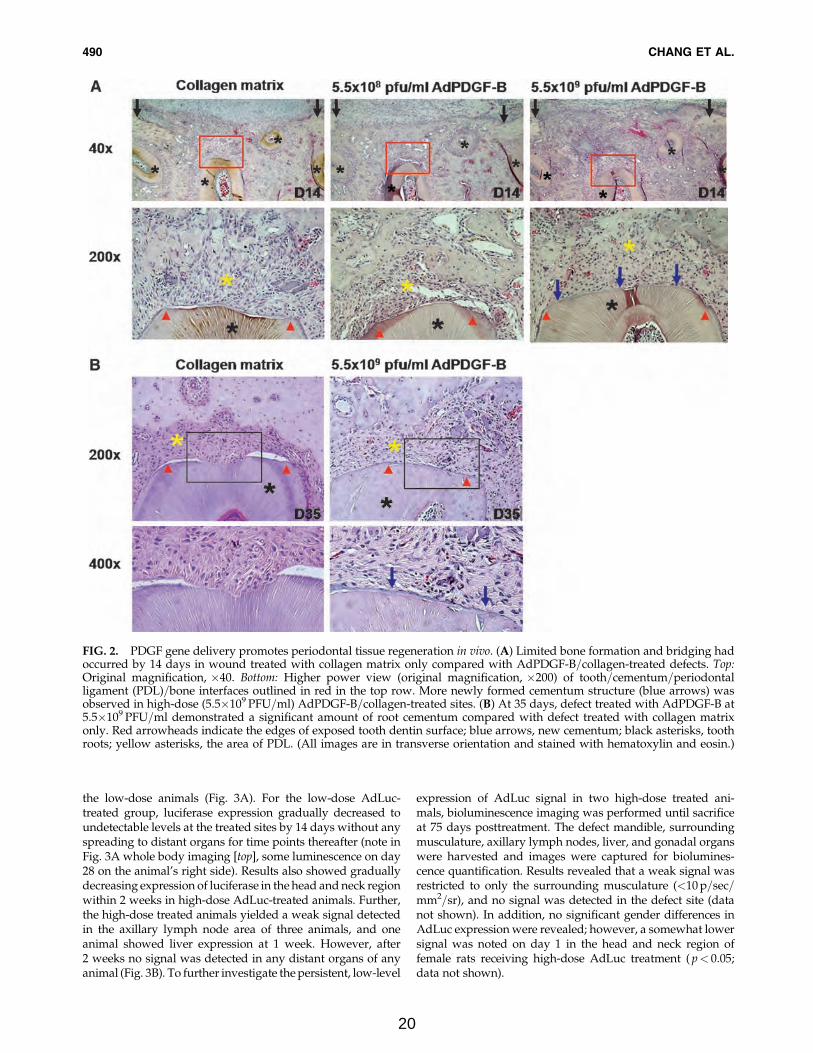
Histology and histopathology
Two weeks after surgery, early bone formation could beobserved within the defect area (Fig. 2A, top). Nearly com-plete bone bridging of the alveolar bone wounds was notedin both AdPDGF-B-treated groups, whereas there was lim-ited bridging in the collagen-only animals. Cementogenesiscould be seen in both AdPDGF-B-treated groups at 2 weeksbut not in the collagen matrix group, and the defects treatedwith high-dose (5.5�109 PFU=ml) AdPDGF-B revealed morecementum formation compared with the other groups (Fig.2A, bottom). At 35 days, the bone had completely bridgedall of the defect area, and the fractions of defect fill be-came consistent in all animals. Animals receiving high-doseAdPDGF-B demonstrated greater evidence of cementogenesisalong the tooth root (Fig. 2B).
Macroscopic evaluations of the harvested organs revealedno meaningful changes except mild enlargement of the sub-mandibular lymph nodes in AdPDGF-B-treated (both high-dose and low-dose) and collagen matrix-only groups withinthe first week postsurgery. Evaluation of histological sectionsshowed occasional but mild inflammatory infiltration inlymph nodes, spleen, and liver in all groups. However, nosignificant histopathological signs were noted beyond thesuspected alterations associated with the surgical operation.In particular, no evidence of viral inclusions was observed forany of the evaluated tissues and organs.
Hematology and blood chemistry
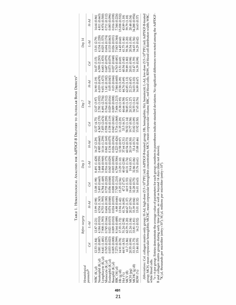
Blood was analyzed from each animal before surgery andthrough 35 days postoperation (Fig. 1A). Also, blood from sixanimals in the no-treatment group was collected for com-parison. All parameters for hematology and blood chemistrywere consistent among groups and were generally within thenormal range. Although there were some minor changes, wefound no significant differences in complete blood count(CBC) and clinical chemistry parameters in any treatmentgroup throughout the period of observation (Tables 1 and 2).There were several animals in both the high-dose and low-dose groups that revealed significant changes in amylase;however, the majority of the values were within the normalrange. On day 28, animals in the low-dose group demon-strated significant elevation in serum glucose, but those levelsreturned to the baseline range by day 35.
Vector expression by bioluminescence
Whole body image analysis of animals treated withAdLuc=collagen matrix revealed a transduction and distribu-tion profile from adenoviral gene delivery over the course ofthe experiment. Bioluminescent luciferase expression was de-tected in the head and neck region for all AdLuc=collagen-treated animals (n¼ 6 per group), with the level of expressionhigher in animals receiving high-dose AdLuc compared with
SAFETY PROFILE OF AdPDGF-B IN ALVEOLAR BONE DEFECTS 489
19
the low-dose animals (Fig. 3A). For the low-dose AdLuc-treated group, luciferase expression gradually decreased toundetectable levels at the treated sites by 14 days without anyspreading to distant organs for time points thereafter (note inFig. 3A whole body imaging [top], some luminescence on day28 on the animal’s right side). Results also showed graduallydecreasing expression of luciferase in the head and neck regionwithin 2 weeks in high-dose AdLuc-treated animals. Further,the high-dose treated animals yielded a weak signal detectedin the axillary lymph node area of three animals, and oneanimal showed liver expression at 1 week. However, after2 weeks no signal was detected in any distant organs of anyanimal (Fig. 3B). To further investigate the persistent, low-level
expression of AdLuc signal in two high-dose treated ani-mals, bioluminescence imaging was performed until sacrificeat 75 days posttreatment. The defect mandible, surroundingmusculature, axillary lymph nodes, liver, and gonadal organswere harvested and images were captured for biolumines-cence quantification. Results revealed that a weak signal wasrestricted to only the surrounding musculature (<10 p=sec=mm2=sr), and no signal was detected in the defect site (datanot shown). In addition, no significant gender differences inAdLuc expression were revealed; however, a somewhat lowersignal was noted on day 1 in the head and neck region offemale rats receiving high-dose AdLuc treatment ( p< 0.05;data not shown).
FIG. 2. PDGF gene delivery promotes periodontal tissue regeneration in vivo. (A) Limited bone formation and bridging hadoccurred by 14 days in wound treated with collagen matrix only compared with AdPDGF-B=collagen-treated defects. Top:Original magnification, �40. Bottom: Higher power view (original magnification, �200) of tooth=cementum=periodontalligament (PDL)=bone interfaces outlined in red in the top row. More newly formed cementum structure (blue arrows) wasobserved in high-dose (5.5�109 PFU=ml) AdPDGF-B=collagen-treated sites. (B) At 35 days, defect treated with AdPDGF-B at5.5�109 PFU=ml demonstrated a significant amount of root cementum compared with defect treated with collagen matrixonly. Red arrowheads indicate the edges of exposed tooth dentin surface; blue arrows, new cementum; black asterisks, toothroots; yellow asterisks, the area of PDL. (All images are in transverse orientation and stained with hematoxylin and eosin.)
490 CHANG ET AL.
20
Ta
bl
e1.
He
ma
to
lo
gic
al
An
al
yse
sfo
rA
dP
DG
F-B
De
liv
er
yt
oA
lv
eo
la
rB
on
eD
efe
ct
sa
Hem
atol
ogic
alpa
ram
eter
b
Bef
ore
surg
ery
Day
3D
ay7
Day
14
Col
L-A
dH
-Ad
Col
L-A
dH
-Ad
Col
L-A
dH
-Ad
Col
L-A
dH
-Ad
WB
C(K=ml
)12
.53
(1.8
4)12
.87
(2.2
1)13
.99
(2.9
8)13
.08
(1.9
8)8.
491
(1.4
28)
16.2
7(2
.29)
12.5
7(4
.75)
12.0
7(3
.97)
16.9
0(2
.19)
16.0
7(3
.15)
13.0
1(2
.79)
14.6
4(0
.86)
Neu
tro
ph
il(K=ml
)3.
081
(0.8
87)
4.18
4(0
.910
)4.
534
(1.3
43)
3.49
3(0
.665
)2.
448
(0.5
59)
4.98
5(0
.660
)4.
365
(2.1
70)
2.78
1(1
.032
)6.
019
(0.6
78)
6.59
9(2
.293
)4.
134
(1.2
28)
4.81
1(0
.663
)L
ym
ph
ocy
te(K=ml
)8.
641
(1.4
81)
7.90
8(1
.593
)8.
753
(1.5
95)
8.78
4(1
.449
)5.
484
(0.9
49)
10.1
6(1
.259
)7.
455
(2.6
74)
8.49
1(2
.754
)9.
651
(1.6
73)
8.68
3(1
.870
)8.
025
(1.5
75)
8.97
4(0
.500
)M
on
ocy
te(K=ml
)0.
765
(0.2
39)
0.74
5(0
.166
)0.
604
(0.1
80)
0.73
5(0
.220
)0.
516
(0.1
75)
0.84
1(0
.169
)0.
558
(0.2
99)
0.76
4(0
.312
)1.
141
(0.1
82)
0.68
7(0
.079
)0.
694
(0.1
74)
0.71
1(0
.112
)E
osi
no
ph
il(K=ml
)0.
033
(0.0
23)
0.02
1(0
.015
)0.
074
(0.0
48)
0.05
6(0
.048
)0.
043
(0.0
29)
0.13
4(0
.075
)0.
158
(0.1
19)
0.02
9(0
.016
)0.
05(0
.043
)0.
073
(0.0
79)
0.13
3(0
.063
)0.
083
(0.0
96)
Bas
op
hil
(K=ml
)0.
005
(0.0
08)
0.00
9(0
.010
)0.
024
(0.0
35)
0.00
8(0
.010
)0.
003
(0.0
05)
0.02
6(0
.036
)0.
043
(0.0
41)
0.00
9(0
.008
)0.
03(0
.044
)0.
027
(0.0
48)
0.02
6(0
.031
)0.
016
(0.0
38)
RB
C(M
=ml
)7.
273
(0.5
99)
8.16
4(0
.488
)6.
88(0
.646
)7.
745
(1.2
10)
6.70
9(0
.506
)6.
223
(0.4
26)
5.71
6(1
.068
)7.
606
(1.2
13)
7.34
4(0
.600
)6.
763
(0.4
81)
7.04
3(0
.344
)7.
344
(0.2
24)
Hb
(g=d
l)14
.91
(0.7
8)17
.35
(1.7
2)13
.94
(1.4
2)15
.25
(2.5
6)13
.29
(1.1
0)12
.58
(0.9
7)11
.35
(2.2
9)15
.38
(1.9
5)15
.58
(1.0
9)13
.51
(0.6
9)14
.45
(0.6
0)15
.038
(0.5
32)
Hct
(%)
44.9
1(4
.53)
51.2
4(2
.98)
41.7
6(4
.09)
47.2
(7.6
7)40
.43
(2.8
8)37
.64
(2.3
1)33
.5(6
.37)
44.6
6(6
.58)
44.7
4(5
.69)
40.5
4(3
.40)
41.7
5(1
.46)
43.8
9(1
.18)
MC
V(fl
)61
.7(1
.86)
62.7
9(1
.20)
60.6
7(1
.37)
60.9
(1.5
3)60
.31
(1.7
4)60
.54
(0.7
3)58
.52
(1.1
4)58
.85
(1.3
7)62
.85
(1.1
3)59
.89
(1.8
0)59
.36
(1.2
0)59
.79
(1.1
8)M
CH
(pg
)20
.55
(0.9
9)21
.24
(1.0
4)20
.27
(0.7
6)19
.69
(0.8
1)19
.8(0
.37)
20.1
9(0
.30)
19.7
7(0
.58)
20.3
3(1
.27)
21.2
3(0
.47)
20.0
1(0
.98)
20.5
4(1
.09)
20.4
8(0
.34)
MC
HC
(g=d
l)33
.33
(1.7
5)33
.81
(1.8
1)33
.39
(0.8
4)32
.28
(0.7
5)32
.86
(1.0
6)33
.4(0
.71)
33.8
2(1
.09)
34.5
5(1
.61)
34.0
3(1
.03)
33.4
7(1
.59)
34.6
5(1
.56)
34.2
88(0
.954
)R
DW
(%)
15.4
4(0
.55)
16.2
(0.5
2)15
.01
(0.3
2)16
.01
(0.6
3)15
.76
(0.6
1)15
.88
(0.4
6)15
.92
(0.5
0)17
.65
(0.7
6)16
.89
(0.6
7)16
.39
(0,9
8)16
.29
(0.4
2)16
.63
(0.3
7)
Abb
revia
tion
s:C
ol.
coll
agen
mat
rix-
on
lyg
rou
p;
H-A
d,
hig
h-d
ose
(5.5�
109
PF
U=m
l)A
dP
DG
F-B
-tre
ated
gro
up
;H
b,
hem
og
lob
in;
Hct
,h
emat
ocr
it;
L-A
d,
low
-do
se(5
.5�
108
PF
U=m
l)A
dP
DG
F-B
-tre
ated
gro
up
;MC
H,m
ean
corp
usc
ula
rh
emo
glo
bin
;MC
HC
,mea
nco
rpu
scu
lar
hem
og
lob
inco
nce
ntr
atio
n;M
CV
,mea
nco
rpu
scu
lar
vo
lum
e;R
BC
,red
blo
od
cell
s;R
DW
,red
blo
od
cell
dis
trib
uti
on
wid
th;W
BC
,w
hit
eb
loo
dce
lls.
an¼
6p
erg
rou
p.E
ntr
ies
dem
on
stra
teth
eav
erag
ev
alu
eo
fp
aram
eter
sfo
rea
chg
rou
p;n
um
ber
sin
par
enth
eses
ind
icat
est
and
ard
dev
iati
on
s.N
osi
gn
ifica
nt
dif
fere
nce
sw
ere
no
ted
amo
ng
the
Ad
PD
GF
-B
and
coll
agen
mat
rix
gro
up
sd
uri
ng
earl
yti
me
po
ints
,as
wel
las
bey
on
d14
day
s(d
ata
no
tsh
ow
n).
bK=ml
,th
ou
san
ds
per
mic
roli
ter
(en
try�
103);
M=ml
,m
illi
on
sp
erm
icro
lite
r(e
ntr
y�
106).
491
21
Ta
bl
e2.
Clin
ic
al
Ch
em
istr
yA
na
lyses
Fo
rA
d-P
DG
F-B
De
liv
er
ya
Cli
nic
alch
emis
try
par
amet
er
Bef
ore
surg
ery
Day
3D
ay7
Day
14
Col
L-A
dH
-Ad
Col
L-A
dH
-Ad
Col
L-A
dH
-Ad
Col
L-A
dH
-Ad
Alb
um
in(g=d
l)2.
814
(0.1
35)
2.76
3(0
.130
)2.
657
(0.1
81)
2.65
(0.6
48)
2.65
7(0
.172
)2.
825
(0.1
16)
2.54
3(0
.113
)2.
95(0
.141
)2.
788
(0.1
81)
2.78
6(0
.177
)2.
913
(0.1
25)
3.13
3(0
.234
)A
LP
ase
(U=
lite
r)26
0.43
(23.
52)
255
(53.
18)
239
(31.
09)
185.
5(5
9.34
)23
8.43
(35.
45)
166.
63(2
9.61
)20
0.86
(37.
66)
205.
75(5
8.94
)15
3.38
(27.
69)
232.
29(2
9.19
)23
2.63
(42.
45)
251.
67(4
3.48
)A
LT
(U=li
ter)
67.1
4(8
.30)
89(8
.45)
87.2
5(7
.50)
72.8
8(8
.92)
79.8
6(4
.06)
79.8
8(8
.15)
100.
57(1
0.47
)79
.38
(9.9
6)81
(5.0
4)84
.86
(5.0
5)86
.5(7
.58)
79.5
(19.
99)
Am
yla
se(U=li
ter)
1881
.14
(186
.95)
1831
(188
.36)
1554
.17
(267
.61)
1905
.38
(388
.61)
1857
.43
(544
.49)
1770
(251
.95)
2494
.86
b(8
44.4
0)17
05.7
5(3
10.8
8)17
85.1
3(3
28.2
2)19
90.7
1(5
25.5
8)18
79.3
8(1
95.6
0)20
85.3
3(4
4.00
4)A
ST
(U=
lite
r)71
.86
(9.9
1)79
.38
(9.3
2)79
.88
(8.9
7)69
.5(2
0.76
)83
.5(1
6.55
)69
.88
(5.1
9)12
6.14
(111
.67)
77.5
7(1
3.23
)70
.5(1
1.43
)66
.43
(11.
77)
90.5
(26.
46)
93.2
5(1
6.34
)B
ilir
ub
in(m
g=d
l)20
.14
(3.3
4)19
.5(3
.70)
19(2
.16)
21.2
5(2
.49)
20(2
)22
.13
(2.5
9)24
.86
(1.8
6)21
.38
(2.3
9)19
.13
(1.6
4)22
.57
(1.6
2)23
.75
(3.4
5)24
.17
(1.3
3)C
alci
um
(mg=d
l)10
.11
(0.2
5)10
.29
(0.1
9)10
.16
(0.2
8)10
.54
(1.3
0)10
.23
(0.2
8)10
.49
(0.1
5)10
.41
(0.2
5)10
.35
(0.2
3)10
.35
(0.1
2)10
.86
(0.2
4)10
.28
(0.1
8)10
.83
(0.2
3)C
ho
lest
ero
l(m
g=
dl)
83.2
9(7
.13)
78(9
.20)
63.8
6(1
1.81
)78
.71
(29.
37)
80.2
9(8
.16)
90(7
.01)
80(6
.90)
80.3
8(7
.03)
81.1
3(2
.80)
84.4
3(1
0.88
)77
.38
(10.
32)
82.3
3(5
.86)
Cre
atin
ek
inas
e(U=
lite
r)16
6.67
(28.
25)
190.
63(5
1.90
)17
6.86
(47.
36)
178.
5(7
0.13
)25
9.83
(133
.13)
142.
63(3
3.18
)15
6.20
(38.
46)
244.
57(1
06.6
9)21
9.38
(64.
16)
121.
71(2
8.96
)18
6.88
(61.
42)
197.
75(6
1.41
)
Cre
atin
ine
(mg=d
l)0.
4(0
.058
)0.
35(0
.053
)0.
329
(0.0
49)
0.36
3(0
.052
)0.
386
(0.0
38)
0.4
(0)
0.38
6(0
.038
)0.
388
(0.0
35)
0.3
(0.0
53)
0.4
(0)
0.38
8(0
.035
)0.
629
(0.3
99)
Glu
cose
(mg=d
l)23
0.43
(18.
39)
227.
88(2
7.22
)22
9(3
5.77
)21
7.65
(24.
22)
221.
57(1
9.15
)19
1(3
3.81
5)24
5.29
(53.
94)
232
(29.
99)
229.
88(3
0.19
)19
2.14
(7.6
7)21
2.5
(39.
75)
200.
4(1
0.69
)P
ho
sph
oru
s(m
g=d
l)7.
657
(0.6
60)
7.4
(0.4
90)
6.74
3(0
.395
)7.
225
(0.5
85)
6.21
4(0
.157
)6.
663
(0.4
34)
6.67
1(0
.340
)7.
65(0
.537
)7.
45(0
.407
)5.
9(2
.62)
7.32
5(0
.486
)6.
833
(0.1
15)
T.
bil
iru
bin
(mg=d
l)0.
1(0
)0.
1(0
)0.
1(0
)0.
275
(0.4
56)
0.11
4(0
.038
)0.
113
(0.0
35)
0.1
(0)
0.17
5(0
.139
)0.
213
(0.2
10)
0.1
(0)
0.11
3(0
.035
)0.
1(0
)T
ota
lp
rote
in(g=
dl)
5.62
9(0
.325
)5.
625
(0.2
05)
5.55
7(0
.276
)5.
95(0
.680
)5.
529
(0.2
50)
5.92
5(0
.128
)5.
586
(0.2
19)
5.86
3(0
.250
)5.
8(0
.278
)5.
943
(0.1
90)
5.93
8(0
.262
)6.
443
(0.3
51)
Glo
bu
lin
(g=d
l)2.
814
(0.2
27)
2.83
8(0
.106
)2.
886
(0.1
07)
3.28
8(1
.272
)2.
957
(0.1
62)
3.1
(0.0
93)
3.04
3(0
.151
)2.
925
(0.1
28)
3.02
5(0
.128
)3.
157
(0.1
40)
3.01
3(0
.146
)3.
25(0
.152
)
Abb
rev
iati
ons:
AL
Pas
e,al
kal
ine
ph
osp
hat
ase;
AL
T,
alan
ine
tran
sam
inas
e;A
ST
,as
par
tate
tran
sam
inas
e;C
ol,
coll
agen
mat
rix
-on
lyg
rou
p;
H-A
d,
hig
h-d
ose
(5.5�
109
PF
U=m
l)A
dP
DG
F-B
-tre
ated
gro
up
;L
-Ad
,lo
w-d
ose
(5.5�
108
PF
U=m
l)A
dP
DG
F-B
-tre
ated
gro
up
;T
.b
ilir
ub
in,
tota
lb
ilir
ub
in.
aA
llco
mp
aris
on
sar
em
ade
wit
hre
fere
nce
toth
eco
llag
enm
atri
xg
rou
p.
En
trie
sd
emo
nst
rate
the
mea
nv
alu
eo
fp
aram
eter
sfo
rea
chg
rou
p;
nu
mb
ers
inp
aren
thes
esin
dic
ates
stan
dar
dd
evia
tio
ns.
Ser
um
amy
lase
for
bo
thA
dP
DG
F-B
-tre
ated
gro
up
sre
vea
led
sig
nifi
can
td
iffe
ren
ces
wit
hre
spec
tto
the
coll
agen
mat
rix
gro
up
,an
dw
asw
ith
inth
en
orm
alra
ng
efo
rti
me
po
ints
bey
ou
nd
14d
ays.
bS
ign
ifica
nt
dif
fere
nce
fro
mco
llag
enm
atri
xg
rou
p(p<
0.05
;n¼
6p
erg
rou
p).
492
22
Biodistribution by quantitative PCR
The specificity of our PCR primers and the sensitivity of theassay were determined before analysis of the study samples.We measured no primer cross-reaction with adenovirus en-coding bone sialoprotein, bone morphogenetic protein-7, lu-ciferase, noggin, PDGF-A, PDGF-1308, or GFP (data notshown). The sensitivity and detection limit of our PCR assayswas 30 virus copies per 500 ng of DNA. Within the AdPDGF-B-treated area, viral vector could be detected within the first
week in DNA from both high-dose and low-dose treated an-imals. The number of vector copies gradually decreased toundetectable levels after 2 weeks (Table 3). Vector copiesmeasured in the blood were below the detection limit for allanimals over the total period of observation. The PCR assaymeasured a low level of vector within spleen DNA of oneanimal at 3 days posttreatment, and within the lung of anotheranimal at 2 weeks posttreatment; however, no significantvector DNA was detected in organs or tissues from the treat-ment groups for the remainder of the experimental time points
FIG. 3. Vector transduction efficiency and systemic distribution of bioluminescence. (A) Most of the luciferin signal isrestricted to the alveolar bone defect region, with minimal systemic involvement. Signals in distant organs were absentafter 14 days for both dose level groups. (B) Mild vector expression was noted during the first 3–7 days in animals treatedwith AdLuc at 5.5�108 PFU=ml. (C) Animals treated with AdLuc at 5.5�109 PFU=ml demonstrated significant vectorexpression during the first 14 days, followed by a decrease in vector expression in the head and neck region over time. Thehigh-dose group also showed modest vector expression in liver (one of six positive on day 14) and axillary lymph nodes(one of six positive on day 3, and two of six positive on both days 7 and 10). Group size: n¼ 6 (three per gender). If theintensity of bioluminescence within the region of interest was less than 5000 p=sec=cm2=sr, that region was defined as‘‘negative’’.
SAFETY PROFILE OF AdPDGF-B IN ALVEOLAR BONE DEFECTS 493
23
(Table 3). These values were below the detection limit andcompared similarly with vector values at the defect site, whichwere low to below the detection level. On examination ofhistological sections from the tissues (spleen and lung) posi-tive for AdPDGF-B DNA, we found no inflammation-relatedphenotype or other pathological findings when comparedwith tissue sections from collagen matrix-treated animals.
Discussion
PDGF-BB protein has demonstrated its strong potential forsoft and hard tissue repair and is available for clinical use(Nevins et al., 2005; Hollinger et al., 2008). However, becauseof the high degradation rate and transient persistence in vivo,the treatment outcome is not entirely predictable for clinicalapplications (Kaigler et al., 2006). Gene delivery using an ad-enoviral vector provides sustained and stable transduc-tion efficiency in vitro (Chen and Giannobile, 2002). Thesedata confirm and extend those of Jin and colleagues (2004)demonstrating significant enhancement of tooth-supportingalveolar bone and cementum regeneration in vivo, using gene-activated matrices containing AdPDGF-B.
Although a number of studies focus on the safety profile ofadenovirus-mediated gene therapy, few of them have ad-dressed the local delivery of vectors using a gene-activatedmatrix and none are related to the periodontium or localizedbone defects. Studies have shown that direct systemic ad-ministration of adenoviral vectors can result in acute toxicityand hepatic pathology (Nunes et al., 1999; Lenaerts et al., 2005;Ni et al., 2005). Systemic dissemination can be reduced and theefficacy-to-toxicity ratio can be improved by local gene de-livery (Wang et al., 2005). With localized delivery, the vectorlikely enters the systemic circulation via the leaky micro-vessels and systemically disseminates within 10 min (Wanget al., 2005), with the inflammatory infiltrate within liverobserved after 15 min in mice (Ni et al., 2005). In this study,
we employed matrix (collagen)-enabled gene delivery for lo-calized administration to alveolar bone defects. The vectordissemination in our animals beyond the alveolar bone areawas limited, demonstrating well-contained localization of thegene-activated matrix.
Studies have shown that nearly 99% of systemically de-livered adenoviral vectors will eventually accumulate in theliver, and are rapidly taken up by Kupffer cells and hepato-cytes (Hackett et al., 2000; Manickan et al., 2006). The Kupffercells might distribute to the lung and spleen via the circula-tion, but in this study we did not detect any significant vectorquantities in those organs. No significant elevation of theenzymes specific to those organs further demonstrates thelimited systemic influence of this approach. Although trans-gene luciferase expression was found in the axillary lymphnodes, spleen, and lungs of a few adenoviral vector-treatedanimals at 2 weeks postadministration (with no expression inthese organs at later time points), the level was only slightlygreater than background and no accompanying toxicologicalsigns or histopathological changes were found. We also notedno treatment-related toxicity throughout the 35-day period.Most of the hematological and clinical chemistry parameterswere within normal ranges and the only significant differencewas noted for amylase (derived primarily from the pancreasand parotid gland, with some from the liver), which is oneof the major enzymes to digest starch into simple sugars.Changes in serum amylase may represent a normal physio-logic process, acute or chronic pancreatitis, or concomitantongoing diseases (Garrison, 1986). However, lipase is a moresensitive and specific marker with which to diagnose pan-creatitis (Tietz et al., 1986), and the lipase level in all of theanimals did not change significantly. However, it is quitepossible that the amylase came from the parotid salivarygland that was located in close proximity to the surgical field.The parotid gland in rats is nonencapsulated, as comparedwith the gland in humans. We cannot rule out this area at
Table 3. AdPDGF-B PCR Results in Bloodstream and Distant Organs
Organ=tissue Treatment No treatment Day 3 Day 7 Day 14 Day 21 Day 28 Day 35
Whole tissuefrom osseousdefect
Collagen matrix N N N N N N N5.5�108 PFU=ml AdPDGF-B N 3=3 (301) 2=3 (137) 1=3 (84) N N N5.5�109 PFU=ml AdPDGF-B N 3=3 (45,930) 3=3 (6,097) N N N N
Blood Collagen matrix N N N N N N N5.5�108 PFU=ml AdPDGF-B N N N N N N N5.5�109 PFU=ml AdPDGF-B N N N N N N N
Lung Collagen matrix N N N N N N N5.5�108 PFU=ml AdPDGF-B N N N 1=3 (38) N N N5.5�109 PFU=ml AdPDGF-B N N N N N N N
Spleen Collagen matrix N N N N N N N5.5�108 PFU=ml AdPDGF-B N 1=3 (31) N N N N N5.5�109 PFU=ml AdPDGF-B N N N N N N N
Collagen matrix N N N N N N N5.5�108 PFU=ml AdPDGF-B N N N N N N N5.5�109 PFU=ml AdPDGF-B N N N N N N N
Abbreviations: ALN, axillary lymph nodes; N, negative; PFU, plaque-forming units; SLN, submandibular lymph nodes.an¼ 3 per group (for organ analyses) and 23 per group (for blood analyses). Test sample DNAs yielding signals below the limit of detection
(<30 vector particles per 500 ng of DNA) are reported as negative. Entries demonstrate ‘‘positive’’ animals in each group and entries inparentheses indicate the mean vector copy number per 500 ng of DNA from the positive animals.
494 CHANG ET AL.
24
early time points. At later time points when we measured theluciferase signal from the harvested organs, no detectablesignal was found in any of the parotid glands, but mainlyin the surrounding musculature (Fig. 3). In vivo biolumines-cence generated by expression of the luciferase transgenepermitted quantification and localization of transgene ex-pression and provided noninvasive, dynamic, and compre-hensive monitoring of vector expression at the whole bodylevel (Wood et al., 1999; Johnson et al., 2006). As little as 104
luciferase-expressing recombinant adenoviruses are capableof producing luminescence in the liver (Honigman et al., 2001),which is significantly higher in sensitivity than is possiblewith qPCR ( Johnson et al., 2006), making bioluminescence amore sensitive mode of evaluation of biodistribution andsubsequent vector activity. In the early time periods we de-tected vector in the defect area of adenovirus-treated animals,which reached undetectable levels by day 14. This resultsupports those reported by Jin and colleagues (2004), showingthat the luciferase signal decreased to 20% by day 14 andreached an undetectable level by day 28 compared with theexpression on day 1. Moreover, given that PDGF is expressedin vivo over about 10 days in periodontal wounds after injury(Green et al., 1997), this gene therapy approach demonstrates asimilar expression profile that may be favorable for thera-peutic application.
In summary, the results of our experiments demonstratethat local administration of AdPDGF-B with gene-activatedmatrix is safe when delivered to tooth-supporting alveo-lar bone defects. No treatment-related toxicity or systemic in-volvement was found. Although vector particle DNA wasdetectable during the first 2 weeks, primarily in the osseousdefects, the titer was low and quickly attenuated at subsequenttime points. These results support the further clinical devel-opment of AdPDGF-B for regeneration therapy for oral andcraniofacial bone application.
Acknowledgments
The authors thank Anna Colvig for performing hema-tological and clinical chemical examinations, AmandaWelton for assistance with bioluminescence, Dr. John E.Wilkinson for assistance with veterinary pathology, andChristopher Strayhorn for assistance with histological pro-cessing. This study was supported in part by grants from theAO Foundation (Davos, Switzerland) and NIH=NIDCR R01-DE13397.
Author Disclosure Statement
Drs. Sosnowski and Chandler are employees of TissueRepair Co. The University of Michigan will benefit financiallyby clinical development of this technology.
References
Andrae, J., Gallini, R., and Betsholtz, C. (2008). Role of platelet-derived growth factors in physiology and medicine. GenesDev. 22, 1276–1312.
Chen, Q.P., and Giannobile, W.V. (2002). Adenoviral genetransfer of PDGF downregulates gas gene product PDGFaRand prolongs ERK and Akt=PKB activation. Am. J. Physiol.Cell Physiol. 282, C538–C544.
Cooke, J.W., Sarment, D.P., Whitesman, L.A., Miller, S.E., Jin, Q.,Lynch, S.E., and Giannobile, W.V. (2006). Effect of rhPDGF-BBdelivery on mediators of periodontal wound repair. TissueEng. 12, 1441–1450.
Cotrim, A.P., and Baum, B.J. (2008). Gene therapy: Some history,applications, problems, and prospects. Toxicol. Pathol. 36, 97–103.
Cotrim, A.P., Sowers, A., Mitchell, J.B., and Baum, B.J. (2007).Prevention of irradiation-induced salivary hypofunction bymicrovessel protection in mouse salivary glands. Mol. Ther.15, 2101–2106.
Fiedler, J., Etzel, N., and Brenner, R.E. (2004). To go or not to go:Migration of human mesenchymal progenitor cells stimulatedby isoforms of PDGF. J. Cell Biochem. 93, 990–998.
Garrison, R. (1986). Amylase. Emerg. Med. Clin. North Am. 4,315–327.
Giannobile, W.V., Finkelman, R.D., and Lynch, S.E. (1994).Comparison of canine and nonhuman primate models forperiodontal regenerative therapy: results following a singleadministration of PDGF=IGF-I. J. Periodontol. 65, 1158–1168.
Giannobile, W.V., Hernandez, R.A., Finkelman, R.D., Ryan, S.,Kiritsy, C.P., D’Andrea, M.D., and Lynch, S.E. (1996). Com-parative Effects of PDGF-BB, IGF-I singularly and in combi-nation on periodontal regeneration in Macaca fascicularis.J. Periodont. Res. 31, 301–312.
Green, R.J., Usui, M.L., Hart, C.E., Ammons, W.F., and Nar-ayanan, A.S. (1997). Immunolocalization of platelet-derivedgrowth factor A and B chains and PDGF-a and b receptors inhuman gingival wounds. J. Periodont. Res. 32, 209–214.
Gu, D.L., Nguyen, T., Gonzalez, A.M., Printz, M.A., Pierce, G.F.,Sosnowski, B.A., Phillips, M.L., and Chandler, L.A. (2004).Adenovirus encoding human platelet-derived growth factor-Bdelivered in collagen exhibits safety, biodistribution, and im-munogenicity profiles favorable for clinical use. Mol. Ther. 9,699–711.
Haase, H.R., Clarkson, R.W., Waters, M.J., and Bartold, P.M.(1998). Growth factor modulation of mitogenic responses andproteoglycan synthesis by human periodontal fibroblasts.J. Cell Physiol. 174, 353–361.
Hackett, N.R., El Sawy, T., Lee, L.Y., Silva, I., O’Leary, J.,Rosengart, T.K., and Crystal, R.G. (2000). Use of quantitativeTaqMan real-time PCR to track the time-dependent distribu-tion of gene transfer vectors in vivo. Mol. Ther. 2, 649–656.
Hollinger, J.O., Hart, C.E., Hirsch, S.N., Lynch, S., and Fried-laender, G.E. (2008). Recombinant human platelet-derivedgrowth factor: Biology and clinical applications. J. Bone JointSurg. Am. 90(Suppl. 1):48–54.
Honigman, A., Zeira, E., Ohana, P., Abramovitz, R., Tavor, E., Bar,I., Zilberman, Y., Rabinovsky, R., Gazit, D., Joseph, A., Panet, A.,Shai, E., Palmon, A., Laster, M., and Galun, E. (2001). Imagingtransgene expression in live animals. Mol. Ther. 4, 239–249.
Howell, T.H., Fiorellini, J.P., Paquette, D.W., Offenbacher, S.,Giannobile, W.V., and Lynch, S.E. (1997). A phase I=II clinicaltrial to evaluate a combination of recombinant human platelet-derived growth factor-BB and recombinant human insulin-likegrowth factor-I in patients with periodontal disease. J. Peri-odontol. 68, 1186–1193.
Jin, Q., Anusaksathien, O., Webb, S.A., Rutherford, R.B., andGiannobile, W.V. (2003). Gene therapy of bone morphogeneticprotein for periodontal tissue engineering. J. Periodontol. 74,202–213.
SAFETY PROFILE OF AdPDGF-B IN ALVEOLAR BONE DEFECTS 495
25
Jin, Q., Anusaksathien, O., Webb, S.A., Printz, M.A., and Gian-nobile, W.V. (2004). Engineering of tooth-supporting structuresby delivery of PDGF gene therapy vectors. Mol. Ther. 9, 519–526.
Johnson, M., Huyn, S., Burton, J., Sato, M., and Wu, L. (2006).Differential biodistribution of adenoviral vector in vivo asmonitored by bioluminescence imaging and quantitativepolymerase chain reaction. Hum. Gene Ther. 17, 1262–1269.
Kaigler, D., Cirelli, J.A., and Giannobile, W.V. (2006). Growthfactor delivery for oral and periodontal tissue engineering.Exp. Opin. Drug Deliv. 3, 647–662.
Lenaerts, L., Verbeken, E., De Clercq, E., and Naesens, L. (2005).Mouse adenovirus type 1 infection in SCID mice: An experi-mental model for antiviral therapy of systemic adenovirusinfections. Antimicrob. Agents Chemother. 49, 4689–4699.
Manickan, E., Smith, J.S., Tian, J., Eggerman, T.L., Lozier, J.N.,Muller, J., and Byrnes, A.P. (2006). Rapid Kupffer cell deathafter intravenous injection of adenovirus vectors. Mol. Ther.13, 108–117.
Nevins, M., Giannobile, W.V., McGuire, M.K., Kao, R.T., Mel-lonig, J.T., Hinrichs, J.E., McAllister, B.S., Murphy, K.S.,McClain, P.K., Nevins, M.L., Paquette, D.W., Han, T.J., Reddy,M.S., Lavin, P.T., Genco, R.J., and Lynch, S.E. (2005). Platelet-derived growth factor stimulates bone fill and rate of attach-ment level gain: Results of a large multicenter randomizedcontrolled trial. J. Periodontol. 76, 2205–2215.
Ni, S., Bernt, K., Gaggar, A., Li, Z.Y., Kiem, H.P., and Lieber, A.(2005). Evaluation of biodistribution and safety of adenovirusvectors containing group B fibers after intravenous injectioninto baboons. Hum. Gene Ther. 16, 664–677.
Nishimura, F., and Terranova, V.P. (1996). Comparative study ofthe chemotactic responses of periodontal ligament cells andgingival fibroblasts to polypeptide growth factors. J. Dent.Res. 75, 986–992.
Nunes, F.A., Furth, E.E., Wilson, J.M., and Raper, S.E. (1999).Gene transfer into the liver of nonhuman primates withE1-deleted recombinant adenoviral vectors: Safety of re-administration. Hum. Gene Ther. 10, 2515–2526.
Park, Y.J., Lee, Y.M., Park, S.N., Sheen, S.Y., Chung, C.P., andLee, S.J. (2000). Platelet derived growth factor releasing chi-tosan sponge for periodontal bone regeneration. Biomaterials21, 153–159.
Ramseier, C.A., Abramson, Z.R., Jin, Q., and Giannobile, W.V.(2006). Gene therapeutics for periodontal regenerative medi-cine. Dent. Clin. North Am. 50, 245–263, ix.
Ronnstrand, L., and Heldin, C.H. (2001). Mechanisms of platelet-derived growth factor-induced chemotaxis. Int. J. Cancer 91,757–762.
Southwood, L.L., Frisbie, D.D., Kawcak, C.E., and McIlwraith,C.W. (2004). Delivery of growth factors using gene therapy toenhance bone healing. Vet. Surg. 33, 565–578.
Tietz, N.W., Huang, W.Y., Rauh, D.F., and Shuey, D.F. (1986).Laboratory tests in the differential diagnosis of hyper-amylasemia. Clin. Chem. 32, 301–307.
Voutetakis, A., Zheng, C., Metzger, M., Cotrim, A.P., Donahue,R.E., Dunbar, C.E., and Baum, B.J. (2008). Sorting of transgenicsecretory proteins in rhesus macaque parotid glands follow-ing adenoviral mediated gene transfer. Hum. Gene Ther. 19,1401–1405.
Wang, Y., Yang, Z., Liu, S., Kon, T., Krol, A., Li, C.Y., and Yuan,F. (2005). Characterisation of systemic dissemination of non-replicating adenoviral vectors from tumours in local genedelivery. Br. J. Cancer 92, 1414–1420.
Wood, M., Perrotte, P., Onishi, E., Harper, M.E., Dinney, C.,Pagliaro, L., and Wilson, D.R. (1999). Biodistribution of anadenoviral vector carrying the luciferase reporter gene fol-lowing intravesical or intravenous administration to a mouse.Cancer Gene Ther. 6, 367–372.
Address reprint requests to:Dr. William V. Giannobile
Received for publication July 25, 2008;accepted after revision January 23, 2009.
Published online: April 1, 2009.
496 CHANG ET AL.
26
27
RESULTS, DISCUSSIONS, and CONCLUSIONS
B: PDGF-B gene therapy accelerates bone engineering and oral implant osseointegration. (Chang et al. Gene Ther. 2009 (in press)) Results and Discussion are on pages 28 ~ 34.
ORIGINAL ARTICLE
PDGF-B gene therapy accelerates bone engineeringand oral implant osseointegration
P-C Chang1,2,8, Y-J Seol1,3,8, JA Cirelli1,4, G Pellegrini1,5, Q Jin1, LM Franco1, SA Goldstein2,6,
LA Chandler7, B Sosnowski7 and WV Giannobile1,2
1Department of Periodontics and Oral Medicine, School of Dentistry, University of Michigan, Ann Arbor, MI, USA; 2Department ofBiomedical Engineering, College of Engineering, University of Michigan, Ann Arbor, MI, USA; 3Department of Periodontology, Schoolof Dentistry, Seoul National University, Seoul, Korea; 4Department of Periodontology, School of Dentistry at Araraquara, StateUniversity of Sao Paulo, Araraquara, Sao Paulo, Brazil; 5Department of Periodontology, Clinics Hospital Mangiagalli, University of Milan,Milan, Italy; 6Department of Orthopaedic Surgery, School of Medicine, University of Michigan, Ann Arbor, MI, USA and 7Tissue RepairCompany, San Diego, CA, USA
Platelet-derived growth factor-BB (PDGF-BB) stimulatesrepair of healing-impaired chronic wounds such as diabeticulcers and periodontal lesions. However, limitations inpredictability of tissue regeneration occur due, in part, totransient growth factor bioavailability in vivo. Here, we reportthat gene delivery of PDGF-B stimulates repair of oralimplant extraction socket defects. Alveolar ridge defectswere created in rats and were treated at the time oftitanium implant installation with a collagen matrix contain-ing an adenoviral (Ad) vector encoding PDGF-B (5.5� 108
or 5.5� 109 pfu ml�1), Ad encoding luciferase (Ad-Luc;5.5� 109 pfu ml�1; control) or recombinant human PDGF-BB protein (rhPDGF-BB, 0.3 mg ml�1). Bone repair andosseointegration were measured through backscattered
scanning electron microscopy, histomorphometry, micro-computed tomography and biomechanical assessments.Furthermore, a panel of local and systemic safety assess-ments was performed. Results indicated that bone repairwas accelerated by Ad-PDGF-B and rhPDGF-BB deliverycompared with Ad-Luc, with the high dose of Ad-PDGF-Bmore effective than the low dose. No significant dissemina-tion of the vector construct or alteration of systemic param-eters was noted. In summary, gene delivery of Ad-PDGF-Bshows regenerative and safety capabilities for bone tissueengineering and osseointegration in alveolar bone defectscomparable with rhPDGF-BB protein delivery in vivo.Gene Therapy (2010) 17, 95–104; doi:10.1038/gt.2009.117;published online 10 September 2009
Oral implants are widely accepted in dental medicine asa reconstructive treatment modality for tooth replace-ment due to disease, injury or congenital defects. Inclinical situations exhibiting limited alveolar bone avail-ability, growth factor application has been advocatedto improve osteogenesis and osseointegration.1 However,as a result of the transient action and the highdegradation rate of recombinant proteins in vivo,2 thesustained bioactivity of gene therapy vectors has beenpurported to be an effective alternative for the deliveryof growth factor proteins.3,4 Adenoviral (Ad) vectorshave been shown to exhibit a high in vivo transductionefficiency,5 with a relatively short expression periodcompared with other viral-based gene delivery methods,and their effectiveness for promoting initial woundhealing without eliciting long-term health concerns inwound healing models).6,7
Platelet-derived growth factor (PDGF) is a potentmitogen that facilitates wound healing8 and stimulates
bone repair by expanding osteoblastic precursor cells.9,10
PDGF-BB is Food and Drug Administration-approved foruse in the treatment of localized periodontal defects anddiabetic ulcers11–13 Ad-mediated PDGF-B (Ad-PDGF-B)gene delivery has been shown to enhance periodontaltissue regeneration of tooth-supporting wounds.6,14
Limited information is available regarding the potential ofPDGF-BB on promoting osseointegration of oral implants. Inaddition, the influence of PDGF-B on the mechanicalintegrity of an implant interface is unknown. The purposeof this study was to investigate the effects of rhPDGF-BBand Ad-PDGF-B delivered in a collagen matrix on theosteogenesis and osseointegration of dental implants in anin vivo osseointegration model. This approach shows theability of Ad-PDGF-B to accelerate oral implant osseointe-gration. The data support the concept that Ad-PDGF-B genedelivery may be an effective and safe mode of therapycomparable with PDGF-BB application to promote dentalimplant osseointegration and oral bone repair.
Results
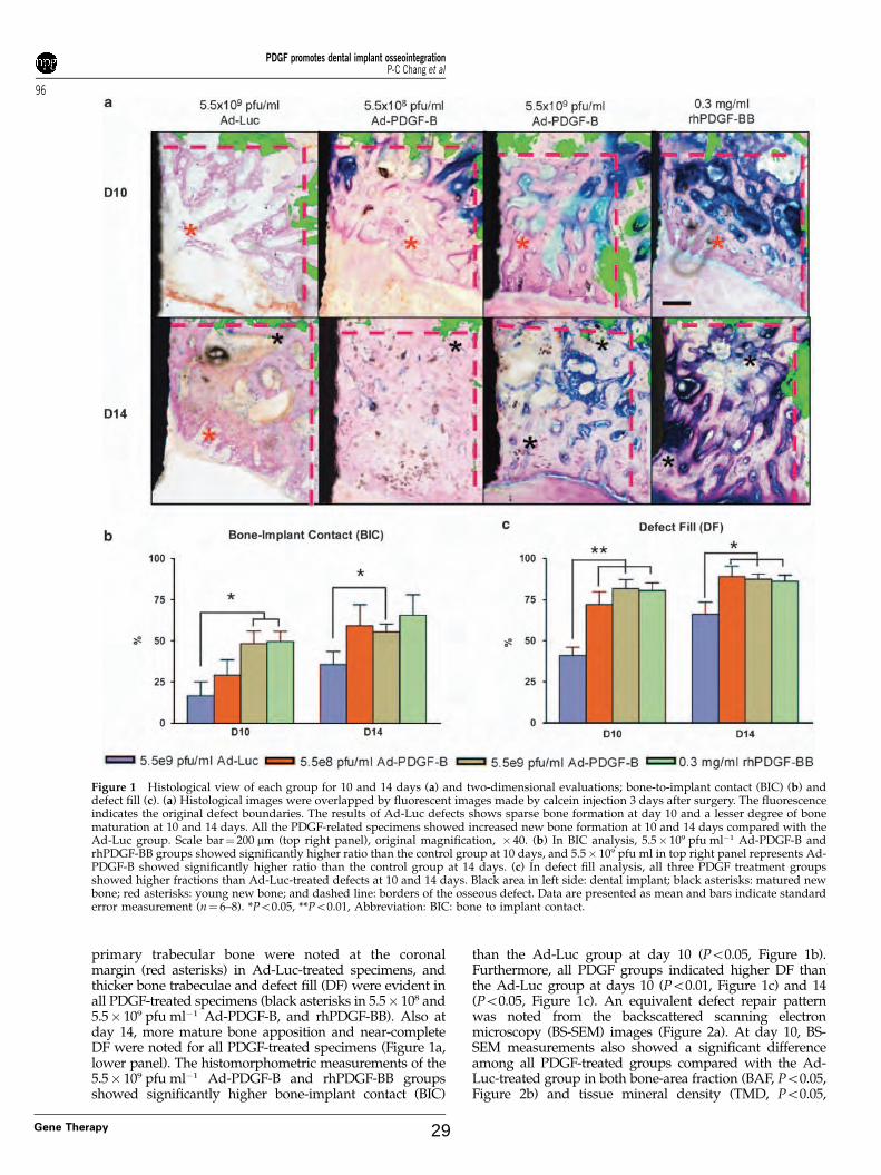
Ad-PDGF-B and rhPDGF-BB enhanceosteogenesis in vivoOn the basis of the descriptive histology (Figure 1a), byday 10 a gradual defect resolution was observed overtime in all groups. At days 10 and 14, woven bone and
Received 13 April 2009; revised 23 June 2009; accepted 23 June 2009;published online 10 September 2009
Correspondence: Professor WV Giannobile, Department ofPeriodontics and Oral Medicine, School of Dentistry, University ofMichigan, 1011 N. University Ave., Ann Arbor, MI 48109, USA.E-mail: [email protected] authors contributed equally to this work.
Gene Therapy (2010) 17, 95–104& 2010 Macmillan Publishers Limited All rights reserved 0969-7128/10 $32.00
primary trabecular bone were noted at the coronalmargin (red asterisks) in Ad-Luc-treated specimens, andthicker bone trabeculae and defect fill (DF) were evident inall PDGF-treated specimens (black asterisks in 5.5� 108 and5.5� 109 pfu ml�1 Ad-PDGF-B, and rhPDGF-BB). Also atday 14, more mature bone apposition and near-completeDF were noted for all PDGF-treated specimens (Figure 1a,lower panel). The histomorphometric measurements of the5.5� 109 pfu ml�1 Ad-PDGF-B and rhPDGF-BB groupsshowed significantly higher bone-implant contact (BIC)
than the Ad-Luc group at day 10 (Po0.05, Figure 1b).Furthermore, all PDGF groups indicated higher DF thanthe Ad-Luc group at days 10 (Po0.01, Figure 1c) and 14(Po0.05, Figure 1c). An equivalent defect repair patternwas noted from the backscattered scanning electronmicroscopy (BS-SEM) images (Figure 2a). At day 10, BS-SEM measurements also showed a significant differenceamong all PDGF-treated groups compared with the Ad-Luc-treated group in both bone-area fraction (BAF, Po0.05,Figure 2b) and tissue mineral density (TMD, Po0.05,
Figure 1 Histological view of each group for 10 and 14 days (a) and two-dimensional evaluations; bone-to-implant contact (BIC) (b) anddefect fill (c). (a) Histological images were overlapped by fluorescent images made by calcein injection 3 days after surgery. The fluorescenceindicates the original defect boundaries. The results of Ad-Luc defects shows sparse bone formation at day 10 and a lesser degree of bonematuration at 10 and 14 days. All the PDGF-related specimens showed increased new bone formation at 10 and 14 days compared with theAd-Luc group. Scale bar¼ 200 mm (top right panel), original magnification, � 40. (b) In BIC analysis, 5.5� 109 pfu ml�1 Ad-PDGF-B andrhPDGF-BB groups showed significantly higher ratio than the control group at 10 days, and 5.5� 109 pfu ml in top right panel represents Ad-PDGF-B showed significantly higher ratio than the control group at 14 days. (c) In defect fill analysis, all three PDGF treatment groupsshowed higher fractions than Ad-Luc-treated defects at 10 and 14 days. Black area in left side: dental implant; black asterisks: matured newbone; red asterisks: young new bone; and dashed line: borders of the osseous defect. Data are presented as mean and bars indicate standarderror measurement (n¼ 6–8). *Po0.05, **Po0.01, Abbreviation: BIC: bone to implant contact.
PDGF promotes dental implant osseointegrationP-C Chang et al
96
Gene Therapy 29
Figure 2c). A significant difference between rhPDGF-BBand Ad-Luc groups in TMD was also noted at day 14(Po0.05, Figure 3c). Completion of the DF was noted in allthe animals by day 21, and no significant differences forany BS-SEM or histomorphometric parameters could befound among all the groups (data not shown).
Both Ad-PDGF-B and rhPDGF-BB promoteosseointegrationThe consequence of push-out testing was reflected fromthe osseointegration index (OI), with all PDGF-treatedspecimens showing higher scores than Ad-Luc, with
significant differences noted between rhPDGF-BB andAd-Luc at both days 10 and 14 (Po0.05, Figure 3a).PDGF application tended to improve the interfacialstiffness (IS) and maximum removal loading (MRL)compared with the Ad-Luc group. The rhPDGF-BBtreatment indicated significantly higher IS than all othergroups at days 10 and 14 (Po0.05, Figure 3b), and higherMRL than all other groups at day 10 (Po0.05, Figure 3c).At day 14, the MRL of rhPDGF-BB was signifi-cantly higher compared with both the Ad-Luc and the5.5� 109 pfu ml�1 Ad-PDGF-B groups (Po0.05, Figure 3c).Significant improvement of IS using 5.5� 108 pfu ml�1
Ad-PDGF-B treatment versus Ad-Luc (Po0.05, Figure 3b)
Figure 2 Backscattered SEM (BS-SEM) images (a) and two-dimensional evaluations, bone-area fraction (b), and tissue mineral density (c).(a) BS-SEM images were merged with fluorescent images (dashed line: borders of the osseous defect.). The BS-SEM images show mineralizedtissue against the oral implant surface. (original magnification, � 42) (b) The three PDGF treatment groups showed a significant difference inbone area fraction at 10 days compared with the control group. (c) The three PDGF groups also showed significant differences in tissuemineral density at 10 days and the rhPDGF-BB group showed significance at 14 days compared with Ad-Luc defects. Data are presented asmean and bars indicate standard error measurement (n¼ 6–8). *Po0.05.
PDGF promotes dental implant osseointegrationP-C Chang et al
97
Gene Therapy30
was also seen at day 10. Most day 21 specimensexperienced cortical bone fractures during the push-outtesting (suggestive of strong osseointegration), and nosignificant differences among all the groups in IS and OIscores were noted (data not shown).
Micro-computed tomography (micro-CT) images wereanalyzed after implant removal, and both the5.5� 109 pfu ml�1 Ad-PDGF-B and rhPDGF-BB groupsshowed significantly higher bone volume fraction (BVF)and TMD than the 5.5� 108 pfu ml�1 Ad-PDGF-B andAd-Luc groups at day 10 (Po0.05, Figure 3d and e). Asignificant difference in BVF was found between5.5� 109 pfu ml�1 Ad-PDGF-B and Ad-Luc groups atday 14 (Po0.05, Figure 3d). Both the 5.5� 109 pfu ml�1
Ad-PDGF-B and rhPDGF-BB groups showed equi-valent extents of functional composite tissue apparentmodulus (FCAM), which was significantly stiffer thanthe 5.5� 108 pfu ml�1 Ad-PDGF-B or Ad-Luc groupat day 10 (Po0.05, Figure 3f). At day 14, there wereno FCAM differences between any of the treatmentgroups.
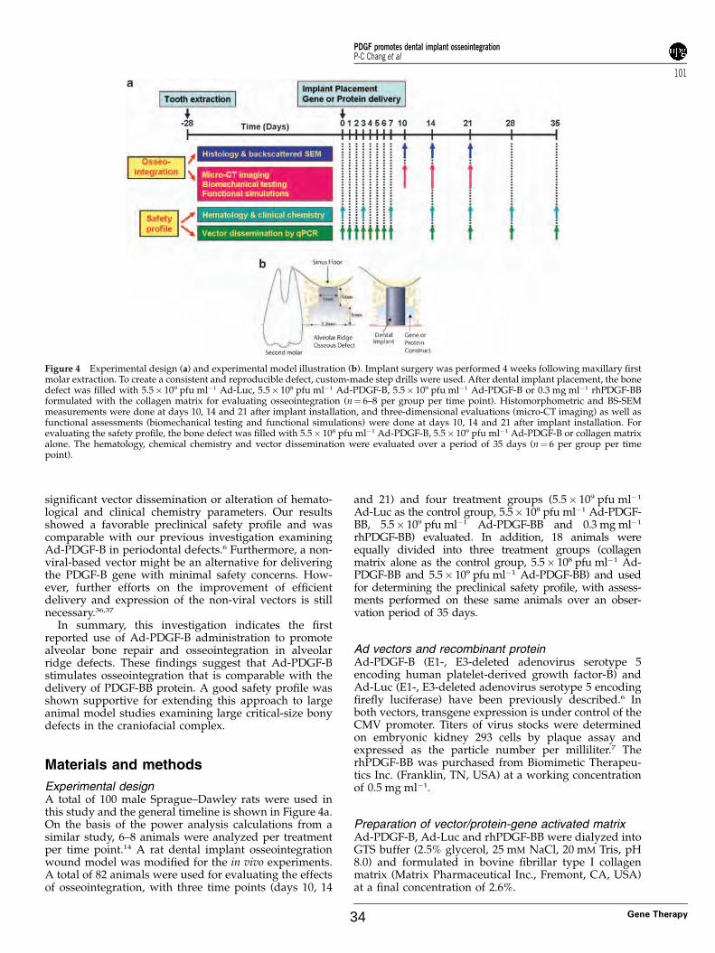
Local delivery of Ad-PDGF-B shows acceptable safetyprofiles in vivoIn a separate study of systemic safety, animals weretreated with collagen alone (control) or collagen contain-ing Ad-PDGF-B (5.5� 108 or 5.5� 109 pfu ml�1). Bloodsamples were taken at various time points for hemato-logical and clinical chemistry analyses and PCR analysesfor vector sequence. All animals survived until the day ofkilling, with no progressive swelling or symptoms noted.
The majority of hematological and clinical chemistryparameters were within their normal ranges with nosignificant differences between Ad-PDGF-B and col-lagen-only treatments (Tables 1 and 2).
Vector-specific quantitative PCR6 was carried outon blood samples taken at baseline, days 1, 2, 3, 4, 5, 7,14, 21, 28, and 35 after treatment. Ad-PDGF-B was notdetected in the bloodstream over the 35-day observationperiod (data not shown).
Discussion
This study shows that both Ad-PDGF-B gene andrhPDGF-BB protein delivery promote the accelerationof neo-osteogenesis of peri-implant bony defects in vivo.The effect on bone apposition was examined through DFfrom histomorphometry (Figure 1c), BAF from BS-SEM(Figure 2b) and BVF from micro-CT (Figure 3d). Fromthese results, all treatment groups, especially the5.5� 109 pfu/ml Ad-PDGF-B and rhPDGF-BB groups,showed significantly greater bone formation comparedwith the Ad-Luc vector control group at 10 days.Regarding bone maturation, the Ad-Luc-treated defectsshowed sparse and limited new bone formation andslower bone formation within the defect area comparedwith the other three groups. By day 14, in the Ad-Lucgroup, new bone near the base of the defect (Figure 1a)showed thick trabeculae and bone marrow formation,suggesting greater maturation, whereas the thin trabe-culae and primary woven bone-like structures at thecoronal portion of the defects suggests early-stage
Figure 3 Biomechanical and micro-CT/functional stimulations show that Ad-PDGFB and PDGF-BB improve osseointegration in vivo.Osseointegration index (a), interfacial stiffness (b), and maximum removing load (c) showed significant differences between rhPDGF-BBtreatment and the other three groups. Bone volume fractions (d), tissue mineral density (e) and functional tissue modulus (f) show that5.5� 109 pfu ml�1 Ad-PDGF-B and rhPDGF-BB groups showed significant differences compared with the 5.5� 108 pfu ml�1 AD-PDGF-B andAd-Luc groups. There were no significant differences in tissue mineral density and functional composite tissue apparent modulus (FCAM) atday 14. Data are presented as mean and bars indicate standard error measurement (n¼ 6–8). *Po0.05.
PDGF promotes dental implant osseointegrationP-C Chang et al
98
Gene Therapy 31
Ta
ble
1H
emat
olo
gic
alan
aly
ses
for
Ad
-PD
GF
-Bd
eliv
ery
a
Hem
atol
ogic
alpa
ram
eter
sB
efor
esu
rger
yD
ay3
Day
7D
ay14
Col
L-A
dH
-Ad
Col
L-A
dH
-Ad
Col
L-A
dH
-Ad
Col
L-A
dH
-Ad
WB
C(K
ml�
1)
11.8
7(2
.99)
10.5
5(1
.58)
12.1
5(2
.69)
9.67
(2.8
2)11
.04
(1.4
9)11
.81
(1.6
7)14
.70
(5.2
2)11
.97
(4.4
4)12
.15
(2.7
8)10
.90
(3.9
8)11
.36
(3.0
2)12
.23
(3.2
5)N
eutr
op
hil
(Kml�
1)
2.98
8(0
.909
)2.
462
(0.9
14)
3.51
2(0
.995
)2.
807
(1.1
61)
4.54
2(1
.397
)3.
323
(0.7
78)
4.43
8(1
.994
)4.
340
(2.9
13)
3.88
7(0
.878
)3.
343
(1.6
00)
4.54
7(2
.489
)3.
527
(1.2
72)
Ly
mp
ho
cyte
(Kml�
1)
8.16
0(1
.355
)7.
487
(0.6
99)
7.84
0(1
.511
)6.
452
(2.9
62)
5.94
3(0
.918
)7.
768
(1.3
91)
9.40
0(3
.051
)6.
905
(1.2
34)
7.65
8(2
.086
)6.
933
(2.1
03)
6.16
2(0
.785
)7.
988
(1.8
45)
Mo
no
cyte
(Kml�
1)
0.63
5(0
.311
)0.
560
(0.1
39)
0.55
0(0
.179
)0.
305
(0.0
91)
0.48
8(0
.128
)0.
648
(0.1
47)
0.64
3(0
.299
)0.
707
(0.1
28)
0.49
3(0
.307
)0.
537
(0.3
07)
0.59
3(0
.227
)0.
540
(0.1
47)
Eo
sin
op
hil
(Kml�
1)
0.07
3(0
.039
)0.
048
(0.0
19)
0.19
0(0
.158
)0.
100
(0.0
82)
0.05
8(0
.034
)0.
057
(0.0
28)
0.16
5(0
.128
)0.
157
(0.2
24)
0.10
2(0
.122
)0.
085
(0.0
60)
0.04
8(0
.018
)0.
160
(0.1
18)
Bas
op
hil
(Kml�
1)
0.00
7(0
.012
)0.
003
(0.0
05)
0.05
2(0
.064
)0.
015
(0.0
23)
0.01
5(0
.023
)0.
007
(0.0
10)
0.05
5(0
.053
)0.
035
(0.0
67)
0.00
2(0
.004
)0
(0)
0.00
7(0
.010
)0.
013
(0.0
14)
RB
C(M
ml�
1)
8.71
3(0
.305
)8.
315
(0.4
05)
7.38
8(0
.783
)8.
033
(0.5
85)
8.30
0(0
.893
)8.
082
(0.4
49)
7.55
8(0
.493
)7.
502
(0.3
29)
7.92
5(0
.344
)7.
277
(1.2
57)
7.93
3(0
.701
)7.
963
(0.4
92)
Hb
(gp
er10
0m
l)16
.03
(0.5
6)15
.53
(0.4
0)15
.20
(0.6
4)15
.05
(0.6
2)15
.13
(1.7
2)14
.63
(0.7
8)13
.85
(1.0
7)13
.65
(0.4
6)14
.38
(0.5
8)14
.37
(1.8
6)15
.68
(1.4
2)14
.67
(0.2
3)H
ct(%
)51
.68
(2.2
2)48
.27
(2.7
6)42
.97
(4.5
1)47
.50
(3.6
8)48
.033
(4.8
8)47
.88
(2.3
1)45
.02
(3.1
4)43
.60
(1.7
1)47
.32
(1.8
8)43
.95
(8.2
7)46
.77
(4.3
5)48
.23
(2.1
5)M
CV
(fl)
59.3
3(2
.25)
58.0
7(1
.47)
58.1
8(1
.64)
59.1
3(2
.29)
57.9
0(1
.56)
59.3
0(1
.43)
59.5
8(2
.23)
58.2
0(0
.85)
59.7
2(1
.59)
60.2
5(2
.48)
58.9
7(2
.05)
60.6
5(1
.72)
MC
H(p
g)
18.4
2(0
.74)
18.7
0(0
.87)
20.7
7(2
.30)
18.7
8(0
.89)
18.2
7(1
.15)
18.1
3(0
.64)
18.3
3(1
.14)
18.2
3(0
.74)
18.1
3(0
.55)
19.9
2(1
.40)
19.7
7(0
.38)
18.4
7(1
.18)
MC
HC
(gp
er10
0m
l)31
.05
(0.9
4)32
.25
(1.5
9)35
.68
(3.7
9)31
.75
(1.4
7)31
.52
(1.6
4)30
.53
(0.5
5)30
.77
(1.0
7)31
.30
(1.0
7)30
.40
(0.2
6)33
.12
(2.9
5)33
.55
(0.8
1)30
.47
(1.4
9)R
DW
(%)
14.0
5(0
.42)
13.9
7(0
.53)
14.1
0(0
.57)
14.2
3(0
.49)
14.2
5(0
.72)
14.1
0(0
.64)
15.2
7(0
.72)
15.0
5(0
.88)
14.5
0(0
.59)
15.9
0(0
.43)
15.7
7(0
.55)
15.5
5(0
.38)
Ab
bre
via
tio
ns:
Co
l,co
llag
enm
atri
xo
nly
gro
up
,Hb
,hem
og
lob
in;H
ct,h
emat
ocr
it;L
-Ad
,5.5�
108
pfu
ml�
1A
d-P
DG
F-B
-tre
ated
gro
up
,H-A
d,5
.5�
109
pfu
ml�
1A
d-P
DG
F-B
-tre
ated
gro
up
;MC
H,m
ean
corp
usc
ula
rh
emo
glo
bin
;M
CH
C,
mea
nco
rpu
scu
lar
hem
og
lob
inco
nce
ntr
atio
n;
MC
V,
mea
nco
rpu
scu
lar
vo
lum
e;R
DW
,re
db
loo
dce
lld
istr
ibu
tio
nw
idth
;R
BC
,re
db
loo
dce
ll;
WB
C,
wh
ite
blo
od
cell
.aA
llco
mp
aris
on
sto
the
coll
agen
gro
up
(n¼
6p
erg
rou
p).
Th
en
um
ber
inth
ista
ble
sho
ws
the
aver
age
val
ue
of
par
amet
ers
fro
mth
eea
chg
rou
pan
dth
en
um
ber
inth
ep
aren
thes
esre
fers
toth
est
and
ard
dev
iati
on
s.N
eith
ersi
gn
ifica
nt
dif
fere
nce
sn
or
val
ue
ou
to
fn
orm
alra
ng
ew
ere
no
ted
amo
ng
the
Ad
-PD
GF
-Ban
dco
llag
enm
atri
xg
rou
ps
du
rin
gea
rly
tim
ep
oin
ts,a
sw
ell
asb
eyo
nd
14d
ays
(dat
an
ot
sho
wn
).
Ta
ble
2C
lin
ical
chem
ical
anal
yse
sfo
rA
d-P
DG
F-B
del
iver
ya
Cli
nic
alch
emic
alpa
ram
eter
sB
efor
esu
rger
yD
ay3
Day
7D
ay14
Col
L-A
dH
-Ad
Col
L-A
dH
-Ad
Col
L-A
dH
-Ad
Col
L-A
dH
-Ad
Alb
um
in(g
per
100
ml)
2.90
0(0
.200
)3.
100
(0.2
10)
2.75
0(0
.055
)2.
733
(0.2
16)
2.78
3(0
.223
)2.
917
(0.2
23)
2.66
7(0
.216
)2.
600
(0.1
26)
2.91
7(0
.117
)2.
750
(0.2
43)
2.70
0(0
.420
)2.
783
(0.1
94)
AL
P(U
l�1)
200.
67(2
9.49
)25
3.50
(28.
81)
207.
50(3
6.30
)18
3.67
(45.
70)
195.
50(4
5.05
)14
1.17
(30.
64)
177.
83(4
1.80
)16
3.50
(28.
39)
192.
17(4
0.63
)20
4.00
(46.
43)
227.
83(5
1.34
)20
0.00
(36.
78)
AL
T(U
l�1)
89.6
7(7
.74)
88.1
7(6
.68)
90.3
3(8
.55)
75.0
0(8
.60)
76.6
7(1
3.31
)69
.50
(3.7
8)87
.50
(22.
82)
85.5
0(7
.23)
89.8
3(1
5.96
)85
.83
(10.
46)
78.8
3(8
.11)
89.6
7(1
1.27
)A
my
lase
(Ul�
1)
2182
.17
(119
.59)
2054
.5(3
33.8
4)20
19.6
7(2
09.9
3)17
06.6
7(2
56.0
8)13
35.0
0(2
46.3
3)14
87.5
0(1
55.9
6)17
79.0
0(1
89.7
4)15
89.5
0(2
32.5
2)17
64.1
7(1
88.1
3)18
93.1
7(2
26.8
3)17
42.0
0(5
04.3
2)19
45.6
7(2
19.4
6)A
ST
(Ul�
1)
81.3
3(1
6.67
)78
.33
(9.4
2)80
.83
(12.
95)
91.5
0(1
2.42
)11
5.00
(42.
68)
88.3
3(1
7.10
)97
.83
(23.
70)
71.5
0(1
0.88
)98
.00
(11.
51)
73.0
0(9
.38)
85.5
0(1
0.58
)83
.50
(14.
15)
Bil
iru
bin
(mg
per
100
ml)
19.6
7(1
.37)
21.8
3(1
.47)
23.3
3(1
.86)
23.3
3(2
.25)
24.0
0(1
.67)
20.6
7(1
.03)
21.6
7(1
.63)
19.3
3(1
.03)
22.8
3(1
.47)
21.6
7(1
.86)
29.6
7(1
4.60
)22
.33
(2.0
7)C
alci
um
(mg
per
100
ml)
11.1
8(0
.70)
10.7
8(0
.23)
10.6
3(0
.15)
10.2
8(0
.16)
10.3
2(0
.23)
10.6
0(0
.23)
10.4
7(0
.22)
10.5
7(0
.29)
10.3
5(0
.25)
10.5
2(0
.22)
10.5
8(0
.26)
10.8
0(0
.27)
Ch
ole
ster
ol
(mg
per
100
ml)
87.1
7(1
7.97
)87
.50
(13.
07)
81.0
0(7
.69)
99.3
3(1
4.31
)10
6.83
(13.
12)
95.3
3(7
.31)
83.0
0(2
0.95
)83
.83
(9.8
1)82
.33
(7.4
5)90
.50
(17.
07)
93.1
7(1
6.10
)84
.33
(15.
34)
Cre
atin
ek
inas
e(U
l�1)
105.
83(1
1.86
)94
.67
(12.
04)
104.
50(3
6.54
)42
6.5
(72.
45)
403.
50(1
46.0
6)15
3.00
(119
.30)
302.
50(1
32.6
3)11
5.67
(55.
85)
346.
33(1
17.0
8)83
.50
(35.
80)
244.
83(1
10.4
9)94
.83
(22.
48)
Cre
atin
ine
(mg
per
100
ml)
0.28
3(0
.041
)0.
383
(0.0
41)
0.38
3(0
.041
)0.
383
(0.0
41)
0.40
0(0
.063
)0.
333
(0.0
82)
0.43
3(0
.234
)0.
367
(0.0
52)
0.40
0(0
)0.
350
(0.0
55)
1.70
0(3
.184
)0.
400
(0)
Glu
cose
(mg
per
100
ml)
181.
00(1
8.98
)18
7.33
(3.4
4)28
3.33
(52.
30)
225.
17(4
8.06
)25
1.00
(77.
69)
226.
33(3
9.62
)24
3.17
(127
.82)
275.
83(3
3.58
)20
9.83
(23.
20)
255.
83(5
8.81
)22
3.83
(62.
07)
295.
83(4
0.92
)P
ho
sph
oru
s(m
gp
er10
0m
l)5.
983
(0.3
13)
5.76
7(0
.497
)5.
300
(0.4
98)
5.56
7(0
.383
)5.
533
(0.5
24)
5.70
0(0
.704
)5.
733
(0.6
25)
5.25
0(0
.367
)5.
983
(0.4
17)
5.70
0(0
.228
)6.
500
(1.9
42)
5.46
7(0
.372
)T
.b
ilir
ub
in(m
gp
er10
0m
l)0.
167
(0.0
52)
0.11
7(0
.041
)0.
167
(0.0
82)
0.11
7(0
.041
)0.
217
(0.2
40)
0.40
0(0
.642
)0.
250
(0.2
07)
0.16
7(0
.103
)0.
183
(0.0
75)
0.20
0(0
.155
)0.
150
(0.1
22)
0.21
7(0
.117
)T
ota
lp
rote
in(g
per
100
ml)
6.51
7(0
.256
)6.
550
(0.2
17)
6.01
7(0
.117
)6.
217
(0.3
43)
6.26
7(0
.344
)6.
467
(0.4
84)
6.23
3(0
.207
)5.
850
(0.2
07)
6.30
0(0
.141
)6.
183
(0.2
04)
6.15
0(0
.689
)6.
067
(0.3
14)
Glo
bu
lin
(gp
er10
0m
l)3.
617
(0.4
07)
3.48
3(0
.041
)3.
283
(0.1
47)
3.46
7(0
.186
)3.
483
(0.1
33)
3.56
7(0
.273
)3.
533
(0.6
95)
3.25
0(0
.152
)3.
400
(0.6
32)
3.38
3(0
.117
)3.
560
(0.1
14)
3.30
0(0
.141
)
Ab
bre
via
tio
ns:
AL
P,al
kal
ine
ph
osp
hat
ase;
AL
T,a
lan
ine
tran
sam
inas
e;A
ST
,asp
arta
tetr
ansa
min
ase;
Co
l,co
llag
enm
atri
xo
nly
gro
up
,L
-Ad
,5.
5�
108
pfu
ml�
1A
d-P
DG
F-B
-tre
ated
gro
up
,H
-Ad
:5.
5�
109
pfu
ml�
1A
d-P
DG
F-B
-tre
ated
gro
up
;T
.b
ilir
ub
in,
tota
lb
ilir
ub
in.
aA
llco
mp
aris
on
sto
the
coll
agen
gro
up
(n¼
6p
erg
rou
p).
Th
en
um
ber
inth
ista
ble
sho
ws
the
aver
age
val
ue
of
par
amet
ers
fro
mth
eea
chg
rou
pan
dth
en
um
ber
inth
ep
aren
thes
esre
fers
toth
est
and
ard
dev
iati
on
s.N
eith
ersi
gn
ifica
nt
dif
fere
nce
sn
or
val
ue
ou
to
fn
orm
alra
ng
ew
ere
no
ted
amo
ng
the
Ad
-PD
GF
-Ban
dco
llag
enm
atri
xg
rou
ps
du
rin
gea
rly
tim
ep
oin
ts,a
sw
ell
asb
eyo
nd
14d
ays
(dat
an
ot
sho
wn
).
PDGF promotes dental implant osseointegrationP-C Chang et al
99
Gene Therapy32
osteogenesis. However, in all PDGF-treated groups,advanced bone maturation throughout the defect area,especially in the higher dose Ad-PDGF-B and rhPDGF-BB groups, indicates that new bone formation initiatedearlier in those two groups compared with controls.Taken together, these results strongly suggest that PDGFdelivery, through both the protein and the gene transfervector, significantly accelerated and enhanced new boneformation in the peri-implant defects, and the higher doseof Ad-PDGF-B showed more favorable results than lowerdosage, suggesting a dose-dependent effect on osseointe-gration.
We also presented FCAM predicting the functionalcontribution of the newly-formed bone through the FEoptimization procedures.15 It is more correlated with theimplant interfacial resistance than any single structuralparameter. Significantly, higher FCAM from the5.5� 109 pfu ml�1 Ad-PDGF-B and rhPDGF-BB treat-ment groups at day 10 indicates that both PDGF proteinand gene delivery stimulate not only osteogenesis butalso favorable initial implant function.
Two- and three-dimensional quantification resultsbetween rhPDGF-BB and higher dose Ad-PDGF-B werealso comparable (Figures 1–3). However, the biomecha-nical analyses did not show equivalent trends, whereasrhPDGF-BB showed significant improvements comparedwith Ad-Luc for most of the parameters (Figure 3a–c).Although the correlation between implant stability andperi-implant structures had been proven in previousresearch,16,17 this finding may be due to the differentdelivery profile of PDGF by either Ad or as a protein.Although the initial response to a bolus administration ofrhPDGF-BB may be robust, the protein’s short half-liferesults in rapid degradation within a few days,2 anda decrease in the mitogenic response. In contrast,Ad-PDGF-B delivery shows a delayed PDGF-BB expres-sion profile that gradually decreases to B20% of thehighest level by day 14 in vivo.14 This finding is consistentwith a previous report whereby Ad-PDGF-B prolongsPDGF signaling, leading to a delay with respect to timingof osteogenic differentiation.18
The effects of PDGF on osseous wound healing havebeen reported mechanistically in previous investigations.It had been shown that PDGF signaling is important forchemotaxis and proliferation of osteoblasts and fibro-blasts.19,20 However, PDGF’s ability to induce osteogeniclineage differentiation is less clear. Tokunaga et al.21
reported that PDGFRb signaling strongly inhibitedosteogenic differentiation of mesenchymal stem cells,and Kono et al.22 further validated that the Erk signaling,which is the subsequent PDGFR pathway, negativelyregulated osteogenesis. On the other hand, otherevidence implies that PDGF contributes to osteogenicdifferentiation through a more downstream mechanism.Huang et al.23 detected PDGF mRNA expression at boththe early proliferation stage and a late differentiationstage of osteoprogenitor cells. Furthermore, Ng et al.24
showed that PDGFR activation was a key step for theosteogenic lineage differentiation of mesenchymal stemcells, whereas inhibition of PDGFbR resulted in de-creased mineralized nodule formation. Kratchmarovaet al.25 reported that PDGF increased new bone formationin vivo despite limited influences in osteogenic differ-entiation in vitro. These results imply that the differentia-tion is promoted at a certain level of expression, such as
dose- or time-dependent reactions.19,20 De Donatis et al.19
reported that a higher concentration of PDGF is favorablefor mitogenesis and lower doses for cell motility. Hsiehand Graves20 found that pulse application of PDGFenhances bone formation, but prolonged exposure toPDGF limited in vitro bone regeneration. As osteogenesisinvolves a cascade of events in vivo, varying strategies ofPDGF delivery must be considered for different indica-tions. Thus, the rhPDGF-BB treatment may be suitablefor the needs of rapid bone fill, where it would quicklyrecruit cells without significantly affecting the time frameof subsequent differentiation (Supplementary Figure 1a).The higher dose of Ad-PDGF-B may be a better choice fora large wound site (that remains to be tested), in whichthe sustained PDGF signal would attract cell progenitorsfor a more extended, but still limited period of time sothat the differentiation and maturation would initiateafter PDGF signaling subsided (Supplementary Figure1b). Given the limited size of the rat maxilla and the highcell proliferative activity, it is necessary to furthervalidate this assumption in a large animal model withmore challenging, critical-size defects.
This use of gene therapy introduced a differentstrategy when compared with traditional scaffold-growth factor delivery. In our approach, the mainfunction of the gene-activated matrix (that is, collagenmatrix) was to mobilize the vector and allow for cellinvasion.26 The vector is then actively transfected into thecells, followed by disintegration of capsid, condensed bythe adenovirus core proteins to enter the nucleus(o40 nm diameter) for the subsequent expression ofthe carrier gene.27 Thus, the rate-limiting step of genedelivery was the vector transduction. High levels ofadenovirus transduction within the first 2 weeks ofdelivery, and favorable regenerative effects have beendocumented in several studies.6,14,28 Further efforts onthe condensation of adenovirus vector may be beneficialfor amplifying the efficiency of the gene therapy.27
The angiogenic effect of PDGF, which are similar to theeffect of vascular endothelial growth factor, may also befavorable for osseous wound repair. During woundhealing, angiogenesis is an important event for newtissue regeneration (that is, providing nutrients andessential signals). The PDGFs have a similar structure tovascular endothelial growth factor,29 and PDGF-BBenhances fibroblast growth factor-2 stimulated vascularendothelial growth factor release.30 PDGFRb also has animportant role in angiogenesis.31 Therefore, it is reason-able to conclude that PDGF-BB also positively affectsangiogenesis and ultimately contributes to bone forma-tion. Considering that dental implant function (with ametallic non-vascularized interface) is largely dependenton the surrounding bone quantity, quality and thewound healing microenvironment, these acceleratingand enhancing bone formation effects of PDGFmay promote greater bone volume for earlier implantplacement and loading.
One important consideration with the use of genetherapy vectors is the potential immune response andrelated sequelae.32,33 In our study, Ad-PDGF-B wasdelivered in a collagen matrix, which potentially masksthe host immune function against Ad vectors invivo.28,18,26,34 Typically, transformation and self-replica-tion is eliminated by removing the E1- and E3-generegions of the adenovirus genome.35 We discovered no
PDGF promotes dental implant osseointegrationP-C Chang et al
100
Gene Therapy 33
significant vector dissemination or alteration of hemato-logical and clinical chemistry parameters. Our resultsshowed a favorable preclinical safety profile and wascomparable with our previous investigation examiningAd-PDGF-B in periodontal defects.6 Furthermore, a non-viral-based vector might be an alternative for deliveringthe PDGF-B gene with minimal safety concerns. How-ever, further efforts on the improvement of efficientdelivery and expression of the non-viral vectors is stillnecessary.36,37
In summary, this investigation indicates the firstreported use of Ad-PDGF-B administration to promotealveolar bone repair and osseointegration in alveolarridge defects. These findings suggest that Ad-PDGF-Bstimulates osseointegration that is comparable with thedelivery of PDGF-BB protein. A good safety profile wasshown supportive for extending this approach to largeanimal model studies examining large critical-size bonydefects in the craniofacial complex.
Materials and methods
Experimental designA total of 100 male Sprague–Dawley rats were used inthis study and the general timeline is shown in Figure 4a.On the basis of the power analysis calculations from asimilar study, 6–8 animals were analyzed per treatmentper time point.14 A rat dental implant osseointegrationwound model was modified for the in vivo experiments.A total of 82 animals were used for evaluating the effectsof osseointegration, with three time points (days 10, 14
and 21) and four treatment groups (5.5� 109 pfu ml�1
Ad-Luc as the control group, 5.5� 108 pfu ml�1 Ad-PDGF-BB, 5.5� 109 pfu ml�1 Ad-PDGF-BB and 0.3 mg ml�1
rhPDGF-BB) evaluated. In addition, 18 animals wereequally divided into three treatment groups (collagenmatrix alone as the control group, 5.5� 108 pfu ml�1 Ad-PDGF-BB and 5.5� 109 pfu ml�1 Ad-PDGF-BB) and usedfor determining the preclinical safety profile, with assess-ments performed on these same animals over an obser-vation period of 35 days.
Ad vectors and recombinant proteinAd-PDGF-B (E1-, E3-deleted adenovirus serotype 5encoding human platelet-derived growth factor-B) andAd-Luc (E1-, E3-deleted adenovirus serotype 5 encodingfirefly luciferase) have been previously described.6 Inboth vectors, transgene expression is under control of theCMV promoter. Titers of virus stocks were determinedon embryonic kidney 293 cells by plaque assay andexpressed as the particle number per milliliter.7 TherhPDGF-BB was purchased from Biomimetic Therapeu-tics Inc. (Franklin, TN, USA) at a working concentrationof 0.5 mg ml�1.
Preparation of vector/protein-gene activated matrixAd-PDGF-B, Ad-Luc and rhPDGF-BB were dialyzed intoGTS buffer (2.5% glycerol, 25 mM NaCl, 20 mM Tris, pH8.0) and formulated in bovine fibrillar type I collagenmatrix (Matrix Pharmaceutical Inc., Fremont, CA, USA)at a final concentration of 2.6%.
Figure 4 Experimental design (a) and experimental model illustration (b). Implant surgery was performed 4 weeks following maxillary firstmolar extraction. To create a consistent and reproducible defect, custom-made step drills were used. After dental implant placement, the bonedefect was filled with 5.5� 109 pfu ml�1 Ad-Luc, 5.5� 108 pfu ml�1 Ad-PDGF-B, 5.5� 109 pfu ml�1 Ad-PDGF-B or 0.3 mg ml�1 rhPDGF-BBformulated with the collagen matrix for evaluating osseointegration (n¼ 6–8 per group per time point). Histomorphometric and BS-SEMmeasurements were done at days 10, 14 and 21 after implant installation, and three-dimensional evaluations (micro-CT imaging) as well asfunctional assessments (biomechanical testing and functional simulations) were done at days 10, 14 and 21 after implant installation. Forevaluating the safety profile, the bone defect was filled with 5.5� 108 pfu ml�1 Ad-PDGF-B, 5.5� 109 pfu ml�1 Ad-PDGF-B or collagen matrixalone. The hematology, chemical chemistry and vector dissemination were evaluated over a period of 35 days (n¼ 6 per group per timepoint).
PDGF promotes dental implant osseointegrationP-C Chang et al
101
Gene Therapy34
Animal model for evaluating therapeutic effectsAll animal procedures followed the guidelines fromthe Committee on Use and Care of Animals of theUniversity of Michigan. The maxillary first molars wereextracted bilaterally 4 weeks before dental implantinstallation. After healing, an osteotomy was created usinga custom drill-bit by a single surgeon (Y-JS). The drill-bitwas designed with a 0.95-mm diameter, 1 mm long-apicalportion and a 2.2-mm diameter, 1 mm long at the coronalaspect (Figure 4b). The apical part of the drill created anosteotomy for initial fixation and the coronal part of thedrill created a circumferential osseous defect before dentalimplant installation. A custom cylinder-type titanium mini-implant (kind gift of Institut Straumann AG, Basel,Switzerland), 1 mm in diameter and 2 mm in depth, waspress-fit into the surgically created socket (Figure 4b).The remaining defect was then filled with the type Icollagen matrix containing 5.5� 109 pfu ml�1 Ad-Luc,5.5� 108 pfu ml�1 Ad-PDGF-B, 5.5� 109 pfu ml�1 Ad-PDGF-B or 0.3 mg ml�1 rhPDGF-BB (Figure 4b). Ad-Luchas not previously exhibited biological activities in den-toalveolar defects14 and served as a control group in thisstudy. The surgical area was covered by gingival tissue andre-approximated using butyl cyanoacrylate (Periacryl,Glustitch Inc., Point Roberts, WA, USA). The vital fluoro-chrome dye, calcein (10 mg kg�1), was injected intramus-cularly after 3 days, and antibiotics (268 mg l�1 ampicillinin 5% dextrose water) were provided during the first 7days after operation.
BS-SEM and histologyCoded maxillae containing the implants were harvestedupon killing, with one side of maxillae taken for BS-SEMand histology, whereas the contralateral maxillae wereused for biomechanical assessments (see followingsection). The specimens were fixed in 50% ethanol forat least 72 h and subsequently embedded in epoxy resin.The specimens were then sectioned in the longitudinaldirection relative to the implants using a diamond sawblade (Crystalite Co., Westerville OH, USA), thenpolished to achieve a 50- to 100-mm final thickness. Thetissue mineralization was evaluated under the back-scattered mode on Qanta F1B SEM with � 45 magnifica-tion, calibrated with aluminum and carbon discs,38
and transferred to physical density using bone subs-titute radiographic phantoms (Gammex Inc., MiddletonWI, USA). The photographs were then segmentedand threshholded by Otsu’s adaptive technique.39
To eliminate any metal scattering effect, the measuredbone-implant interface was defined as the horizontaldistance 5 mm from the outermost homogenous high-intensity area. The defect borders were projected usingthe calcein fluorescent images. BAF (the ratio of newlyformed bone in the defect to the entire defect area) andTMD within the defect (the average grayscale level ofmineralized tissue within the defect area) were measu-red from BS-SEM images. Next, histological stainingby methylene blue was performed, with the acidfuschin used as the counterstain.28 BIC (the ratio of thelength of bone contacting the titanium to the entirelength of titanium interface with the defect area) andDF (the ratio of bone-occupied area to the entiredefect area) were measured by calibrated examinersP-CC and Y-JS).
Biomechanical, three-dimensional radiographic andfunctional evaluationsThe remaining maxillae were used for biomechanicaland micro-CT evaluation and stored in normal salineat �20 1C to preserve the mechanical integrity. Afterthawing at room temperature, the specimens wererapidly secured in acrylic resin. The mini-implants weremeticulously pushed out of each maxilla using an MTSmachine (Model 858, Mini-Bionix II, MTS Systems Corp.,Eden Prairie, MN, USA) at a constant displacement rateof 0.1 mm s�1, while recording the load–displacementrelationship of the top of implant during the push-outprocedures. The region from 20 to 80% of the MRL waschosen and a linear regression was performed tocalculate the IS. A previously described OI based onthe nature of the bone fail during implant push-out testswas also used to further document the interfacialbiomechanical behavior (Supplementary Table S1).15
After implant push-out, micro-CT scans were per-formed using an eXplore Locus SP Micro-CT system (GEHealthCare, London, ON, Canada) and reconstructed tothe voxel size of 18 mm� 18 mm� 18 mm. The spatialrelationship of the mini-implant and surrounding tissueswas then analyzed using a customized MATLAB (Math-works Inc., Natick, MA, USA) algorithm. The imageswere segmented with a threshold determined by Otsu’sadaptive technique,39 and several parameters werequantitatively evaluated within the osseous defect areas:(1) BVF: the volume of mineralized tissue within theosseous wound divided by the volume of osseouswound; (2) TMD: the mineral content of the radio-graphic-defined mineralized tissue within the osseouswound divided by the volume of osseous wound; (3)bone mineral density: the mineral density within theradiographic-defined mineralized tissue in the osseouswound. After micro-CT evaluations, the images weretransferred to create a finite element (FE) mesh, and thefunctional bone modulus (referring to the rigidity ofbone within the area of interest toward dental implant)and FCAM (rigidity of the whole tissue within the area ofinterest toward dental implant) were generated frompreviously described simulation procedures.15
Safety profile determinationA total of 18 male Sprague–Dawley rats had theirfirst maxillary molars extracted, osseous defect createdand implant placement as previously described.28 Theosseous defects were filled with the type I collagenvehicle alone, or containing Ad-PDGF-B (5.5� 108 or5.5� 109 pfu ml�1). Another six animals without anysurgical treatments were also included to provide base-line parameters. Blood was drawn from rat tail veins atbaseline and at 1, 2, 3, 4, 5, 6, 7, 14, 21, 28 and 35 days.Hematological and clinical chemistry parameters (listedin Table 1) were examined at baseline and at 3, 7, 14, 21,28 and 35 days. Vector dissemination was evaluated forall blood draw time points. Genomic DNA was isolatedfrom 50 ml whole blood using QIAamp DNA Blood Minikit (Qiagen Inc., Valencia, CA, USA), and quantitativeTaqMan PCR was used to determine the copies of Ad-PDGF-B in the bloodstream. The primers used for qPCRbridging the vector backbone and PDGF-B prepro regionwere as follows: sense, 50-GGATCTTCGAGTCGACAAGCTT-30; anti-sense, 50-ATCTCATAAAGCTCCTCG
PDGF promotes dental implant osseointegrationP-C Chang et al
102
Gene Therapy 35
GGAAT-30; and internal fluorogenic probe, 50-CGCCCAGCAGCGATTCATGGTGAT-30. The resulting amp-licon was detected by ABI Prism 7700 sequence detectioninstrument (Applied Biosystems, Foster City, CA, USA),and the thermal condition was as follows: 50 1C for2 min, 95 1C at 10 min followed by 45 cycles of 95 1C for15 s and 60 1C for 1 min. The assay sensitivity was 30copies per 500 ng DNA. There was no cross-reaction withAd vector encoding PDGF-A, PDGF-1308 (dominant-negative, PDGF mutant), bone morphogenetic protein-7,noggin, bone sialoprotein, Ad-Luc or green fluorescentprotein.
Statistical analysisOne-way ANOVA with Tukey test was used to analyzethe difference of coded specimens for histomorpho-metric, BS-SEM, micro-CT, biomechanical and functionalparameters between PDGF-treated (collagen containing0.3 mg ml�1 rhPDGF-BB, 5.5� 108 or 5.5� 109 pfu ml�1
Ad-PDGF-B) and non-PDGF-treated (collagen alone)groups at each time point. For evaluating the safetyprofile, the difference of vector replicates, hematologicaland chemical parameters between experimental groups(collagen containing 5.5� 108 or 5.5� 109 pfu ml�1
Ad-PDGF-B) were evaluated for time-dependent dy-namics with the control (collagen alone) group usingBonferroni post-tests, and the significance was assessedby repeated-measures ANOVA. The statistical differencewas considered with a P-value of o0.05.
Conflict of interest
Lois A Chandler and Barbara Sosnowski are employeesof Tissue Repair Company. Steven A Goldstein mayreceive royalties if distributed by the University ofMichigan, and the University of Michigan may benefitfrom the subject of this paper, as a result of thetechnology that was licensed to Tissue Repair Company.William Giannobile has financial interest in BioMimeticTherapeutics, Inc.
Acknowledgements
We thank Valeria Pontelli Navarro Tedeschi for assis-tance with animal surgeries, Dennis Kayner for assistingremoval of the implants, Dr. Noboru Kikuchi forestablishing finite element models and Anna Colvig forperforming hematological and clinical chemical exam-inations. This study was supported in part by the grantsfrom the National Institutes of Health (NIH)/NationalInstitute of Dental and Craniofacial Research (NIDCR)(R01-DE13397) and the AO Foundation Research Advi-sory Council (Davos, Switzerland) to WVG.
References
1 Wikesjo UM, Sorensen RG, Wozney JM. Augmentation ofalveolar bone and dental implant osseointegration: clinicalimplications of studies with rhBMP-2. J Bone Joint Surg Am2001; 83-A (Suppl 1): S136–S145.
2 Lynch SE, de Castilla GR, Williams RC, Kiritsy CP, Howell TH,Reddy MS et al. The effects of short-term application of a
combination of platelet-derived and insulin-like growth factors
on periodontal wound healing. J Periodontol 1991; 62: 458–467.3 Fang J, Zhu YY, Smiley E, Bonadio J, Rouleau JP, Goldstein SA
et al. Stimulation of new bone formation by direct transfer
of osteogenic plasmid genes. Proc Natl Acad Sci USA 1996; 93:
5753–5758.4 Ramseier CA, Abramson ZR, Jin Q, Giannobile WV. Gene
therapeutics for periodontal regenerative medicine. Dent ClinNorth Am 2006; 50: 245–263, ix.
5 Ghosh SS, Gopinath P, Ramesh A. Adenoviral vectors: a
promising tool for gene therapy. Appl Biochem Biotechnol 2006;
133: 9–29.6 Chang PC, Cirelli JA, Jin Q, Seol YJ, Sugai JV, D’Silva NJ et al.
Adenovirus encoding human platelet-derived growth factor-B
delivered to alveolar bone defects exhibits safety and biodis-
tribution profiles favorable for clinical use. Hum Gene Ther 2009;
blast signal transduction. J Periodontal Res 2008; 43: 440–449.19 De Donatis A, Comito G, Buricchi F, Vinci MC, Parenti A, Caselli
A et al. Proliferation versus migration in platelet-derived growth
factor signaling: the key role of endocytosis. J Biol Chem 2008;
283: 19948–19956.
PDGF promotes dental implant osseointegrationP-C Chang et al
103
Gene Therapy36
20 Hsieh SC, Graves DT. Pulse application of platelet-derivedgrowth factor enhances formation of a mineralizing matrixwhile continuous application is inhibitory. J Cell Biochem 1998;69: 169–180.
21 Tokunaga A, Oya T, Ishii Y, Motomura H, Nakamura C,Ishizawa S et al. PDGF receptor beta is a potent regulator ofmesenchymal stromal cell function. J Bone Miner Res 2008; 23:1519–1528.
22 Kono SJ, Oshima Y, Hoshi K, Bonewald LF, Oda H, Nakamura Ket al. Erk pathways negatively regulate matrix mineralization.Bone 2007; 40: 68–74.
23 Huang Z, Nelson ER, Smith RL, Goodman SB. The sequentialexpression profiles of growth factors from osteoprogenitors(correction of osteroprogenitors) to osteoblasts in vitro. TissueEng 2007; 13: 2311–2320.
24 Ng F, Boucher S, Koh S, Sastry KS, Chase L, Lakshmipathy Uet al. PDGF, TGF-beta, and FGF signaling is importantfor differentiation and growth of mesenchymal stem cells(MSCs): transcriptional profiling can identify markers andsignaling pathways important in differentiation of MSCs intoadipogenic, chondrogenic, and osteogenic lineages. Blood 2008;112: 295–307.
25 Kratchmarova I, Blagoev B, Haack-Sorensen M, Kassem M,Mann M. Mechanism of divergent growth factor effectsin mesenchymal stem cell differentiation. Science 2005; 308:1472–1477.
27 Pouton CW, Wagstaff KM, Roth DM, Moseley GW, Jans DA.Targeted delivery to the nucleus. Adv Drug Deliv Rev 2007; 59:698–717.
28 Dunn CA, Jin Q, Taba Jr M, Franceschi RT, Bruce Rutherford R,Giannobile WV. BMP gene delivery for alveolar boneengineering at dental implant defects. Mol Ther 2005; 11:294–299.
29 Andrae J, Gallini R, Betsholtz C. Role of platelet-derivedgrowth factors in physiology and medicine. Genes Dev 2008;22: 1276–1312.
30 Tokuda H, Takai S, Hanai Y, Harada A, Matsushima-NishiwakiR, Kato H et al. Potentiation by platelet-derived growth factor-BBof FGF-2-stimulated VEGF release in osteoblasts. J Bone MinerMetab 2008; 26: 335–341.
31 Zhang J, Cao R, Zhang Y, Jia T, Cao Y, Wahlberg E. Differentialroles of PDGFR-{alpha} and PDGFR-{beta} in angiogenesis andvessel stability. FASEB J 2008; 23: 153–163.
32 Douglas JT. Adenoviral vectors for gene therapy. Mol Biotechnol2007; 36: 71–80.
33 Hartman ZC, Appledorn DM, Amalfitano A. Adenovirus vectorinduced innate immune responses: impact upon efficacy andtoxicity in gene therapy and vaccine applications. Virus Res 2008;132: 1–14.
34 Sonobe J, Okubo Y, Kaihara S, Miyatake S, Bessho K.Osteoinduction by bone morphogenetic protein 2-expressingadenoviral vector: application of biomaterial to mask the hostimmune response. Hum Gene Ther 2004; 15: 659–668.
35 Wang Y, Yang Z, Liu S, Kon T, Krol A, Li CY et al.Characterisation of systemic dissemination of nonreplicatingadenoviral vectors from tumours in local gene delivery. Br JCancer 2005; 92: 1414–1420.
36 Paleos CM, Tziveleka LA, Sideratou Z, Tsiourvas D. Genedelivery using functional dendritic polymers. Expert Opin DrugDeliv 2009; 6: 27–38.
37 Ditto AJ, Shah PN, Gump LR, Yun YH. Nanospheres formulatedfrom l-tyrosine polyphosphate exhibiting sustained release ofpolyplexes and in vitro controlled transfection properties. MolPharm 2009; 6: 986–995.
38 Traini T, Degidi M, Iezzi G, Artese L, Piattelli A. Comparativeevaluation of the peri-implant bone tissue mineral densityaround unloaded titanium dental implants. J Dent 2007; 35:84–92.
39 Otsu N. Threshold selection method from gray-level histograms.IEEE Trans Syst Man Cybern 1979; 9: 62–66.
Supplementary Information accompanies the paper on Gene Therapy website (http://www.nature.com/gt)
PDGF promotes dental implant osseointegrationP-C Chang et al