In patients suspected of having ACD and referred for patch testing, the positive patch test rates ranging from 14% to 70% and 56% to 93% were of current relevance. In a study by Seidenari and colleagues21 in Italian children, the highest percentage of positive responses was found in children less than 3 years of age, suggesting a higher sensitization rate in small children. In a study designed to look specifically at infants and young children, Bruckner and colleagues22 found that 24.5% of asymptomatic children aged 6 months to 5 years were sensitized to one or more contact allergens. Approximately one half of the sensitized chil-dren were younger than 18 months. In the adolescent age group, females have significantly higher rates of ACD on the face. This is likely to be explained by increased exposure to nickel in pierc-ing and to preservative and fragrance in cosmetic products. A US-based study showed nickel, fragrance, cobalt, thimerosal, Balsam of Peru, potassium dichromate, neomycin, lanolin, thiuram mix and p-phenylenediamine (PPD) to be common aller-gens in children.5 Less common, but emerging allergens include cocamidopropyl betaine in ‘no tears’ shampoos, baby washes and cleansers and disperse dyes in clothing materials. In a dif-ferent study looking at age-related specific allergens, thiomerosal (19%),mercury (19%) and thiuram mix (7%) were most common in patients between 0 and 5 years of age, thimerosal (22%), mercury (18%) and nickel sulfate (12%) in patients between 6 and 10 years of age, and nickel (28%), thimerosal (21%) and mercury (18%) in children between 11 and 15 years of age, showing that with increasing age, nickel takes the place of mercurials as the principal allergen.23 The distribution between males and females was equal. With respect to race, in a large study of more than 9000 individuals, De Leo and colleagues24 found no difference in the overall response rate to allergens on patch testing between white and black patients, although reactivity to specific allergens differed, likely reflecting differences in exposures rather than a genetic basis.
Pathogenesis
Irritant Contact Dermatitis
Irritant CD results from contact with agents that abrade or irritate the skin. Irritation is usually a cytotoxic event produced by a wide variety of chemicals, detergents, solvents, alcohol, creams, lotions, ointments, and powders and by environmental factors such as wetting, drying, perspiration, and temperature extremes. A major finding after exposure to skin irritants is per-turbation of the skin barrier with an associated increase in
Contact dermatitis (CD) includes a spectrum of inflammatory skin reactions induced by exposure to external agents. Clinically, CD most commonly manifests as a dermatitis or eczema, but it can present as urticaria, erythroderma, phototoxic or photo-allergic reactions, hypopigmentation or hyperpigmentation, and even acneiform. The more common type of CD results from tissue damage caused by contact with irritants (irritant CD), whereas contact with allergens causes allergic contact dermatitis (ACD). The former is seen commonly in infants as diaper der-matitis, whereas nickel and poison ivy are more frequent causes of ACD in the pediatric population.1
An estimated 85 000 chemicals exist in the world, and the majority of these agents, when applied to the skin, induce an irritant CD. Approximately 2800 substances have been identi-fied as contact allergens.2 The majority of these agents, when applied to the skin can induce an irritant CD.3 Identifying the responsible agent is essential for the appropriate manage-ment of patients with CD. The diagnosis is usually inferred by the clinical presentation, which must then be supported by a history of exposure to the offending agent. It is only when the implicated agent is identified and strictly avoided that resolution occurs.
Epidemiology
While ACD has been considered to occur less frequently in chil-dren, possibly because of reduced exposure to contact allergens or because the immune system in children may be less suscepti-ble to contact allergens, recent studies show that ACD may affect as many as 20% of the pediatric population.4 In some general population based US studies, positive patch testing (PT) was seen in 13–24% and the relevance rate reported in one study was 7%.5 A cross-sectional study of 1501 children aged 12 to 16 years using questionnaires, examination, and patch testing found that the point prevalence of contact allergy was 15.2% (girls [19.4%] > boys [10.3%]; P < 0.001), and present or past ACD was found in 7.2% (girls [11.3%] > boys [2.5%]).6 In other studies looking specifically at pediatric populations, up to 52% showed positive reactions on patch testing.7–18 ACD is considered rare in the first few months of life but has been reported as early as 1 week of age from a hospital ID bracelet.19 The prevalence increases with increasing age and by 10 years of age, the incidence reaches that seen in adults. Subsequently, variations for some allergens depend on the patterns of exposure. With advancing age, ACD diminishes in severity and in the loss of allergic response in previously sensitized individuals.20
Section H Allergic Skin and Eye Diseases
Sec
tion
H A
llerg
ic S
kin
and
Eye
Dis
ease
s
586
endothelial cells, leading to an accumulation of even more den-dritic cells at the site of antigen contact. In addition, the release of IL-1β by epidermal Langerhans cells promotes their egress from the epidermis. After the uptake of antigen, Langerhans cells process it while migrating to regional lymph nodes, where they present it to naïve T cells. An important property of Langerhans cells and dendritic cells is their ability to present exogenous antigens on both MHC class I and class II molecules. This cross-priming leads to the activation of both CD4+ and CD8+ hapten-specific T cells.32 Although classic delayed-type hypersensitivity reactions are mediated primarily by CD4+ cells, CD to haptens is mediated primarily by CD8+ cells with a Th1-type cytokine profile.33,34
On subsequent contact of the skin with a hapten, that is, during the elicitation phase of ACD, other antigen-presenting cells (APCs), including macrophages and dermal dendritic cells may stimulate antigen-specific memory T cells and contribute to the initiation of the local inflammatory response (the dermatitis reaction). The sensitized T cells home in on the hapten-provoked skin site, releasing their inflammatory mediators, which results in epidermal spongiosis (‘eczema’). Secondary or subsequent hapten exposure shortens the period of latency from contact to appearance of the rash.
Of interest, recent evidence suggests that a significant number of nickel-specific T cells isolated from allergic subjects can be directly activated by the metal in the absence of professional APCs.35 T-T nickel presentation was MHC class II restricted, independent of CD28 triggering, and was followed by CD25, CD80, CD86 up-regulation, cytokine release, and cell prolifera-tion. The results demonstrate that the epitopes recognized by APC-independent T cell clones do not require processing. Thus in T-T presentation the epitopes were generated by a direct inter-action of the hapten with MHC class II molecules expressed on T cells. Nevertheless, not all of the processing-independent clones belonged to the APC-independent subtype, suggesting that inde-pendence from APC processing was necessary but not sufficient for T-T presentation. It is likely that fewer epitopes are generated by T cells on interaction with nickel, whereas professional APCs may display a broader spectrum of nickel epitopes. These data suggest that in the efferent phase of ACD, T lymphocytes can simultaneously act as effector cells and APCs. In particular, the subset of APC-independent lymphocytes may play a role in the initiation and rapid amplification of the cutaneous allergic reac-tion, representing a subset of nickel-reactive T cells not requiring professional APCs for complete functional activation.
Keratinocyte Apoptosis and Eczema
Spongiosis is a well-established histologic hallmark of the epi-dermis in eczema. It is characterized by the diminution and rounding of keratinocytes (condensation), and widening of inter-cellular spaces resulting in a spongelike appearance of the epi-dermis that progresses sometimes to the formation of small intraepidermal vesicles. The function and integrity of the epider-mis are dependent on specific cell surface adhesion molecules. Trautmann and colleagues36 made the important observation that activated T cells infiltrating the skin in eczematous derma-toses induced keratinocyte apoptosis, resulting in spongiosis. They investigated the effects of immunomodulatory agents on an in vitro model of eczematous dermatitis using keratinocyte/T cell cocultures. In addition, these authors performed in vivo studies in ACD, demonstrating resolution of epidermal spongi-osis and cellular infiltrate in skin successfully treated with both topical corticosteroids and tacrolimus 0.1% ointment.37
transepidermal water loss. The mechanism associated with this barrier perturbation may include disorganization of the lipid bilayers in the epidermis.25 In addition, these changes can stimu-late an array of proinflammatory cytokine production in the epidermis.26
Although allergens are not implicated in irritant CD, the skin-associated immune system is clearly involved, and historically few differences were noted when irritant and ACD were com-pared immunohistopathologically.27 An important difference between the two forms of CD is that the irritant form does not require prior sensitization and immunologic memory is not involved in the clinical manifestation. The cellular infiltrate includes CD4+ T cells with a T helper cell type 1 (Th1)-type profile.28 A number of studies have identified the epidermal keratinocyte as a key effector cell in the initiation and propaga-tion of contact irritancy. Keratinocytes, which make up the majority of cells in the epidermis, can release both preformed and newly synthesized cytokines, as well as up-regulate major histo-compatibility complex (MHC) class II molecules and induce adhesion molecules in response to irritants.29 These mediators can cause direct tissue damage, activating the underlying mast cells, which in turn release their proinflammatory mediators. This is recognized by the increase in dermal mast cell density, and their mediators are thought to be largely responsible for the vasodilation that occurs in the early stages of acute irritant der-matitis. Other mast cell pleiotropic proinflammatory cytokines are thought to stimulate leukocyte recruitment and activation in the acute inflammatory responses. The ‘final’ cellular damage results from inflammatory mediators released by activated, non-sensitized T cells. The inflammatory response is dose and time dependent. Any impairment to the epidermal barrier layer (e.g. fissuring, overhydration) renders the skin more susceptible to an irritant effect. The clinical presentation of irritant CD is usually restricted to the skin site directly in contact with the offending agents, with little or no extension beyond the site of contact. The evolution and resolution of irritant CD are less predictable than those of ACD.
Allergic Contact Dermatitis
ACD is recognized as the prototypic cutaneous cell-mediated hypersensitivity reaction in which a distinct type of dendritic cell, the epidermal Langerhans cells, plays a pivotal role.30 The offending agent, acting as an antigen, initiates the immunologic reaction at the site of contact with the skin. Most environmental allergens are haptens (>500 daltons) that bind to carrier proteins to form complete antigens before they can cause sensitization. The thickness and integrity of the skin influence the allergic response. Thus thinner sites such as the eyelids, earlobes, and genital skin are most vulnerable, whereas the thicker palms and soles are more resistant. Exposure patterns determine the clinical appearance and course of the dermatitis. Recently, an association of filaggrin gene (FGN) mutations with contact sensitization to nickel and contact sensitization to nickel combined with intoler-ance to fashion jewelry, but not with other contact allergens, was demonstrated.31 Thus, FLG deficiency may represent a risk factor for contact sensitization to allergens.
The immune response of ACD requires completion of both an afferent and an efferent limb. The afferent limb consists of the hapten gaining entrance to the epidermis, activating keratino-cytes to release inflammatory cytokines and chemokines includ-ing tumor necrosis factor (TNF)-α, GM-CSF, interleukin (IL)-1β, IL-10, and macrophage inflammatory protein (MIP)-2. The latter in turn activate Langerhans cells, other dendritic cells, and
Contact D
ermatitis
CH
AP
TER
56
587
which blocks the maturation of dendritic cells including IL-12 release, thus impairing their capacity to activate specific T effec-tor lymphocytes. Thus regulatory T cells may limit excessive tissue damage and participate in the resolution of ACD.
Evaluation and Management
Differential Diagnosis
A number of both eczematous and noneczematous dermatoses should be considered in the evaluation of a child with suspected CD (Box 56-1). Seborrheic and atopic dermatitis occur commonly, whereas psoriasis and zinc deficiency are less common.
Spectrum of Contact Dermatitis
Contact dermatitis could be allergic (20%) or irritant (80%). The innate allergenicity or irritancy of the allergen, the site of contact, the degree of contact, the exposure time to contactants, the thick-ness and integrity of the skin involved, the environmental condi-tions, the immunocompetency of the patient and genetics affect the type, severity and location of the CD. However, there is fre-quent overlap between ACD and irritant CD because many aller-gens at high enough concentrations can also act as irritants. Impairment to the epidermal barrier layer such as fissuring may increase allergen entry into the epidermis.
Diagnosis of Contact Dermatitis
History
A careful, thorough, and comprehensive, age-appropriate history should include possible contact exposure of the child such as diapers, hygiene products, perfume-containing products, mois-turizers, cosmetics, sun blocks, tattoos, body piercing, textiles with dyes and fire retardant, medications, pets and pet products, school projects, recreational exposure sports, work, etc. Irritant CD may be the cause of the dermatitis or an aggravating factor. The frequent hand-washing, use of water, and soaps, detergents
T Cell Recruitment in Allergic Contact Dermatitis
The recruitment of T cells into the skin is regulated by the expres-sion of the specific skin homing receptor, cutaneous lymphocyte-associated antigen (CLA), which mediates rolling of T cells over activated endothelial cells expressing E-selectin.38 In addition, chemokine receptors have been proposed as important regulators of the tissue targeting of T cells. In this respect, CLA+ T cells co-express the chemokine receptor CCR4, the ligand for thymus and activation-regulated chemokine TARC (CCL17) and macrophage-derived chemokine (CCL22). CCR4 triggered by TARC exposed on the endothelial cell surface during inflammatory skin disorders is thought to augment integrin-dependent firm adhesion of T cells to endothelial intercellular adhesion molecule (ICAM)-1.39 T cell migration into peripheral tissues mostly depends on their chemo-kine receptor profiles. Th1-type cells express high levels of CCR5 and CXCR3, interacting with MIP-1β (CCL4) and interferon gamma (IFN-γ)-inducible protein 10 (CXCL10), respectively, whereas Th2-type cells express primarily CCR3, CCR4, and CCR8 and interact with eotaxin (CCL11), TARC and MDC, and I-309 (CCL1).40
Epidermal keratinocytes have been shown to be an important source of inflammatory mediators for the initiation and amplifi-cation of skin immune responses. Treatment with IFN-γ or IFN-γ plus TNF-α induces keratinocytes to express ICAM-1 and MHC class II molecules and to release a number of chemokines and cytokines, including IL-1, TNF-α, and GM-CSF.41 IL-17, modu-lates many of the effects induced by IFN-γ. Of note, IL-4, a Th2 cytokine, acts synergistically with the Th1 cytokine IFN-γ to enhance keratinocyte ICAM-1 expression and release of the CXCR3 agonistic chemokines, IP-10, monokines induced by IFN-γ (Mig; CXCL9), and IFN-inducible T cell α-chemoattractant (I-TAC; CXCL11), thus augmenting both recruitment and reten-tion of Th1-type cells in lesional skin.42
Effector T Cells in Allergic Contact Dermatitis
Both CD4 and CD8 T cells participate in ACD, with CD8 T cells predominating in effector mechanisms of tissue damage.43 Budinger and colleagues44 demonstrated that nickel-responsive peripheral T cells from patients with nickel-induced CD showed a significant overexpression of T cell receptor (TCR)-Vβ17, and the frequency of TCR-Vβ17+ T cells correlated significantly with the in vitro reactivity of peripheral blood mononuclear cells to nickel. In addition, the cutaneous infiltrate of nickel-induced patch test reactions consisted primarily of Vβ17+ T cells, suggest-ing that T cells with a restricted TCR-Vβ repertoire predominate in nickel-induced CD and may be crucial in the effector phase of nickel hypersensitivity. Of note, these nickel-specific T cells pro-duced IL-5 but not IFN-γ, consistent with a Th2-type cytokine profile. Other studies have shown nickel-specific T cells with a Th1-type profile;45 in addition, nickel-specific CD4+ Th1-type cells have been shown to be cytotoxic (along with CD8+ T cells) against keratinocytes, whereas Th2-type nickel-reactive T cells were not.46 More recently, IL-17-producing TH17 cells have been shown to play a role in the immunopathology of ACD, including in both innate and adaptive immune responses to nickel.47
Regulatory T Cells in Allergic Contact Dermatitis
Cavani and colleagues48 described nickel-specific CD4+ T cells from nickel-allergic subjects that secrete predominantly IL-10,
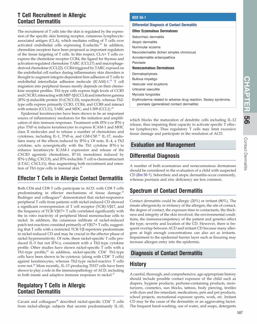
BOX 56-1
Differential Diagnosis of Contact Dermatitis
Other Eczematous Dermatoses
Seborrheic dermatitis
Atopic dermatitis
Nummular eczema
Neurodermatitis (lichen simplex chronicus)
Acrodermatitis enteropathica
Psoriasis
Noneczematous Dermatoses
Dermatophytosis
Bullous impetigo
Vesicular viral eruptions
Urticarial vasculitis
Mycosis fungoides
Erythroderma related to adverse drug reaction, Sezary syndrome, psoriasis (generalized contact dermatitis)
Sec
tion
H A
llerg
ic S
kin
and
Eye
Dis
ease
s
588
clinical presentations of CD that may be overlooked include urti-caria, acneiform, and pigmentary changes. A broader spectrum of irritant CD, including acute, acute delayed, cumulative, traumatic, and subjective, has been described.49
Regional Considerations in Children
Hand DermatitisHand dermatitis deserves special consideration not only because it is extremely common but also because the differential diagno-sis can be challenging. Because the palmar skin is much thicker than the dorsum of the hands, ACD rarely involves the palms, occurring most often on the thinner skin between the fingers and the dorsum of the hands (Figure 56-2).
Hand dermatitis may be due to irritant CD or ACD, atopic dermatitis, dyshidrosis and psoriasis. Because of significant overlap, it may be difficult to distinguish the etiology of the hand dermatitis. Common causes of irritant CD of the hands include water, detergents, and solvents. Patch tests in patients with hand eczema showed relevant allergens including nickel sulfate (17.6%), potassium dichromate (7.2%), rubber elements includ-ing thiuram mix, carba mix, p-phenylenediamine and MBT (19.6%) and cobalt chloride (6.4%).50,51 A Swedish study of 5700 patients showed that specific allergens correlated with eczema localized to different sites on the hand.52 Patients whose entire hands were involved were more likely to react to thiuram mix, p-phenylenediamine, chromate and Balsam of Peru, while those with involvement of the fingers and interdigital spaces or palm were more likely to react to nickel, cobalt, and 5-chloro-2- methyl-4-isothiazolin-3-one/2-methyl-4-isothiazolin-3-one.
The prevalence of hand eczema in patients with atopic derma-titis is 2- to 10-fold higher than in nonatopic individuals. Certain morphologic features can help distinguish the different contrib-uting factors to hand eczema. Involvement of the dorsal aspect of the hand and fingers, combined with volar wrist involvement, suggests atopic dermatitis as a contributing etiologic factor. Irri-tant CD commonly presents as a localized dermatitis without vesicles over webs of fingers extending onto the dorsal and ventral surfaces (‘apron’ pattern), dorsum of the hands, palms and ball of the thumb. In contrast, ACD is often associated with vesicles and tends to favor the fingertips, nail folds, dorsum of the hands and, less commonly, involves the palms. Since irritant CD of the hands can precede ACD, pattern changes such as increasing dermatitis from web spaces to fingertips or from palms to dorsal surfaces should prompt patch testing.53
and cleansers used, should be noted. The evolution of the skin reaction is influenced by many factors, including the patient’s skin, age, color, ambient conditions, the use of topical or other oral medication and response to all prior treatment. Because the majority of contact reactions present as eczematous eruptions, it is essential to note clinical evolution from acute vesiculation to chronic lichenification.
Unfortunately, although history can strongly suggest the cause of CD, relying solely on the history, other than with obvious nickel reactions and a few other allergens, may confirm sensitiza-tion in only 10–20% of patients with ACD. At times, CD may be superimposed on atopic dermatitis. The impaired epidermal barrier layer of all atopics, with or without active dermatitis, subjects them to a greater risk for both allergic sensitization and irritation.
Physical Examination
The diagnosis of ACD is suspected from the clinical presentation of the rash and the possible exposure to a contact allergen. The objective findings found on physical exam include the appropri-ate identification of all the primary and secondary skin lesions. CD can be described as acute, subacute, or chronic. Acute der-matitis can present with erythematous papules, vesicles, and even bullae. Chronic CD is generally pruritic, erythematous and may be associated with crusting, scaling, fissuring, excoriations and lichenification. Irritant CD is usually confined to the area of the skin in direct contact with the offending agents, with little or no extension beyond the site of contact (Figure 56-1). Less common
Figure 56-1 Patch test grading. Figure 56-2 Allergic contact dermatitis of the hand.
Contact D
ermatitis
CH
AP
TER
56
589
flavoring in food products, skin care products, and dentifrices. Balsam of Peru is found in dentifrice, mouthwash, lipstick, and food. Dodecyl gallate is a preservative used to extend the shelf life of oil-based foods such as peanut butter, soups, and pastries. Toothpaste, fluoride mouth washes, chewing gum and other foods may contain cinnamic aldehyde, flavorings and pepper-mint, which are common causes of allergic cheilitis. Thus, an oral antigen screening series should include not only metals but also flavorings, preservatives, medications and dental acrylates. In patients with cheilitis, patch test should include an even more comprehensive panel of flavorings and preservatives. The use-fulness of patch testing in the evaluation orofacial granulomato-sis and recurrent aphthous stomatitis is questionable.58
Flexural Areas of Neck and Axillary DermatitisThe thin intertriginous skin of the neck is vulnerable to irritant reactions from ‘perms’, hair dyes, shampoos, and conditioners. ‘Berloque’ dermatitis from certain perfumes or nail polish presents as localized areas of eczema. Nickel-sensitive individu-als may react to wearing a necklace or to zippers.
ACD can be caused by deodorants but not antiperspirants. These agents generally cause a dermatitis involving the entire axillary vault, whereas textile ACD spares the apex of the vault. Irritant CD can occur from shaving and depilating agents. However, sweat and perspiration may cause increased deodor-ant allergen in the periphery giving a dermatitis that is less intense in the apex of the axillae.
Diaper DermatitisEruptions in the diaper area are the most common dermatologic disorder of infancy.59 Friction, occlusion, maceration and increased exposure to water, moisture, urine and feces60 contrib-ute to irritant CD, and are probably the most common causes of diaper dermatitis. The prevalence of diaper dermatitis, an irritant CD, in infants has been estimated to be 7–35% with a peak inci-dence between ages 9 and 12 months.61 However, a more recent large-scale study in Great Britain demonstrated an incidence of 25% in the first 4 weeks of life alone.62
ACD to rubber chemicals (mercaptobenzothiazole, cyclohexyl thiophathalimide) or glues (p-tertiary butyl phenol-formalde-hyde resin) has been reported and called ‘Lucky Luke’ CD.63 The characteristic dermatitis is predominantly located on the outer buttocks and hips in toddlers (‘gun holster’ pattern) and is caused by the elastic bands that hold tightly on the thighs to prevent leaking. Treatment usually involves increasing the fre-quency of diaper changes, using superabsorbent disposable diapers and applying topical agents such as low-potency corti-costeroids and barrier ointments or creams. When secondary Candida albicans infection is present, a topical antifungal agent is beneficial. There has been a definite decrease in the incidence of diaper dermatitis due to the availability of newer and improved diapers, including those with superabsorbent gel.64
Medication, douches, spermicides, sprays, and cleaners can cause CD in the genital area. Fragrances found in liners, toilet paper, soap, and bubble baths can cause a reaction in sensitized patients. Contraceptive devices can affect rubber and latex- sensitive individuals. Ammonia and/or the acidity of urine may cause an irritant dermatitis, especially in incontinent patients. The ingestion of spices, antibiotics, or laxatives may cause anal itching.
Leg DermatitisShaving agents, moisturizers, and rubber in the elastic of socks can cause allergic reactions in children. Local absorption of the
Face and Eyelid DermatitisEyelid dermatitis may be due to ACD (55–63.5%), irritant CD (15%), atopic dermatitis (<10%) and seborrheic dermatitis (4%).54 The eyelid is susceptible to ACD because of higher exposure to allergens, greater sensitivity to allergens including aeroallergens, and easy accessibility to touch, facilitating the transfer of chemi-cals applied to other areas of the body (e.g. nails, scalp) to the eyelid. Although CD is considered to be the most common cause of eyelid dermatitis, it is believed that 25% of patients with atopic dermatitis may have chronic eyelid dermatitis. In evaluating patients with eyelid dermatitis, one must note if other areas of the body are involved. Pure eyelid dermatitis may be distinct from those with other areas of involvement.55 Common allergens causing eyelid dermatitis are fragrances (facial tissues, cosmet-ics), preservatives, nickel (eyelash curlers), thiuram (rubber sponges, masks, balloons, toys), cocamidopropyl betaine and amidoamine (shampoos), tosylamide formaldehyde resin (nail polish) and gold.56 Facial tissues may contain fragrances, formal-dehyde, or benzalkonium chloride. Shampoos, conditioners, hair sprays, gels, and mousses may cause eyelid dermatitis without causing scalp or forehead lesions. Paraphenylenediamine (PPD) and ammonium persulfate can cause urticaria and/or eyelid edema.
Similar to eyelid dermatitis, facial dermatitis may occur sec-ondary to allergens transferred to the face from other regions of the body. Most commercially available cosmetics are virtually free of sensitizing components, but ACD in response to moistur-izers, sunscreens, foundations, and powders does occur and usually produces a symmetric dermatitis. Rubber-sensitive indi-viduals may react to rubber sponges, masks, balloons, children’s toys, and other products that are in contact with the face.
The scalp skin is relatively resistant to allergens in shampoos and hair dyes, and the dermatitis may be manifest on the face or eyelids. Severe burns of the scalp and hair can be caused by the misuse of hair straighteners and relaxers. The manufacturers of hair dyes recommend patch testing with the product before each application.
Oral Mucous Membranes, Perioral Dermatitis and CheilitisPerioral dermatitis and cheilitis are common in children and are associated with lip licking, lip chewing, thumb sucking, or exces-sive drooling. Objectively, changes may be barely visible or may vary from a mild erythema to a fiery red color, with or without edema. Juices of foods and even chewing gum ingredients may contribute to skin irritation of these areas. Cinnamon flavorings and peppermint are the most common causes of allergic cheilitis from toothpastes.57 Other buccal mucosal diseases include the burning mouth syndrome, stomatitis, oral lichenoid lesions and gingivitis.
Contact allergy of the mucous membrane is rare and use of patch testing to evaluate patients with mucosal involvement is controversial. In a series of 331 patients with different oral dis-eases (burning mouth syndrome, cheilitis, gingivitis, orofacial granulomatosis, perioral dermatitis, lichenoid tissue reaction and recurrent aphthous stomatitis), metals (nickel and gold) were most frequently positive on patch testing.58 Metals, includ-ing mercury, chromate, nickel, gold, cobalt, beryllium, and palladium have been used in orthodontic materials and are important allergens in patients with dental implants or ortho-dontic devices presenting with oral lichenoid lesions. Other aller-gens with a high percentage of positive reactions on patch testing include flavorings and preservatives. Fragrance mix is used as a
Sec
tion
H A
llerg
ic S
kin
and
Eye
Dis
ease
s
590
two most common allergens identified were nickel and balsam of Peru.67 Hjorth reported two children who were patch test positive to balsam of Peru whose eczema flared after oral intake of naturally occurring balsams. Other relevant positive patch test reactions in this patient population include preservatives (formaldehyde, quaternium 15, methyldibromoglutaronitrile/phenoxyethanol, diazolidinyl urea, 2-bromo-2-nitropropane-1, 3-diol, imidazolidinyl urea, and DMDM hydantoin) and propyl-ene glycol. Dyes such as Disperse Blue 106 in synthetic fibers in children’s garments have also been implicated.21
Advising patients to use skin care products without the most frequent, relevant allergens (formaldehyde-releasing preserva-tives, fragrances, and propylene glycol) is one strategy that may be helpful while awaiting definitive patch testing results. However, 8–10% of patients with scattered generalized dermati-tis remain in the unclassified eczema category.66
Systemic ACD is a localized or generalized inflammatory skin disease that occurs in sensitized individuals when they are exposed to the specific allergen orally, transcutaneously, intra-venously or by inhalation. It can manifest as a reactivation of a previous dermatitis, reactivation of a previously positive patch test or a systemic inflammatory skin disease, such as the ‘baboon syndrome’.68–72 Patients allergic to ethylenediamine may react to systemic aminophylline and antihistamines of the piperazine or ethanolamine families. Similar reactions have been reported to glucocorticoids, diphenhydramine, neomycin, penicillin, sulfon-amides (reaction to oral hypoglycemics such as tolbutamide, chlorpropamide), thiuram (reaction to oral Antabuse), coloph-ony, Balsam of Peru, fragrance mix (reaction to spices such as cloves, nutmeg, cinnamon, cayenne pepper). Nickel-sensitive patients may develop systemic reactions from the ingestion of nickel in tap water or foods cooked in nickel utensils and from eating canned foods (Table 56-1).
Patch Testing
Unfortunately, even with an extensive history and physical exam, only about 10–20% of patients with ACD can be diagnosed
topical medication has also been noted to produce an ‘autosen-sitization’, resulting in a generalized ‘id’ reaction.
Foot DermatitisIn their evaluation of CD in children, Romaguera and Vilaplana7 found that foot eczema was the most frequent localization. Irritant dermatitis of the feet may occur in children because of excessive perspiration or the use of synthetic footwear. More commonly, children can also develop allergic sensitization to rubber accelerators (MBT mix, thiuram mix, carba mix, and PPD mix), dichromates (Figure 56-3), or cements used in the manufac-ture of shoes. Other chemicals in footwear (e.g. leather, adhe-sives, glues, and dyes) or in topical medications (e.g. creams, ointments, and antiperspirants) can cause ACD. Chemical agents, such as chrome used in the tanning and dyeing processes of leather, and colophony used in glues in soles and insoles, may be sensitizing. Reactions to nickel sulfate were also frequent with metal present in footwear buckles, eyelets and ornaments.65 The dorsal aspect of the foot and toes, especially the hallux is more commonly involved in ACD. The interdigital areas are rarely involved. Irritant dermatitis can involve either the dorsum or the sole. The majority of patients with CD of the feet also have hyper-hidrosis. ‘Sweaty sock’ dermatitis needs to be distinguished from CD, atopic dermatitis, and tinea pedis. Patients should be encour-aged to wear cotton socks and to change them frequently, along with breathable footwear. Occasionally, a dusting powder (Zea-SORB) may be needed. Other causes of foot dermatitis are tinea pedis, AD, psoriasis, dyshidrosis, and nummular eczema.
Generalized DermatitisDermatitis with scattered generalized distribution is a difficult diagnostic and therapeutic challenge because it lacks the charac-teristic distribution that gives a clue as to the possible diagnosis of ACD. The frequency of positive and relevant patch testing is unknown. Zug and colleagues66 examined the yield of patch testing, relevant allergens and allergen sources in patients with scattered generalized dermatitis referred for patch testing. Approximately 15% of the patients patch tested had scattered generalized dermatitis only and approximately half (49%) had a positive patch test deemed at least possibly relevant to their dermatitis. The prevalence of scattered generalized dermatitis was higher in patients with a history of atopic dermatitis. The
Figure 56-3 Chronic dermatitis on dorsa of feet and toes caused by potassium dichromate allergy from chronic exposure to leather tennis shoes. (From Weston WL, Lane AT, Morelli JG. Dermatitis. In: Color Textbook of Pediatric Dermatology, 4th edn. 2007, Mosby.)
Table 56-1 Reported Causes of Systemic Allergic Contact Dermatitis
fragrance mix (5.1%). For children aged 0 to 18 years, the most frequent relevant positive reactions were to nickel sulfate (26.0%), cobalt (12.4%), neomycin (4.4%), fragrance mix (4.1%), gold (3.6%), and quaternium 15 (3.6%).
34.0% of the children with a relevant positive reaction had atopic dermatitis. Of note, 15% and 39% of children had relevant allergens not included in the NACDG series or T.R.U.E. TEST® (thin-layer rapid use epicutaneous test, Allerderm), respectively. It is important for the clinician to remember that the majority of patients will be allergic to a single allergen or a single group of allergens and that there is a risk of false-positive patch test results. Some patients may benefit more from direct therapeutic intervention (i.e. allergen avoidance) than from patch testing. Ideally, one needs to know the value of all the clinical data before patch testing, in predicting a clinically relevant response to any of the allergens tested. Immunocompromised patients, including those on oral steroids or those on cancer chemotherapy or immu-nosuppressive drugs, are not appropriate candidates for patch testing. Ideally, the dermatitis should be quiescent. The skin site where the patch tests are to be applied should have had no potent steroid applied for 5 to 7 days before testing. Patients should avoid sun or ultraviolet light exposure for 96 hours. Systemic antihistamines have no effect on patch test results.
Sources of Allergens
Commercially available standardized patch test allergens have been calibrated with respect to nonirritant concentrations and compatibility with the test vehicle. Test systems currently avail-able in the USA are the T.R.U.E. TEST® and the standardized allergens loaded in Finn chambers or allergEAZE patch test chambers. Individual chambers are filled with contactants, applied to the skin and held in place by hypoallergenic tape. Certain screening panels such as the NACD Series with a range from 65 to 70 allergens are not FDA-approved but conform to standards of care recommended by CD experts. Commercial sources of customized patch test materials include Smart Practice Canada (1.866.903.2671), Hermal Pharmaceutical Laboratories, Inc. (1.800.HERMAL-1), Dormer Laboratories, Inc. and Trolab Allergens.
Allergens
The ideal number of patch tests to be applied depends on the patient and the usefulness of patch testing is enhanced with the number of allergens tested. Allergens not found on commercially available screening series in the USA frequently give relevant reactions, and personal products are a useful supplement espe-cially in facial or periorbital dermatitis.
The T.R.U.E. TEST® consists of a standard battery of 28 aller-gens and a negative control on three panels (Table 56-2). The allergens in the T.R.U.E. TEST® are in a vehicle attached to an adhesive backing. Comparative results of the T.R.U.E. TEST® and Finn Chamber method have shown a 64–98% concordance in results, depending on the allergen. However, a further study suggested that false-negative results may occur with the T.R.U.E. TEST®, particularly with fragrance mix and rubber additives (thiuram and carba mix).75 In addition, the T.R.U. E. TEST® has higher false-negative reactions to neomycin, cobalt and lanolin. Allergens including gold, bacitracin, MDGN/PE (methyldibro-moglutaronitrile/phenoxyethanol), propylene glycol, bromoni-tropropane, cinnamic aldehyde, DMDM hydantoin, ethylene urea/melamine formaldehyde have a prevalence of more than
accurately without a patch test. Patch testing should be consid-ered for any patient with a chronic, pruritic, or recurrent eczematous dermatitis, especially those with eyelid or hand involvement.73
Virtually any eczematous lesion could be caused or aggravated by a contactant (Box 56-2). Patch testing is needed to identify the responsible allergens, is helpful in young children suspected of ACD and remains the gold standard for confirming ACD. The paradox of patch testing lies in its deceptive simplicity. Although the application of antigens for patch testing is rather simple, antigen selection and patch test interpretation require an experi-enced clinician (Box 56-3).
Selection of Appropriate Subjects to Test
The higher the index of suspicion, the more frequent the diagno-sis of ACD. Indeed, the observation that the greatest abuse of patch testing is its lack of use holds true even for the pediatric population. Most recently, the North American Contact Derma-titis Group (NACDG) sought to determine the frequency of posi-tive and relevant patch tests in children referred for patch testing in North America. In addition, they compared results of patch testing children and adults, as well as results with international data on contact allergy in children. No significant difference in the overall frequency of at least one relevant positive patch test reaction was noted in children (51.2%) compared with adults (54.1%).74 The most frequent positive reactions in children were to nickel (28.3%), cobalt chloride (17.9%), thimerosal (15.3%), neomycin sulfate (8.0%), gold sodium thiosulfate (7.7%), and
BOX 56-2 Key concepts
Evaluation and Management of Contact Dermatitis
• Irritant contact dermatitis is much more common than allergic contact dermatitis.
• Response to a contactant is influenced by factors related to the agent, the host, the exposure, and the environment.
• The higher the index of suspicion for allergic contact dermatitis, the more frequent the correct diagnosis.
• The greatest abuse of patch testing is lack of use.
• Patch testing is indicated for any persistent eczematous eruption on the dorsum of the hands but rarely for palmar rashes.
• Patients with a suggestive history or physical findings but negative results on Thin-layer Rapid-Use Epicutaneous Test should be considered for further evaluation in a patch testing clinic.
BOX 56-3 Key concepts
Approach to Patch Testing in Allergic Contact Dermatitis
• Understand the underlying pathophysiology.
• Select the proper patient to test.
• Never test with an unknown substance.
• Apply the patches properly, and instruct the patient or family in proper care.
• Interpret the patch test results correctly.
• Determine the relevance of the results.
• Counsel the patient and/or family.
Sec
tion
H A
llerg
ic S
kin
and
Eye
Dis
ease
s
592
2% in the NACDG 2004 but are not included in the current T.R.U.E. TEST®. A number of other standardized allergens can be tested individually with the Finn Chamber attached to the back with Scanpor tape or an alternative patch testing system (allergEAZE, SmartPractice Canada). Clinicians need to be aware of the limitations of each system of patch testing for individual allergens.76 Caution should be exercised when testing for non-standardized antigens to avoid adverse effects and false-positive or negative responses. Ideally, at least two control subjects should be tested with any nonstandardized allergen. ‘Leave-on’ cosmetics (make-up, perfume, moisturizer, nail polish), clothing and most foods are tested ‘as is’, whereas ‘wash-off’ cosmetics (soap, shampoo) are tested at 1 : 10-1 : 100 dilution.
Patch testing should never be performed with an unknown substance. Photopatch tests should be performed by a clinician with expertise in ultraviolet radiation if photocontact dermatitis is suspected. Additional guidelines for patch testing including strength of recommendations and quality of evidence have been recently published by the British Association of Dermatologists Therapy Guidelines and Audit Subcommittee.77 The T.R.U.E. TEST® may serve as triage or a screening tool in an allergists’ practice but occupational exposures may benefit from early referral for supplemental testing.
Selection of Allergens in Children
Available data show that the sensitization profile of children does not differ significantly from that of adults. Thimerosal, neo-mycin sulfate, nickel sulfate, mercury, amide chloride, cobalt chloride, fragrance mix, bufexamac, Compositae mix, propylene glycol and turpentine are the substances with the highest sensi-tization rates in childhood.78 It has been suggested that in very young children, allergens such as formaldehyde, formaldehyde-releasing preservatives, mercaptobenzothiazole and thiuram be diluted 50%, and potassium dichromate 25% in petrolatum, to avoid irritant false positive reactions.79,80 However, most studies to date suggest that the same test concentrations as in adults can be used.81
Due to the limited surface available for testing and the poten-tial risk of active sensitization, the German Contact Dermatitis Research Group78 recommends a panel of 12 contact allergens as a standard series in children from 6 to 12 years of age. Based on their frequency and clinical relevance, the allergens included are nickel sulfate, thiuram mix, colophony, mercaptobenzothiazole, fragrance mix I, fragrance mix II, mercapto mix, bufexamac, dibromdicyanobutane, chlormethylisothiazolinone, neomycin, and compositae mix. When history suggests exposure to shoe allergens, p-tert-butylphenol-formaldehyde resin and potassium dichromate are added. Wool alcohols/lanolin 30% is added when there is exposure to skin care products, Disperse blue if clothing dermatitis is suspected and paraphenylenediamine if there is exposure to henna, tattoos and hair dyes. Children under the age of 6 years should be tested if there is a high degree of suspicion and only selectively with the suspected contact aller-gens. Children older than 12 years and adolescents can be tested in the same manner as adults.
Patch Testing Procedure
Standardized criteria for patch testing have been set by the Task Force on Contact Dermatitis of the American Academy of Dermatology. All results are dependent on the recommended protocol for application, removal, and interpretation of results.
Table 56-2 Thin-layer Rapid Use Epicutaneous Test (T.R.U.E. TEST®) Antigens
Quinoline mix Paste bandages; prescription and non prescription topical antibiotics and antifungal creams, lotions, ointments; animal food
Contact D
ermatitis
CH
AP
TER
56
593
can also occur. Common combinations of a positive patch test are: PPD and benzocaine (cross-sensitizer); thiuram mix, carba mix and mercapto mix (rubber products); quarternium 15 and paraben, (quarternium-15, a formaldehyde releaser and formal-dehyde are preservatives that are frequently combined and cosensitize); cobalt and nickel (cobalt is used in alloys with nickel and chromium and cosensitize). Poly sensitization is common in children. Children with and without atopic dermatitis have the same rate of positive patch test.
The repeat open application test (ROAT) or exaggerated use test may be done to confirm the presence or absence of ACD. The suspected allergen (for ‘leave-on’ but not ‘wash-off’ products) is applied to the antecubital fossa twice daily for 7 days, and observed for dermatitis. The absence of a reaction makes CD unlikely. If eyelid dermatitis is considered, ROAT can be carried out on the back of the ear.
Additional tests used less frequently in the diagnosis of CD include skin biopsy to differentiate from other diseases (listed in Box 56-1). Prick or intradermal testing may be helpful, especially in the evaluation of contact urticaria. Contact urticaria can also be evaluated with an ‘open’ patch test as an alternative to the prick or intradermal test. Potassium hydroxide preparation for fungal hyphae or cultures may be needed to identify fungal disease.
Allergens of Particular Importance in Children
Nickel
Nickel is a more common cause of ACD than all other metals combined, even in children. Of 391 children aged 18 years or less who were patch tested between 2001 and 2004 by the North American Contact Dermatitis Group, 28% had a positive patch test to nickel, and 26% were deemed to have a nickel allergy of either current or past relevance.82 In 2008, the American Contact Dermatitis Society selected it as the ‘contact allergen of the year’, pointing to its rise in incidence and high sensitization rate in children. It is more common in adolescents, girls more than boys, and ear piercing is the most important predisposing factor. The prevalence of nickel allergy among those children with pierced ears was 13% compared to 1% among those without pierced ears.83 The risk of sensitization to nickel appears higher when earlobes are pierced before the age of 20 years (p < 0.05)84 and is increased with the number of piercings.85 Thus, some authors recommend that ear piercing be delayed until after 10 years of age, presumably to allow for the development of immune tolerance.86
Nearly 5 million people in the USA and Canada undergo orthodontic treatment. In patients with contact allergy to ortho-dontics, nickel is the most common allergen. Nickel is commonly used in orthodontics but stainless steel, which contains about 8% nickel that is not normally biologically available, is generally considered safe in nickel-allergic patients. However, recent studies suggest that certain flexible titanium-nickel arch wires used in orthodontics release increased amounts of nickel com-pared to stainless steel and may need to be avoided in patients with known nickel sensitivity.87 A Finnish study of adolescents and the effect of age, gender, onset, duration and specific ortho-dontic treatment, and age of ear piercing on the incidence of nickel sensitization, found that 35% of the girls who had their ears pierced prior to orthodontic treatment were nickel-allergic versus none of the girls who had orthodontic treatment prior to ear piercing.88 The mechanism responsible was suggested to be oral tolerance.89 In females, nickel sensitivity may increase the
Patch tests are typically applied to the upper- or mid-back areas (2.5 lateral to a mid-spinal reference point) which must be free of dermatitis and hair and kept in place for 48 hours. After securing the patches with hypoallergenic tape, patients are instructed to keep the area dry and avoid activities that will cause excessive sweating or excessive movement that may cause displacement of the patches. In infants and small children, the patch tests can be covered with fabric adhesive tape or a stocki-net vest. Patch tests are removed after 48 hours and read 30 minutes after to allow resolution of erythema and irritative effect from the tape and/or chamber if present. A second reading should be done 3 to 5 days after the initial application. 30% of relevant allergens negative at the 48-hour reading become posi-tive in 96 hours. Irritant reactions tend to disappear by 96 hours. Metals (gold, potassium dichromate, nickel, cobalt), topical anti-biotics (neomycin, bacitracin), topical corticosteroids and PPD may become positive after 7 days. More than 50% of positive patch test to gold was delayed for about 1 week.
The American Contact Dermatitis Society has established a grading system that is almost universally recognized (Table 56-3). Alternative grading with the T.R.U.E. TEST® is shown in Figure 56-1. Relevance of positive reactions to the clinical presentation needs to be carefully evaluated. Conversely, patients with nega-tive results may need to be referred for more complete testing to a patch test clinic.
Determining Clinical Relevance
The relevance of positive reactions to clinical ACD can only be established by carefully correlating the history, including expo-sure to the allergen. A positive patch test reaction may be rele-vant to present or previous dermatitis; multiple true-positives can occur and mild responses may still represent allergic reac-tion. Thus, understanding the sources of antigen in the patient’s environment is required to be able to advise the patient ade-quately regarding avoidance and alternatives in ACD. A positive patch test is considered to be a ‘definite’ reaction of ACD if the result of a ‘use test’ with the suspected item was positive or the reaction of the patch test with the object or product was positive. It would be ‘probable’ if the antigen could be verified as present in known skin contactants and the clinical presentation was con-sistent; and ‘possible’ if the patient is exposed to circumstances in which skin contact with materials known to contain allergen was likely. Multiple sensitivities are possible if different aller-gens are present in different products used simultaneously, or concomitant sensitization occurs if allergens are present in the same products and both induce sensitization. Cross-sensitization
Table 56-3 Patch Test Interpretation
Grade Patch Test GradingCLINICAL Interpretation of Grading
0 No reaction No evidence for contact allergy
+/− Mild erythema only Doubtful for contact allergy
1+ 50% of patch test site erythematous with edema
Possible (versus false-positive) contact allergy
2+ 50% of patch test site with erythematous papules
Probable contact allergy
3+ 50% of patch test site with vesicles or bullae
Definite contact allergy
Sec
tion
H A
llerg
ic S
kin
and
Eye
Dis
ease
s
594
2009’ by the American Contact Dermatitis Society. Large quanti-ties of thioureas have been shown to leach from neoprene com-pounds and the levels were found to be sufficient to elicit ACD.102 This allergen mixture has been found to have one of the highest relevancy rates in the NACDG database. ACD from the allergens in neoprene include cases caused by orthopedic braces, prosthe-ses, splints, and foot supports; athletic shoes; rubber masks, swim goggles and wet suits; computer wrist rests; neoprene gloves; and rubber-based materials in automobiles.
Special Considerations
Plant Dermatitis (Phytodermatoses)
A number of plants can cause irritant reactions through mechani-cal or chemical injury. Most mechanical injury from plants is trivial, although inoculation of cactus hairs can give rise to pru-ritus. Implanted cacti spines can be removed by applying sticky tape to the skin and gently peeling it off. ‘Itching powder’ from rose hip hairs has caused maculopapular, and sometimes pustu-lar, eruptions at sites of contact. Chemical irritants caused by oxalate crystals results from contact with mustard, horseradish, and capsaicinoids in chili peppers. Contact with stinging nettles injects a mixture of inflammatory mediators, including hista-mine, causing a hive, and an unidentified neurotoxin that causes localized numbness and tingling.
Plants of the Toxicodendron group, including poison ivy and poison oak, are the most common causes of allergic plant derma-titis in children in the USA. Even newborns can be sensitized to the oleoresin (urushiol). Because this is a potent antigen, the clinical reaction typically results in vesicles and bullae, often with a characteristic linear appearance. Of note, the fluid content of vesicles is not antigenic. On the other hand, the oleoresin can be transferred by handling exposed animals, clothing, or sports equipment, even pet dander. Soap and water inactivate the antigen. Urishiol is also found in cashew nut trees, Japanese lacquer, Ginkgo biloba, and mango skin, and the ingestion of cashews or contact with mango skin can cause a similar rash. Rhus patch testing is not recommended because it has a signifi-cant sensitizing capacity.
The Compositae family (Asteraceae), the second largest plant family, represents approximately 10% of the world’s flowering plants. ACD to compositae may manifest as acute or chronic dermatitis of exposed sites. Although typically seen in florists, farmers, and professional gardeners, recent studies indicate that ACD to compositae may be more common in children than pre-viously believed. Positive patch test reactions on screening with two different Compositae mixes detected 4.2% and 2.6% posi-tives among children and adolescent, with significantly higher positive results in children with atopic dermatitis reported.103 The dermatitis has an airborne contact pattern distribution in the exposed areas of the hands and face with symptoms worse in late spring or summer and worse after picking daisies, dandeli-ons or playing outdoors. Belloni Fortina and colleagues104 sug-gested adding Compositae mix to the pediatric screening series when investigating dermatitis of air-exposed areas in children with atopic dermatitis; however, this carries the risk of false posi-tive results or sensitization. Cross-reactivity between fragrance terpenes and Compositae plant extracts may be a cause of false positive patch test to Compositae.105 In summary, ACD to Com-positae should be suspected in children with a family or personal history of atopy, summer-related or -exacerbated dermatitis, and a history of plant exposure.
risk of developing hand eczema.90 The presence of releasable nickel from the surface of any object can be detected using the di-methylglyoxime spot test; a pink color indicates the presence of releasable nickel. Despite some studies suggesting benefit,91,92 the evidence for dietary avoidance of nickel is not strong (quality of evidence IV, strength of recommendation C).77
Chromate
Chromate is found in leather, especially shoes, where chromium salts are used in the tanning process. Metallic chromium is not an allergen. Chromate sensitivity can be associated with hand or foot dermatitis, which can persist even after chromate avoidance.
Thimerosal
Thimerosal is a mercuric derivative of thiosalicylic acid that has been used as a preservative in vaccines, cosmetics, tattoo inks, eye drops, and contact lens solutions as well as a disinfectant (e.g. merthiolate).93,94 It may cross-react with mercury, which is used as preservative material in shoe manufacturing and in some antiseptic solutions. Although many children react to it on patch testing, these are rarely of clinical significance and individuals reacting to thimerosal typically have no reactions when given vaccines containing this preservative.5,95–97 Data from the North American Contact Dermatitis Group reported thimerosal as the fifth most common allergen, inducing allergic reactions in 11% of patch-tested patients.98 However, in only 17% of patients with sensitivity to thimerosal was the patch test result considered clinically relevant to their dermatitis, ranking thimerosal last in relevance among the 50 allergens tested. Thimerosal was named the contact (non)allergen of the year in 2002 with recommenda-tions for removal from the allergy patch testing screening tray.99 Because of its potential toxicity and allergenicity in children, precautionary measures are underway to remove thiomerosal from vaccines.100 Systemic reactions manifesting as an exanthem that preferentially involves the buttocks and flexural aspects of extremities, and known as the baboon syndrome, have been observed in children, mainly after exposure to metallic mercury vapors.68 Aside from thimerosal, reactions to adjuvants (e.g. alu-minum hydroxide), stabilizers (e.g. gelatin), preservatives, and antibiotics (e.g. neomycin) in vaccines have been reported.
Aluminum
Aluminum may cause cutaneous granulomas in response to vac-cines containing aluminum hydroxide. These tend to resolve spontaneously, although children subsequently have positive patch tests to metallic aluminum or their salts.101 The aluminum sensitivity appears to be lost with time as it occurs rarely in adults.
Rubber Chemicals
Rubber chemicals, including thiuram mix, mercaptobenzothia-zole, and mercapto mix are used in the manufacturing of rubber products, including dipped (e.g. balloons, gloves) and molded (e.g. pacifiers, handle bars) products. Mixed dialkyl thioureas (MDTU), a mixture of two thiourea chemicals used for rubber acceleration and as antioxidants in the manufacturing of neo-prene, was designated as the ‘contact allergen of the year for
Contact D
ermatitis
CH
AP
TER
56
595
Preservatives are present in most aqueous-based cosmetics and personal hygiene products to prevent rancidity. These pre-servatives are grouped into two broad categories: formaldehyde releasers and nonformaldehyde releaser (Table 56-4). Individuals who are allergic to formaldehyde cannot use any of the formal-dehyde releasers. Common sensitizers that are formaldehyde releasers include quaternium-15, whereas thimerosal, benzalko-nium chloride, and parabens are nonformaldehyde releasers. Paraben is the most commonly used preservatives in cosmetic, pharmaceutical and industrial products because of its broad spectrum of activity against yeasts, molds, and bacteria.
Excipients, including propylene glycol, ethylenediamine, and lanolin are inert substances that make up the base of a product and serve to solubilize, sequester, thicken, foam, or lubricate the active component in a product. They can cause ACD or, in higher concentrations, can act as irritants. Lanolin is a common compo-nent of consumer products. Unfortunately, its composition has not been fully characterized. Medicaments containing lanolin are more sensitizing than lanolin-containing cosmetics. Lanolin is a weak sensitizer when applied on normal skin but a stronger sensitizer on damaged skin.
Hair products are second only to skin care products as the most common cause of cosmetic allergy. In addition to routine hair care products, intermittent cosmetic hair products such as permanent or semi-permanent hair dye and permanent wave solutions are commonly used.
p-Phenylenediamine (PPD)New trends in permanent and temporary tattoos have emerged in our adolescent population. The use of black henna mixtures, containing indigo, henna and PPD and/or diaminotoluens to temporarily paint the skin is even used in some cultures prima-rily before major events. The addition of PPD to give henna (auburn to red color) a darker shade of brown to black is increas-ingly used in body painting and the need for a policy for use in children has been suggested.108 Adolescents working in hair salons may be exposed to PPD, the most common allergen affect-ing hairdressers. A number of chemicals may cross-react with PPD (Table 56-5).109
Glycerol thioglycolate is the active ingredient in permanent wave solutions. Cocamidopropyl betaine, amphoteric surfactant in shampoos, bath products and cleansers can also cause ACD from hair products.
Nail cosmetics and glues have become increasingly popular and fashionable. There are several varieties of sculpting nails and the currently marketed products contain various methacrylate ester monomers, dimethacrylates, and trimethacrylates as well as cyanoacrylate-based glues. Clinical allergy to acrylics in nails
Ambrosia species, which include ragweed, can cause allergic plant dermatitis when pollinating, in both atopic and nonatopic individuals. Repeated contact with ornamental cut flowers, including Alstromeria (the lily and tulip family of plants), can result in an ACD that presents with a fissured dermatitis of the fingertips. Finally, plants that contain furocoumarins (psoralens), including parsley, parsnips, and wild carrots, can cause phyto-toxic reactions, especially in summer when psoralens are most abundant in the plants where children are playing. These reac-tions occur when the skin, contaminated with psoralens, is exposed to ultraviolet A light. They occur typically during the summer months, when psoralens are most abundant in wild garden plants and children are playing outdoors.
Dermatitis from Topical Medications
The topical application of anesthetics, antihistamines, antibiotics, and even antiinflammatory drugs along with preservatives or fragrances has been implicated in sensitization and contact reac-tions. Neomycin is a frequent and potent sensitizer in children, as is diphenhydramine in topical form. Contact allergy to topical corticosteroids can be difficult to diagnose.106 It should be sus-pected in any patient whose skin condition worsens with the application of a corticosteroid. There have been a number of reports of contact allergy in the nasal mucosa to budesonide nasal spray and stomatitis with budesonide for oral inhalation.107 Patch testing to corticosteroids is not standardized, is compli-cated by the concurrent antiinflammatory actions of the medica-tion, and should be performed by clinicians familiar with this problem. Corticosteroids representative of different groupings typically used in patch testing include tixocortol, triamcinolone, dexamethasone, and budesonide. Sensitized patients must be instructed to avoid the systemic use of those drugs.
Contact Dermatitis to Cosmetics
An average adult applies 12 personal hygiene products daily and in the course of using these products, is exposed to 168 discrete chemicals. Children, and especially adolescents, may be exposed to similar numbers. Exposure to multiple potential allergens occurs repeatedly with the use of cosmetics and it is not unusual for these products to manifest as contact allergy distant from the sites on which the agent is applied. This phenomenon is termed ectopic contact dermatitis and requires diligent evaluation to elucidate the cause of the eruption.
Fragrances are one of the most common causes of ACD from the use of cosmetics in the USA. They can be found in cosmetics and personal hygiene products such as shampoo/shower gels, detergents, diapers, moisturizers, and even scented toys, either overtly to add an appealing scent or to mask unpleasant odors.
The term unscented can erroneously suggest that a product does not contain fragrance when, in fact, a masking fragrance can be present. Fragrance-free products are typically free of classic fragrance ingredients and generally acceptable for the allergic patient. However, if a fragrance-based chemical is added to a product for a purpose other than to act as a fragrance (such as the preservative benzyl alcohol), the product can still claim that it is fragrance-free. The addition of botanical and natural chemi-cals can also alter the smell of the product. Diagnosing fragrance allergy is essential for appropriate avoidance. The fragrance mix that is popularly used for patch testing contains eight different fragrances and will diagnose approximately 85% of fragrance-allergic individuals.
Note: Paraben, quarternium-15 and formaldehyde preservatives are frequently combined and cosensitize.
Sec
tion
H A
llerg
ic S
kin
and
Eye
Dis
ease
s
596
CD in soccer or football players is usually caused by equip-ment or chemicals used on the field. ‘Cement burns’ presenting as erythematous, edematous plaques, bullae, and erosions on the upper inner thighs are due to the lime component used in field markings. The characteristic rash, a history of exposure to wet field lines, and worsening of symptoms after taking a hot shower, point to the diagnosis. Patch testing performed on a few of the athletes has been negative. Treatment includes removing con-taminated clothing, cleaning the areas with water and applying topical antibiotics or petroleum jelly. Urea-formaldehyde resin in shin pads has caused ACD in soccer players.
Like runners, soccer players may develop ACD from topical anesthetic creams, epoxy resins, nickel in certain athletic shoes and tincture of benzoin used in conjunction with athletic tape.110
Ball handling in baseball and basketball can cause both irritant CD and ACD. ‘Basketball pebble fingers’ manifesting as small petechiae and abrasions on a shiny denuded surface of the fin-gertips and pads is an irritant CD resulting from mechanical irritation from the ball’s pebbled surface. An eczematous rash on both palms, the palmar fingertips and base of the thumbs may be due to rubber allergy.116 Protective knee padding and adhe-sives in athletic tape contain rubber accelerators and formalde-hyde resins.
Tennis players can develop irritant CD due to friction of the medial thighs and this manifests as erythematous eruptions over the opposing areas. ACD can be caused by isophorone diamine and epoxy resin used in the manufacture of tennis rackets, neo-prene splints to prevent or relieve tennis elbow, squash balls with N-isopropyl-N-phenylparaphenylenediamine and rubber and medications and anesthetic sprays with ethyl chloride.
Fiberglass in hockey sticks, epoxy resin adhesives in a face mask and dyes used in the manufacture of hockey gloves have caused ACD.64,117 Weightlifters have developed ACD to the nickel and palladium found in weights or bars118 and the chalk used to achieve a better grip.119 In summary, the young athlete is constantly exposed to allergens in clothing, equipment, envi-ronment and medications. The unique presentation of the rash, a careful sports-directed history and allergen-directed patch testing enhances the ability to diagnose and care for the young athlete with dermatitis.
Treatment and Prevention
The identification of the allergen to improve avoidance of contact to the allergen and education of patients and/or families is the mainstay of treatment for ACD. All other measures are palliative and temporary. Compliance with allergen avoidance is fre-quently difficult.
Once the offending agent is identified, patients and/or care-givers must be educated regarding the nature of the dermatitis, triggering agents, and irritant factors. A list of potential expo-sure alternatives and substitutes to cosmetics should be offered to the patient to increase compliance. (usually listed in textbooks, such as Fisher’s Contact Dermatitis120 or Marks and DeLeo’s Contact and Occupational Dermatitis121). The American Contact Dermatitis Society maintains a topical skin care product database called the Contact Allergen Replacement Database (CARD). After patch testing, patients can be provided with a comprehen-sive list of skin care products that are free of their identified allergens.
After removal of the offending agent, topical therapy may be used. Topical antiinflammatory agents, primarily corticosteroids (CSs), are most effective when treating localized dermatitis.
can present locally at the distal digit or ectopically on the eyelids and face. Patch testing to a variety of acrylates and nail polish resin may be necessary to delineate the causative agent.
Sunscreens are frequently present in cosmetics such as mois-turizers, lip preparations, and foundations. As a group they are the most common cause of photoallergic CD. Chemical-free sun blocks use physical blocking agents instead of photoactive chem-icals and include titanium dioxide and zinc oxide, which are rarely sensitizers.
Contact Dermatitis in Athletes
The skin of athletes is exposed to repeated trauma, heat, mois-ture, and numerous allergens and chemicals and is predisposed to irritant CD or ACD. Early recognition can facilitate appropri-ate therapy and prevention.110
In swimmers, chemicals used to disinfect swimming pools such as chlorine can cause both irritant CD and ACD. In such cases, the dermatitis may spare the area of the skin under the swimwear. However, swimwear and equipment including goggles, nose plugs, nose clips, ear plugs, fins and swim caps may also cause CD. Although ACD from swimming goggles usually presents with well-demarcated, bilateral periorbital edema and erythema with varying degrees of pruritus, exudate, and scaling, conjunctival injection and hypopigmentation have been reported.111,112 Patch testing to rubber products should ideally include a piece of material taken from the offending goggles. Allergens include rubber and chemicals used in manu-facturing; neoprene, benzoyl peroxide, phenol-formaldehyde resin, thioureas, and antioxidants are frequently used in rubber production.
‘Jogger’s nipples’ are painful, erythematous, and crusted ero-sions and represent irritant dermatitis caused by friction from the running shirt.113 Other skin and nail problems are associated with irritant CD and ACD from the shoes, shirts and topical medications. Contact allergens include components of rubber, leather, glues, or dyes used in the manufacture of running shoes. Sweat helps leach out the chemicals from the shoes responsible for causing ACD. Rubber insoles from tennis shoes containing mercaptobenzothiazole and dibenzothiazyl disulfide have been reported to cause recurrent eczematous eruptions of the feet and can be prevented by switching to new innersoles such as poly-urethane.114 Analgesic sprays, topical salicylates, and antiinflam-matory creams and gels have also caused ACD in athletes. In a large case series of student athletes, benzocaine and lanolin (found in topical anesthetics and massage creams) were the most prevalent allergens responsible for CD in runners.115
Table 56-5 Chemicals that May Cross-React with p-Phenylenediamine
PABA and padimate OSulfonamides and p-aminosalicylic acidThiazidesBenzocaine and related ‘caines’Azo dyesSulfonylureasCelecoxibN-isopropyl-N’-phenyl-p-phenylenediamine
Contact D
ermatitis
CH
AP
TER
56
597
Conclusions
CD includes irritant and allergic forms and can affect patients of any age. Identification and avoidance of the allergen is key to the successful treatment of ACD. Patch testing remains the gold standard for diagnosis of ACD, and negative results in the face of a convincing clinical presentation should prompt considera-tion for further evaluation by a specialist in CD. A limited number of interventions effectively prevent or treat irritant and allergic CD, but well-controlled, outcome-blinded studies, par-ticularly in the area of ACD prevention, are needed.130 New insights into the immune mechanisms involved may lead to better treatment strategies, including induction of tolerance, especially with difficult-to-avoid allergens.
Summary and Recommendations
The following may decrease risk of developing allergic and irri-tant CD:
1. Use mild detergents in children.2. Use products generally free of fragrances and preservatives.3. Limit the use of topical antibiotics to decrease sensitization
and the development of resistance strains.4. Delay ear piercing possibly until after 10 years of age.5. ‘Natural’ herbal supplements are not necessarily safe and
without adverse effects.6. Start strategies of avoidance of allergen exposure as early as
possible.7. There is good and fair-quality evidence that barrier creams
containing dimethicone or perfluoropolyethers, short-term use of high-lipid content moisturizers, use of cotton liners if occlusive gloves are worn, and use of softened fabrics, can prevent the development of ICD.
8. There is good- and fair-quality evidence that lipid-rich moisturizers can effectively treat ICD.
Helpful WebsitesAmerican Contact Dermatitis Society website includes ‘Find a
Physician’ for Patch Testing (www.contactderm.org)The American Academy of Dermatology website (www.aad.org)T.R.U.E. TEST® website (www.truetest.com)
References1. Mortz CG, Andersen KE. Allergic contact dermatitis in children and
Immunol 1997;78:160–73;quiz 174–6.3. De Groot AC. Patch testing: test concentrations and vehicles for 3700
chemicals. 2nd ed. Amsterdam, the Netherlands: Elsevier; 1994.4. Militello G, Jacob SE, Crawford GH. Allergic contact dermatitis in chil-
dren. Curr Opin Pediatr 2006;18:385–90.5. Jacob SE, Brod B, Crawford GH. Clinically relevant patch test reactions
in children: a United states Based Study. Pediatric Dermatol 2008;25: 520–7.
6. Mortz CG, Lauritsen JM, Bindslev-Jensen C, et al. Prevalence of atopic dermatitis, asthma, allergic rhinitis, and hand and contact dermatitis in adolescents. The Odense Adolescence Cohort Study on Atopic Diseases and Dermatitis. Br J Dermatol 2001;144:523–32.
7. Romaguera C, Vilaplana J. Contact dermatitis in children: 6 years experi-ence (1992–1997). Contact Dermatitis 1998;39:277–80.
8. Manzini BM, Ferdani G, Simonetti V, et al. Contact sensitization in chil-dren. Pediatr Dermatol 1998;15:12–7.
9. Shah M, Lewis FM, Gawkrodger DJ. Patch testing in children and ado-lescents: five years’ experience and follow-up. J Am Acad Dermatol 1997;37:964–8.
Topical CS is the first-line treatment for ACD. The formulation of topical CS prescribed will depend on the location and extent of the dermatitis. Low-potency CSs are recommended for the thinner skin of the face and flexural areas, and high-potency CSs are indicated for thickened, lichenified lesions. Ointments are generally more potent, more occlusive and contain fewer sensi-tizing preservatives than creams and lotions. Patients with sen-sitivity to preservatives can use preservative-free CSs such as fluocinolone (Synalar®) ointment, triamcinolone (Aristocort®) ointment or betamethasone dipropionate (Diprosone®) ointment. Of note, high-potency CSs should not be used for diaper derma-titis, yet the results of one survey revealed that a combination antifungal-corticosteroid product containing betamethasone dipropionate was used in 6% of encounters.122
Cool compresses are usually soothing and mildly antipruritic. The addition of aluminum subacetate (Burrow’s solution), cala-mine, or colloidal oatmeal may help acute, oozing lesions. In chronic eruptions, emollients, lubricants, and moisturizers may be used, but they should be nonsensitizing and fragrance-free. Excessive hand washing should be discouraged in patients with hand dermatitis and nonirritating or sensitizing moisturizers must be used after washing. Soaps and nonalkaline cleansers should be avoided. Rarely, antibiotics may be needed for second-ary infection.
For extensive and severe CD, systemic corticosteroids may offer relief within 12 to 24 hours.
Topical calcineurin inhibitors, approved for intermittent use in children with atopic dermatitis 2 years of age and older, have been used in both animal models and patients with ACD.123,124 These immunomodulatory agents do not induce skin atrophy and may be especially valuable in treating facial or eyelid der-matitis, although use in ACD or irritant CD would be off-label at the present time. Burning or stinging has been the primary adverse reaction seen with these agents.
Antihistamines may offer some benefit in contact urticaria. Sedating antihistamines may offer some relief from pruritus. Oral diphenhydramine should not be used in patients with ACD to diphenhydramine in a calamine base (Caladryl®) or hydrox-yzine hydrochloride (Atarax®) in ethylenediamine-sensitive patients. Other treatments include ultraviolet light as well as immunomodulating agents such as methotrexate, azathioprine and mycophenolate mofetil.
Whereas mechanical barriers against contact, such as protec-tive gloves, clothing, and barrier creams, are helpful in some cases (nickel allergy), results are often disappointing. For nickel-allergic patients, barriers such as gloves, covers for metal buttons and identification of nickel by the dimethyl-gloxime test can be prescribed but results can be disappointing.125
For diaper dermatitis, allergic, irritant and infectious causes have to be considered. Frequency of diaper changes using improved product design features, such as superabsorbent dis-posable diapers, are believed to explain the decline in diaper dermatitis among infants.126 Low-potency CSs and barrier oint-ments or creams can be used for a limited period of time.127 When secondary C. albicans infection is present, a topical antifungal agent should be used. Possible treatments include minimizing diaper use and using disposable diapers, barrier creams, mild topical cortisones, and antifungal agents. A gentle cleansing routine, frequent diaper changes, and a thick barrier cream help control this condition.128
Patient education regarding the nature of the dermatitis, trig-gering agents, irritant factors plus instruction for avoidance, and appropriate substitutes will not only aid in clearing the derma-titis but also prevent or minimize recurrences. At present, hypo-sensitization of patients with ACD is not a viable therapy.129
41. Albanesi C, Scarponi C, Cavani A, et al. Interleukin-17 is produced by both Th1 and Th2 lymphocytes, and modulates interferongamma- and interleukin-4-induced activation of human keratinocytes. Invest Derma-tol 2000;115:81–7.
42. Cavani A, Albanesi C, Traidl C, et al. Effector and regulatory T cells in allergic contact dermatitis. Trends Immunol 2001;22:118–20.
43. Martin S, Lappin MB, Kohler J, et al. Peptide immunization indicates that CD8+ T cells are the dominant effector cells in trinitrophenyl-specific contact hypersensitivity. J Invest Dermatol 2000;115:260–6.
44. Budinger L, Neuser N, Totzke U, et al. Preferential usage of TCR-Vbeta17 by peripheral and cutaneous T cells in nickel-induced contact dermatitis. J Immunol 2001;167:6038–44.
45. Kapsenberg ML, Wierenga EA, Stiekema FE, et al. Th1 lymphokine pro-duction profiles of nickel-specific CD4+T lymphocyte clones from nickel contact allergic and non-allergic individuals. J Invest Dermatol 1992;98:59–63.
46. Cavani A, Mei D, Guerra E, et al. Patients with allergic contact dermatitis to nickel and nonallergic individuals display different nickel-specific T cell responses: evidence for the presence of effector CD8+ and regulatory CD4+ T cells. J Invest Dermatol 1998;111:621–8.
47. Larsen JM, Bonefeld CM, Poulsen SS. IL-23 and TH17-mediated inflam-mation in human allergic contact dermatitis. J Allergy Clin Immunol 2009;123:486–92.
48. Cavani A, Nasorri F, Prezzi C, et al. Human CD4+ T lymphocytes with remarkable regulatory functions on dendritic cells and nickel-specific Th1 immune responses. J Invest Dermatol 2000;114:295–302.
49. Iliev D, Elsner P. Clinical irritant contact dermatitis syndromes. Immunol Allergy Clin North Am 1997;17:365–75.
50. Duarte I, Terumi Nakano J, Lazzarini R. Hand eczema: evaluation of 250 patients. Am J of Contact Derm 1998;9:216–23.
51. Warshaw EM, Ahmed RL, Belsito DV, et al. Contact dermatitis of the hands: cross-sectional analyses of North American Contact Dermatitis Group Data, 1994–2004. J Am Acad Dermatol 2007;57:301–14.
52. Edman B. Statistical relations between hand eczema and contact aller-gens. In: Menne T, Maibach HI, editors. Hand eczema. 2nd ed. Boca Raton (LA): CRC Press; 2000:75–83.
53 Warshaw E, Lee G, Storrs FJ. Hand dermatitis: a review of clinical fea-tures, therapeutic options, and long-term outcomes. Dermatitis 2003;14: 119–28.
54. Ayala F, Fabbrocini G, Bacchilega R, et al. Eyelid dermatitis: an evalua-tion of 447 patients. Dermatitis 2003;14:69–74.
55. Guin JD. Eyelid dermatitis: experience in 203 cases. J Am Acad Dermatol 2002;47:755–65.
56. Rietschel RL, Warshaw EM, Sasseville D, et al. Common contact aller-gens associated with eyelid dermatitis: data from the North American Contact Dermatitis Group 2003–2004 Study Period. Dermatitis 2007;18: 78–81.
57. Sainio EL, Kanerva L. Contact allergens in toothpastes and a review of their hypersensitivity. Contact Dermatitis 1995;33:100–5.
58 Torgerson RR, Davis MDP, Bruce AJ, et al. Contact allergy in oral disease. J Am Acad Dermatol Aug 2007;57:315–21.
59. Jordon WE, Lawson KD, Berg RW, et al. Diaper dermatitis: frequency and severity among a general infant population. Pediatr Dermatol 1986;3:198–207.
60. Bonifazi E. Napkin dermatitis-causative factors. In: Harper J, Oranje A, Prose N, editors. Textbook of pediatric dermatology. London: Blackwell Scientific Publications; 2000.
61. Scheinfeld, N. Diaper dermatitis: a review and brief survey of eruptions of the diaper area: therapy in practice. Am J Clin Dermatol 2005;6(5): 273–81.
62. Philipp R, Hughes A, Golding J. Getting to the bottom of nappy rash. ALSPAC Survey Team. Avon Longitudinal Study of Pregnancy and Childhood. Br J Gen Pract 1997;47:493–7.
63. Belhadjali H, Giordano-Labadie F, Rance F, et al. ‘Lucky Luke’contact dermatitis from diapers: a new allergen? Contact Dermatitis 2001; 44:248.
64. Reid CM, van Grutten M, Rycroft RJ. Allergic contact dermatitis from ethylbutylthiourea in neoprene. Contact Dermatitis 1993;n28:193.
65. Lazzarini R, Duarte I, Marzagao C. Contact dermatitis of the feet: a study of 53 cases. Dermatitis September 2004;15:125–30.
66. Zug KA, Rietschel RL, Warshaw EM, et al. The value of patch testing patients with a scattered generalized distribution of dermatitis: retrospective cross-sectional analyses of North American Contact Dermatitis Group data, 2001 to 2004. J Am Acad Dermatol 2008;59: 426–31.
67. Hjorth N. Eczematous allergy to balsams, allied perfumes and flavoring agents. Copenhagen: Munkgaard; 1961. p. 134.
68. Moreno-Ramirez D, Garcia-Bravo B, Pichardo AR, et al. Baboon syn-drome in childhood: easy to avoid, easy to diagnose, but the problem continues. Pediatr Dermatol 2004;21:250–3.
69. Goossens C, Sass U, Song M. Baboon syndrome. Dermatology 1997;194:421–22.
10. Stables GI, Forsyth A, Lever RS. Patch testing in children. Contact Der-matitis 1996;34:341–4.
11. Wilkowska A, Grubska-Suchanek E, Karwacka I, et al. Contact allergy in children. Cutis 1996;58:176–80.
12. Rudzki E, Rebandel P. Contact dermatitis in children. Contact Dermatitis 1996;34:66–7.
13. Katsarou A, Koufou V, Armenaka M, et al. Patch tests in children: a review of 14 years experience. Contact Dermatitis 1996;34:70–1.
14. Wantke F, Hemmer W, Jarisch R, et al. Patch test reactions in children, adults and the elderly: a comparative study in patients with suspected allergic contact dermatitis. Contact Dermatitis 1996;34:316–9.
15. Sevila A, Romaguera C, Vilaplana J, et al. Contact dermatitis in children. Contact Dermatitis 1994;30:292–4.
16. Goncalo S, Goncalo M, Azenha A, et al. Allergic contact dermatitis in children: a multicenter study of the Portuguese Contact Dermatitis Group (GPEDC). Contact Dermatitis 1992;26:112–5.
17. Rademaker M, Forsyth A. Contact dermatitis in children. Contact Der-matitis 1989;20:104–7.
18. Balato N, Lembo G, Patruno C, et al. Patch testing in children. Contact Dermatitis 1989;20:305–7.
19. Fisher AA. Allergic contact dermatitis in early infancy. Cutis 1994;54:300–2.
20. Kwangsukstith C, Maibach HI. Effect of age and sex on the induction and elicitation of allergic contact dermatitis. Contact dermatitis 1995;33:289–98.
21. Seidenari S, Giusti F, Pepe P, et al. Contact sensitization in 1094 children undergoing patch testing over a 7 year period. Pediatr Dermatol 2005;22:1–5.
22. Bruckner AL, Weston WL, Morelli JG. Does sensitization to contact aller-gens begin in infancy? Pediatrics 2000;105:e3.
24. DeLeo VA, Taylor SC, Belsito DV, et al. The effect of race and ethnicity on patch test results. J Am Acad Dermatol 2002;46:S107–12.
25. Fartasch M, Schnetz E, Diepgen TL. Characterization of detergent-induced barrier alterations: effect of barrier cream on irritation. J Invest Dermatol Symp Proc 1998;3:121–7.
26. Wood LC, Jackson SM, Elias PM, et al. Cutaneous barrier perturbation stimulates cytokine production in the epidermis of mice. J Clin Invest 1992;90:482–7.
27. Brasch J, Burgard J, Sterry W. Common pathogenetic pathways in allergic and irritant contact dermatitis. J Invest Dermatol 1992;98: 166–70.
28. Hoefakker S, Caubo M, van’t Erve EH, et al. In vivo cytokine profiles in allergic and irritant contact dermatitis. Contact Dermatitis 1995;33: 258–66.
29. Lisby S, Baadsgaard O. Mechanisms of irritant contact dermatitis. In: Rycroft RJG, Menne T, Frosch PJ, et al., editors. Textbook of contact dermatitis. London: Springer-Verlag; 2001.
30. Novak N, Bieber T. The skin as a target for allergic diseases. Allergy 2000;55:103–7.
31. Novak N, Baurecht H, Schafer T, et al. Loss-of-function mutations in the filaggrin gene and allergic contact sensitization to nickel. J Invest Der-matol 2008;128:1430–5.
32. Krasteva M, Kehren J, Horand F, et al. Dual role of dendritic cells in the induction and down-regulation of antigen-specific cutaneous inflamma-tion. J Immunol 1998;160:1181–90.
33. Grabbe S, Schwarz T. Immunoregulatory mechanisms involved in elici-tation of allergic contact hypersensitivity. Immunol Today 1998;19: 37–44.
34. Xu H, DiIulio NA, Fairchild RL. T cell populations primed by hapten sensitization in contact sensitivity are distinguished by polarized pat-terns of cytokine production: interferon gamma-producing (Tc1) effector CD8+ T cells and interleukin (Il) 4/Il-10-producing (Th2) negative regu-latory CD4+ T cells. J Exp Med 1996;183:1001–12.
35. Nasorri F, Sebastiani S, Mariani V, et al. Activation of nickel-specific CD4+ T lymphocytes in the absence of professional antigen-presenting cells. J Invest Dermatol 2002;118:172–9.
36. Trautmann A, Akdis M, Kleemann D, et al. T cell-mediated Fas-induced keratinocyte apoptosis plays a key pathogenetic role in eczematous der-matitis. J Clin Invest 2000;106:25–35.
37. Trautmann A, Akdis M, Schmid-Grendelmeier P, et al. Targeting kerat-inocyte apoptosis in the treatment of atopic dermatitis and allergic contact dermatitis. J Allergy Clin Immunol 2001;108:839–46.
38. Robert C, Kupper TS. Inflammatory skin diseases, T cells, and immune surveillance. N Engl J Med 1999;341:1817–28.
39. Campbell JJ, Haraldsen G, Pan J, et al. The chemokine receptor CCR4 in vascular recognition by cutaneous but not intestinal memory T cells. Nature 1999;400:776–80.
40. Sallusto F, Mackay CR, Lanzavecchia A. The role of chemokine receptors in primary, effector, and memory immune responses. Annu Rev Immunol 2000;18:593–620.
Contact D
ermatitis
CH
AP
TER
56
599
99. Belsito DV. Thimerosal: contact (non)allergen of the year. Am J Contact Dermat 2002;13:1–2.
100. Van’t Veen AJ. Vaccines without thiomersal: why so necessary, why so long coming? Drugs 2001;61:565–72.
101. Kaaber K, Nielsen AO, Veien NK. Vaccination granulomas and alumi-num allergy: course and prognostic factors. Contact Dermatitis 1992;26:304–6.
102 Anderson BE. Mixed dialkyl thioureas. Dermatitis 2009;20:3–5.103. Paulsen E, Otkjaer A, Andersen KE. Sesquiterpene lactone dermatitis in
the young: is atopy a risk factor? Contact Dermatitis 2008;59:1–6.104. Fortina AB, Romano I, Peserico A. Contact sensitization to compositae
mix in children. J Am Acad Dermatol 2005;53:877–80.105. Paulsen E, Andersen KE. Colophonium and Compositae mix as markers
of fragrance allergy: cross reactivity between fragrance terpenes, colo-phonium and compositae plant extracts. Contact Dermatitis 2005;53: 285–91.
106. Matura M, Goossens A. Contact allergy to corticosteroids. Allergy 2000;55:698–704.
107. Gonzalo Garijo MA, Bobadilla Gonzalez P. Cutaneous-mucosal allergic contact reaction due to topical corticosteroids. Allergy 1995;50:833–6.
108. Jacob SE, Zapolanski T, Chayavichitsilp P, et al. p-Phenylenediamine in black henna tattoos: a practice in need of policy in children. Arch Pediatr Adolesc Med 2008;162:790–2.
109. De Leo V. p-Phephenylenediamine. Dermatitis 2006;17:53–5.110. Kockentiet B, Adams BB. Contact dermatitis in athletes. J Am Acad
Dermatol 2007;56:1048–55.111. Goette DK. Raccoon-like periorbital leukoderma from contact with swim
titis caused by swimming goggles. Ann Allergy Asthma Immunol 2003;90:672–3.
113. Mailler EA, Adams BB. The wear and tear of 26.2: dermatological injuries reported on marathon day. Br J Sports Med 2004;38:498–501.
114. Jung JH, McLaughlin JL, Stannard J, et al. Isolation, via activity-directed fractionation, of mercaptobenzothiazole and dibenzothiazyl disulfide as 2 allergens responsible for tennis shoe dermatitis. Contact Dermatitis 1988;19:254–9.
115. Ventura MT, Dagnello M, Matino MG, et al. Contact dermatitis in stu-dents practicing sports: incidence of rubber sensitisation. Br J Sports Med 2001;35:100–2.
116. Rodriguez-Serna M, Molinero J, Febrer I, et al. Persistent hand eczema in a child. Am J Contact Dermat 2002;13:35–6.
118. Guerra L, Misciali C, Borrello P, et al. Sensitization to palladium. Contact Dermatitis 1988;19:306–7.
119. Scott MJ Jr, Scott NI, Scott LM. Dermatologic stigmata in sports: weight-lifting. Cutis 1992;50:141–5.
120. Fisher AA, editors. Contact dermatitis. Philadelphia: Lea & Febiger; 1986.121. Marks JG Jr, DeLeo VA. Contact and occupational dermatology. St Louis:
Mosby; 1992.122. Ward DB, Fleischer AB Jr, Feldman SR, et al. Characterization of diaper
dermatitis in the United States. Arch Pediatr Adolesc Med 2000;154: 943–6.
123. Queille-Roussel C, Graeber M, Thurston M, et al. SDZ ASM 981 is the first non-steroid that suppresses established nickel contact dermatitis elicited by allergen challenge. Contact Dermatitis 2000;42:349–50.
124. Gupta AK, Adamiak A, Chow M. Tacrolimus: a review of its use for the management of dermatoses. J Eur Acad Dermatol Venereol 2002;16:100–14.
125. Marks JG Jr, Elsner P, DeLeo VA. Contact and occupational dermatology. 3rd ed. Mosby Inc; 2002.
126. Runeman B. Skin interaction with absorbent hygiene roducts. Clin Der-matol 2008;26:45–51.
127. Atherton DJ. A review of the pathophysiology, prevention and treatment of irritant diaper dermatitis. Curr Med Res Opin 2004;20:645–9.
128. Shin HT. Diaper dermatitis that does not quit. Dermatol Ther 2005;18: 124–35.
129. Marks JG Jr, Trautlein JJ, Epstein WL, et al. Oral hyposensitization to poison ivy and poison oak. Arch Dermatol 1987;123:476–8.
130. Saary J, Qureshi R, Palda V, et al. A systematic review of contact derma-titis treatment and prevention. J Am Acad Dermatol 2005;53:845.
70. Audicana M, Bernedo N, Gonzalez I, et al. An unusual case of baboon syndrome due to mercury present in a homeopathic medicine. Contact Dermatitis 2001;45:185.
71. Alegre M, Pujol RM, Alomar A. A generalized itchy flexural eruption in a 7-year-old boy. Arch Dermatol 2000;136:1055–60.
72. Bartolome B, Cordoba S, Sanchez-Perez J, et al. Baboon syndrome of unusual origin. Contact Dermatitis 2000;43:113.
73. Beattie PE, Green C, Lowe G, et al. Which children should we patch test? Clin Exp Dermatol 2007;32:6–11.
74. Zug KA, McGinley-Smith D, Warshaw EM, et al. Contact allergy in children referred for patch testing: North American Contact Dermatitis Group data, 2001–2004. Arch Dermatol 2008;144:1329–36.
75. Sherertz EF, Fransway AF, Belsito DV, et al. Patch testing discordance alert: false-negative findings with rubber additives and fragrances. J Am Acad Dermatol 2001;45:313–4.
76. Suneja T, Belsito DV. Comparative study of Finn Chambers and T.R.U.E. test methodologies in detecting the relevant allergens inducing contact dermatitis. J Am Acad Dermatol 2001;45:836–9.
77. Bourke J, Coulson I, English J, et al. Guidelines for the management of contact dermatitis: an update. Br J Dermatol 2009;60:946–54.
78. Heine G, Schnuch A, Uter W, et al. Frequency of contact allergy in German children and adolescents patch tested between 1995 and 2002: results from the Information Network of Departments of Dermatology and the German Contact Dermatitis Research Group. Contact Dermatitis 2004;51:111–7.
79. Fisher AA. Allergic contact dermatitis in early infancy. Cutis 1994;54: 387–8.
80. Carder KR. Hypersensitivity reactions in neonates and infants. Dermatol Ther 2005;18:160–75.
81. Weston WL, Weston JA, Kinoshita J, et al. Prevalence of positive epicu-taneous tests among infants, children, and adolescents. Pediatrics 1986;78:1070–4.
82. Kornik R, Zug KA. Nickel. Dermatitis 2008;19:3–8.83. Larsson-Stymne B, Widstrom L. Ear piercing: a cause of nickel allergy in
schoolgirls. Contact Dermatitis 1985;3:289–93.84. Rystedt I, Fischer T. Relationship between nickel and cobalt sensitization
in hard metal workers. Contact Dermatitis 1983;9:95–200.85. Dotterud LK, Falk ES. Metal allergy in north Norwegian schoolchildren
and its relationship with ear piercing and atopy. Contact Dermatitis 1994;31:308–13.
86. Kütting B, Brehler R, Traupe H. Allergic contact dermatitis in children: strategies of prevention and risk management. Eur J Dermatol 2004;14: 80–5.
87. Rahilly G, Price N. Nickel allergy and orthodontics. J Orthod 2003;30: 171–4.
88. Kerosuo H, Kullaa A, Kerosuo E, et al. Nickel allergy in adolescents in relation to orthodontic treatment and piercing of ears. Am J Orthod Dentofacial Orthop 1996;109:148–54.
89. White JM, Goon AT, Jowsey IR, et al. Oral tolerance to contact allergens: a common occurrence? A review. Contact Dermatitis 2007;56:247–54.
90. Christensen OB, Moller H. Nickel allergy and hand eczema. Contact Dermatitis 1975;1:129–35.
91. Veien NK, Hattel T, Laurberg G. Low nickel diet: an open, prospective trial. J Am Acad Dermatol 1993;29:1002–7.
92. Antico A, Soana R. Chronic allergic-like dermatopathies in nickel sensi-tive patients: results of dietary restrictions and challenge with nickel salts. Allergy Asthma Proc 1999;20:235–42.
93. Breithaupt A, Jacob SE. Thimerosal and the relevance of patch-test reac-tions in children. Dermatitis 2008;19:275–7.
94. Gon Valo M, Figueiredo A, Gon Valo S. Hypertensitivity to thiomersal: the sensitizing moiety. Contact Dermatitis 1996;34:201–03.
95. Wantke F, Demmer CM, Gotz M, et al. Contact dermatitis from thime-rosal: 2 years’ experience with ethylmercuric chloride in patch testing thimerosal-sensitive patients. Contact Dermatitis 1994;30:115–7.
96. Suneja T, Belsito D. Thiomersal in the detection of clinically relevant allergic contact reactions. J Am Acad Dermatol 2001;45:23–7.
97. Osawa J, Kitamura K, Ikezawa Z, et al. A probable role for vaccines containing thiomersal in thiomersal hypersensitivity. Contact Dermatitis 1991;24:178–82.
98. Marks JG Jr, Belsito DV, DeLeo VA, et al. North American Contact Der-matitis Group patch-test results, 1996–1998. Arch Dermatol 2000;136: 272–3.