38
DR. MOHIT GOEL JR1 18 SEPT. 2012
| Date post: | 15-Jul-2015 |
| Category: |
Health & Medicine |
| Upload: | anish-choudhary |
| View: | 302 times |
| Download: | 5 times |
DR MOHIT GOEL
JR1
18 SEPT 2012
Congenital lung lesions
1 Cystic Adenomatoid Malformation
2 Pulmonary Sequestration
3 Bronchogenic Cyst
4 Congenital Lobar Emphysema
5 Congenital Diaphragmatic Hernia
6 Bronchial Atresia
7 Scimitar syndrome
Neonatal Chest Issues
1 Surfactant Deficient Disease
2 Meconium Aspiration Syndrome
3 Transient Tachypnea of the Newborn
4 Pulmonary Interstitial Emphysema
5 Bronchopulmonary Dysplasia
Congenital pulmonary airway malformation
(congenital cystic adenomatoid malformation (CCAM))
Congenital lung lesions
o Multicystic mass with air in cysts
o Imaging appearance depends upon size of cysts and whether cysts fluid filled
o Cysts communicate with bronchial tree at birth and fill with air early in life
Location
o No lobar predilection
o Most lesions confined to single lobe
o Most lesions solitary
Three types based on size of cysts in lesion at imagingpathology ndash
bull CCAM type 1 (50) 1 or more large (2-10 cm) cysts
bull CCAM type 2 (40) Numerous small cysts (lt 2 cm) of uniform size
bull CCAM type 3 (10) Appears solid on gross inspection and imaging but have
microcysts
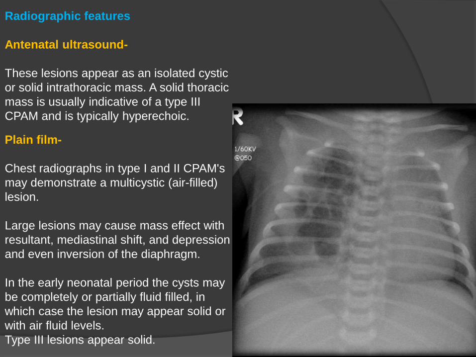
Radiographic features
Antenatal ultrasound-
These lesions appear as an isolated cystic
or solid intrathoracic mass A solid thoracic
mass is usually indicative of a type III
CPAM and is typically hyperechoic
Plain film-
Chest radiographs in type I and II CPAMs
may demonstrate a multicystic (air-filled)
lesion
Large lesions may cause mass effect with
resultant mediastinal shift and depression
and even inversion of the diaphragm
In the early neonatal period the cysts may
be completely or partially fluid filled in
which case the lesion may appear solid or
with air fluid levels
Type III lesions appear solid
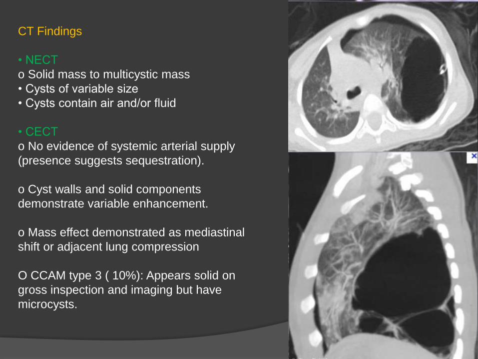
CT Findings
bull NECT
o Solid mass to multicystic mass
bull Cysts of variable size
bull Cysts contain air andor fluid
bull CECT
o No evidence of systemic arterial supply
(presence suggests sequestration)
o Cyst walls and solid components
demonstrate variable enhancement
o Mass effect demonstrated as mediastinal
shift or adjacent lung compression
O CCAM type 3 ( 10) Appears solid on
gross inspection and imaging but have
microcysts
PULMONARY SEQUESTRATION
bull Congenital area of abnormal lung that does
not connect to the bronchial tree or pulmonary
arteries
bull Involved lung is dysplastic and nonfunctioning
bull Arterial supply is typically from systemic
source arising from descending aorta
bull Divided into intralobar and extralobar types-
o Intralobar type has venous drainage into inferior pulmonary vein
o Extralobar type has venous drainage often systemic however drainage
variable
bull May occur in conjunction with other congenital lung lesions such as congenital cystic
adenomatoid malformation
bull diagnostic clue- Persistent lung opacity over multiple presentations with pneumonia-
like symptoms
Location
o Most common location is left lower lobe followed by right lower lobe
o Systemic arterial supply most commonly arises from descending aorta
May arise from below the hemidiaphragm in 20 of cases
bull Diagnostic feature Systemic artery arising from the aorta and feeding sequestration
Radiographic Findings
Radiography
o Often seen as persistent lower lobe opacity that is unchanged over multiple
radiographs
o Does not typically contain air unless infected
o Does not appear as air-containing mass during neonatal period
o If infected may appear as multi-cystic air-containing mass
CT Findings
bull NECT
o Opacification of lower lobe lung parenchyma
o May have cystic air-filled components if infected
or if occurring in conjunction with congenital cystic
adenomatoid malformation
bull CECT
o Systemic arterial supply demonstrated
o Typically artery arises from descending aorta
and extends into abnormal area of lung
o Systemic artery may arise from other systemic
sources as well
Grayscale Ultrasound
o Ultrasound may be used in newborns to
demonstrate systemic arterial supply via Doppler
o Abnormal lung is often opacified providing
acoustic window
Coronal (MIP) image in soft tissue
window The sequestration is seen as an
abnormal enhancing soft tissue mass at
the left lower lobe The large caliber artery
(1) is arising from the abdomen The left
inferior pulmonary vein (LIPV) is seen
draining the sequestration
BRONCHOGENIC CYST
They are developmental lesions that result from abnormal ventral budding of the
tracheobronchial tree between the 26th and 40th days of gestation
bull Location
Mediastinal location is more common than pulmonary
o Mediastinal 65-90
Majority in the middle mediastinum
Typically para tracheal carinal or hilar
Pericarinal most common
o Pulmonary Majority in the medial third of the lungs More frequent in the lower lobes
Typically do not communicate with airway and do not contain air Air presence indicates
infection
CT Findings
bull NECT
o Homogeneous well circumscribed lesion
o Cyst contents variable Water to proteinaceous
o Hence CT attenuation is variable
bull CECT
o Well-defined typically with nonenhancing or minimally enhancing thin wall
o More prominent wall enhancement and wall thickening may be seen with infection
o No central enhancement
MR Findings
bull TlWI o Well-circumscribed lesion
o Homogeneous signal intensity unless infected
o Variable signal due to varying amounts of proteinaceous material but usually
water signal
o Imperceptible wall
bull T2WI Signal is almost always equal to or greater than cerebrospinal fluid (CSF)
bull STIR Markedly increased signal equal to or greater than CSF
bull Tl C+ o May have a thin rim of mild enhancement
o Thicker enhancing wall implies infection
o No central enhancement
(Left) Axial T2WI MR shows homogeneous well circumscribed ovoid mass (arrow)
with signal greater than CSF (curved arrow)
(Right) AP radiograph shows large smooth homogeneous left retrocardiac
parenchymal mass (arrows)
CONGENITAL LOBAR EMPHYSEMA
A congenital lobar emphysema (CLE) refers to an over inflation of one or more lung
lobes presumably due to various factors including a possible obstructive check valve
mechanism at a bronchial level
Location
bull Left upper lobe 42
bull Right middle lobe 35
bull Right upper lobe 21
bullChest radiograph
o Initially after birth lobe may be filled with fetal lung fluid and appear as radiodensity
o May have a reticular pattern as fluid is cleared via distended lymphatics
o Fluid eventually replaced by air
bull Hyperlucent hyperexpanded lobe
o Deviation of mediastinum to contralateral side
o Increased retrosternal lucency on lateral view
o Pulmonary vessels may appear attenuated and displaced
o Classic progression Radiodense lobe that becomes progressively hyperlucent and
hyperexpanded
CT Findings
bullLucency caused by air in distended alveoli - Hyperlucent lung
bullVessels attenuated Smaller than those in adjacent lung
bullMediastinal and tracheal deviation
bullCompression of remainder of ipsilateral lung
(Left) Axial HRCT shows involvement of the medial basal segment the right lower lobe
which is hyperlucent (white arrows) The pulmonary vessels in this region are
attenuated (open arrows) being appreciably smaller than those in the remainder of
the right lower lobe (black arrows)
(Right) Anteroposterior radiograph shows hyperinflation of the right lower lobe (arrows)
with deviation of the mediastinum to the left
CONGENITAL DIAPHRAGMATIC HERNIA
bull Location
o More common on left than right (51)
o May contain variable abdominal contents Stomach small and large bowel liver
Radiography
o Radiographic appearance depends on hernia contents and whether air present within
herniated bowel
o XR initially after birth may show hernia as radiodense (prior to air introduced into
bowel)
o Later when air introduced into bowel Appears as air-containing cystic mass
resembling bowel
o Decreased bowel gas in abdomen
o Right-sided hernia often contains liver and not bowel (soft tissue density)
o Mediastinal shift away from hernia
o Low volumes of ipsilateral or contralateral lung (from hypoplasia)
o Abnormal position of support apparatus may be clue to diagnosis-
bull NG tube lodged with tip at esophagogastric junction
bull NG tube above diaphragm documenting stomach in hernia
NECT
o Shows multiple loops of bowel in chest
o Oral contrast documents bowel-containing nature of hernia
Chest X-ray shows left-sided
congenital diaphragmatic hernia with
herniating loops of large bowel into
the right hemithorax
BRONCHIAL ATRESIA
Bronchial atresia is a congenital abnormality resulting from focal interruption of a lobar
segmental or subsegmental bronchus with associated peripheral mucus impaction
(bronchocele mucocele) and associated hyperinflation of the obstructed lung segment
Location
o Most common location Apicoposterior segment left upper lobe
o Next most likely Right upper lobe right middle lobe lower lobe bronchi rare
Morphology
o Round or branching tubular mass of dilated fluid-filled bronchi distal to an atretic
proximal segmental bronchus
Radiographic Findings
bull Round or ovoid mass adjacent to the hilum (bronchocele)
bull Branching tubular opacities (mucoid impaction of dilated bronchi) distal to segmental
bronchus
bull Distal lung hyperinflated
bull Diminished vascularity
bull Neonates lobe or segment may be fluid filled gradually replaced by air
CECT
o Central low to intermediate attenuation rounded or branching tubular mass
o Hyperinflated distal lung with decreased vascularity
bull HRCT Air-trapping confirmed in hyperlucent distal lung on expiratory images
(Left) Axial CECT shows a rounded lesion centrally adjacent to the right upper lobe
bronchus (open arrow) The distal lung is hyperlucent with decrease in pulmonary
vascularity (arrow)
(Right) Axial CECT shows branching tubular structure in the left upper lobe (open
arrows) Distal to these dilated fluid filled bronchi is hyperlucent lung with decreased
vascularity (arrows)
(Right) Inspiratory radiograph obtained shows a bronchocele in the left upper lobe
(arrow)
(Left) Exhalation radiograph shows an area of hyperlucency () surrounding the area of
increased opacity a finding indicative of air trapping
Scimitar syndrome(also known as pulmonary venolobar syndrome or hypogenetic lung syndrome)
Pathology
It is essentially a combination of pulmonary hypoplasia and partial anomalous pulmonary
venous return (PAPVR) It almost exclusively occurs on the right side
Haemodynamically there is an acyanotic left to right shunt
The anomalous vein usually drains into ndash
bullIVC most common
bullright atrium or portal vein
The lung is frequently perfused by the aorta but the bronchial tree is still connected and
thus the lung is not sequestered
Radiographic features
The diagnosis is made by by CT or MR angiography
Plain film
CXR findings are that of a small lung with ipsilateral mediastinal shift
The anomalous draining vein may be seen as a tubular structure paralleling the
right heart border in the shape of a Turkish sword (ldquoscimitarrdquo)
Coronal reformat contrast-enhanced CT demonstrates right pulmonary venous drainage
into the inferior vena cava at the level of the diaphragm
Respiratory distress syndrome (RDS)
Lung disease occurring in the premature infants due to lack of surfactant
o Microatelectasis abnormal pulmonary compliance are hallmarks of disease
Radiography
o Initial Features-
bull Low lung volumes secondary to micro-collapse
bull Diffuse granular opacities represent collapsed alveoli interspersed with open alveoli
bull Peripherally extending air bronchograms Air bronchograms demonstrate patent
bronchi in abnormal lung
Potential complications include
Pulmonary interstitial emphysema pneumo-mediastinum pneumothorax superimposed
pneumonia pulmonary hemorrhage bronchopulmonary dysplasia
o Features after surfactant administration
bull Clearing of granular opacities and increased lung volumes
o Findings after several days
bull Localized areas of atelectasis
bull Focal hyperinflation
bull Intubation and ventilatory support changes the imaging appearance
bull High incidence of patent ductus arteriosus (PDA) which shows pulmonary edema
(white out of lungs with cardiomegaly)
o Bronchopulmonary dysplasia in 17-55 of premature infants
(Chronic lung disease characterized by focal areas of atelectasis focal hyperinflation)
bull HRCT
o Not typically used to make diagnosis of SDD
o Bronchopulmonary dysplasia (BPD) demonstrates bilateral disease -
bull Peribronchial thickening and prominent interlobular septum
bull Subpleural parenchymal bands
bull Hyperexpanded cyst-like areas cobblestone appearance
bull Mosaic attenuation with airtrapping
Severe respiratory distress syndrome (RDS) Reticulogranular opacities are present
throughout both lungs with prominent air bronchograms and total obscuration of the
cardiac silhouette Cystic areas in the right lung may represent dilated alveoli or early
pulmonary interstitial emphysema (PIE)
Complication of respiratory distress syndrome (RDS) After receiving ventilation therapy
this premature infant with RDS developed pulmonary interstitial emphysema (PIE) with
discrete linear and cystic radiolucent air collections throughout the right lung
MECONIUM ASPIRATION SYNDROME
Radiography
bullImaging findings Bilateral diffuse grossly patchy coarse opacities (atelectasis and
consolidation)
bullRope-like perihilar densities
bullHyperinflation of lungs
bullAreas of emphysema (air-trapping)
bullSpontaneous pneumothorax and pneumomediastinum
bull25 requiring no therapy
bullSmall pleural effusions (20)
bullRapid clearing usually within 48 hours
Frontal chest shows diffuse grossly patchy densities in
a post-mature infant consistent with Meconium Aspiration Syndrome
TRANSIENT TACHYPNEA OF THE NEWBORN
(Wet lung disease)
Definitions
bull Transient tachypnea occurs when liquid in the fetal lung is removed slowly or
incompletely from newborn lung and there is increased absorption by lymphatics
and capillaries
o Lack of normal thoracic compression that normally occurs during vaginal delivery and
is bipassed via C-sections
o Lack of normal breathing may occur with sedated infants
Best diagnostic clue Prominent interstitial pattern in lung with history of C-section
The lungs usually are affected diffusely and symmetrically and the condition is
commonly accompanied by a small pleural effusion
Chest radiographs
bull Findings similar to pulmonary edema
bull Prominent intersitial markings with normal heart size
bull Diffuse bilateral and somewhat symmetric increase in lung markings
bull Pleural effusions may be present
bull Fluid in the fissures
bull Normal to hyperinflated lung volumes
bull Interstitial pattern and other findings resolve and is normal within 72 hours Clearing
continues from peripheral to central and from upper to lower lung
bullThe radiographic appearance can mimic the diffuse granular appearance of hyaline
membrane disease but without the pulmonary underaeration
Frontal radiograph of the chest of a term newborn (left) shows streaky perhilar linear
densities (white circles) indistinctness of the blood vessels and fluid in the minor fissure
(black arrow) all signs of increased fluid in the lungs Three days later (right) a frontal
radiograph of the same baby shows complete clearing of the fluid and a normal chest
radiograph
PULMONARY INTERSTITIAL EMPHYSEMA
Definitions
bull Abnormal location of pulmonary air within the interstitium and lymphatics usually
secondary to barotrauma This collection develops as a result of alveolar and terminal
bronchiolar rupture It is more frequent in premature infants who require mechanical
ventilation for severe lung disease
Radiography
o Bubble-like or linear lucencies within the lung
o Lucencies typically uniform in size
o Often radiate from hilum
o May be focal (one lobe) or diffuse and bilateral
CT findings
Air surrounds pulmonary arterial branches which are seen as soft tissue linear or
dot-like densities surrounded by abnormal gas collections
o This pattern of central linear and dot-like densities is characteristic for PIE
(A) Transaxial and (B) coronal sections of the chest CT scan before treatment showing
diffuse pulmonary interstitial emphysema with multiple air lucencies contiguous to
pulmonary vessels and bronchi on both lung fields
This radiograph obtained from a premature infant at 26 weeks gestation shows
characteristic radiographic changes of pulmonary interstitial emphysema (PIE) of the
right lung
BRONCHOPULMONARY DYSPLASIA(chronic lung disease of infancy chronic lung disease of prematurity (CLD))
Bronchopulmonary dysplasia (BPD) is a form of chronic lung disease that develops in
preterm neonates treated with oxygen and positive-pressure ventilation (PPV)
Radiography
o Early
bull Homogeneously increased opacities bilaterally primarily related to retained fluid andor
patent ductus arteriosus
Pathology
Occurs from a paradoxical combination of hypoxia and oxygen toxicity There is Initial
capillary wall damage leading to interstitial fluid seepage
o Subsequently
bull Heterogeneous appearance with focal lucencies separated by coarse reticular and
band-like opacities of fibrosis and atelectasis
bull More opacities in the upper lobes with hyperinflation at the bases
HRCT
o Mosaic attenuation
o Foci of air trapping on expiratory images
o Subpleural triangular opacities
o Linear and reticular opacities
o Reduced bronchial lumen Pulmonary arterial ratio
Complication of ventilation therapy Bronchopulmonary dysplasia AP chest radiograph in
a 4-week-old premature infant with history of RDS and receiving mechanical ventilation
shows moderate pulmonary hyperinflation coarse interstitial opacities throughout both
lungs and atelectasis in the right upper lobe
Thank U
Congenital lung lesions
1 Cystic Adenomatoid Malformation
2 Pulmonary Sequestration
3 Bronchogenic Cyst
4 Congenital Lobar Emphysema
5 Congenital Diaphragmatic Hernia
6 Bronchial Atresia
7 Scimitar syndrome
Neonatal Chest Issues
1 Surfactant Deficient Disease
2 Meconium Aspiration Syndrome
3 Transient Tachypnea of the Newborn
4 Pulmonary Interstitial Emphysema
5 Bronchopulmonary Dysplasia
Congenital pulmonary airway malformation
(congenital cystic adenomatoid malformation (CCAM))
Congenital lung lesions
o Multicystic mass with air in cysts
o Imaging appearance depends upon size of cysts and whether cysts fluid filled
o Cysts communicate with bronchial tree at birth and fill with air early in life
Location
o No lobar predilection
o Most lesions confined to single lobe
o Most lesions solitary
Three types based on size of cysts in lesion at imagingpathology ndash
bull CCAM type 1 (50) 1 or more large (2-10 cm) cysts
bull CCAM type 2 (40) Numerous small cysts (lt 2 cm) of uniform size
bull CCAM type 3 (10) Appears solid on gross inspection and imaging but have
microcysts
Radiographic features
Antenatal ultrasound-
These lesions appear as an isolated cystic
or solid intrathoracic mass A solid thoracic
mass is usually indicative of a type III
CPAM and is typically hyperechoic
Plain film-
Chest radiographs in type I and II CPAMs
may demonstrate a multicystic (air-filled)
lesion
Large lesions may cause mass effect with
resultant mediastinal shift and depression
and even inversion of the diaphragm
In the early neonatal period the cysts may
be completely or partially fluid filled in
which case the lesion may appear solid or
with air fluid levels
Type III lesions appear solid
CT Findings
bull NECT
o Solid mass to multicystic mass
bull Cysts of variable size
bull Cysts contain air andor fluid
bull CECT
o No evidence of systemic arterial supply
(presence suggests sequestration)
o Cyst walls and solid components
demonstrate variable enhancement
o Mass effect demonstrated as mediastinal
shift or adjacent lung compression
O CCAM type 3 ( 10) Appears solid on
gross inspection and imaging but have
microcysts
PULMONARY SEQUESTRATION
bull Congenital area of abnormal lung that does
not connect to the bronchial tree or pulmonary
arteries
bull Involved lung is dysplastic and nonfunctioning
bull Arterial supply is typically from systemic
source arising from descending aorta
bull Divided into intralobar and extralobar types-
o Intralobar type has venous drainage into inferior pulmonary vein
o Extralobar type has venous drainage often systemic however drainage
variable
bull May occur in conjunction with other congenital lung lesions such as congenital cystic
adenomatoid malformation
bull diagnostic clue- Persistent lung opacity over multiple presentations with pneumonia-
like symptoms
Location
o Most common location is left lower lobe followed by right lower lobe
o Systemic arterial supply most commonly arises from descending aorta
May arise from below the hemidiaphragm in 20 of cases
bull Diagnostic feature Systemic artery arising from the aorta and feeding sequestration
Radiographic Findings
Radiography
o Often seen as persistent lower lobe opacity that is unchanged over multiple
radiographs
o Does not typically contain air unless infected
o Does not appear as air-containing mass during neonatal period
o If infected may appear as multi-cystic air-containing mass
CT Findings
bull NECT
o Opacification of lower lobe lung parenchyma
o May have cystic air-filled components if infected
or if occurring in conjunction with congenital cystic
adenomatoid malformation
bull CECT
o Systemic arterial supply demonstrated
o Typically artery arises from descending aorta
and extends into abnormal area of lung
o Systemic artery may arise from other systemic
sources as well
Grayscale Ultrasound
o Ultrasound may be used in newborns to
demonstrate systemic arterial supply via Doppler
o Abnormal lung is often opacified providing
acoustic window
Coronal (MIP) image in soft tissue
window The sequestration is seen as an
abnormal enhancing soft tissue mass at
the left lower lobe The large caliber artery
(1) is arising from the abdomen The left
inferior pulmonary vein (LIPV) is seen
draining the sequestration
BRONCHOGENIC CYST
They are developmental lesions that result from abnormal ventral budding of the
tracheobronchial tree between the 26th and 40th days of gestation
bull Location
Mediastinal location is more common than pulmonary
o Mediastinal 65-90
Majority in the middle mediastinum
Typically para tracheal carinal or hilar
Pericarinal most common
o Pulmonary Majority in the medial third of the lungs More frequent in the lower lobes
Typically do not communicate with airway and do not contain air Air presence indicates
infection
CT Findings
bull NECT
o Homogeneous well circumscribed lesion
o Cyst contents variable Water to proteinaceous
o Hence CT attenuation is variable
bull CECT
o Well-defined typically with nonenhancing or minimally enhancing thin wall
o More prominent wall enhancement and wall thickening may be seen with infection
o No central enhancement
MR Findings
bull TlWI o Well-circumscribed lesion
o Homogeneous signal intensity unless infected
o Variable signal due to varying amounts of proteinaceous material but usually
water signal
o Imperceptible wall
bull T2WI Signal is almost always equal to or greater than cerebrospinal fluid (CSF)
bull STIR Markedly increased signal equal to or greater than CSF
bull Tl C+ o May have a thin rim of mild enhancement
o Thicker enhancing wall implies infection
o No central enhancement
(Left) Axial T2WI MR shows homogeneous well circumscribed ovoid mass (arrow)
with signal greater than CSF (curved arrow)
(Right) AP radiograph shows large smooth homogeneous left retrocardiac
parenchymal mass (arrows)
CONGENITAL LOBAR EMPHYSEMA
A congenital lobar emphysema (CLE) refers to an over inflation of one or more lung
lobes presumably due to various factors including a possible obstructive check valve
mechanism at a bronchial level
Location
bull Left upper lobe 42
bull Right middle lobe 35
bull Right upper lobe 21
bullChest radiograph
o Initially after birth lobe may be filled with fetal lung fluid and appear as radiodensity
o May have a reticular pattern as fluid is cleared via distended lymphatics
o Fluid eventually replaced by air
bull Hyperlucent hyperexpanded lobe
o Deviation of mediastinum to contralateral side
o Increased retrosternal lucency on lateral view
o Pulmonary vessels may appear attenuated and displaced
o Classic progression Radiodense lobe that becomes progressively hyperlucent and
hyperexpanded
CT Findings
bullLucency caused by air in distended alveoli - Hyperlucent lung
bullVessels attenuated Smaller than those in adjacent lung
bullMediastinal and tracheal deviation
bullCompression of remainder of ipsilateral lung
(Left) Axial HRCT shows involvement of the medial basal segment the right lower lobe
which is hyperlucent (white arrows) The pulmonary vessels in this region are
attenuated (open arrows) being appreciably smaller than those in the remainder of
the right lower lobe (black arrows)
(Right) Anteroposterior radiograph shows hyperinflation of the right lower lobe (arrows)
with deviation of the mediastinum to the left
CONGENITAL DIAPHRAGMATIC HERNIA
bull Location
o More common on left than right (51)
o May contain variable abdominal contents Stomach small and large bowel liver
Radiography
o Radiographic appearance depends on hernia contents and whether air present within
herniated bowel
o XR initially after birth may show hernia as radiodense (prior to air introduced into
bowel)
o Later when air introduced into bowel Appears as air-containing cystic mass
resembling bowel
o Decreased bowel gas in abdomen
o Right-sided hernia often contains liver and not bowel (soft tissue density)
o Mediastinal shift away from hernia
o Low volumes of ipsilateral or contralateral lung (from hypoplasia)
o Abnormal position of support apparatus may be clue to diagnosis-
bull NG tube lodged with tip at esophagogastric junction
bull NG tube above diaphragm documenting stomach in hernia
NECT
o Shows multiple loops of bowel in chest
o Oral contrast documents bowel-containing nature of hernia
Chest X-ray shows left-sided
congenital diaphragmatic hernia with
herniating loops of large bowel into
the right hemithorax
BRONCHIAL ATRESIA
Bronchial atresia is a congenital abnormality resulting from focal interruption of a lobar
segmental or subsegmental bronchus with associated peripheral mucus impaction
(bronchocele mucocele) and associated hyperinflation of the obstructed lung segment
Location
o Most common location Apicoposterior segment left upper lobe
o Next most likely Right upper lobe right middle lobe lower lobe bronchi rare
Morphology
o Round or branching tubular mass of dilated fluid-filled bronchi distal to an atretic
proximal segmental bronchus
Radiographic Findings
bull Round or ovoid mass adjacent to the hilum (bronchocele)
bull Branching tubular opacities (mucoid impaction of dilated bronchi) distal to segmental
bronchus
bull Distal lung hyperinflated
bull Diminished vascularity
bull Neonates lobe or segment may be fluid filled gradually replaced by air
CECT
o Central low to intermediate attenuation rounded or branching tubular mass
o Hyperinflated distal lung with decreased vascularity
bull HRCT Air-trapping confirmed in hyperlucent distal lung on expiratory images
(Left) Axial CECT shows a rounded lesion centrally adjacent to the right upper lobe
bronchus (open arrow) The distal lung is hyperlucent with decrease in pulmonary
vascularity (arrow)
(Right) Axial CECT shows branching tubular structure in the left upper lobe (open
arrows) Distal to these dilated fluid filled bronchi is hyperlucent lung with decreased
vascularity (arrows)
(Right) Inspiratory radiograph obtained shows a bronchocele in the left upper lobe
(arrow)
(Left) Exhalation radiograph shows an area of hyperlucency () surrounding the area of
increased opacity a finding indicative of air trapping
Scimitar syndrome(also known as pulmonary venolobar syndrome or hypogenetic lung syndrome)
Pathology
It is essentially a combination of pulmonary hypoplasia and partial anomalous pulmonary
venous return (PAPVR) It almost exclusively occurs on the right side
Haemodynamically there is an acyanotic left to right shunt
The anomalous vein usually drains into ndash
bullIVC most common
bullright atrium or portal vein
The lung is frequently perfused by the aorta but the bronchial tree is still connected and
thus the lung is not sequestered
Radiographic features
The diagnosis is made by by CT or MR angiography
Plain film
CXR findings are that of a small lung with ipsilateral mediastinal shift
The anomalous draining vein may be seen as a tubular structure paralleling the
right heart border in the shape of a Turkish sword (ldquoscimitarrdquo)
Coronal reformat contrast-enhanced CT demonstrates right pulmonary venous drainage
into the inferior vena cava at the level of the diaphragm
Respiratory distress syndrome (RDS)
Lung disease occurring in the premature infants due to lack of surfactant
o Microatelectasis abnormal pulmonary compliance are hallmarks of disease
Radiography
o Initial Features-
bull Low lung volumes secondary to micro-collapse
bull Diffuse granular opacities represent collapsed alveoli interspersed with open alveoli
bull Peripherally extending air bronchograms Air bronchograms demonstrate patent
bronchi in abnormal lung
Potential complications include
Pulmonary interstitial emphysema pneumo-mediastinum pneumothorax superimposed
pneumonia pulmonary hemorrhage bronchopulmonary dysplasia
o Features after surfactant administration
bull Clearing of granular opacities and increased lung volumes
o Findings after several days
bull Localized areas of atelectasis
bull Focal hyperinflation
bull Intubation and ventilatory support changes the imaging appearance
bull High incidence of patent ductus arteriosus (PDA) which shows pulmonary edema
(white out of lungs with cardiomegaly)
o Bronchopulmonary dysplasia in 17-55 of premature infants
(Chronic lung disease characterized by focal areas of atelectasis focal hyperinflation)
bull HRCT
o Not typically used to make diagnosis of SDD
o Bronchopulmonary dysplasia (BPD) demonstrates bilateral disease -
bull Peribronchial thickening and prominent interlobular septum
bull Subpleural parenchymal bands
bull Hyperexpanded cyst-like areas cobblestone appearance
bull Mosaic attenuation with airtrapping
Severe respiratory distress syndrome (RDS) Reticulogranular opacities are present
throughout both lungs with prominent air bronchograms and total obscuration of the
cardiac silhouette Cystic areas in the right lung may represent dilated alveoli or early
pulmonary interstitial emphysema (PIE)
Complication of respiratory distress syndrome (RDS) After receiving ventilation therapy
this premature infant with RDS developed pulmonary interstitial emphysema (PIE) with
discrete linear and cystic radiolucent air collections throughout the right lung
MECONIUM ASPIRATION SYNDROME
Radiography
bullImaging findings Bilateral diffuse grossly patchy coarse opacities (atelectasis and
consolidation)
bullRope-like perihilar densities
bullHyperinflation of lungs
bullAreas of emphysema (air-trapping)
bullSpontaneous pneumothorax and pneumomediastinum
bull25 requiring no therapy
bullSmall pleural effusions (20)
bullRapid clearing usually within 48 hours
Frontal chest shows diffuse grossly patchy densities in
a post-mature infant consistent with Meconium Aspiration Syndrome
TRANSIENT TACHYPNEA OF THE NEWBORN
(Wet lung disease)
Definitions
bull Transient tachypnea occurs when liquid in the fetal lung is removed slowly or
incompletely from newborn lung and there is increased absorption by lymphatics
and capillaries
o Lack of normal thoracic compression that normally occurs during vaginal delivery and
is bipassed via C-sections
o Lack of normal breathing may occur with sedated infants
Best diagnostic clue Prominent interstitial pattern in lung with history of C-section
The lungs usually are affected diffusely and symmetrically and the condition is
commonly accompanied by a small pleural effusion
Chest radiographs
bull Findings similar to pulmonary edema
bull Prominent intersitial markings with normal heart size
bull Diffuse bilateral and somewhat symmetric increase in lung markings
bull Pleural effusions may be present
bull Fluid in the fissures
bull Normal to hyperinflated lung volumes
bull Interstitial pattern and other findings resolve and is normal within 72 hours Clearing
continues from peripheral to central and from upper to lower lung
bullThe radiographic appearance can mimic the diffuse granular appearance of hyaline
membrane disease but without the pulmonary underaeration
Frontal radiograph of the chest of a term newborn (left) shows streaky perhilar linear
densities (white circles) indistinctness of the blood vessels and fluid in the minor fissure
(black arrow) all signs of increased fluid in the lungs Three days later (right) a frontal
radiograph of the same baby shows complete clearing of the fluid and a normal chest
radiograph
PULMONARY INTERSTITIAL EMPHYSEMA
Definitions
bull Abnormal location of pulmonary air within the interstitium and lymphatics usually
secondary to barotrauma This collection develops as a result of alveolar and terminal
bronchiolar rupture It is more frequent in premature infants who require mechanical
ventilation for severe lung disease
Radiography
o Bubble-like or linear lucencies within the lung
o Lucencies typically uniform in size
o Often radiate from hilum
o May be focal (one lobe) or diffuse and bilateral
CT findings
Air surrounds pulmonary arterial branches which are seen as soft tissue linear or
dot-like densities surrounded by abnormal gas collections
o This pattern of central linear and dot-like densities is characteristic for PIE
(A) Transaxial and (B) coronal sections of the chest CT scan before treatment showing
diffuse pulmonary interstitial emphysema with multiple air lucencies contiguous to
pulmonary vessels and bronchi on both lung fields
This radiograph obtained from a premature infant at 26 weeks gestation shows
characteristic radiographic changes of pulmonary interstitial emphysema (PIE) of the
right lung
BRONCHOPULMONARY DYSPLASIA(chronic lung disease of infancy chronic lung disease of prematurity (CLD))
Bronchopulmonary dysplasia (BPD) is a form of chronic lung disease that develops in
preterm neonates treated with oxygen and positive-pressure ventilation (PPV)
Radiography
o Early
bull Homogeneously increased opacities bilaterally primarily related to retained fluid andor
patent ductus arteriosus
Pathology
Occurs from a paradoxical combination of hypoxia and oxygen toxicity There is Initial
capillary wall damage leading to interstitial fluid seepage
o Subsequently
bull Heterogeneous appearance with focal lucencies separated by coarse reticular and
band-like opacities of fibrosis and atelectasis
bull More opacities in the upper lobes with hyperinflation at the bases
HRCT
o Mosaic attenuation
o Foci of air trapping on expiratory images
o Subpleural triangular opacities
o Linear and reticular opacities
o Reduced bronchial lumen Pulmonary arterial ratio
Complication of ventilation therapy Bronchopulmonary dysplasia AP chest radiograph in
a 4-week-old premature infant with history of RDS and receiving mechanical ventilation
shows moderate pulmonary hyperinflation coarse interstitial opacities throughout both
lungs and atelectasis in the right upper lobe
Thank U
Radiographic features
Antenatal ultrasound-
These lesions appear as an isolated cystic
or solid intrathoracic mass A solid thoracic
mass is usually indicative of a type III
CPAM and is typically hyperechoic
Plain film-
Chest radiographs in type I and II CPAMs
may demonstrate a multicystic (air-filled)
lesion
Large lesions may cause mass effect with
resultant mediastinal shift and depression
and even inversion of the diaphragm
In the early neonatal period the cysts may
be completely or partially fluid filled in
which case the lesion may appear solid or
with air fluid levels
Type III lesions appear solid
CT Findings
bull NECT
o Solid mass to multicystic mass
bull Cysts of variable size
bull Cysts contain air andor fluid
bull CECT
o No evidence of systemic arterial supply
(presence suggests sequestration)
o Cyst walls and solid components
demonstrate variable enhancement
o Mass effect demonstrated as mediastinal
shift or adjacent lung compression
O CCAM type 3 ( 10) Appears solid on
gross inspection and imaging but have
microcysts
PULMONARY SEQUESTRATION
bull Congenital area of abnormal lung that does
not connect to the bronchial tree or pulmonary
arteries
bull Involved lung is dysplastic and nonfunctioning
bull Arterial supply is typically from systemic
source arising from descending aorta
bull Divided into intralobar and extralobar types-
o Intralobar type has venous drainage into inferior pulmonary vein
o Extralobar type has venous drainage often systemic however drainage
variable
bull May occur in conjunction with other congenital lung lesions such as congenital cystic
adenomatoid malformation
bull diagnostic clue- Persistent lung opacity over multiple presentations with pneumonia-
like symptoms
Location
o Most common location is left lower lobe followed by right lower lobe
o Systemic arterial supply most commonly arises from descending aorta
May arise from below the hemidiaphragm in 20 of cases
bull Diagnostic feature Systemic artery arising from the aorta and feeding sequestration
Radiographic Findings
Radiography
o Often seen as persistent lower lobe opacity that is unchanged over multiple
radiographs
o Does not typically contain air unless infected
o Does not appear as air-containing mass during neonatal period
o If infected may appear as multi-cystic air-containing mass
CT Findings
bull NECT
o Opacification of lower lobe lung parenchyma
o May have cystic air-filled components if infected
or if occurring in conjunction with congenital cystic
adenomatoid malformation
bull CECT
o Systemic arterial supply demonstrated
o Typically artery arises from descending aorta
and extends into abnormal area of lung
o Systemic artery may arise from other systemic
sources as well
Grayscale Ultrasound
o Ultrasound may be used in newborns to
demonstrate systemic arterial supply via Doppler
o Abnormal lung is often opacified providing
acoustic window
Coronal (MIP) image in soft tissue
window The sequestration is seen as an
abnormal enhancing soft tissue mass at
the left lower lobe The large caliber artery
(1) is arising from the abdomen The left
inferior pulmonary vein (LIPV) is seen
draining the sequestration
BRONCHOGENIC CYST
They are developmental lesions that result from abnormal ventral budding of the
tracheobronchial tree between the 26th and 40th days of gestation
bull Location
Mediastinal location is more common than pulmonary
o Mediastinal 65-90
Majority in the middle mediastinum
Typically para tracheal carinal or hilar
Pericarinal most common
o Pulmonary Majority in the medial third of the lungs More frequent in the lower lobes
Typically do not communicate with airway and do not contain air Air presence indicates
infection
CT Findings
bull NECT
o Homogeneous well circumscribed lesion
o Cyst contents variable Water to proteinaceous
o Hence CT attenuation is variable
bull CECT
o Well-defined typically with nonenhancing or minimally enhancing thin wall
o More prominent wall enhancement and wall thickening may be seen with infection
o No central enhancement
MR Findings
bull TlWI o Well-circumscribed lesion
o Homogeneous signal intensity unless infected
o Variable signal due to varying amounts of proteinaceous material but usually
water signal
o Imperceptible wall
bull T2WI Signal is almost always equal to or greater than cerebrospinal fluid (CSF)
bull STIR Markedly increased signal equal to or greater than CSF
bull Tl C+ o May have a thin rim of mild enhancement
o Thicker enhancing wall implies infection
o No central enhancement
(Left) Axial T2WI MR shows homogeneous well circumscribed ovoid mass (arrow)
with signal greater than CSF (curved arrow)
(Right) AP radiograph shows large smooth homogeneous left retrocardiac
parenchymal mass (arrows)
CONGENITAL LOBAR EMPHYSEMA
A congenital lobar emphysema (CLE) refers to an over inflation of one or more lung
lobes presumably due to various factors including a possible obstructive check valve
mechanism at a bronchial level
Location
bull Left upper lobe 42
bull Right middle lobe 35
bull Right upper lobe 21
bullChest radiograph
o Initially after birth lobe may be filled with fetal lung fluid and appear as radiodensity
o May have a reticular pattern as fluid is cleared via distended lymphatics
o Fluid eventually replaced by air
bull Hyperlucent hyperexpanded lobe
o Deviation of mediastinum to contralateral side
o Increased retrosternal lucency on lateral view
o Pulmonary vessels may appear attenuated and displaced
o Classic progression Radiodense lobe that becomes progressively hyperlucent and
hyperexpanded
CT Findings
bullLucency caused by air in distended alveoli - Hyperlucent lung
bullVessels attenuated Smaller than those in adjacent lung
bullMediastinal and tracheal deviation
bullCompression of remainder of ipsilateral lung
(Left) Axial HRCT shows involvement of the medial basal segment the right lower lobe
which is hyperlucent (white arrows) The pulmonary vessels in this region are
attenuated (open arrows) being appreciably smaller than those in the remainder of
the right lower lobe (black arrows)
(Right) Anteroposterior radiograph shows hyperinflation of the right lower lobe (arrows)
with deviation of the mediastinum to the left
CONGENITAL DIAPHRAGMATIC HERNIA
bull Location
o More common on left than right (51)
o May contain variable abdominal contents Stomach small and large bowel liver
Radiography
o Radiographic appearance depends on hernia contents and whether air present within
herniated bowel
o XR initially after birth may show hernia as radiodense (prior to air introduced into
bowel)
o Later when air introduced into bowel Appears as air-containing cystic mass
resembling bowel
o Decreased bowel gas in abdomen
o Right-sided hernia often contains liver and not bowel (soft tissue density)
o Mediastinal shift away from hernia
o Low volumes of ipsilateral or contralateral lung (from hypoplasia)
o Abnormal position of support apparatus may be clue to diagnosis-
bull NG tube lodged with tip at esophagogastric junction
bull NG tube above diaphragm documenting stomach in hernia
NECT
o Shows multiple loops of bowel in chest
o Oral contrast documents bowel-containing nature of hernia
Chest X-ray shows left-sided
congenital diaphragmatic hernia with
herniating loops of large bowel into
the right hemithorax
BRONCHIAL ATRESIA
Bronchial atresia is a congenital abnormality resulting from focal interruption of a lobar
segmental or subsegmental bronchus with associated peripheral mucus impaction
(bronchocele mucocele) and associated hyperinflation of the obstructed lung segment
Location
o Most common location Apicoposterior segment left upper lobe
o Next most likely Right upper lobe right middle lobe lower lobe bronchi rare
Morphology
o Round or branching tubular mass of dilated fluid-filled bronchi distal to an atretic
proximal segmental bronchus
Radiographic Findings
bull Round or ovoid mass adjacent to the hilum (bronchocele)
bull Branching tubular opacities (mucoid impaction of dilated bronchi) distal to segmental
bronchus
bull Distal lung hyperinflated
bull Diminished vascularity
bull Neonates lobe or segment may be fluid filled gradually replaced by air
CECT
o Central low to intermediate attenuation rounded or branching tubular mass
o Hyperinflated distal lung with decreased vascularity
bull HRCT Air-trapping confirmed in hyperlucent distal lung on expiratory images
(Left) Axial CECT shows a rounded lesion centrally adjacent to the right upper lobe
bronchus (open arrow) The distal lung is hyperlucent with decrease in pulmonary
vascularity (arrow)
(Right) Axial CECT shows branching tubular structure in the left upper lobe (open
arrows) Distal to these dilated fluid filled bronchi is hyperlucent lung with decreased
vascularity (arrows)
(Right) Inspiratory radiograph obtained shows a bronchocele in the left upper lobe
(arrow)
(Left) Exhalation radiograph shows an area of hyperlucency () surrounding the area of
increased opacity a finding indicative of air trapping
Scimitar syndrome(also known as pulmonary venolobar syndrome or hypogenetic lung syndrome)
Pathology
It is essentially a combination of pulmonary hypoplasia and partial anomalous pulmonary
venous return (PAPVR) It almost exclusively occurs on the right side
Haemodynamically there is an acyanotic left to right shunt
The anomalous vein usually drains into ndash
bullIVC most common
bullright atrium or portal vein
The lung is frequently perfused by the aorta but the bronchial tree is still connected and
thus the lung is not sequestered
Radiographic features
The diagnosis is made by by CT or MR angiography
Plain film
CXR findings are that of a small lung with ipsilateral mediastinal shift
The anomalous draining vein may be seen as a tubular structure paralleling the
right heart border in the shape of a Turkish sword (ldquoscimitarrdquo)
Coronal reformat contrast-enhanced CT demonstrates right pulmonary venous drainage
into the inferior vena cava at the level of the diaphragm
Respiratory distress syndrome (RDS)
Lung disease occurring in the premature infants due to lack of surfactant
o Microatelectasis abnormal pulmonary compliance are hallmarks of disease
Radiography
o Initial Features-
bull Low lung volumes secondary to micro-collapse
bull Diffuse granular opacities represent collapsed alveoli interspersed with open alveoli
bull Peripherally extending air bronchograms Air bronchograms demonstrate patent
bronchi in abnormal lung
Potential complications include
Pulmonary interstitial emphysema pneumo-mediastinum pneumothorax superimposed
pneumonia pulmonary hemorrhage bronchopulmonary dysplasia
o Features after surfactant administration
bull Clearing of granular opacities and increased lung volumes
o Findings after several days
bull Localized areas of atelectasis
bull Focal hyperinflation
bull Intubation and ventilatory support changes the imaging appearance
bull High incidence of patent ductus arteriosus (PDA) which shows pulmonary edema
(white out of lungs with cardiomegaly)
o Bronchopulmonary dysplasia in 17-55 of premature infants
(Chronic lung disease characterized by focal areas of atelectasis focal hyperinflation)
bull HRCT
o Not typically used to make diagnosis of SDD
o Bronchopulmonary dysplasia (BPD) demonstrates bilateral disease -
bull Peribronchial thickening and prominent interlobular septum
bull Subpleural parenchymal bands
bull Hyperexpanded cyst-like areas cobblestone appearance
bull Mosaic attenuation with airtrapping
Severe respiratory distress syndrome (RDS) Reticulogranular opacities are present
throughout both lungs with prominent air bronchograms and total obscuration of the
cardiac silhouette Cystic areas in the right lung may represent dilated alveoli or early
pulmonary interstitial emphysema (PIE)
Complication of respiratory distress syndrome (RDS) After receiving ventilation therapy
this premature infant with RDS developed pulmonary interstitial emphysema (PIE) with
discrete linear and cystic radiolucent air collections throughout the right lung
MECONIUM ASPIRATION SYNDROME
Radiography
bullImaging findings Bilateral diffuse grossly patchy coarse opacities (atelectasis and
consolidation)
bullRope-like perihilar densities
bullHyperinflation of lungs
bullAreas of emphysema (air-trapping)
bullSpontaneous pneumothorax and pneumomediastinum
bull25 requiring no therapy
bullSmall pleural effusions (20)
bullRapid clearing usually within 48 hours
Frontal chest shows diffuse grossly patchy densities in
a post-mature infant consistent with Meconium Aspiration Syndrome
TRANSIENT TACHYPNEA OF THE NEWBORN
(Wet lung disease)
Definitions
bull Transient tachypnea occurs when liquid in the fetal lung is removed slowly or
incompletely from newborn lung and there is increased absorption by lymphatics
and capillaries
o Lack of normal thoracic compression that normally occurs during vaginal delivery and
is bipassed via C-sections
o Lack of normal breathing may occur with sedated infants
Best diagnostic clue Prominent interstitial pattern in lung with history of C-section
The lungs usually are affected diffusely and symmetrically and the condition is
commonly accompanied by a small pleural effusion
Chest radiographs
bull Findings similar to pulmonary edema
bull Prominent intersitial markings with normal heart size
bull Diffuse bilateral and somewhat symmetric increase in lung markings
bull Pleural effusions may be present
bull Fluid in the fissures
bull Normal to hyperinflated lung volumes
bull Interstitial pattern and other findings resolve and is normal within 72 hours Clearing
continues from peripheral to central and from upper to lower lung
bullThe radiographic appearance can mimic the diffuse granular appearance of hyaline
membrane disease but without the pulmonary underaeration
Frontal radiograph of the chest of a term newborn (left) shows streaky perhilar linear
densities (white circles) indistinctness of the blood vessels and fluid in the minor fissure
(black arrow) all signs of increased fluid in the lungs Three days later (right) a frontal
radiograph of the same baby shows complete clearing of the fluid and a normal chest
radiograph
PULMONARY INTERSTITIAL EMPHYSEMA
Definitions
bull Abnormal location of pulmonary air within the interstitium and lymphatics usually
secondary to barotrauma This collection develops as a result of alveolar and terminal
bronchiolar rupture It is more frequent in premature infants who require mechanical
ventilation for severe lung disease
Radiography
o Bubble-like or linear lucencies within the lung
o Lucencies typically uniform in size
o Often radiate from hilum
o May be focal (one lobe) or diffuse and bilateral
CT findings
Air surrounds pulmonary arterial branches which are seen as soft tissue linear or
dot-like densities surrounded by abnormal gas collections
o This pattern of central linear and dot-like densities is characteristic for PIE
(A) Transaxial and (B) coronal sections of the chest CT scan before treatment showing
diffuse pulmonary interstitial emphysema with multiple air lucencies contiguous to
pulmonary vessels and bronchi on both lung fields
This radiograph obtained from a premature infant at 26 weeks gestation shows
characteristic radiographic changes of pulmonary interstitial emphysema (PIE) of the
right lung
BRONCHOPULMONARY DYSPLASIA(chronic lung disease of infancy chronic lung disease of prematurity (CLD))
Bronchopulmonary dysplasia (BPD) is a form of chronic lung disease that develops in
preterm neonates treated with oxygen and positive-pressure ventilation (PPV)
Radiography
o Early
bull Homogeneously increased opacities bilaterally primarily related to retained fluid andor
patent ductus arteriosus
Pathology
Occurs from a paradoxical combination of hypoxia and oxygen toxicity There is Initial
capillary wall damage leading to interstitial fluid seepage
o Subsequently
bull Heterogeneous appearance with focal lucencies separated by coarse reticular and
band-like opacities of fibrosis and atelectasis
bull More opacities in the upper lobes with hyperinflation at the bases
HRCT
o Mosaic attenuation
o Foci of air trapping on expiratory images
o Subpleural triangular opacities
o Linear and reticular opacities
o Reduced bronchial lumen Pulmonary arterial ratio
Complication of ventilation therapy Bronchopulmonary dysplasia AP chest radiograph in
a 4-week-old premature infant with history of RDS and receiving mechanical ventilation
shows moderate pulmonary hyperinflation coarse interstitial opacities throughout both
lungs and atelectasis in the right upper lobe
Thank U
CT Findings
bull NECT
o Solid mass to multicystic mass
bull Cysts of variable size
bull Cysts contain air andor fluid
bull CECT
o No evidence of systemic arterial supply
(presence suggests sequestration)
o Cyst walls and solid components
demonstrate variable enhancement
o Mass effect demonstrated as mediastinal
shift or adjacent lung compression
O CCAM type 3 ( 10) Appears solid on
gross inspection and imaging but have
microcysts
PULMONARY SEQUESTRATION
bull Congenital area of abnormal lung that does
not connect to the bronchial tree or pulmonary
arteries
bull Involved lung is dysplastic and nonfunctioning
bull Arterial supply is typically from systemic
source arising from descending aorta
bull Divided into intralobar and extralobar types-
o Intralobar type has venous drainage into inferior pulmonary vein
o Extralobar type has venous drainage often systemic however drainage
variable
bull May occur in conjunction with other congenital lung lesions such as congenital cystic
adenomatoid malformation
bull diagnostic clue- Persistent lung opacity over multiple presentations with pneumonia-
like symptoms
Location
o Most common location is left lower lobe followed by right lower lobe
o Systemic arterial supply most commonly arises from descending aorta
May arise from below the hemidiaphragm in 20 of cases
bull Diagnostic feature Systemic artery arising from the aorta and feeding sequestration
Radiographic Findings
Radiography
o Often seen as persistent lower lobe opacity that is unchanged over multiple
radiographs
o Does not typically contain air unless infected
o Does not appear as air-containing mass during neonatal period
o If infected may appear as multi-cystic air-containing mass
CT Findings
bull NECT
o Opacification of lower lobe lung parenchyma
o May have cystic air-filled components if infected
or if occurring in conjunction with congenital cystic
adenomatoid malformation
bull CECT
o Systemic arterial supply demonstrated
o Typically artery arises from descending aorta
and extends into abnormal area of lung
o Systemic artery may arise from other systemic
sources as well
Grayscale Ultrasound
o Ultrasound may be used in newborns to
demonstrate systemic arterial supply via Doppler
o Abnormal lung is often opacified providing
acoustic window
Coronal (MIP) image in soft tissue
window The sequestration is seen as an
abnormal enhancing soft tissue mass at
the left lower lobe The large caliber artery
(1) is arising from the abdomen The left
inferior pulmonary vein (LIPV) is seen
draining the sequestration
BRONCHOGENIC CYST
They are developmental lesions that result from abnormal ventral budding of the
tracheobronchial tree between the 26th and 40th days of gestation
bull Location
Mediastinal location is more common than pulmonary
o Mediastinal 65-90
Majority in the middle mediastinum
Typically para tracheal carinal or hilar
Pericarinal most common
o Pulmonary Majority in the medial third of the lungs More frequent in the lower lobes
Typically do not communicate with airway and do not contain air Air presence indicates
infection
CT Findings
bull NECT
o Homogeneous well circumscribed lesion
o Cyst contents variable Water to proteinaceous
o Hence CT attenuation is variable
bull CECT
o Well-defined typically with nonenhancing or minimally enhancing thin wall
o More prominent wall enhancement and wall thickening may be seen with infection
o No central enhancement
MR Findings
bull TlWI o Well-circumscribed lesion
o Homogeneous signal intensity unless infected
o Variable signal due to varying amounts of proteinaceous material but usually
water signal
o Imperceptible wall
bull T2WI Signal is almost always equal to or greater than cerebrospinal fluid (CSF)
bull STIR Markedly increased signal equal to or greater than CSF
bull Tl C+ o May have a thin rim of mild enhancement
o Thicker enhancing wall implies infection
o No central enhancement
(Left) Axial T2WI MR shows homogeneous well circumscribed ovoid mass (arrow)
with signal greater than CSF (curved arrow)
(Right) AP radiograph shows large smooth homogeneous left retrocardiac
parenchymal mass (arrows)
CONGENITAL LOBAR EMPHYSEMA
A congenital lobar emphysema (CLE) refers to an over inflation of one or more lung
lobes presumably due to various factors including a possible obstructive check valve
mechanism at a bronchial level
Location
bull Left upper lobe 42
bull Right middle lobe 35
bull Right upper lobe 21
bullChest radiograph
o Initially after birth lobe may be filled with fetal lung fluid and appear as radiodensity
o May have a reticular pattern as fluid is cleared via distended lymphatics
o Fluid eventually replaced by air
bull Hyperlucent hyperexpanded lobe
o Deviation of mediastinum to contralateral side
o Increased retrosternal lucency on lateral view
o Pulmonary vessels may appear attenuated and displaced
o Classic progression Radiodense lobe that becomes progressively hyperlucent and
hyperexpanded
CT Findings
bullLucency caused by air in distended alveoli - Hyperlucent lung
bullVessels attenuated Smaller than those in adjacent lung
bullMediastinal and tracheal deviation
bullCompression of remainder of ipsilateral lung
(Left) Axial HRCT shows involvement of the medial basal segment the right lower lobe
which is hyperlucent (white arrows) The pulmonary vessels in this region are
attenuated (open arrows) being appreciably smaller than those in the remainder of
the right lower lobe (black arrows)
(Right) Anteroposterior radiograph shows hyperinflation of the right lower lobe (arrows)
with deviation of the mediastinum to the left
CONGENITAL DIAPHRAGMATIC HERNIA
bull Location
o More common on left than right (51)
o May contain variable abdominal contents Stomach small and large bowel liver
Radiography
o Radiographic appearance depends on hernia contents and whether air present within
herniated bowel
o XR initially after birth may show hernia as radiodense (prior to air introduced into
bowel)
o Later when air introduced into bowel Appears as air-containing cystic mass
resembling bowel
o Decreased bowel gas in abdomen
o Right-sided hernia often contains liver and not bowel (soft tissue density)
o Mediastinal shift away from hernia
o Low volumes of ipsilateral or contralateral lung (from hypoplasia)
o Abnormal position of support apparatus may be clue to diagnosis-
bull NG tube lodged with tip at esophagogastric junction
bull NG tube above diaphragm documenting stomach in hernia
NECT
o Shows multiple loops of bowel in chest
o Oral contrast documents bowel-containing nature of hernia
Chest X-ray shows left-sided
congenital diaphragmatic hernia with
herniating loops of large bowel into
the right hemithorax
BRONCHIAL ATRESIA
Bronchial atresia is a congenital abnormality resulting from focal interruption of a lobar
segmental or subsegmental bronchus with associated peripheral mucus impaction
(bronchocele mucocele) and associated hyperinflation of the obstructed lung segment
Location
o Most common location Apicoposterior segment left upper lobe
o Next most likely Right upper lobe right middle lobe lower lobe bronchi rare
Morphology
o Round or branching tubular mass of dilated fluid-filled bronchi distal to an atretic
proximal segmental bronchus
Radiographic Findings
bull Round or ovoid mass adjacent to the hilum (bronchocele)
bull Branching tubular opacities (mucoid impaction of dilated bronchi) distal to segmental
bronchus
bull Distal lung hyperinflated
bull Diminished vascularity
bull Neonates lobe or segment may be fluid filled gradually replaced by air
CECT
o Central low to intermediate attenuation rounded or branching tubular mass
o Hyperinflated distal lung with decreased vascularity
bull HRCT Air-trapping confirmed in hyperlucent distal lung on expiratory images
(Left) Axial CECT shows a rounded lesion centrally adjacent to the right upper lobe
bronchus (open arrow) The distal lung is hyperlucent with decrease in pulmonary
vascularity (arrow)
(Right) Axial CECT shows branching tubular structure in the left upper lobe (open
arrows) Distal to these dilated fluid filled bronchi is hyperlucent lung with decreased
vascularity (arrows)
(Right) Inspiratory radiograph obtained shows a bronchocele in the left upper lobe
(arrow)
(Left) Exhalation radiograph shows an area of hyperlucency () surrounding the area of
increased opacity a finding indicative of air trapping
Scimitar syndrome(also known as pulmonary venolobar syndrome or hypogenetic lung syndrome)
Pathology
It is essentially a combination of pulmonary hypoplasia and partial anomalous pulmonary
venous return (PAPVR) It almost exclusively occurs on the right side
Haemodynamically there is an acyanotic left to right shunt
The anomalous vein usually drains into ndash
bullIVC most common
bullright atrium or portal vein
The lung is frequently perfused by the aorta but the bronchial tree is still connected and
thus the lung is not sequestered
Radiographic features
The diagnosis is made by by CT or MR angiography
Plain film
CXR findings are that of a small lung with ipsilateral mediastinal shift
The anomalous draining vein may be seen as a tubular structure paralleling the
right heart border in the shape of a Turkish sword (ldquoscimitarrdquo)
Coronal reformat contrast-enhanced CT demonstrates right pulmonary venous drainage
into the inferior vena cava at the level of the diaphragm
Respiratory distress syndrome (RDS)
Lung disease occurring in the premature infants due to lack of surfactant
o Microatelectasis abnormal pulmonary compliance are hallmarks of disease
Radiography
o Initial Features-
bull Low lung volumes secondary to micro-collapse
bull Diffuse granular opacities represent collapsed alveoli interspersed with open alveoli
bull Peripherally extending air bronchograms Air bronchograms demonstrate patent
bronchi in abnormal lung
Potential complications include
Pulmonary interstitial emphysema pneumo-mediastinum pneumothorax superimposed
pneumonia pulmonary hemorrhage bronchopulmonary dysplasia
o Features after surfactant administration
bull Clearing of granular opacities and increased lung volumes
o Findings after several days
bull Localized areas of atelectasis
bull Focal hyperinflation
bull Intubation and ventilatory support changes the imaging appearance
bull High incidence of patent ductus arteriosus (PDA) which shows pulmonary edema
(white out of lungs with cardiomegaly)
o Bronchopulmonary dysplasia in 17-55 of premature infants
(Chronic lung disease characterized by focal areas of atelectasis focal hyperinflation)
bull HRCT
o Not typically used to make diagnosis of SDD
o Bronchopulmonary dysplasia (BPD) demonstrates bilateral disease -
bull Peribronchial thickening and prominent interlobular septum
bull Subpleural parenchymal bands
bull Hyperexpanded cyst-like areas cobblestone appearance
bull Mosaic attenuation with airtrapping
Severe respiratory distress syndrome (RDS) Reticulogranular opacities are present
throughout both lungs with prominent air bronchograms and total obscuration of the
cardiac silhouette Cystic areas in the right lung may represent dilated alveoli or early
pulmonary interstitial emphysema (PIE)
Complication of respiratory distress syndrome (RDS) After receiving ventilation therapy
this premature infant with RDS developed pulmonary interstitial emphysema (PIE) with
discrete linear and cystic radiolucent air collections throughout the right lung
MECONIUM ASPIRATION SYNDROME
Radiography
bullImaging findings Bilateral diffuse grossly patchy coarse opacities (atelectasis and
consolidation)
bullRope-like perihilar densities
bullHyperinflation of lungs
bullAreas of emphysema (air-trapping)
bullSpontaneous pneumothorax and pneumomediastinum
bull25 requiring no therapy
bullSmall pleural effusions (20)
bullRapid clearing usually within 48 hours
Frontal chest shows diffuse grossly patchy densities in
a post-mature infant consistent with Meconium Aspiration Syndrome
TRANSIENT TACHYPNEA OF THE NEWBORN
(Wet lung disease)
Definitions
bull Transient tachypnea occurs when liquid in the fetal lung is removed slowly or
incompletely from newborn lung and there is increased absorption by lymphatics
and capillaries
o Lack of normal thoracic compression that normally occurs during vaginal delivery and
is bipassed via C-sections
o Lack of normal breathing may occur with sedated infants
Best diagnostic clue Prominent interstitial pattern in lung with history of C-section
The lungs usually are affected diffusely and symmetrically and the condition is
commonly accompanied by a small pleural effusion
Chest radiographs
bull Findings similar to pulmonary edema
bull Prominent intersitial markings with normal heart size
bull Diffuse bilateral and somewhat symmetric increase in lung markings
bull Pleural effusions may be present
bull Fluid in the fissures
bull Normal to hyperinflated lung volumes
bull Interstitial pattern and other findings resolve and is normal within 72 hours Clearing
continues from peripheral to central and from upper to lower lung
bullThe radiographic appearance can mimic the diffuse granular appearance of hyaline
membrane disease but without the pulmonary underaeration
Frontal radiograph of the chest of a term newborn (left) shows streaky perhilar linear
densities (white circles) indistinctness of the blood vessels and fluid in the minor fissure
(black arrow) all signs of increased fluid in the lungs Three days later (right) a frontal
radiograph of the same baby shows complete clearing of the fluid and a normal chest
radiograph
PULMONARY INTERSTITIAL EMPHYSEMA
Definitions
bull Abnormal location of pulmonary air within the interstitium and lymphatics usually
secondary to barotrauma This collection develops as a result of alveolar and terminal
bronchiolar rupture It is more frequent in premature infants who require mechanical
ventilation for severe lung disease
Radiography
o Bubble-like or linear lucencies within the lung
o Lucencies typically uniform in size
o Often radiate from hilum
o May be focal (one lobe) or diffuse and bilateral
CT findings
Air surrounds pulmonary arterial branches which are seen as soft tissue linear or
dot-like densities surrounded by abnormal gas collections
o This pattern of central linear and dot-like densities is characteristic for PIE
(A) Transaxial and (B) coronal sections of the chest CT scan before treatment showing
diffuse pulmonary interstitial emphysema with multiple air lucencies contiguous to
pulmonary vessels and bronchi on both lung fields
This radiograph obtained from a premature infant at 26 weeks gestation shows
characteristic radiographic changes of pulmonary interstitial emphysema (PIE) of the
right lung
BRONCHOPULMONARY DYSPLASIA(chronic lung disease of infancy chronic lung disease of prematurity (CLD))
Bronchopulmonary dysplasia (BPD) is a form of chronic lung disease that develops in
preterm neonates treated with oxygen and positive-pressure ventilation (PPV)
Radiography
o Early
bull Homogeneously increased opacities bilaterally primarily related to retained fluid andor
patent ductus arteriosus
Pathology
Occurs from a paradoxical combination of hypoxia and oxygen toxicity There is Initial
capillary wall damage leading to interstitial fluid seepage
o Subsequently
bull Heterogeneous appearance with focal lucencies separated by coarse reticular and
band-like opacities of fibrosis and atelectasis
bull More opacities in the upper lobes with hyperinflation at the bases
HRCT
o Mosaic attenuation
o Foci of air trapping on expiratory images
o Subpleural triangular opacities
o Linear and reticular opacities
o Reduced bronchial lumen Pulmonary arterial ratio
Complication of ventilation therapy Bronchopulmonary dysplasia AP chest radiograph in
a 4-week-old premature infant with history of RDS and receiving mechanical ventilation
shows moderate pulmonary hyperinflation coarse interstitial opacities throughout both
lungs and atelectasis in the right upper lobe
Thank U

Location
o Most common location is left lower lobe followed by right lower lobe
o Systemic arterial supply most commonly arises from descending aorta
May arise from below the hemidiaphragm in 20 of cases
bull Diagnostic feature Systemic artery arising from the aorta and feeding sequestration
Radiographic Findings
Radiography
o Often seen as persistent lower lobe opacity that is unchanged over multiple
radiographs
o Does not typically contain air unless infected
o Does not appear as air-containing mass during neonatal period
o If infected may appear as multi-cystic air-containing mass
CT Findings
bull NECT
o Opacification of lower lobe lung parenchyma
o May have cystic air-filled components if infected
or if occurring in conjunction with congenital cystic
adenomatoid malformation
bull CECT
o Systemic arterial supply demonstrated
o Typically artery arises from descending aorta
and extends into abnormal area of lung
o Systemic artery may arise from other systemic
sources as well
Grayscale Ultrasound
o Ultrasound may be used in newborns to
demonstrate systemic arterial supply via Doppler
o Abnormal lung is often opacified providing
acoustic window
Coronal (MIP) image in soft tissue
window The sequestration is seen as an
abnormal enhancing soft tissue mass at
the left lower lobe The large caliber artery
(1) is arising from the abdomen The left
inferior pulmonary vein (LIPV) is seen
draining the sequestration
BRONCHOGENIC CYST
They are developmental lesions that result from abnormal ventral budding of the
tracheobronchial tree between the 26th and 40th days of gestation
bull Location
Mediastinal location is more common than pulmonary
o Mediastinal 65-90
Majority in the middle mediastinum
Typically para tracheal carinal or hilar
Pericarinal most common
o Pulmonary Majority in the medial third of the lungs More frequent in the lower lobes
Typically do not communicate with airway and do not contain air Air presence indicates
infection
CT Findings
bull NECT
o Homogeneous well circumscribed lesion
o Cyst contents variable Water to proteinaceous
o Hence CT attenuation is variable
bull CECT
o Well-defined typically with nonenhancing or minimally enhancing thin wall
o More prominent wall enhancement and wall thickening may be seen with infection
o No central enhancement
MR Findings
bull TlWI o Well-circumscribed lesion
o Homogeneous signal intensity unless infected
o Variable signal due to varying amounts of proteinaceous material but usually
water signal
o Imperceptible wall
bull T2WI Signal is almost always equal to or greater than cerebrospinal fluid (CSF)
bull STIR Markedly increased signal equal to or greater than CSF
bull Tl C+ o May have a thin rim of mild enhancement
o Thicker enhancing wall implies infection
o No central enhancement
(Left) Axial T2WI MR shows homogeneous well circumscribed ovoid mass (arrow)
with signal greater than CSF (curved arrow)
(Right) AP radiograph shows large smooth homogeneous left retrocardiac
parenchymal mass (arrows)
CONGENITAL LOBAR EMPHYSEMA
A congenital lobar emphysema (CLE) refers to an over inflation of one or more lung
lobes presumably due to various factors including a possible obstructive check valve
mechanism at a bronchial level
Location
bull Left upper lobe 42
bull Right middle lobe 35
bull Right upper lobe 21
bullChest radiograph
o Initially after birth lobe may be filled with fetal lung fluid and appear as radiodensity
o May have a reticular pattern as fluid is cleared via distended lymphatics
o Fluid eventually replaced by air
bull Hyperlucent hyperexpanded lobe
o Deviation of mediastinum to contralateral side
o Increased retrosternal lucency on lateral view
o Pulmonary vessels may appear attenuated and displaced
o Classic progression Radiodense lobe that becomes progressively hyperlucent and
hyperexpanded
CT Findings
bullLucency caused by air in distended alveoli - Hyperlucent lung
bullVessels attenuated Smaller than those in adjacent lung
bullMediastinal and tracheal deviation
bullCompression of remainder of ipsilateral lung
(Left) Axial HRCT shows involvement of the medial basal segment the right lower lobe
which is hyperlucent (white arrows) The pulmonary vessels in this region are
attenuated (open arrows) being appreciably smaller than those in the remainder of
the right lower lobe (black arrows)
(Right) Anteroposterior radiograph shows hyperinflation of the right lower lobe (arrows)
with deviation of the mediastinum to the left
CONGENITAL DIAPHRAGMATIC HERNIA
bull Location
o More common on left than right (51)
o May contain variable abdominal contents Stomach small and large bowel liver
Radiography
o Radiographic appearance depends on hernia contents and whether air present within
herniated bowel
o XR initially after birth may show hernia as radiodense (prior to air introduced into
bowel)
o Later when air introduced into bowel Appears as air-containing cystic mass
resembling bowel
o Decreased bowel gas in abdomen
o Right-sided hernia often contains liver and not bowel (soft tissue density)
o Mediastinal shift away from hernia
o Low volumes of ipsilateral or contralateral lung (from hypoplasia)
o Abnormal position of support apparatus may be clue to diagnosis-
bull NG tube lodged with tip at esophagogastric junction
bull NG tube above diaphragm documenting stomach in hernia
NECT
o Shows multiple loops of bowel in chest
o Oral contrast documents bowel-containing nature of hernia
Chest X-ray shows left-sided
congenital diaphragmatic hernia with
herniating loops of large bowel into
the right hemithorax
BRONCHIAL ATRESIA
Bronchial atresia is a congenital abnormality resulting from focal interruption of a lobar
segmental or subsegmental bronchus with associated peripheral mucus impaction
(bronchocele mucocele) and associated hyperinflation of the obstructed lung segment
Location
o Most common location Apicoposterior segment left upper lobe
o Next most likely Right upper lobe right middle lobe lower lobe bronchi rare
Morphology
o Round or branching tubular mass of dilated fluid-filled bronchi distal to an atretic
proximal segmental bronchus
Radiographic Findings
bull Round or ovoid mass adjacent to the hilum (bronchocele)
bull Branching tubular opacities (mucoid impaction of dilated bronchi) distal to segmental
bronchus
bull Distal lung hyperinflated
bull Diminished vascularity
bull Neonates lobe or segment may be fluid filled gradually replaced by air
CECT
o Central low to intermediate attenuation rounded or branching tubular mass
o Hyperinflated distal lung with decreased vascularity
bull HRCT Air-trapping confirmed in hyperlucent distal lung on expiratory images
(Left) Axial CECT shows a rounded lesion centrally adjacent to the right upper lobe
bronchus (open arrow) The distal lung is hyperlucent with decrease in pulmonary
vascularity (arrow)
(Right) Axial CECT shows branching tubular structure in the left upper lobe (open
arrows) Distal to these dilated fluid filled bronchi is hyperlucent lung with decreased
vascularity (arrows)
(Right) Inspiratory radiograph obtained shows a bronchocele in the left upper lobe
(arrow)
(Left) Exhalation radiograph shows an area of hyperlucency () surrounding the area of
increased opacity a finding indicative of air trapping
Scimitar syndrome(also known as pulmonary venolobar syndrome or hypogenetic lung syndrome)
Pathology
It is essentially a combination of pulmonary hypoplasia and partial anomalous pulmonary
venous return (PAPVR) It almost exclusively occurs on the right side
Haemodynamically there is an acyanotic left to right shunt
The anomalous vein usually drains into ndash
bullIVC most common
bullright atrium or portal vein
The lung is frequently perfused by the aorta but the bronchial tree is still connected and
thus the lung is not sequestered
Radiographic features
The diagnosis is made by by CT or MR angiography
Plain film
CXR findings are that of a small lung with ipsilateral mediastinal shift
The anomalous draining vein may be seen as a tubular structure paralleling the
right heart border in the shape of a Turkish sword (ldquoscimitarrdquo)
Coronal reformat contrast-enhanced CT demonstrates right pulmonary venous drainage
into the inferior vena cava at the level of the diaphragm
Respiratory distress syndrome (RDS)
Lung disease occurring in the premature infants due to lack of surfactant
o Microatelectasis abnormal pulmonary compliance are hallmarks of disease
Radiography
o Initial Features-
bull Low lung volumes secondary to micro-collapse
bull Diffuse granular opacities represent collapsed alveoli interspersed with open alveoli
bull Peripherally extending air bronchograms Air bronchograms demonstrate patent
bronchi in abnormal lung
Potential complications include
Pulmonary interstitial emphysema pneumo-mediastinum pneumothorax superimposed
pneumonia pulmonary hemorrhage bronchopulmonary dysplasia
o Features after surfactant administration
bull Clearing of granular opacities and increased lung volumes
o Findings after several days
bull Localized areas of atelectasis
bull Focal hyperinflation
bull Intubation and ventilatory support changes the imaging appearance
bull High incidence of patent ductus arteriosus (PDA) which shows pulmonary edema
(white out of lungs with cardiomegaly)
o Bronchopulmonary dysplasia in 17-55 of premature infants
(Chronic lung disease characterized by focal areas of atelectasis focal hyperinflation)
bull HRCT
o Not typically used to make diagnosis of SDD
o Bronchopulmonary dysplasia (BPD) demonstrates bilateral disease -
bull Peribronchial thickening and prominent interlobular septum
bull Subpleural parenchymal bands
bull Hyperexpanded cyst-like areas cobblestone appearance
bull Mosaic attenuation with airtrapping
Severe respiratory distress syndrome (RDS) Reticulogranular opacities are present
throughout both lungs with prominent air bronchograms and total obscuration of the
cardiac silhouette Cystic areas in the right lung may represent dilated alveoli or early
pulmonary interstitial emphysema (PIE)
Complication of respiratory distress syndrome (RDS) After receiving ventilation therapy
this premature infant with RDS developed pulmonary interstitial emphysema (PIE) with
discrete linear and cystic radiolucent air collections throughout the right lung
MECONIUM ASPIRATION SYNDROME
Radiography
bullImaging findings Bilateral diffuse grossly patchy coarse opacities (atelectasis and
consolidation)
bullRope-like perihilar densities
bullHyperinflation of lungs
bullAreas of emphysema (air-trapping)
bullSpontaneous pneumothorax and pneumomediastinum
bull25 requiring no therapy
bullSmall pleural effusions (20)
bullRapid clearing usually within 48 hours
Frontal chest shows diffuse grossly patchy densities in
a post-mature infant consistent with Meconium Aspiration Syndrome
TRANSIENT TACHYPNEA OF THE NEWBORN
(Wet lung disease)
Definitions
bull Transient tachypnea occurs when liquid in the fetal lung is removed slowly or
incompletely from newborn lung and there is increased absorption by lymphatics
and capillaries
o Lack of normal thoracic compression that normally occurs during vaginal delivery and
is bipassed via C-sections
o Lack of normal breathing may occur with sedated infants
Best diagnostic clue Prominent interstitial pattern in lung with history of C-section
The lungs usually are affected diffusely and symmetrically and the condition is
commonly accompanied by a small pleural effusion
Chest radiographs
bull Findings similar to pulmonary edema
bull Prominent intersitial markings with normal heart size
bull Diffuse bilateral and somewhat symmetric increase in lung markings
bull Pleural effusions may be present
bull Fluid in the fissures
bull Normal to hyperinflated lung volumes
bull Interstitial pattern and other findings resolve and is normal within 72 hours Clearing
continues from peripheral to central and from upper to lower lung
bullThe radiographic appearance can mimic the diffuse granular appearance of hyaline
membrane disease but without the pulmonary underaeration
Frontal radiograph of the chest of a term newborn (left) shows streaky perhilar linear
densities (white circles) indistinctness of the blood vessels and fluid in the minor fissure
(black arrow) all signs of increased fluid in the lungs Three days later (right) a frontal
radiograph of the same baby shows complete clearing of the fluid and a normal chest
radiograph
PULMONARY INTERSTITIAL EMPHYSEMA
Definitions
bull Abnormal location of pulmonary air within the interstitium and lymphatics usually
secondary to barotrauma This collection develops as a result of alveolar and terminal
bronchiolar rupture It is more frequent in premature infants who require mechanical
ventilation for severe lung disease
Radiography
o Bubble-like or linear lucencies within the lung
o Lucencies typically uniform in size
o Often radiate from hilum
o May be focal (one lobe) or diffuse and bilateral
CT findings
Air surrounds pulmonary arterial branches which are seen as soft tissue linear or
dot-like densities surrounded by abnormal gas collections
o This pattern of central linear and dot-like densities is characteristic for PIE
(A) Transaxial and (B) coronal sections of the chest CT scan before treatment showing
diffuse pulmonary interstitial emphysema with multiple air lucencies contiguous to
pulmonary vessels and bronchi on both lung fields
This radiograph obtained from a premature infant at 26 weeks gestation shows
characteristic radiographic changes of pulmonary interstitial emphysema (PIE) of the
right lung
BRONCHOPULMONARY DYSPLASIA(chronic lung disease of infancy chronic lung disease of prematurity (CLD))
Bronchopulmonary dysplasia (BPD) is a form of chronic lung disease that develops in
preterm neonates treated with oxygen and positive-pressure ventilation (PPV)
Radiography
o Early
bull Homogeneously increased opacities bilaterally primarily related to retained fluid andor
patent ductus arteriosus
Pathology
Occurs from a paradoxical combination of hypoxia and oxygen toxicity There is Initial
capillary wall damage leading to interstitial fluid seepage
o Subsequently
bull Heterogeneous appearance with focal lucencies separated by coarse reticular and
band-like opacities of fibrosis and atelectasis
bull More opacities in the upper lobes with hyperinflation at the bases
HRCT
o Mosaic attenuation
o Foci of air trapping on expiratory images
o Subpleural triangular opacities
o Linear and reticular opacities
o Reduced bronchial lumen Pulmonary arterial ratio
Complication of ventilation therapy Bronchopulmonary dysplasia AP chest radiograph in
a 4-week-old premature infant with history of RDS and receiving mechanical ventilation
shows moderate pulmonary hyperinflation coarse interstitial opacities throughout both
lungs and atelectasis in the right upper lobe
Thank U

CT Findings
bull NECT
o Opacification of lower lobe lung parenchyma
o May have cystic air-filled components if infected
or if occurring in conjunction with congenital cystic
adenomatoid malformation
bull CECT
o Systemic arterial supply demonstrated
o Typically artery arises from descending aorta
and extends into abnormal area of lung
o Systemic artery may arise from other systemic
sources as well
Grayscale Ultrasound
o Ultrasound may be used in newborns to
demonstrate systemic arterial supply via Doppler
o Abnormal lung is often opacified providing
acoustic window
Coronal (MIP) image in soft tissue
window The sequestration is seen as an
abnormal enhancing soft tissue mass at
the left lower lobe The large caliber artery
(1) is arising from the abdomen The left
inferior pulmonary vein (LIPV) is seen
draining the sequestration
BRONCHOGENIC CYST
They are developmental lesions that result from abnormal ventral budding of the
tracheobronchial tree between the 26th and 40th days of gestation
bull Location
Mediastinal location is more common than pulmonary
o Mediastinal 65-90
Majority in the middle mediastinum
Typically para tracheal carinal or hilar
Pericarinal most common
o Pulmonary Majority in the medial third of the lungs More frequent in the lower lobes
Typically do not communicate with airway and do not contain air Air presence indicates
infection
CT Findings
bull NECT
o Homogeneous well circumscribed lesion
o Cyst contents variable Water to proteinaceous
o Hence CT attenuation is variable
bull CECT
o Well-defined typically with nonenhancing or minimally enhancing thin wall
o More prominent wall enhancement and wall thickening may be seen with infection
o No central enhancement
MR Findings
bull TlWI o Well-circumscribed lesion
o Homogeneous signal intensity unless infected
o Variable signal due to varying amounts of proteinaceous material but usually
water signal
o Imperceptible wall
bull T2WI Signal is almost always equal to or greater than cerebrospinal fluid (CSF)
bull STIR Markedly increased signal equal to or greater than CSF
bull Tl C+ o May have a thin rim of mild enhancement
o Thicker enhancing wall implies infection
o No central enhancement
(Left) Axial T2WI MR shows homogeneous well circumscribed ovoid mass (arrow)
with signal greater than CSF (curved arrow)
(Right) AP radiograph shows large smooth homogeneous left retrocardiac
parenchymal mass (arrows)
CONGENITAL LOBAR EMPHYSEMA
A congenital lobar emphysema (CLE) refers to an over inflation of one or more lung
lobes presumably due to various factors including a possible obstructive check valve
mechanism at a bronchial level
Location
bull Left upper lobe 42
bull Right middle lobe 35
bull Right upper lobe 21
bullChest radiograph
o Initially after birth lobe may be filled with fetal lung fluid and appear as radiodensity
o May have a reticular pattern as fluid is cleared via distended lymphatics
o Fluid eventually replaced by air
bull Hyperlucent hyperexpanded lobe
o Deviation of mediastinum to contralateral side
o Increased retrosternal lucency on lateral view
o Pulmonary vessels may appear attenuated and displaced
o Classic progression Radiodense lobe that becomes progressively hyperlucent and
hyperexpanded
CT Findings
bullLucency caused by air in distended alveoli - Hyperlucent lung
bullVessels attenuated Smaller than those in adjacent lung
bullMediastinal and tracheal deviation
bullCompression of remainder of ipsilateral lung
(Left) Axial HRCT shows involvement of the medial basal segment the right lower lobe
which is hyperlucent (white arrows) The pulmonary vessels in this region are
attenuated (open arrows) being appreciably smaller than those in the remainder of
the right lower lobe (black arrows)
(Right) Anteroposterior radiograph shows hyperinflation of the right lower lobe (arrows)
with deviation of the mediastinum to the left
CONGENITAL DIAPHRAGMATIC HERNIA
bull Location
o More common on left than right (51)
o May contain variable abdominal contents Stomach small and large bowel liver
Radiography
o Radiographic appearance depends on hernia contents and whether air present within
herniated bowel
o XR initially after birth may show hernia as radiodense (prior to air introduced into
bowel)
o Later when air introduced into bowel Appears as air-containing cystic mass
resembling bowel
o Decreased bowel gas in abdomen
o Right-sided hernia often contains liver and not bowel (soft tissue density)
o Mediastinal shift away from hernia
o Low volumes of ipsilateral or contralateral lung (from hypoplasia)
o Abnormal position of support apparatus may be clue to diagnosis-
bull NG tube lodged with tip at esophagogastric junction
bull NG tube above diaphragm documenting stomach in hernia
NECT
o Shows multiple loops of bowel in chest
o Oral contrast documents bowel-containing nature of hernia
Chest X-ray shows left-sided
congenital diaphragmatic hernia with
herniating loops of large bowel into
the right hemithorax
BRONCHIAL ATRESIA
Bronchial atresia is a congenital abnormality resulting from focal interruption of a lobar
segmental or subsegmental bronchus with associated peripheral mucus impaction
(bronchocele mucocele) and associated hyperinflation of the obstructed lung segment
Location
o Most common location Apicoposterior segment left upper lobe
o Next most likely Right upper lobe right middle lobe lower lobe bronchi rare
Morphology
o Round or branching tubular mass of dilated fluid-filled bronchi distal to an atretic
proximal segmental bronchus
Radiographic Findings
bull Round or ovoid mass adjacent to the hilum (bronchocele)
bull Branching tubular opacities (mucoid impaction of dilated bronchi) distal to segmental
bronchus
bull Distal lung hyperinflated
bull Diminished vascularity
bull Neonates lobe or segment may be fluid filled gradually replaced by air
CECT
o Central low to intermediate attenuation rounded or branching tubular mass
o Hyperinflated distal lung with decreased vascularity
bull HRCT Air-trapping confirmed in hyperlucent distal lung on expiratory images
(Left) Axial CECT shows a rounded lesion centrally adjacent to the right upper lobe
bronchus (open arrow) The distal lung is hyperlucent with decrease in pulmonary
vascularity (arrow)
(Right) Axial CECT shows branching tubular structure in the left upper lobe (open
arrows) Distal to these dilated fluid filled bronchi is hyperlucent lung with decreased
vascularity (arrows)
(Right) Inspiratory radiograph obtained shows a bronchocele in the left upper lobe
(arrow)
(Left) Exhalation radiograph shows an area of hyperlucency () surrounding the area of
increased opacity a finding indicative of air trapping
Scimitar syndrome(also known as pulmonary venolobar syndrome or hypogenetic lung syndrome)
Pathology
It is essentially a combination of pulmonary hypoplasia and partial anomalous pulmonary
venous return (PAPVR) It almost exclusively occurs on the right side
Haemodynamically there is an acyanotic left to right shunt
The anomalous vein usually drains into ndash
bullIVC most common
bullright atrium or portal vein
The lung is frequently perfused by the aorta but the bronchial tree is still connected and
thus the lung is not sequestered
Radiographic features
The diagnosis is made by by CT or MR angiography
Plain film
CXR findings are that of a small lung with ipsilateral mediastinal shift
The anomalous draining vein may be seen as a tubular structure paralleling the
right heart border in the shape of a Turkish sword (ldquoscimitarrdquo)
Coronal reformat contrast-enhanced CT demonstrates right pulmonary venous drainage
into the inferior vena cava at the level of the diaphragm
Respiratory distress syndrome (RDS)
Lung disease occurring in the premature infants due to lack of surfactant
o Microatelectasis abnormal pulmonary compliance are hallmarks of disease
Radiography
o Initial Features-
bull Low lung volumes secondary to micro-collapse
bull Diffuse granular opacities represent collapsed alveoli interspersed with open alveoli
bull Peripherally extending air bronchograms Air bronchograms demonstrate patent
bronchi in abnormal lung
Potential complications include
Pulmonary interstitial emphysema pneumo-mediastinum pneumothorax superimposed
pneumonia pulmonary hemorrhage bronchopulmonary dysplasia
o Features after surfactant administration
bull Clearing of granular opacities and increased lung volumes
o Findings after several days
bull Localized areas of atelectasis
bull Focal hyperinflation
bull Intubation and ventilatory support changes the imaging appearance
bull High incidence of patent ductus arteriosus (PDA) which shows pulmonary edema
(white out of lungs with cardiomegaly)
o Bronchopulmonary dysplasia in 17-55 of premature infants
(Chronic lung disease characterized by focal areas of atelectasis focal hyperinflation)
bull HRCT
o Not typically used to make diagnosis of SDD
o Bronchopulmonary dysplasia (BPD) demonstrates bilateral disease -
bull Peribronchial thickening and prominent interlobular septum
bull Subpleural parenchymal bands
bull Hyperexpanded cyst-like areas cobblestone appearance
bull Mosaic attenuation with airtrapping
Severe respiratory distress syndrome (RDS) Reticulogranular opacities are present
throughout both lungs with prominent air bronchograms and total obscuration of the
cardiac silhouette Cystic areas in the right lung may represent dilated alveoli or early
pulmonary interstitial emphysema (PIE)
Complication of respiratory distress syndrome (RDS) After receiving ventilation therapy
this premature infant with RDS developed pulmonary interstitial emphysema (PIE) with
discrete linear and cystic radiolucent air collections throughout the right lung
MECONIUM ASPIRATION SYNDROME
Radiography
bullImaging findings Bilateral diffuse grossly patchy coarse opacities (atelectasis and
consolidation)
bullRope-like perihilar densities
bullHyperinflation of lungs
bullAreas of emphysema (air-trapping)
bullSpontaneous pneumothorax and pneumomediastinum
bull25 requiring no therapy
bullSmall pleural effusions (20)
bullRapid clearing usually within 48 hours
Frontal chest shows diffuse grossly patchy densities in
a post-mature infant consistent with Meconium Aspiration Syndrome
TRANSIENT TACHYPNEA OF THE NEWBORN
(Wet lung disease)
Definitions
bull Transient tachypnea occurs when liquid in the fetal lung is removed slowly or
incompletely from newborn lung and there is increased absorption by lymphatics
and capillaries
o Lack of normal thoracic compression that normally occurs during vaginal delivery and
is bipassed via C-sections
o Lack of normal breathing may occur with sedated infants
Best diagnostic clue Prominent interstitial pattern in lung with history of C-section
The lungs usually are affected diffusely and symmetrically and the condition is
commonly accompanied by a small pleural effusion
Chest radiographs
bull Findings similar to pulmonary edema
bull Prominent intersitial markings with normal heart size
bull Diffuse bilateral and somewhat symmetric increase in lung markings
bull Pleural effusions may be present
bull Fluid in the fissures
bull Normal to hyperinflated lung volumes
bull Interstitial pattern and other findings resolve and is normal within 72 hours Clearing
continues from peripheral to central and from upper to lower lung
bullThe radiographic appearance can mimic the diffuse granular appearance of hyaline
membrane disease but without the pulmonary underaeration
Frontal radiograph of the chest of a term newborn (left) shows streaky perhilar linear
densities (white circles) indistinctness of the blood vessels and fluid in the minor fissure
(black arrow) all signs of increased fluid in the lungs Three days later (right) a frontal
radiograph of the same baby shows complete clearing of the fluid and a normal chest
radiograph
PULMONARY INTERSTITIAL EMPHYSEMA
Definitions
bull Abnormal location of pulmonary air within the interstitium and lymphatics usually
secondary to barotrauma This collection develops as a result of alveolar and terminal
bronchiolar rupture It is more frequent in premature infants who require mechanical
ventilation for severe lung disease
Radiography
o Bubble-like or linear lucencies within the lung
o Lucencies typically uniform in size
o Often radiate from hilum
o May be focal (one lobe) or diffuse and bilateral
CT findings
Air surrounds pulmonary arterial branches which are seen as soft tissue linear or
dot-like densities surrounded by abnormal gas collections
o This pattern of central linear and dot-like densities is characteristic for PIE
(A) Transaxial and (B) coronal sections of the chest CT scan before treatment showing
diffuse pulmonary interstitial emphysema with multiple air lucencies contiguous to
pulmonary vessels and bronchi on both lung fields
This radiograph obtained from a premature infant at 26 weeks gestation shows
characteristic radiographic changes of pulmonary interstitial emphysema (PIE) of the
right lung
BRONCHOPULMONARY DYSPLASIA(chronic lung disease of infancy chronic lung disease of prematurity (CLD))
Bronchopulmonary dysplasia (BPD) is a form of chronic lung disease that develops in
preterm neonates treated with oxygen and positive-pressure ventilation (PPV)
Radiography
o Early
bull Homogeneously increased opacities bilaterally primarily related to retained fluid andor
patent ductus arteriosus
Pathology
Occurs from a paradoxical combination of hypoxia and oxygen toxicity There is Initial
capillary wall damage leading to interstitial fluid seepage
o Subsequently
bull Heterogeneous appearance with focal lucencies separated by coarse reticular and
band-like opacities of fibrosis and atelectasis
bull More opacities in the upper lobes with hyperinflation at the bases
HRCT
o Mosaic attenuation
o Foci of air trapping on expiratory images
o Subpleural triangular opacities
o Linear and reticular opacities
o Reduced bronchial lumen Pulmonary arterial ratio
Complication of ventilation therapy Bronchopulmonary dysplasia AP chest radiograph in
a 4-week-old premature infant with history of RDS and receiving mechanical ventilation
shows moderate pulmonary hyperinflation coarse interstitial opacities throughout both
lungs and atelectasis in the right upper lobe
Thank U
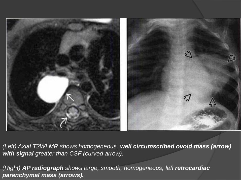
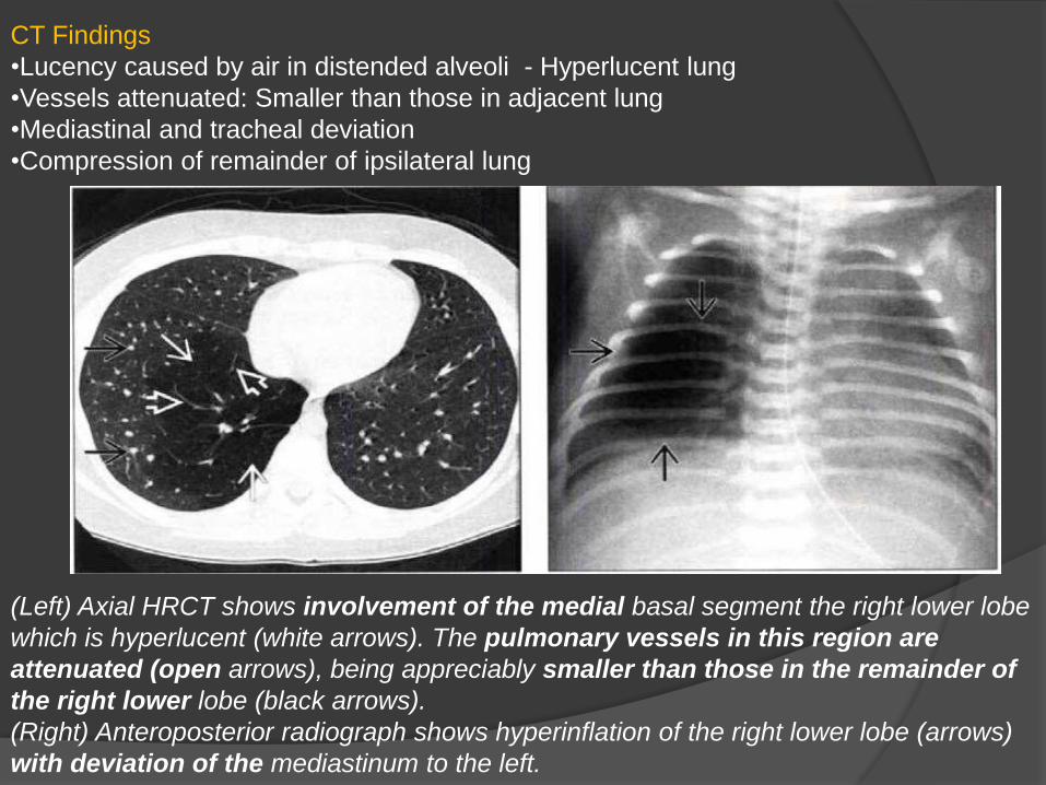
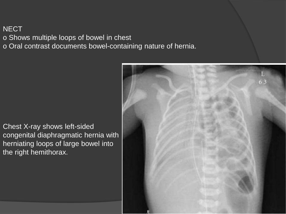
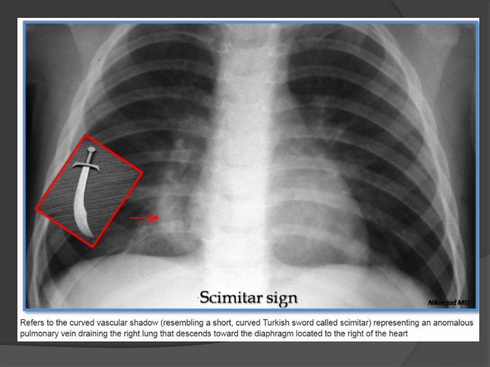
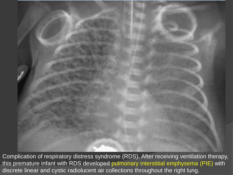
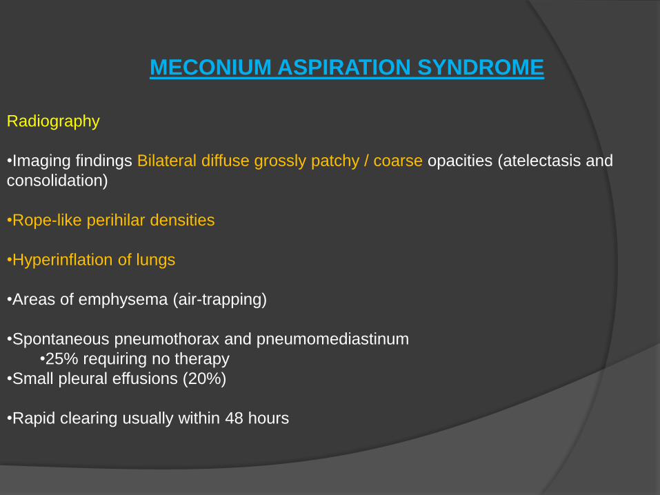
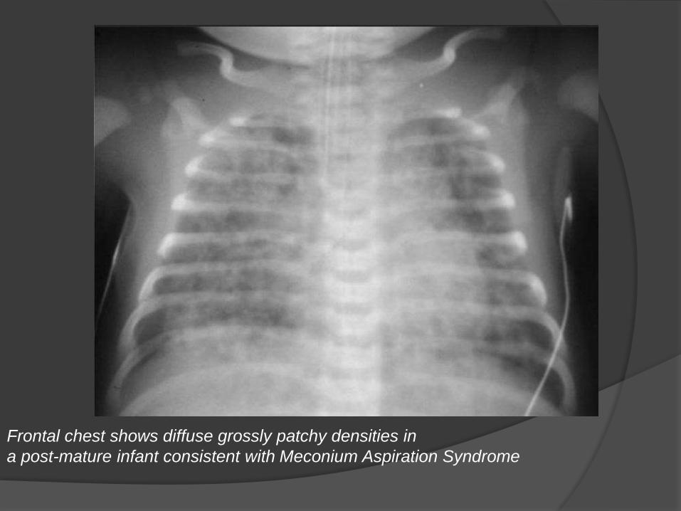
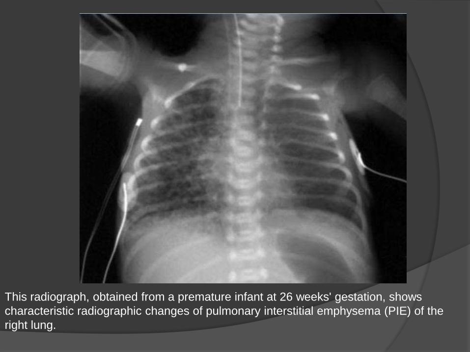
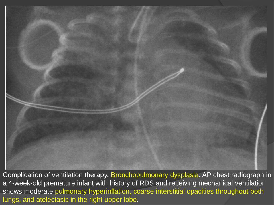
Thank U


