39
Pediatric Fractures & Case Study Dr. Joseph P. McCormick AMG Orthopedic Surgeon
| Date post: | 09-May-2018 |
| Category: |
Documents |
| Upload: | nguyencong |
| View: | 221 times |
| Download: | 2 times |
Pediatric Fractures & Case Study
Dr. Joseph P. McCormick AMG Orthopedic Surgeon
Overview • Unique pediatric traits in fractures
– Salter-Harris classification in physeal fractures
• Wrist injuries • Elbow fractures • Ankle fractures • SCFE
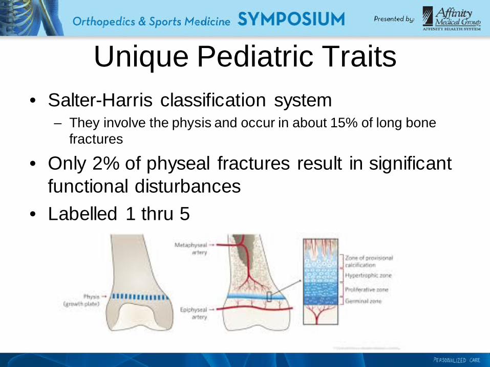
Unique Pediatric Traits • Salter-Harris classification system
– They involve the physis and occur in about 15% of long bone fractures
• Only 2% of physeal fractures result in significant functional disturbances
• Labelled 1 thru 5
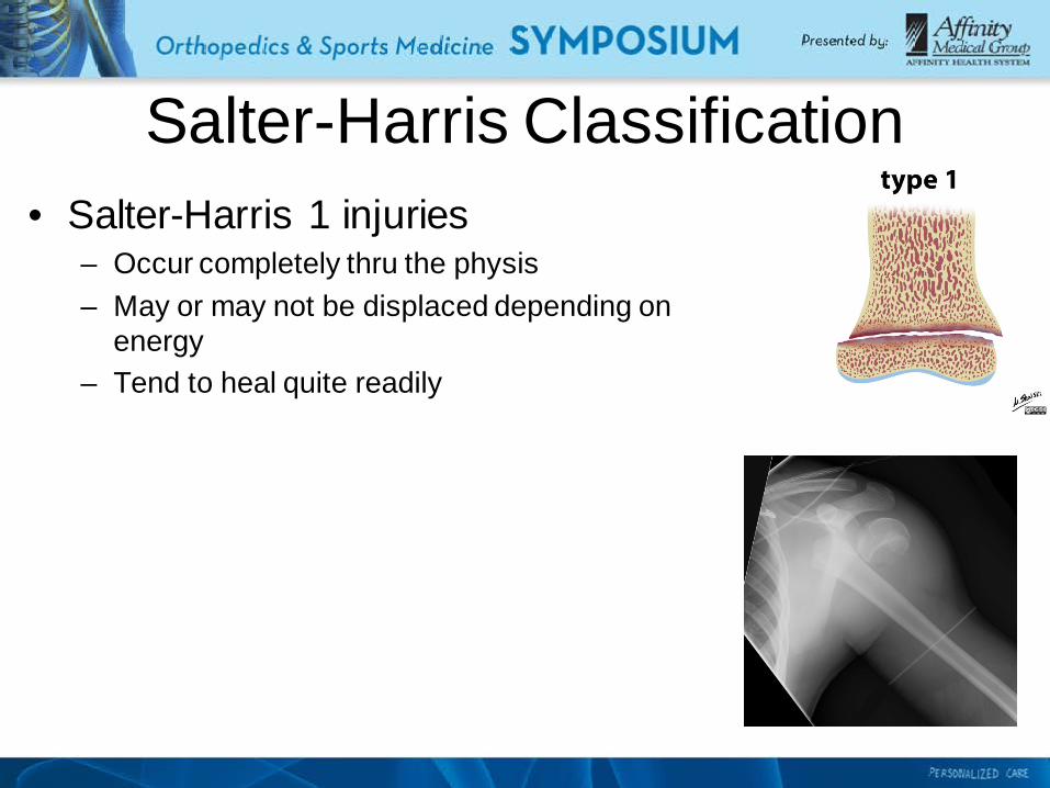
Salter-Harris Classification • Salter-Harris 1 injuries
– Occur completely thru the physis – May or may not be displaced depending on
energy – Tend to heal quite readily

Salter-Harris Classification • Salter-Harris 2 injuries
– Occurs across the physis with exit thru the metaphysis
– May or may not be displaced depending on energy
– Very common version of S-H fractures – Tend to heal quite readily
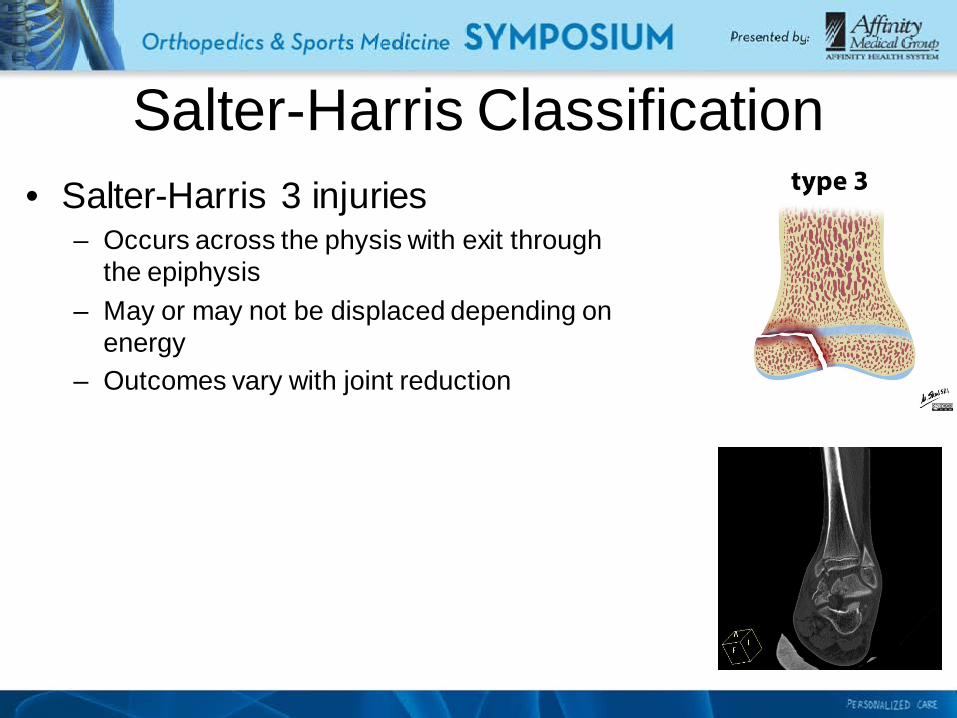
Salter-Harris Classification • Salter-Harris 3 injuries
– Occurs across the physis with exit through the epiphysis
– May or may not be displaced depending on energy
– Outcomes vary with joint reduction

Salter-Harris Classification • Salter-Harris 4 injuries
– Crosses the metaphysis & epiphysis – Through the physeal plate and intra-articular – Least common – Results depend on joint reduction

Salter-Harris Classification • Salter-Harris 5 injuries
Through the physeal plate – Difficult to diagnose – Usually discovered retroactively – Premature physeal closure can be seen
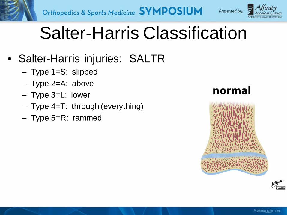
Salter-Harris Classification • Salter-Harris injuries: SALTR
– Type 1=S: slipped – Type 2=A: above – Type 3=L: lower – Type 4=T: through (everything) – Type 5=R: rammed
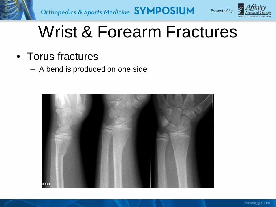
Wrist & Forearm Fractures • Torus fractures
– A bend is produced on one side

Wrist & Forearm Fractures • Torus fractures
– Typically from a fall; pediatric bones (thicker periosteum) allows this to occur
– Heals in a cast, 3-6 weeks – When the cast is removed, the wrist stiffness will go away in 10-
14d – PT is not needed – Parents are urged to avoid playground structures for 3-4 weeks
after

Wrist & Forearm Fractures • Greenstick fractures
– An incomplete fracture pattern
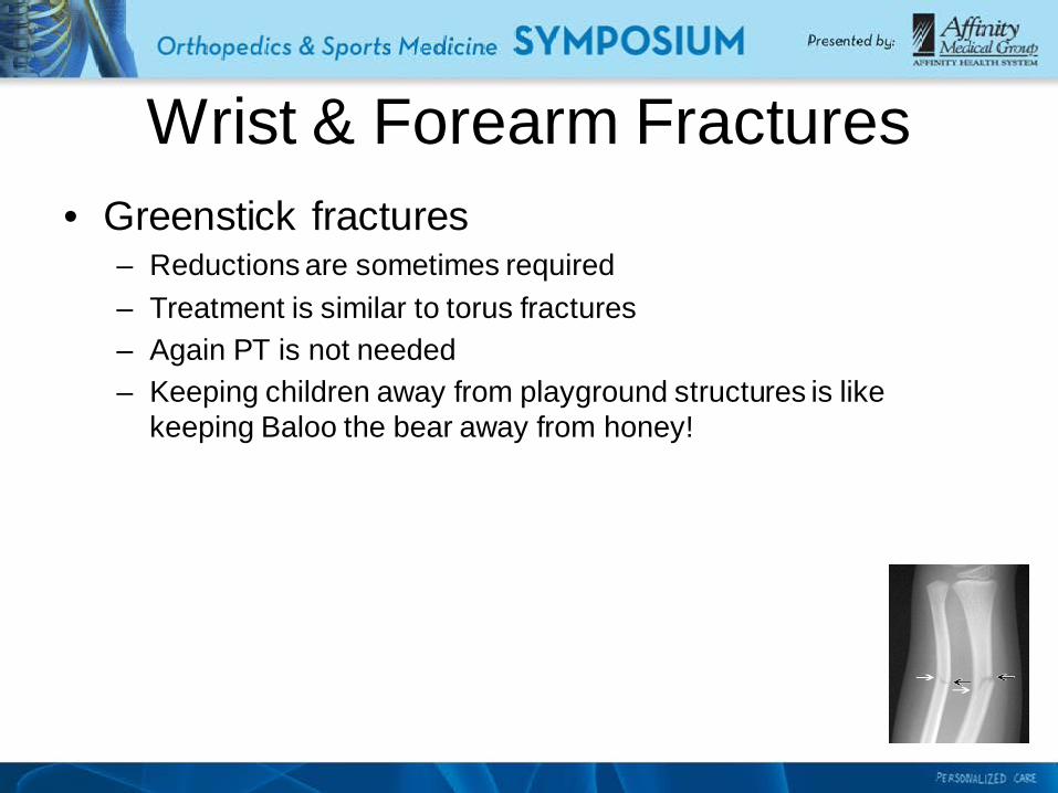
Wrist & Forearm Fractures • Greenstick fractures
– Reductions are sometimes required – Treatment is similar to torus fractures – Again PT is not needed – Keeping children away from playground structures is like
keeping Baloo the bear away from honey!
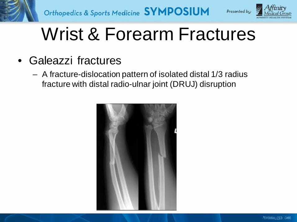
Wrist & Forearm Fractures • Galeazzi fractures
– A fracture-dislocation pattern of isolated distal 1/3 radius fracture with distal radio-ulnar joint (DRUJ) disruption
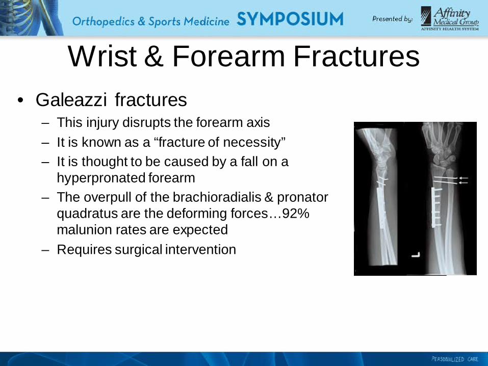
Wrist & Forearm Fractures • Galeazzi fractures
– This injury disrupts the forearm axis – It is known as a “fracture of necessity” – It is thought to be caused by a fall on a
hyperpronated forearm – The overpull of the brachioradialis & pronator
quadratus are the deforming forces…92% malunion rates are expected
– Requires surgical intervention

Wrist & Forearm Fractures • Monteggia fractures
– A fracture-dislocation pattern of the proximal ulnar shaft & radial head dislocation
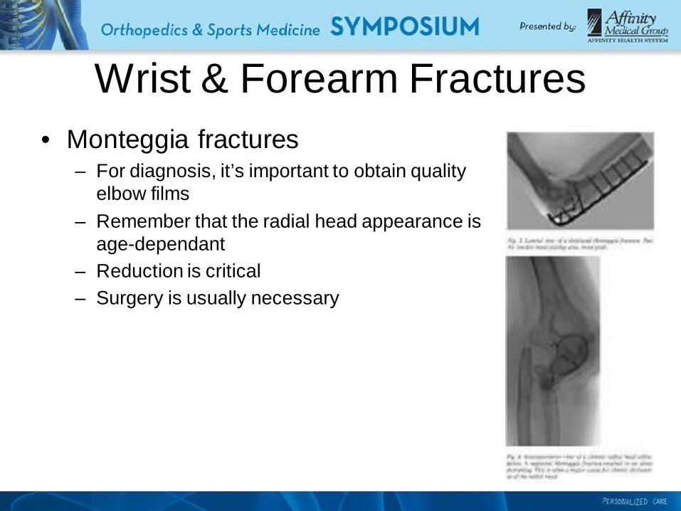
Wrist & Forearm Fractures • Monteggia fractures
– For diagnosis, it’s important to obtain quality elbow films
– Remember that the radial head appearance is age-dependant
– Reduction is critical – Surgery is usually necessary
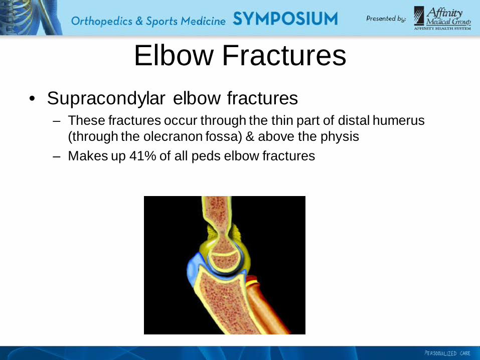
Elbow Fractures • Supracondylar elbow fractures
– These fractures occur through the thin part of distal humerus (through the olecranon fossa) & above the physis
– Makes up 41% of all peds elbow fractures
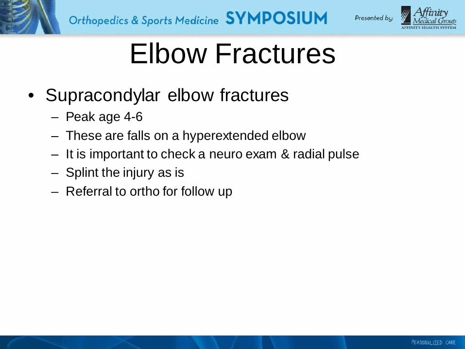
Elbow Fractures • Supracondylar elbow fractures
– Peak age 4-6 – These are falls on a hyperextended elbow – It is important to check a neuro exam & radial pulse – Splint the injury as is – Referral to ortho for follow up
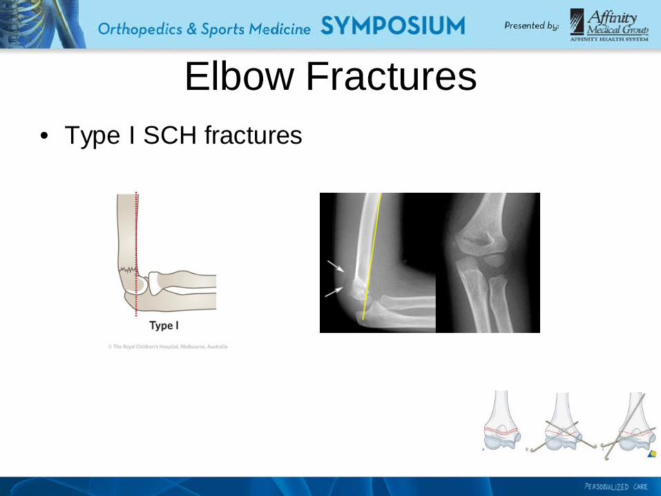
Elbow Fractures • Type I SCH fractures

Elbow Fractures • Type II SCH fractures

Elbow Fractures • Type III SCH fractures

Elbow Fractures • Medial epicondyle fractures
– The injury mechanism is typically a large valgus stress – About ½ are associated with posterior elbow dislocation – 14% of all peds elbow fractures
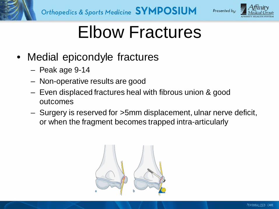
Elbow Fractures • Medial epicondyle fractures
– Peak age 9-14 – Non-operative results are good – Even displaced fractures heal with fibrous union & good
outcomes – Surgery is reserved for >5mm displacement, ulnar nerve deficit,
or when the fragment becomes trapped intra-articularly

Elbow Fractures • Lateral condyle fractures
– Highly missed diagnosis – Remember, these generally represent a S-H 4 fracture pattern – Use 4V elbow radiographs (important to check the obliques) – 17% of all peds elbow fractures
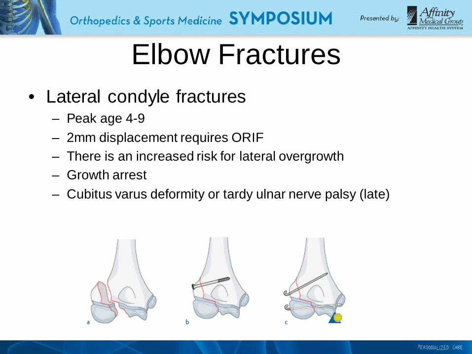
Elbow Fractures • Lateral condyle fractures
– Peak age 4-9 – 2mm displacement requires ORIF – There is an increased risk for lateral overgrowth – Growth arrest – Cubitus varus deformity or tardy ulnar nerve palsy (late)
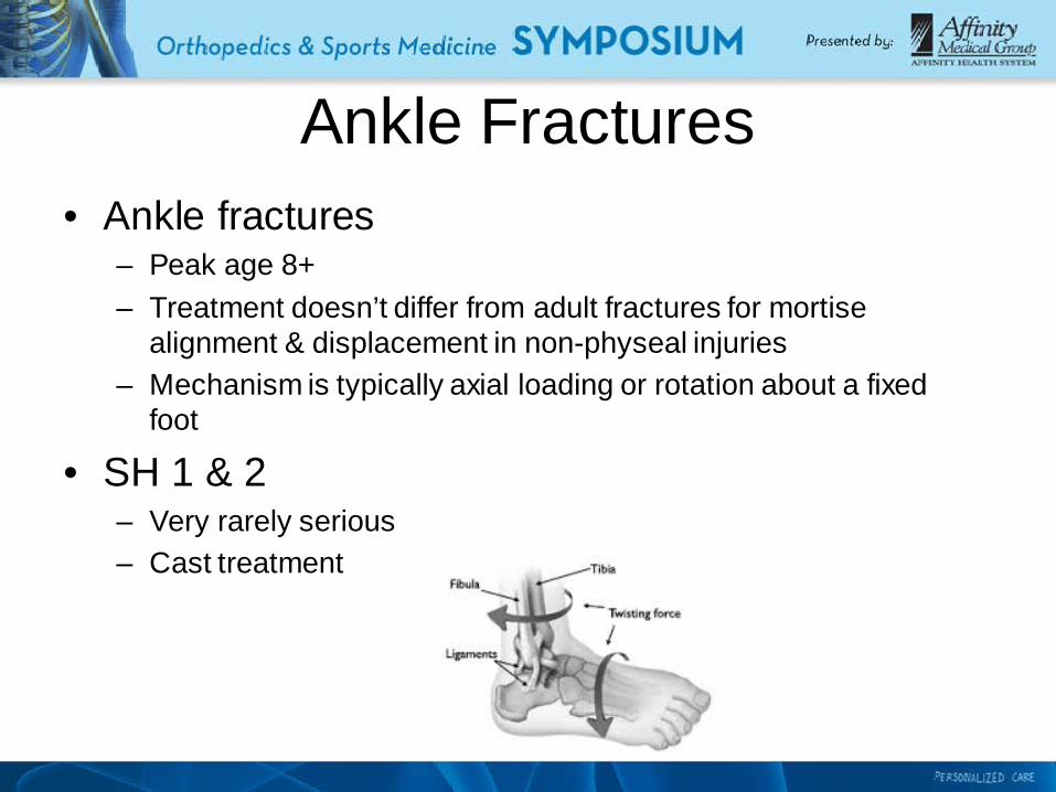
Ankle Fractures • Ankle fractures
– Peak age 8+ – Treatment doesn’t differ from adult fractures for mortise
alignment & displacement in non-physeal injuries – Mechanism is typically axial loading or rotation about a fixed
foot
• SH 1 & 2 – Very rarely serious – Cast treatment

Ankle Fractures • SH 3: Tillaux fracture
– Represents an anterior tibiofibular avulsion – Higher risk for growth plate disturbance – Requires an anatomic reduction – K wire or screw fixation

Ankle Fractures • SH 4: Triplane fracture
– Higher energy torsional fracture – Higher risk for growth plate disturbance – Requires an anatomic reduction – K wire or screw fixation
Slipped Capital Femoral Epiphysis
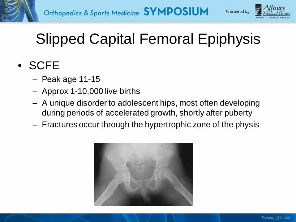
• SCFE – Peak age 11-15 – Approx 1-10,000 live births – A unique disorder to adolescent hips, most often developing
during periods of accelerated growth, shortly after puberty – Fractures occur through the hypertrophic zone of the physis

Slipped Capital Femoral Epiphysis
• SCFE – Slips are acute or subacute (stable or unstable) – 20+% are missed at the first presentation to a medical facility – Good long term outcomes if caught early – At least 1 in 5 are eventually bilateral

Slipped Capital Femoral Epiphysis
• Other risk factors – Endocrine disorders (inc Type I Diabetes) – Renal disease – Cancer treatments – Corticosteroid use
• Diagnosis – 2-3x more common in high BMI males – Pain in the hip or knee – Waddling gait – Restricted IR on exam
Slipped Capital Femoral Epiphysis
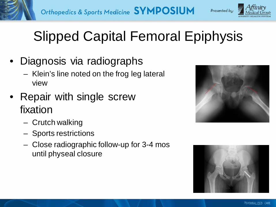
• Diagnosis via radiographs – Klein’s line noted on the frog leg lateral
view
• Repair with single screw fixation – Crutch walking – Sports restrictions – Close radiographic follow-up for 3-4 mos
until physeal closure
Case Study • C.H., a 12 y/o M.S. boy
– Limping – Diagnosis via radiographs – Urgent screw fixation, 2003 – C.H.’s gym teacher required a note – Healing & eventual opposite hip ORIF 2005
• Local High School – 6’3” 284lb offensive lineman, sev state championships & second
team all-state – state wrestling runner-up, heavyweight

Case Study • HWR • UW-
– 270lb OLB, played in the Rose Bowl – Engineering, post-grad honors – Lost 60lbs – Married

Conclusion • Remember SALTR
– Five types of Salter-Harris fractures
• Torus & greenstick wrist fractures – Unique to peds – Heal similarly
• Galeazzi & Monteggia – Require urgent ortho referral
• SCH elbow fractures – Type I-III – Usually splint as is and consult ortho
Conclusion • Medial epicondyle fractures
– Are elbow dislocation variants – Check the ulnar nerve
• Lateral condyle fractures – Can be missed easily – Often described on the phone to me as “something just not right”
• Ankle Fractures – SH 1&2 are low energy & stable – Special consideration for SH 3&4
Conclusion • SCFE
– Higher BMI males with insidious onset hip pain – Waddling limp – May present as knee pain – Good results are expected with early diagnosis & treatment

Pediatric Fractures


