Page 1
1
Pediatric Obstructive Sleep Apnea Syndrome
Christian Guilleminault MD.BiolD.1 Ji Hyun Lee MD1, Allison Chan DO1
Stanford University Sleep Disorders Program, Stanford, CA1
Corresponding author:
Christian Guilleminault MD.BiolD
Stanford University Sleep Disorders Clinic
401 Quarry Rd suite 3301
Stanford, CA, 94305
Phone: 1 650 723 6601
Fax: 1 650 725 8910
[email protected]
Page 2
2
ABSTRACT
Objective To review evidence-based knowledge of pediatric obstructive sleep apnea syndrome
(OSAS).
Methods Review of published articles regarding pediatric OSAS; extraction of clinical symptoms,
syndromes, polysomnographic findings and variables, and treatment options; and authors’
recommendations.
Results Many complaints and syndromes are associated with pediatric OSAS. This diagnosis
should be considered in patients who report the presence of such symptoms and syndromes.
Orthodontic and craniofacial abnormalities related to pediatric OSAS are commonly ignored despite
their impact on public health. One area of controversy involves the use of a Respiratory Disturbance
Index (RDI) to define various pathologies, but apneas and hypopneas are not the only abnormalities
obtained on polysomnograms, which can be diagnostic for sleep disordered breathing.
Adenotonsillectomy is often considered the treatment of choice for pediatric OSAS. However,
many clinicians may not discern which patient population is most appropriate for this type of
intervention; the isolated finding of small tonsils is not sufficient to rule out the need for surgery.
Nasal CPAP can be an effective treatment option, but it entails cooperation and training of both the
child and the family. A valid but often overlooked alternative, orthodontic treatment may
complement adenotonsillectomy.
Page 3
3
Introduction
Understanding obstructive sleep apnea syndrome (OSAS) in children requires knowledge of
the physiology of sleep and breathing. There is an immediate increase in upper airway resistance
with sleep onset, with an initial “overshoot” in this resistance that decreases very quickly. Still, this
resistance during established sleep is mildly higher than during wakefulness.1 There is also a slight
decrease in tidal volume with sleep. This decrease will be more pronounced with the occurrence of
REM sleep. These mild decreases will be compensated by a very slight increase in breathing
frequency to keep minute ventilation normal. Breathing frequency decreases during the first two
years of life but stays the same thereafter; it has been calculated to be between a maximum of 16 to
18 breaths per minute in NREM sleep and 17 to 19 breaths per minute during REM sleep.2-3
The obesity epidemic, evident in the United States and industrialized countries, has
complicated the investigation of obstructive sleep apnea and related syndromes. Fat distribution
varies depending on genetic, gender, and hormonal patterns as well as the inherent relationship
between these three factors. It is common for the fat to deposit in the abdominal region. Such
abdominal obesity will lead to chest-bellows impairment, as seen in restrictive thoracic disorders.
Although it may not lead to upper airway obstruction, abdominal obesity may worsen the poor gas
exchange that may already exist because of OSAS. Sleep will always worsen the gas exchange in
these subjects when they are in the supine position and when they achieve REM sleep. During REM
sleep, the associated atonia eliminates contractions of the accessory respiratory muscles and the
abdominal muscles, which engage in active expiration.2-3 REM sleep is also associated with further
flattening of the diaphragm’s position.2 These physiologic changes worsen gas exchange in subjects
with abdominal obesity and may even lead to REM sleep-related hypoventilation with some degree
of carbon dioxide (CO2) retention. Upper airway impairment, per se, is not directly related to this
CO2 retention. It has, however, been hypothesized that abnormal gas exchange during sleep may
impair the coordination of time-related contractions of both upper airway dilator muscles and
inspiratory muscles.
Page 4
4
OSAS was described in children in 1976.4 Although children may present with OSAS, the
literature had established, by 1982, that children had other abnormal respiratory effort patterns
during sleep that were frequently associated with snoring and clinical symptoms.5
Epidemiology
There is no definitive population-based study evaluating the presence of OSAS in children.
Previous studies were performed in different settings and implemented a variety of tools. Some
considered regular nocturnal snoring as a marker of chronic obstructive breathing during sleep. The
percentage of individuals younger than 18 years of age who have been reported with regular heavy
snoring oscillated between 8 and 12%. Other studies polygraphically monitored subjects but were
limited in terms of sample size and testing difficulties; initial studies estimated OSAS prevalence
between 1 and 3%.6-15 More recently, many specialists have quoted the OSAS prevalence between 5
and 6%. Although better monitoring techniques during polysomnography have shown that more
abnormal breathing events are present,16 the definitive data are still lacking.
Clinical symptoms
Abnormal narrowing in the nose, nasopharynx, oropharynx, or hypopharynx causes
abnormal air exchange during sleep, which in turn leads to clinical symptoms. These symptoms will
vary with age. Recognition of the problem is often only noted in older children, who are able to
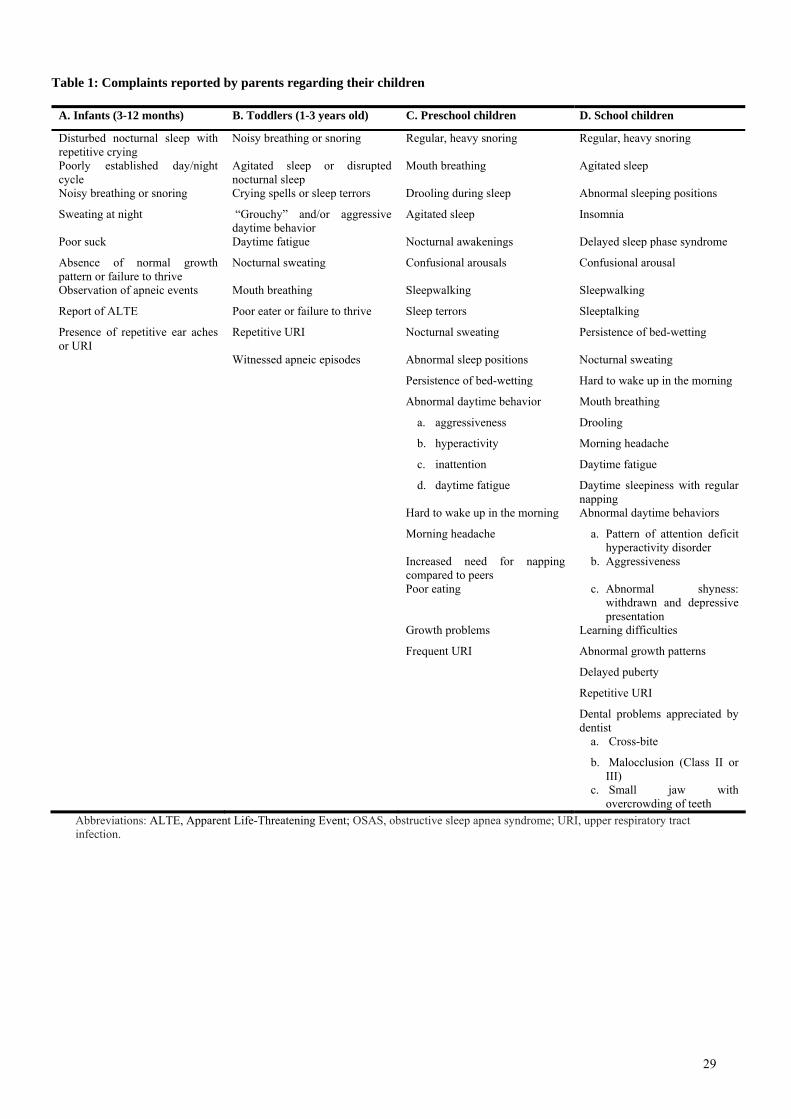
articulate complaints. Table 1 (A-D) indicates the parental complaints of children seen at sleep
clinics over time.17-25
Abnormal breathing during sleep has been associated with specific clinical problems and
findings. The clinical interview of a child suspected of having sleep disordered breathing (SDB)
must lead to systematic questioning of the parents regarding their child’s symptoms; the parents
may not associate the occurrence of these symptoms with abnormal breathing during sleep. Table 2
outlines syndromes that have been shown to be related to SDB and are subsequently controlled after
the appropriate treatment of the breathing disorder has been initiated.20, 24, 27-51 Interestingly, some
of the syndromes are related to the maxillomandibular development and are more connected to
Page 5
5
orthodontic practice. Pediatricians do not traditionally consider orthodontic problems as part of a
child’s health issues, but in light of the related health cost and syndromic association, they should.
Clinical Evaluation and Diagnosis of Sleep Disordered Breathing
SDB in a child will be suspected based on the parental complaints. The presence of one of
the syndromes listed in Table 2 should lead to a thorough interview of the behavior during sleep as
well as sleep-related factors associated with SDB.17-50
The suspicion of SDB indicates the need for not only a general pediatric evaluation but for a
thorough evaluation of the upper airway anatomy. Clinically, it involves a comprehensive
examination of its successive segments. Starting with the nose, one should look for asymmetry of
the nares, a large septal base, collapse of nasal valves during inspiration, a deviated septum or
enlargement of inferior nasal turbinates (Figure1). Next, the oropharynx should be examined for the
position of the uvula in relation to the tongue. The Mallampati scale may help evaluate this
position.52 The size of the tonsils should be compared to the size of the airway; application of a
standardized scale is useful.53 The presence of a high and narrow hard palate, overlapping incisors,
cross-bite, and an important (> 2 mm) overjet (the horizontal distance between the upper and lower
teeth) are indicative of a small jaw and/or abnormal maxillomandibular development. This clinical
evaluation provides important details of the upper airway anatomy and identifies anatomical risk
factors which can predispose one to developing abnormal breathing during sleep.
The results of this examination must be summarized, as the different anatomical narrowings
have additive effects. The apparent sizes of tonsils and adenoids are not the only anatomical
findings which determine whether or not sleep disordered breathing is present. Change in flow due
to an abnormal nose, secondary development of turbulence and the increased collapsibility at
specific vulnerable points in the upper airway are elements to consider.
There is a complex interaction between nasal breathing and maxillomandibular growth.
Abnormal nose breathing in very young individuals leads to an increase in nasal resistance and
Page 6
6
mouth breathing with secondary impairment of maxillomandibular growth54-62 as shown
experimentally in young rhesus monkeys.63 The first four years of age are of particular importance,
as 60% of the adult face is built during that period.64 Otolaryngologic and orthodontic data have
clearly demonstrated the impact of enlarged tonsils, adenoids, enlarged nasal turbinates, and upper
airway allergies on orofacial growth in children.21,55--70
Other factors may be considered. Neck circumference and the presence of fatty infiltration
should be noted, but no scale correlates neck circumference with age or pathology. The overall
aspect of the face can be appreciated. The frontal aspect of the face can be subdivided into superior,
middle, and inferior portions. They are approximately the same length in a normal child. The upper
part of the bridge of the nose and the part just below the nare represent the middle third of the face.
In individuals with a maxillomandibular risk factor for OSA, the lower third of the face may be
longer than expected. The terms “long face” and “long face syndrome” have been used.21, 26
Objective confirmation of SDB
Testing during sleep is the only way to affirm the presence of SDB. There is controversy
over the need for and type of test to be performed. Some of the measures employed for this testing
include questionnaires and scales, home monitoring, and polysomnograms (PSGs).71-74
Questionnaires with specific emphasis on the common symptoms associated with SDB have
been implemented. Although questionnaires may be helpful in directing the attention of parents to
the diurnal and nocturnal symptoms of SDB, the sensitivity and specificity of questionnaires are not
sufficient for affirming the presence of SDB.23,75-77
Home monitoring with or without videotaping has also been used. Ambulatory monitoring
with recording of cardiac and respiratory variables has been suggested as the first diagnostic step in
testing for SDB. These devices can detect the presence of oxygen saturation (SaO2) drops, apneas,
and hypopneas; affirm the diagnosis of SDB; and lead to treatment. Associated videotaping may
confirm abnormal breathing behavior. This approach may recognize severe SDB but fails to identify
Page 7
7
the presence of associated sleep disorders and partially obstructed breaths. A negative test does not
rule out the diagnosis of SDB and must be followed by a PSG; however, a positive finding may lead
to faster treatment.78-80
A PSG is the only test that may exclude the diagnosis of SDB. It must always include
monitoring of sleep/wake states through electroencephalogram (EEG), electro-oculogram, chin and
leg electromyography (EMG), ECG, body position, and appropriate monitoring of breathing. Nasal
cannula-pressure transducer, oral thermistor, chest and abdominal belts, neck microphone, and
pulse oximetry are recommended, but variable montages are used.
Respiratory efforts can be investigated by a variety of means during the PSG. Although
infrequently used, the best approach involves measuring esophageal pressure (Pes) movements. A
less reliable approach is to monitor intercostal/diaphragmatic EMG. A recently developed analysis
of this signal appears promising but needs further testing in children.81 CO2 may be monitored using
a nasal cannula with measurement of end-tidal CO2. But the combination of two cannulas in the
nose of a child may disturb sleep and negatively impact nasal breathing; thus, a transcutaneous CO2
electrode will often be needed for this measurement.16, 82-83
SDB has consequences related to the repetitive changes induced by a decrease in size of the
upper airway during sleep. As a compensatory first step, there will be an increase in breathing
frequency (tachypnea) and an increase in respiratory efforts.5,84-85 The selected response is related to
both the decrease in size of the upper airway as well as the age of the subject. Following the classic
“breathing frequency × tidal volume = minute ventilation,” tachypnea is a more common finding in
young children with small and relatively unstable chests; this population has mild to moderate
breathing impairment during sleep.5, 84 Despite better chest stability, this response will also be seen
in older children. Tachypnea and an increase in inspiratory efforts have been seen in the same
children in association with airflow limitation. The mechanisms behind a specific response and the
relationship with sleep state are unknown.
Page 8
8
The repetitive challenges resulting from a reduction of upper airway size have negative
consequences on a child’s well-being. However, the normative data for many of the studied
variables are still unclear. The polygraphic normative data on sleep duration and sleep stages are
available in children aged 7 years and older.86 But the frequency of short arousals during sleep (i.e.,
EEG arousals lasting ≥ 3 seconds that can be reliably scored by 3 years of age87) is unknown for
different age group. But abnormal breathing patterns during sleep have been identified (Table 3).85
Interpretation of the PSG
There are controversies concerning PSGs,71,84 because many existing criteria are based on
information obtained from small studies. Other recommendations were taken “by consensus,”
which means they were not necessarily based on data; still others were based on information
collected with outdated technology. The specificity and sensitivity of many indices used have never
been calculated. Only one study has looked at polygraphic respiratory patterns; their frequency of
occurrence; their change in frequency with treatments; and the impact on the clinical outcome
associated with polygraphic changes in pre-pubertal children.84
One of the most debated issues today is what type of respiratory event should be scored and
tabulated. Another issue is determining when “pathology” is present.23,76,84 Historically, the
presence of OSA was easy to recognize with simple albeit relatively insensitive equipment
(thermistors). Based on the variability of breathing frequency between birth and two years of age,
an “apnea” was defined as “longer than 2 breaths.” For many years, there was a consensus that
OSA, a complete cessation of air exchange at nose and mouth, was associated with clinical
symptoms. It was shown that removal of the obstructive apnea (OA) led to improvement of the
symptoms. Initial criterion for an abnormal finding with polysomnography was “≥ 1 OA/hour of
sleep.”88
But pathology was also seen without complete absence of air exchange. To improve the
scoring system, clinicians used the term “hypopnea,” but there is no consensus of what a
“hypopnea” is. Following adult criteria and utilizing thermistors with limited sensitivity, clinicians
Page 9
9
suggested that a “hypopnea” should last “longer than 2 breaths.” Also, the airflow signal from the
combined nasal/oral thermistors should decrease by at least 50% of normal baseline breathing.
Hypopneas should be terminated with either an EEG arousal or a drop of SaO2 of at least 3%.23, 76
Using these criteria, pathology was considered to be present if the OA index was ≥ 1 or if the AH
index (AHI) was ≥ 5 events/hour.
Some children with very noisy breathing at night and enlarged tonsils and adenoids had a
normal score at PSG, but had clinical symptoms89,90 that led to adenotonsillectomy. Also, other
sleep-disordered breathing syndromes without an associated abnormal AHI but with an elevated
“respiratory disturbance index” were controlled with nasal CPAP or upper airway surgery.85
Although an AHI ≥ 5 was considered pathological, there was the recognition that “apnea and
hypopnea” as defined did not encompass all pathological breathing during sleep. Hence, an arousal
index was calculated; thus, snoring sequences which were terminated with an EEG arousal were
scored. The association of apnea-hypopnea and other measurements led to the usage of the term
“respiratory disturbance index (RDI).” This term acknowledges that the defined PSG patterns did
not encompass all abnormal breathing events.
The introduction of the nasal cannula/pressure transducer system16,91 allowed a more
accurate recognition of abnormal breathing during sleep, as this technique based on nasal flow is
semi-quantitative. It allows better recognition of partially obstructed breaths. A “hypopnea” was
defined when flow decreased by 30% of a normal breath. But many still require an “EEG arousal”
and/or an SaO2 drop, despite prior demonstration that clinical consequences can be obtained without
a change in SaO2. An RDI > 5 events was used based on prior habits.
A minority of sleep clinics monitored respiratory efforts using esophageal pressure (Pes).
These groups showed that snoring without “hypopneas” was associated with abnormal efforts and
an induction of EEG arousals. Based on Pes recordings,83,85 specific patterns were recognized and
defined, such as “Pes crescendos,” “sustained respiratory effort,” and “Pes reversals.” Some
evidence suggests that these patterns were frequently, but not always, seen with abnormal nasal
Page 10
10
flow on the nasal cannula/pressure transducer recording. But a “flow limitation” between normal
and a 30% decrease at the nasal cannula was usually seen with these patterns. The nasal flow
limitation was described as a “flattening” of the nasal flow curve; several patterns of abnormal
curves have been described. It may be easier to visually recognize a change of the Pes pressure than
a “flattening of the nasal curve.”83-85
Application of these Pes-related definitions showed that children who had no
apneas/hypopneas, SaO2 drop of 3% or more, or EEG arousals presented with clinical complaints
and clinical sleep-related syndromes, primarily parasomnias.32-33 Applying the above criteria, a
clinical outcome study performed at the Stanford University Sleep Disorders Clinic focused on
clinical complaints and the presence of clinical symptoms and signs. Complete treatment of the
sleep-related upper airway problem with resolution of symptoms and signs was associated with less
than one of the events included in the RDI.84 Persistence of symptoms and signs was associated
with the continued presence of an “event” that was not necessarily an “apnea” or a “hypopnea.”
Instead, the breathing event was noted to be either a “flow limitation with an increase in respiratory
effort” or merely an increase in respiratory effort; a cut-off point for RDI at ≥ 1.5 events/hour of
sleep was found.84 But an RDI ≥ 1.5 events/hour is based on only one outcome study, even if
several clinical studies have indicated the validity of such a cut-off point.32,45,84
Changes in autonomic nervous system (ANS) and breathing patterns during sleep
An increase in respiratory efforts is associated with changes in ANS settings. These changes
will affect the cardiovascular system in an individual with a normal ANS.49 One may want to
evaluate these changes to recognize an abnormal pattern and determine if they may be detrimental.
Two types of responses can be seen when an increase in respiratory effort occurs during sleep:
“activation” or “arousal with cortical involvement.”
“Activation” is a clinical neurophysiology term defined by Moruzzi92 during the course of
his study of the “ascending reticular formation;” it is related to the recruitment of sensory inputs
Page 11
11
that will lead to a polysynaptic motor response after relay of sensory input in the brainstem and
subcortical structures. The nucleus ambiguous receives information that simultaneously leads to
efferent responses through the nucleus tractus solitarius (NTS). This relay leads to a simultaneous
ANS stimulation, and an autonomic activation will lead to an increase in sympathetic tone.
An ANS response may be seen with brainstem reflexes leading to full reopening of the
upper airway without EEG cortical arousal, or it may be seen as the consequence of an EEG cortical
arousal. The presence of cortical arousals will be associated with clinical symptoms, e.g.,
complaints of excessive daytime somnolence, irritability, or unrefreshing sleep. The role of
repetitive “activation” is unknown in children.
The determination of how much airway size change and the duration of the change needed
to lead to cortical arousal are unknown. Sleep stages may play a role in the type of response seen,
but no definitive information is available in prepubertal children.
The pulse transmit time (PTT), which measures the transit time of the pulse wave from
approximately the aortic valve to the wrist, and the peripheral arterial tonometry (PAT), are two
variables that were added to polysomnography to help recognize an “arousal”.93-96- None of these
devices can distinguish between a brainstem reflex and a cortical arousal response. The importance
of the sympathetic response could be a relatively accurate indicator of cortical involvement, but the
studies to validate such distinction have not yet been published. Based on a commercially designed
algorithm involving both heart rate and finger plethysmography, the PAT does not really reflect the
balance between the sympathetic and parasympathetic systems during sleep. The PTT also has
limitations of interpretation. When used to identify cortical arousals related to SDB, both techniques
have false positives and false negatives, which limit the accuracy of interpretation.96 Monitoring of
these different variables has, however, shown that repetitive snoring can be associated with
activation and/or EEG arousal.
Page 12
12
Changes in EEG sleep patterns with re-opening of upper airway
Historically, an EEG alpha or alpha and beta arousal lasting three seconds at the termination
of an abnormal breathing event was requested to score an “event.” But several studies have shown
that limited upper airway occlusion may end with a burst of delta waves or a K complex.97 The
usage of a sleep scoring system, based on analysis of the “cyclic alternating pattern” (CAP),
demonstrated the negative effect of these bursts.98-100 The CAP scoring system is based on recurrent
bursts of delta and K complexes with or without superimposed alpha waves within a time period of
60 seconds intertwined with low EEG amplitude. CAP is a normal phenomenon that occurs
between wakefulness and slow wave sleep (SWS) or, at the end of night, between REM sleep and
well-established, repetitive sleep-spindle sleep. It indicates a transition from one stable state to
another stable state and is not seen in REM sleep. CAP is typically a transient period during which a
greater instability of sleep may occur with greater chance to enter a light sleep or even to awaken.
An abnormal CAP rate, defined in different age groups in children,99-100 indicates an “instability of
NREM sleep” as well as a difficulty in reaching a new stable state.98 CAP is associated with
autonomic activation and may lead to awakening and large sympathetic discharge. Chervin et al 101-
102 have also reported a novel approach to evaluate EEG with abnormal breathing during sleep,
based on an algorithm investigating the EEG changes seen with each abnormal inspiration
associated with increased effort. The algorithm recognizes the changes in brain wave activity with
increased inspiratory effort. When adenotonsillectomy is successful in relieving abnormal breathing
during sleep, the abnormal EEG pattern disappears. Furthermore, the daytime symptoms,
particularly sleepiness, abate. This analytic technique needs to be tested further.
Genetic Risk Factors of SDB
Both genetic64,103 -111 and environmental risk factors have been identified in the development
of SDB; they are associated to variable degrees. Oral mucosa thickness has been identified as an
ethnic risk factor in African-Americans, and skull base length has been noted to be an ethnic risk
Page 13
13
factor in Far East Asians. -107-108 African-American and Far East Asian populations have been
shown to have significantly higher risk than Caucasians when age, sex and BMI were
considered.64,103-111 The familial trait of dolichocephaly (or narrow face) has also been implicated as
a risk factor, independent of ethnicity.51,110 Familial cases of SDB are seen in all ethnic groups.
Genetic investigations are performed although there is currently no clear indication for a specific
gene location responsible for increased risk. The strongest current indicators have been related to
facial morphotype.64 Clearly, there is an increased risk of SDB in a family in which a member is
affected.103-105,109-111 Pediatricians should, therefore, systematically question other family members
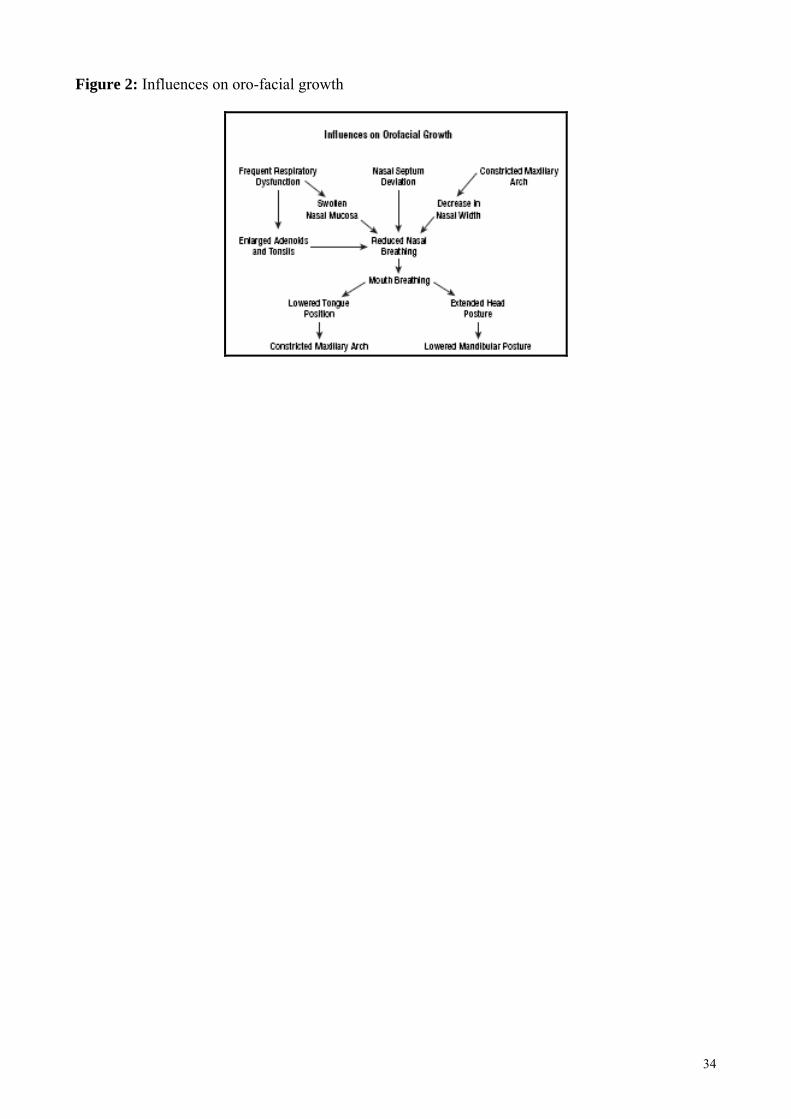
about sleep-related problems when there is a positive history of SDB (Figure 2).
Treatment
There is an overall consensus that children with SDB should be evaluated by an
otolaryngologist for surgical treatment. It is also clear that the well-described but extremely
complex interaction between nasal breathing and facial growth is important, even if rarely
investigated.51
Treatment for short-term outcomes indicates that adenotonsillectomy with or without
radiofrequency (RF) treatment of nasal inferior turbinates is the first approach to consider.85,112-113
Independent of the size of tonsils or adenoids, adenotonsillectomy will provide more airway space.
Two points must be emphasized. First, outcome investigation has shown that isolated tonsillectomy
or adenoidectomy is not as effective as adenotonsillecomy.84,114 Also, RF of the nasal turbinates
should be performed at the same time as the child is under general anesthesia if enlarged turbinates
are present. Performance of adenotonsillectomy without performance of nasal turbinate treatment
may negatively impact the outcome.84 Outcomes of adenotonsillectomy have been reviewed,112 but
no review addresses the reasons for failure. A recent study examined the short-term outcomes to
understand why results were incomplete.114 Surgeons often utilize techniques that are not aimed at
maximally opening the airway; they may fail to treat the nose simultaneously with
Page 14
14
adenotonsillectomy; and others simply do not recognize the craniofacial changes that contributed to
the sleep disordered breathing.
Only two studies have looked at the long-term outcome of regular adenotonsillectomy
performed in prepubertal children.115-116 Evaluating outcome to a minimum of 10 years later, both
studies indicated that there was failure to control the problem due to the presence of hypopneas and
apneas at the long-term follow-up recordings. Demonstration of absence of apnea-hypopneas within
6 weeks to 3 months after surgery was requested in one of the two studies.115 The long-term
outcome in that study linked the recurrence of abnormal breathing during sleep to both the absence
of dealing with a narrow maxilla and/or mandible at the time of the initial surgery as well as the
later occurrence of tongue/mucosal enlargement at the time of puberty, when 90% of oro-facial
adult growth had already occurred.
Association of adenotonsillectomy with orthodontic treatment has been done.117 Rapid
maxillary distraction (RMD) is an orthodontic technique that is based on bone formation process. A
distractor anchored on two molars on both sides applies daily pressure, pushing apart the two half of
the maxilla; bone grows from the borders of the cartilage.117-118 This technique pushes the soft
tissues laterally, decreases the height of the soft palate and enlarges the nasal orifices.117 RMD may
be associated with distraction of the mandible, but as no mid-cartilage is present, there is very
limited widening. This fact may limit the degree of maxillary widening with RMD (Figures 1, 3 and
4). Slow maxillary distraction is based on similar principles and optimizes the degree of widening at
the different growth periods that occur in prepubertal children. Both rapid and slow maxillary
distractions are performed between 5 and 11 years of age. Distraction results in widening of both
the palate and the nose; thus, this procedure remedies nasal occlusion related to a deviated septum,
for which little can be done before 14-16 years of age. But even in association with
adenotonsillectomy, orthodontics may not control all SDB. Abnormal mandibular or
maxillomandibular anteroposterior development is a bigger challenge. Nasal CPAP will be the
recommended treatment until further orthognathic surgery119 can be performed.
Page 15
15
Home nasal CPAP has been used in infants, pre-pubertal children, and pubertal children.
The first report of its usefulness in children in 1986 was a prospective study that followed 5
children, age 3 to 11 years, for 10 months.120 Similar findings were reported in several large
retrospective studies.110,121-126 These studies primarily involved children older than 12 months of
age. Infants 8 to 18 weeks of age were followed from the onset of treatment through the first 12
months of age in a study in 1995;123 this was replicated in 1999.125
The difficulty in application of nasal CPAP relates to training of the family and child as well
as finding the appropriate nasal interface. Children often need to be trained to tolerate the facial
interface; behavioral modification techniques and daytime training may help with this training.
CPAP is very useful when the SDB is related to major craniofacial deformities or other illnesses. If
the upper airway problem is complicated by neuromuscular disease, nasal bilevel may be used.
Regular follow-up should be performed within the first three months to evaluate mask
fitting.123 Due to rapid craniofacial growth of young children, CPAP pressure should be evaluated
every 6 months. An annual craniofacial specialist visit should occur to affirm that headgear and
mask do not worsen a maxillary growth deficiency.127 Clinicians should encourage the use of
humidification, aggressively treat allergies and/or rhinitis and check nasal patency. In light of
children’s favorable response to surgery with or without orthodontic and anti-allergic treatment,
nasal CPAP should only be a second consideration.
Orthognathic surgery entails shifting bones and disrupting the bone growth structures.119
Such an approach is normally postponed until 10-13 years of age. Two surgical techniques used in
SDB patients include mandibular distraction osteogenesis and maxillomandibular advancement.
Mandibular distraction osteogenesis is very similar to RMD, but it is applied to the mandible
when a maxillary and a mandibular widening are needed and when the slow mandibular orthodontic
distraction cannot achieve the needed result.119 A vertical transection of the maxilla is performed
between the 2 central incisors, and a distractor is used as in RMD. Twelve to 14 mm of widening
can easily be obtained in three weeks. Orthodontic treatment is similar to the one described with
Page 16
16
RMD. At that age, both procedures can be simultaneously performed to provide an anterior
displacement of the tongue and enlargement of the retro-lingual airway space.119
Maxillomandibular advancement is a very successful procedure. Nevertheless, it is a major
surgery that should be performed after there has been appropriate orthodontic treatment. Surgeons
who perform this procedure must have a good understanding of upper airway mechanics as well as
dental problems. It may be performed at any time during childhood, but it is often post-pone till 11-
12 years of age.
A controversial issue is how early to perform adenotonsillectomy. Most will agree that
adenotonsillectomy is often performed by 24 months of age. But OSA has been noted as early as
three weeks of age, and cases of heavy snoring and clinical symptoms in children aged 6 to 24
months are actually common. Adenotonsillectomy has been performed as early as 6 months of
age.128
Several advances have been made in sleep medicine. Apneas and hypopneas are not the only
indicators of abnormal breathing during sleep. But in this rapidly evolving field, it has been
challenging to establish new scoring criteria, despite the availability of new technologies. However,
the clinical findings and the polysomnographic results should be used to determine the diagnosis
and guide treatment recommendations.
Page 17
17
References
1. Worsnop C, Kay A, Kim Y, Trinder J, Pierce R. Effect of age on sleep onset-related changes in
respiratory pump and upper airway muscle function. J Appl Physiol. 2000; 88: 1831-1839.
2. Orem J, Montplaisit J, Dement WC. Changes in the activity of respiratory neurons during sleep.
Brain Res. 1974; 82: 309-315.
3. Orem J. Control of the upper airway during sleep and the hypersomnia sleep apnea syndrome. In:
Orem J, Barnes CD, eds. Physiology in sleep. New York Academic Press; 1980: 273-313.
4. Guilleminault C, Eldridge FL, Simmons FB, Dement WC. Sleep apnea in eight children.
Pediatrics. 1976; 58: 23-30.
5. Guilleminault C, Winkle R, Korobkin R, Simmons FB. Children and nocturnal snoring:
evaluation of the effects of sleep related respiratory resistive load and daytime functioning. Eur J
Pediatr. 1982; 139,165-171.
6. Teculescu DB, Caillier I, Perrin P, Rebstock E, Rauch A. Snoring in French preschool children.
Pediatr Pulmonol. 1992; 13: 239-244.
7. Owen GO, Canter RJ, Robinson A. Snoring, apnoea and ENT symptoms in the paediatric
community. Clin Otolaryngol. 1996; 21: 130-134.
8. Lu LR, Peat JK, Sullivan CE. Snoring in preschool children: prevalence and association with
nocturnal cough and asthma. Chest. 2003; 124: 587-593.
9. Shin C, Joo S, Kim J, Kim T. Prevalence and correlates of habitual snoring in high school
students. Chest. 2003; 124: 1709-1715.
10. Urschitz MS, Guenther A, Eitner S, et al. Risk factors and natural history of habitual snoring.
Chest. 2004; 126: 790-800.
11. Castronovo V, Zucconi M, Nosetti L, et al. Prevalence of habitual snoring and sleep-disordered
breathing in preschool-aged children in an Italian community. J Pediatr. 2003; 142: 377-382.
Page 18
18
12. Amuntasaree W, Rookkapan K, Kuasirikul S, Thongsuksay P. Snoring, and obstructive sleep
apnea in Thai school-age children: prevalence and predictive factors. Pediatr Pulmonol. 2001; 32:
222-227.
13. Gislason T, Benediktsdottir B. Snoring, apneic episodes and nocturnal hypoxemia among
children 6 months to 6 years old: an epidemiologic study of lower limit of prevalence. Chest. 1995;
107: 963-966.
14. Brunetti L, Rana S, Lospalluti ML, et al. Prevalence of obstructive sleep apnea in a cohort of
1207 children from the south of Italy. Chest. 2001; 120: 1030-1936.
15. Rosen C, Larkin E, Kirchner H, et al. Prevalence and risk factors for sleep disordered breathing
in 8 to 11 year-old children: association with race and prematurity. J Pediatr. 2003; 142: 383-389.
16. Trang H, Leske V, Gaultier C. Use of nasal cannula for detecting sleep apneas and hypopneas in
infants and children. Am J Respir Crit Care Med. 2002; 166: 464-468.
17. Guilleminault C, Korobkin R, Winkle R. A review of 50 children with obstructive sleep apnea
syndrome. Lung. 1981; 159: 275-287.
18. Goodwin JL, Babar SI, Kaemingk KL, et al. Symptoms related to sleep-disordered breathing in
white and Hispanic children: the Tucson Children's Assessment of Sleep Apnea Study. Chest. 2003;
124: 196-203.
19. Ali NJ, Pison D, Stradling JR. Snoring, sleep disturbances and behavior in 4-5 years old. Arch
Dis Child. 1993; 68: 360-366.
20. Guilleminault C, Khramtsov A. Upper airway resistance syndrome in children: a clinical
review. Semin Pediatr Neurol. 2001; 8: 207-215.
21. Contencin P, Guilleminault C, Manach Y. Long-term follow-up and mechanisms of obstructive
sleep apnea (OSA) and related syndromes through infancy and childhood. Int J Pediatr
Otorhinolaryngol. 2003; 67 (Suppl 1): S119-S123.
22. Marcus CL. Sleep-disordered breathing in children. Am J Respir Crit Care Med. 2001; 164: 16-
30.
Page 19
19
23. American Academy of Pediatrics. Clinical practice guidelines: diagnosis and management of
childhood obstructive sleep apnea syndrome. Pediatrics. 2002; 109: 704-712.
24. Goldstein NA, Pugazhendhi V, Rao SM, et al. Clinical assessment of pediatric obstructive sleep
apnea. Pediatrics. 2004; 114: 33-43.
25. Tarasiuk A, Simon T, Regev U, Reuveni H. Does frequency of nocturnal urination reflect the
severity of sleep-disordered breathing? J Sleep Res. 2004; 13:173-176.
26. Kolar JC, Salter EM. Cranio-Facial Anthropometry: Practical measurement of the head and
face for clinical, surgical, and research use. Charles C. Thomas, Publisher Ltd; 1997: 334.
27. Cinar U, Vural C, Cakir B, Topuz E, Karaman MI, Turgut S. Nocturnal enuresis and upper
airway obstruction. Int J Pediatr Otorhinolaryngol. 2001; 59: 115-118.
28. Weider DJ, Sateia MJ, West RP. Nocturnal enuresis in children with upper airway obstruction.
Otolaryngol Head Neck Surg. 1991; 105: 427-432.
29. Marcus CL, Carroll JL, Koerner CB, Hamer A, Lutz J, Loughlin GM. Determinants of growth
in children with the obstructive sleep apnea syndrome. J Pediatr. 1994; 125: 556-562.
30. Nieminen P, Löppönen T, Tolonen U, Lanning P, Knip M, Löppönen H. Growth and
biochemical markers of growth in children with snoring and obstructive sleep apnea. Pediatrics.
2002; 109: 55-61.
31. Bar A, Tarasiuk A, Segev Y, Phillip M, Tal A. The effect of adenotonsillectomy on serum
insulin-like growth factor-I and growth in children with obstructive sleep apnea syndrome. J
Pediatr. 1999; 135: 76-80.
32. Guilleminault C, Palombini L, Pelayo R, Chervin RD. Sleepwalking and sleep terrors in
prepubertal children: what triggers them? Pediatrics. 2003; 111: 17-25.
33. Goodwin JL, Kaemingk KL, Fregosi RF, et al. Parasomnias and sleep disordered breathing in
Caucasian and Hispanic children - the Tucson children's assessment of sleep apnea study. BMC
Med. 2004; 2: 14.
Page 20
20
34. Chervin RD, Dillon JE, Bassetti C, Ganoczy DA, Pituch KJ. Symptoms of sleep disorders,
inattention, and hyperactivity in children. Sleep. 1997; 20: 1185-1192.
35. Owens J, Spirito A, Marcotte A, McGuinn M, Berkelhammer L. Neuropsychological and
behavioral correlates of obstructive sleep apnea syndrome in children: a preliminary study. Sleep
Breath. 2000; 4: 67-78.
36. Gozal D, Pope DW Jr. Snoring during early childhood and academic performance at ages
thirteen to fourteen years. Pediatrics. 2001; 107: 1394-1399.
37. Chervin RD, Archbold KH, Dillon JE, et al. Inattention, hyperactivity, and symptoms of sleep-
disordered breathing. Pediatrics. 2002; 109: 449-456.
38. Urschitz MS, Guenther A, Eggebrecht E, et al. Snoring, intermittent hypoxia and academic
performance in primary school children. Am J Respir Crit Care Med. 2003; 168: 464-468.
39. Kaemingk KL, Pasvogel AE, Goodwin JL, et al. Learning in children and sleep disordered
breathing: findings of the Tucson Children's Assessment of Sleep Apnea (TuCASA) prospective
cohort study. J Int Neuropsychol Soc. 2003; 9: 1016-1026.
40. Melendres MC, Lutz JM, Rubin ED, Marcus CL. Daytime sleepiness and hyperactivity in
children with suspected sleep-disordered breathing. Pediatrics. 2004; 114: 768-775.
41. O'Brien LM, Mervis CB, Holbrook CR, et al. Neurobehavioral implications of habitual snoring
in children. Pediatrics. 2004; 114: 44-49.
42. Marcus CL, Greene MG, Carroll JL. Blood pressure in children with obstructive sleep apnea.
Am J Respir Crit Care Med. 1998; 157: 1098-1103.
43. Enright PL, Goodwin JL, Sherrill DL, Quan JR, Quan SF. Tucson Children's Assessment of
Sleep Apnea study. Blood pressure elevation associated with sleep-related breathing disorder in a
community sample of white and Hispanic children: the Tucson Children's Assessment of Sleep
Apnea study. Arch Pediatr Adolesc Med. 2003; 157: 901-904.
44. Kwok KL, Ng DK, Cheung YF. BP and arterial distensibility in children with primary snoring.
Chest. 2003; 123: 1561-1566.
Page 21
21
45. Guilleminault C, Khramsov A, Stoohs RA, et al. Abnormal blood pressure in prepubertal
children with sleep-disordered breathing. Pediatr Res. 2004; 55: 76-84
46. Amin RS, Carroll JL, Jeffries JL, et al. Twenty-four-hour ambulatory blood pressure in children
with sleep-disordered breathing. Am J Respir Crit Care Med. 2004; 169: 950-956.
47. Shiomi T, Guilleminault C, Stoohs R, Schnittger I. Obstructed breathing in children during
sleep monitored by echocardiography. Acta Paediatr. 1993; 82: 863-871.
48. Cloward TV, Walker JM, Farney RJ, Anderson JL. Left ventricular hypertrophy is a common
echocardiographic abnormality in severe obstructive sleep apnea and reverses with nasal continuous
positive airway pressure. Chest. 2003; 124: 594-601.
49. Ozdemir H, Altin R, Sogut A, et al. Craniofacial differences according to AHI scores of
children with obstructive sleep apnoea syndrome: cephalometric study in 39 patients. Pediatr
Radiol. 2004; 34: 393-399.
50. Redline S, Tishler PV, Schluchter M, Aylor J, Clark K, Graham G. Risk factors for sleep-
disordered breathing in children. Am J Respir Crit Care Med. 1999; 159: 1527-1532.
51. Guilleminault C, Quo SD. Sleep-disordered breathing. A view at the beginning of the new
millennium. Dent Clin North Am. 2001; 45: 643-656.
52. Mallampati SR, Gatt SP, Gugino LD, Desai SP, Waraksa B, Liu PL. A clinical sign to predict
difficult tracheal intubation: a prospective study. Can Anaesth Soc J. 1985; 32: 429-434.
53. Friedman M, Tanyeri H, La Rosa M, et al. Clinical predictors of obstructive sleep apnea.
Laryngoscope. 1999; 109: 1901-1907.
54. O'Ryan FS, Gallagher DM, LaBanc JP, Epker BN. The relation between nasorespiratory
function and dentofacial morphology: a review. Am J Orthod. 1982; 82: 403-410.
55. Linder-Aronson S. Effects of adenoidectomy on the dentition and facial skeleton over a period
of five years. In: Cook JT, ed. Transactions of the third international orthodontic congress.
London: Crosby Lockwood Staples. 1975: 85-100.
Page 22
22
56. McNamara JA. Influence of respiratory pattern on craniofacial growth. Angle Orthod. 1981; 51:
269-300.
57. Linder-Aronson S, Woodside DG, Lundstrom A. Mandibular growth direction following
adenoidectomy. Am J Orthod. 1986; 89: 273-284.
58. Subtelny JD. Oral respiration: facial maldevelopment and corrective dentofacial orthopedics.
Angle Orthod. 1980; 50: 147-164.
59. Cheng MC, Enlow DH, Papsidero M, Broadbent Jr BH, Oyen O, Sabat M. Developmental
effect of impaired breathing in the face of the growing child. Angle Ortho. 1988; 58: 309-320.
60. Behlfelt K, Linder-Aronson S, McWilliam J, Neander P, Laage-Hellman J. Dentition in children
with enlarged tonsils compared to control children. Eur J Orthod. 1989; 11: 416-429.
61. Woodside DG, Linder-Aronson S, Lundstrom A, McWilliam J. Mandibular and maxillary
growth after changed mode of breathing. Am J Orthod Dentofacial Orthop. 1991; 100: 1-18.
62. Behlfelt K, Linder-Aronson S, Neander P. Posture of the head, the hyoid bone, and the tongue
in children with and without enlarged tonsils. Eur J Orthod. 1990; 12: 458-467.
63. Rosen CL. Obstructive sleep apnea syndrome in children: controversies in diagnosis and
treatment. Pediatr Clin North Am. 2004; 51: 153-167.
64. Whiteford L, Fleming P, Henderson AJ. Who should have a sleep study for sleep related
breathing disorders? Arch Dis Child. 2004; 89: 851-855.
65. Tarasiuk A, Simon T, Regev U, Reuveni H. Willingness to pay for polysomnography in
children with obstructive sleep apnea syndrome: a cost-benefit analysis. Sleep 2003; 26: 1016-1021.
66. Praud JP. Snoring in children: still many questions, only a few answers. Pediatr Pulmonol Suppl.
2004; 26:169-171.
67. Carroll JL, McColley SA, Marcus CL, Curtis S, Loughlin GM. Inability of clinical history to
distinguish primary snoring from obstructive sleep apnea syndrome in children. Chest. 1995; 108:
610-618.
Page 23
23
68. American Thoracic Society. Standards and indications for cardiopulmonary sleep studies in
children. Am J Respir Crit Care Med. 1996; 153: 866-878.
69. American Thoracic Society. Cardiorespiratory sleep studies in children. Establishment of
normative data and polysomnographic predictors of morbidity. Am J Respir Crit Care Med. 1999;
160: 1381-1387.
70. Flemons WW, Littner MR, Rowley JA, et al. Home diagnosis of sleep apnea: a systematic
review of the literature. An evidence review cosponsored by the American Academy of Sleep
Medicine, the American College of Chest Physicians, and the American Thoracic Society. Chest.
2003 ; 124: 1543-1579.
71. Brouillette RT, Morielli A, Leimanis A, Waters KA, Luciano R, Ducharme FM. Nocturnal
pulse oximetry as an abbreviated testing modality for pediatric obstructive sleep apnea. Pediatrics.
2000; 105: 405-412.
72. Nixon GM, Kermack AS, Davis GM, Manoukian JJ, Brown KA, Brouillette RT. Planning
adenotonsillectomy in children with obstructive sleep apnea: the role of overnight oximetry.
Pediatrics. 2004; 113: 19-25.
73. Stoohs RA, Blum HC, Knaak I, Guilleminault C. Non-invasive estimation of esophageal
pressure based on intercostals EMG monitoring. IEEE IMB 2004.
74. D’Andrea LA. Diagnostic studies in the assessment of pediatric sleep-disordered-breathing:
techniques and indications. Pediatr Clin N Am. 2004; 51: 169-186.
75. Guilleminault C, Poyares D, Palombini L, Koester U, Pelin Z, Black J. Variability of respiratory
effort in relationship with sleep stages in normal controls and upper airway resistance syndrome
patients. Sleep Med. 2001 ; 2: 397-406.
76. Guilleminault C, Li KK, Khramtsov A, Pelayo R, Martinez S. Sleep disordered breathing:
surgical outcome in prepubertal children. Laryngoscope. 2004; 114: 132-137.
77. Guilleminault C, Li K, Khramtsov A, Palombini L, Pelayo R. Breathing patterns in prepubertal
children with sleep-related breathing disorders. Arch Pediatr Adolesc Med. 2004; 158: 153-161.
Page 24
24
78. Ohayon MM, Carskadon MA, Guilleminault C, Vitiello MV. Meta-analysis of quantitative
sleep parameters from childhood to old age: Developing normative values across the life span.
Sleep. 2004; 27: 1265-1274.
79. Wong TK, Galster P, Lau TS, Lutz JM, Marcus CL. Reliability of scoring arousals in normal
children and children with obstructive sleep apnea syndrome. Sleep. 2004; 27: 1139-1145.
80. Marcus CL, Omlin KJ, Basinki D et al. Normal polysomnographic values for children and
adolescents. Am Rev Respir Dis. 1992; 146: 61-80.
81. Guilleminault C, Pelayo R. …And if the polysomnogram was faulty? Pediatr Pulmonol. 1998;
26: 1-3.
82. Goldstein NA, Pugazhendhi V, Rao SM, et.al. Clinical assessment of pediatric obstructive sleep
apnea. Pediatrics. 2004; 14: 33-43.
83. Serebrisky D, Cordero R, Mandeli J, Kattan M, Lamm C. Assessment of inspiratory flow
limitation in children with sleep-disordered breathing by a nasal cannula pressure transducer
system. Pediatric Pulmonol. 2002; 33: 380-387.
84. Moruzzi G, Magoun HW. Brainstem reticular formation and activation of EEG.
Electroencephalogr Clin Neurophysiol. 1949; 1; 455-473.
85. Katz ES, Lutz J, Black C, Marcus CL. Pulse transit time as a measure of arousal and respiratory
effort in children with sleep-disordered breathing. Pediatr Res. 2003; 53: 580-588.
86. Tauman R, O’Brien LM, Mast BT, Holbrook CR, Gozal D. Peripheral arterial tonometry events
and electroencephalographic arousals in children. Sleep. 2004; 27: 502-506.
87. Pillar G, Bar A, Betito M, et al. An autonomic ambulatory device for detection of AASM
defined arousals from sleep: the WP100. Sleep Med. 2003; 4: 207-212.
88. Poyares D, Guilleminault C, Rosa A, Ohayon M, Koester U. Arousal, EEG spectral power and
pulse transit time in UARS and mild OSAS subjects. Clin Neurophysiol. 2002; 113: 1598-1606.
Page 25
25
89. Black JE, Guilleminault C, Colrain IM, Carillo O. Upper airway resistance syndrome. Central
electroencephalographic power and changes in breathing effort. Am J Respir Crit Care Med. 2000;
162: 406-411.
90. Terzano MG, Parrino L, Chervin R, et al. Atlas, rules and recording techniques for the scoring
of the cyclical alternating pattern (CAP) in human sleep. Sleep Med. 2001; 2: 537-554.
91. Bruni O, Ferri F, Miano S, et al. Sleep Cyclic Alternating Pattern in normal preschool-aged
children. Sleep 2005; 28: 220-230.
92. Lopes MC, Rosa A, Roizenblatt S, Guilleminault C, Passarelli C, Tufik S. Cyclic Alternating
Pattern in peripubertal Children. Sleep 2005; 28: 215-219.
93. Chervin RD, Burns JW, Subotic NS, Roussi C, Thelen B, Ruzicka DL. Method for detection of
respiratory cycle-related EEG changes in sleep disordered breathing Sleep. 2004; 27: 110-115.
94. Chervin RD, Burns JW, Subotic NS, Roussi C, Thelen B, Ruzicka DL. Correlates of respiratory
cycle-related EEG changes in children with sleep-disordered breathing. Seep. 2004; 27: 116-122.
95. Redline S, Tishler PV, Tosteson TD, et al. The familial aggregation of obstructive sleep apnea.
Am J Respir Crit Care Med. 1995; 151: 682-687.
96. Guilleminault C, Partinen M, Hollman K, Powell NB, Stoohs R. Familial aggregates in
obstructive sleep apnea syndrome. Chest. 1995; 107: 1545-1551.
97. Mathur R, Douglas NJ. Family studies in patients with the sleep apnea-hypopnea syndrome.
Ann Intern Med. 1995; 122: 174-178.
98. Redline S, Tishler PV, Hans MG, Tosteson TD, Strohl KP, Spry K. Racial differences in sleep-
disordered breathing in African-Americans and Caucasians. Am J Respir Crit Care Med. 1997; 155:
186-192.
99. Ng TP, Seow A, Tan WC. Prevalence of snoring and sleep breathing-related disorders in
Chinese, Malay and Indian adults in Singapore. Eur Respir J. 1998; 12: 198-203.
100. Li KK, Kushida C, Adornado B, et al. Obstructive sleep apnea syndrome in the Asian
population. Sleep. 1999; 22: S104-105
Page 26
26
101. Ovchinsky A, Rao M, Lotwin I, Goldstein NA. The familial aggregation of pediatric
obstructive sleep apnea syndrome. Arch Otolaryngol Head Neck Surg. 2002; 128: 815-818.
102. Guilleminault C, Pelayo R, Leger D, Clerk A, Bocian RC. Recognition of sleep disordered
breathing in children. Pediatrics. 1996; 98: 871-882.
103. Pillar G, Lavie P. Assessment of the role of inheritance in sleep apnea syndrome. Am J Respir
Crit Care Med. 1995; 151: 688-691
104. Gaultier C, Guilleminault C. Genetics, control of breathing, and sleep-disordered breathing: a
review. Sleep Med. 2001; 2: 281-295.
105. Harvold EP, Tomer BS, Vargervik K, Chierici G. Primate experiments on oral respiration. Am
J Orthod. 1981; 79: 359-372.
106. Canova CR, Downs SH, Knoblauch A, Andersson M, Tamm M, Leuppi JD. Increased
prevalence of perennial allergic rhinitis in patients with obstructive sleep apnea. Respiration. 2004;
71: 138-143.
107. Rappai M, Collop N, Kemp S, DeShazo R. The nose and sleep-disordered breathing: what we
know and what we do not know. Chest. 2003; 124: 2309-2323.
108. Mansfield LE, Diaz G, Posey CR, Flores-Neder J. Sleep disordered breathing and daytime
quality of life in children with allergic rhinitis during treatment with intranasal budesonide. Ann
Allergy Asthma Immunol. 2004; 92: 240-244.
109. Chang SY, Goh DY, Wang XS, Tan TN, Ong NB. Snoring and atopic disease: a strong
association. Pediatr Pulmonol. 2004; 38: 210-216.
110. Williams EF III, Woo P, Miller R, Kellman RM. The effects of adenotonsillectomy on growth
in young children. Otolaryngol Head Neck Surg. 1991; 104: 509-516.
111. Linder-Aronson S. Effects of adenoidectomy on dentition and nasopharynx. Am J Orthod.
1974; 65: 1-15.
112. Lipton AJ, Gozal D. Treatment of obstructive sleep apnea in children: do we really know how.
Sleep Med Rev. 2003; 7: 61-80.
Page 27
27
113. Zettergren-Wijk L, Linder-Aronson S, Nordlander B, Agren K, Svanborg E. Longitudinal
effect on facial growth after tonsillectomy in children with obstructive sleep apnea. World J Orthod.
2002; 3: 67-72.
114. Guilleminault C, Li KK, Quo S, Inouye R. A prospective study of surgical outcomes of
children with sleep disordered breathing. Sleep. 2004; 27: 95-100
115. Guilleminault C, Partinen M, Praud JP, Quera-Salva MA, Powell N, Riley R. Morphometric
facial changes and obstructive sleep apnea in adolescents. J Pediatr. 1989; 114: 997-999.
116. Tasker C, Crosby JH, Stadling JR. Persistence of upper airway narrowing during sleep, 12
years after adenotonsillectomy. Arch Dis Child. 2002; 86: 34-37.
117. Pirelli P, Saponara M, Guilleminault C. Rapid maxillary expansion in children with obstructive
sleep apnea syndrome. Sleep. 2004; 27: 761-766.
118. Vercellino V, Griffa A, Bello L, et al. Maxillary expansion in pediatric patients with
respiratory problems. World J Orthod. 2003; 4: 126-134.
119. Guilleminault C, Li KK. Maxillomandibular Expansion for the Treatment of Sleep-Disordered
Breathing: Preliminary Result. Laryngoscope. 2004; 114: 893-896.
120. Guilleminault C, Nino-Murcia G, Heldt G, Baldwin R, Hutchinson D. Alternative treatment to
tracheostomy in obstructive sleep apnea syndrome: nasal CPAP in young children. Pediatrics.
1986: 78: 787-802.
121. Waters KA, Everett FM, Bruderer JW, Sullivan CE. Obstructive sleep apnea: the use of nasal
CPAP in 80 children. Am J Respir Crit Care Med. 1995; 152: 780-785.
122. Marcus CL, Ward SL, Mallory GB, et al. Use of nasal continuous airway pressure as the
treatment of childhood obstructive sleep apnea. J Pediatr. 1995; 127: 88-94.
123. Guilleminault C, Pelayo R, Clerk A, Leger D, Bocian RC. Home nasal CPAP in infants with
sleep disordered breathing. J Pediatr. 1995; 127: 905-912.
124. Rains JC. Treatment of obstructive sleep apnea inpediatric patients. Behavioral intervention for
compliance with continuous nasal CPAP intervention. Clin Pediatr.1995; 34: 535-541.
Page 28
28
125. McNamara F, Sullivan CE. Obstructive sleep apnea in infants and its management with nasal
continuous positive airway pressure. Chest. 1999; 116: 10-16.
126. Downey R 3rd, Perkin RM, Mac Quarrie J. Nasal CPAP use in children younger than 2 years of
age. Chest. 2000; 117:1608-1612.
127. Li KK, Riley R, Guilleminault C. An unreported risk in the use of home nasal CPAP and home
nasal ventilation in children: mid-face hypoplasia. Chest. 2000; 117: 916-918.
128. Shatz A. Indications and outcomes of adenoidectomy in infancy. Ann Oto Rhino Laryngol.
2004; 113: 835-838.
129. Arens R, McDonough JM, Costarino AT, et al. Magnetic resonance imaging of the upper
airway structure of children with obstructive sleep apnea syndrome. Am J Respir Crit Care Med.
2001; 164: 698-703.
130. American Academy of Sleep Medicine Task Force. Sleep related breathing disorders in adults:
recommendations for syndrome definitions and measurement techniques in clinical research. 1999.
22: 667-689.
Page 29
29
Table 1: Complaints reported by parents regarding their children
A. Infants (3-12 months) B. Toddlers (1-3 years old) C. Preschool children D. School children
Disturbed nocturnal sleep with repetitive crying
Noisy breathing or snoring Regular, heavy snoring Regular, heavy snoring
Poorly established day/night cycle
Agitated sleep or disrupted nocturnal sleep
Mouth breathing Agitated sleep
Noisy breathing or snoring Crying spells or sleep terrors Drooling during sleep Abnormal sleeping positions
Sweating at night “Grouchy” and/or aggressive daytime behavior
Agitated sleep Insomnia
Poor suck Daytime fatigue Nocturnal awakenings Delayed sleep phase syndrome
Absence of normal growth pattern or failure to thrive
Nocturnal sweating Confusional arousals Confusional arousal
Observation of apneic events Mouth breathing Sleepwalking Sleepwalking
Report of ALTE Poor eater or failure to thrive Sleep terrors Sleeptalking
Presence of repetitive ear aches or URI
Repetitive URI Nocturnal sweating Persistence of bed-wetting
Witnessed apneic episodes Abnormal sleep positions Nocturnal sweating
Persistence of bed-wetting Hard to wake up in the morning
Abnormal daytime behavior Mouth breathing
a. aggressiveness Drooling
b. hyperactivity Morning headache
c. inattention Daytime fatigue
d. daytime fatigue Daytime sleepiness with regular napping
Hard to wake up in the morning Abnormal daytime behaviors
Morning headache a. Pattern of attention deficit hyperactivity disorder
Increased need for napping compared to peers
b. Aggressiveness
Poor eating c. Abnormal shyness: withdrawn and depressive presentation
Growth problems Learning difficulties
Frequent URI Abnormal growth patterns
Delayed puberty
Repetitive URI
Dental problems appreciated by dentist
a. Cross-bite
b. Malocclusion (Class II or III)
c. Small jaw with overcrowding of teeth
Abbreviations: ALTE, Apparent Life-Threatening Event; OSAS, obstructive sleep apnea syndrome; URI, upper respiratory tract infection.
Page 30
30
Table 2: Syndromes related to abnormal breathing during sleep
Chronic snoring
Daytime fatigue
Daytime sleepiness
Sleep maintenance insomnia
Sleep phase delay
Confusional arousal
Sleep talking
Sleep terror
Sleepwalking
Enuresis (primary or secondary)
Morning headache
Nocturnal migraine
Periodic limb movement
Learning or memory problem
Attention deficit hyperactivity disorder
Abnormal social contact (psychologically withdrawn)
Depressive affect
Hypotension with orthostasis
“Fainting” (rare)
Hypertension (rare)
Cor pulmonale (rare)
Nocturnal asthma or nocturnal wheezing
Cross-bite
Pathological overjet
Overcrowding of teeth
Impacted wisdom teeth
Page 31
31
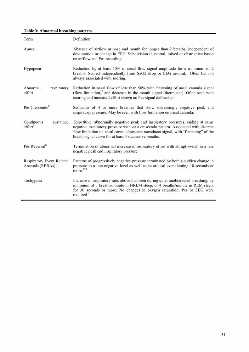
Table 3: Abnormal breathing patterns
Term Definition
Apnea Absence of airflow at nose and mouth for longer than 2 breaths, independent of desaturation or change in EEG. Subdivision in central, mixed or obstructive based on airflow and Pes recording.
Hypopnea Reduction by at least 50% in nasal flow signal amplitude for a minimum of 2 breaths. Scored independently from SaO2 drop or EEG arousal. Often but not always associated with snoring.
Abnormal respiratory effort
Reduction in nasal flow of less than 50% with flattening of nasal cannula signal (flow limitation)7 and decrease in the mouth signal (thermistor). Often seen with snoring and increased effort shown on Pes signal defined as:
Pes Crescendo8 Sequence of 4 or more breathes that show increasingly negative peak end inspiratory pressure. May be seen with flow limitation on nasal cannula.
Continuous sustained effort9
Repetitive, abnormally negative peak end inspiratory pressures, ending at same negative inspiratory pressure without a crescendo pattern. Associated with discrete flow limitation on nasal cannula/pressure transducer signal, with “flattening” of the breath signal curve for at least 4 successive breaths.
Pes Reversal8 Termination of abnormal increase in respiratory effort with abrupt switch to a less negative peak end inspiratory pressure.
Respiratory Event Related Arousals (RERAs)
Patterns of progressively negative pressure terminated by both a sudden change in pressure to a less negative level as well as an arousal event lasting 10 seconds or more.129
Tachypnea Increase in respiratory rate, above that seen during quiet unobstructed breathing, by minimum of 3 breaths/minute in NREM sleep, or 4 breaths/minute in REM sleep, for 30 seconds or more. No changes in oxygen saturation, Pes or EEG were required.11
Page 32
32
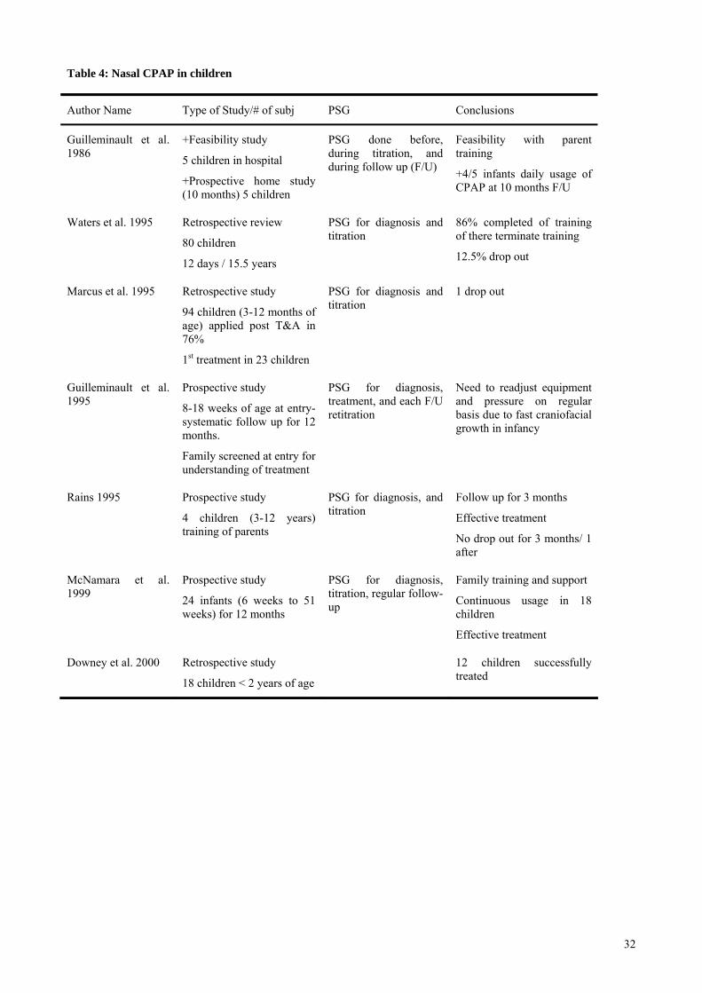
Table 4: Nasal CPAP in children
Author Name Type of Study/# of subj PSG Conclusions
Guilleminault et al. 1986
+Feasibility study
5 children in hospital
+Prospective home study (10 months) 5 children
PSG done before, during titration, and during follow up (F/U)
Feasibility with parent training
+4/5 infants daily usage of CPAP at 10 months F/U
Waters et al. 1995 Retrospective review
80 children
12 days / 15.5 years
PSG for diagnosis and titration
86% completed of training of there terminate training
12.5% drop out
Marcus et al. 1995 Retrospective study
94 children (3-12 months of age) applied post T&A in 76%
1st treatment in 23 children
PSG for diagnosis and titration
1 drop out
Guilleminault et al. 1995
Prospective study
8-18 weeks of age at entry- systematic follow up for 12 months.
Family screened at entry for understanding of treatment
PSG for diagnosis, treatment, and each F/U retitration
Need to readjust equipment and pressure on regular basis due to fast craniofacial growth in infancy
Rains 1995 Prospective study
4 children (3-12 years) training of parents
PSG for diagnosis, and titration
Follow up for 3 months
Effective treatment
No drop out for 3 months/ 1 after
McNamara et al. 1999
Prospective study
24 infants (6 weeks to 51 weeks) for 12 months
PSG for diagnosis, titration, regular follow-up
Family training and support
Continuous usage in 18 children
Effective treatment
Downey et al. 2000 Retrospective study
18 children < 2 years of age
12 children successfully treated
Page 33
33
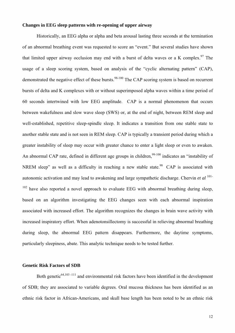
Figure 1: A seven-year old child illustrates many anatomical abnormalities, including asymmetry of the nares, an enlarged septal base, large medial crus, deviation of the septum to the right, and a narrow and high-arched palate. A rapid maxillary distractor has been placed in order to widen the maxillary cavity, decrease the height of the soft palate, and enlarge the bony aspects of the nose
Page 34
34
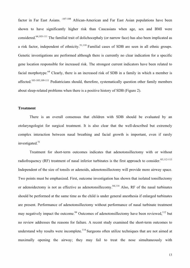
Figure 2: Influences on oro-facial growth
Page 35
35
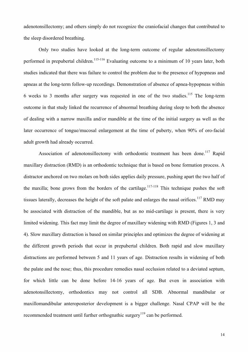
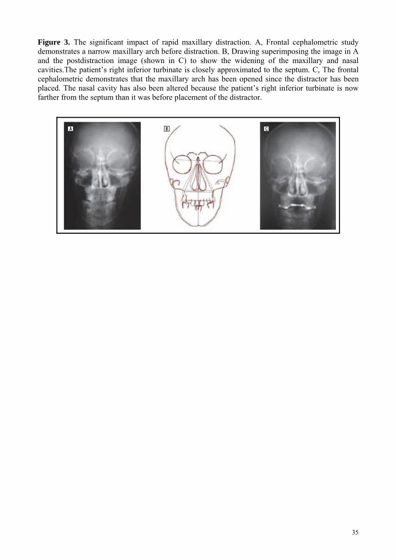
Figure 3. The significant impact of rapid maxillary distraction. A, Frontal cephalometric study demonstrates a narrow maxillary arch before distraction. B, Drawing superimposing the image in A and the postdistraction image (shown in C) to show the widening of the maxillary and nasal cavities.The patient’s right inferior turbinate is closely approximated to the septum. C, The frontal cephalometric demonstrates that the maxillary arch has been opened since the distractor has been placed. The nasal cavity has also been altered because the patient’s right inferior turbinate is now farther from the septum than it was before placement of the distractor.
Page 36
36
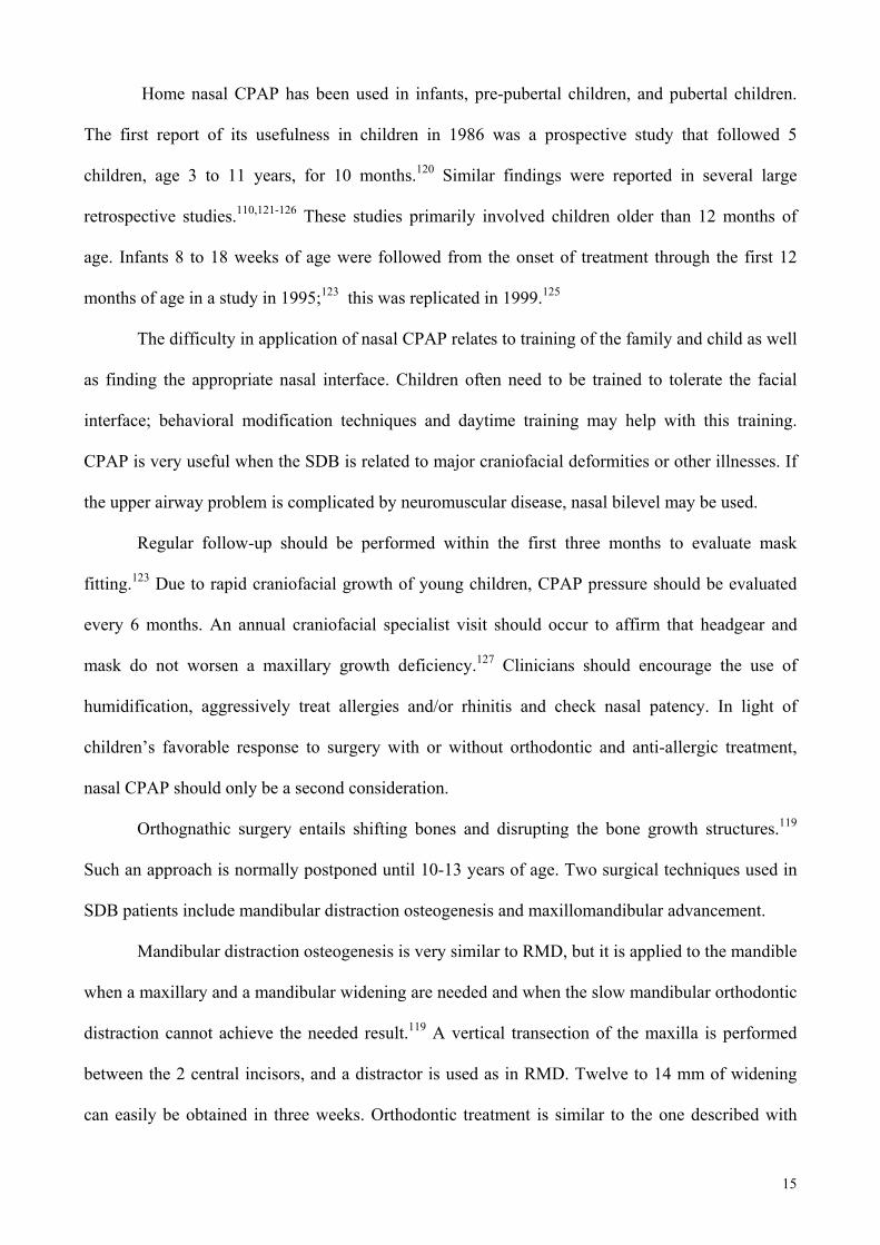
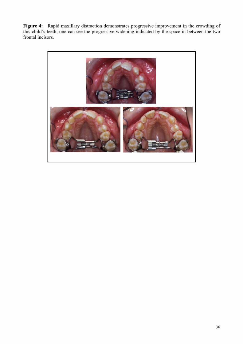
Figure 4: Rapid maxillary distraction demonstrates progressive improvement in the crowding of this child’s teeth; one can see the progressive widening indicated by the space in between the two frontal incisors.