Supportive module 2: Basics of diagnosis, treatment and prevention of major gastroenterological diseases Peptic Ulcer and Other Gastric and Duodenal Ulcers LECTURE IN INTERNAL MEDICINE FOR IV COURSE STUDENTS M. Yabluchansky, L. Bogun, L. Martymianova, O. Bychkova, N. Lysenko, M. Brynza V.N. Karazin National University Medical School’ Internal Medicine Dept.
Transcript
Supportive module 2: Basics of diagnosis, treatment and prevention of major gastroenterological diseases
Peptic Ulcer and Other Gastric and Duodenal Ulcers
LECTURE IN INTERNAL MEDICINE FOR IV COURSE STUDENTS
M. Yabluchansky, L. Bogun, L. Martymianova, O. Bychkova, N. Lysenko, M. Brynza V.N. Karazin National University Medical School’ Internal Medicine Dept.
US MLE TEST
A 41-year-old male who takes NSAIDs regularly for his chronic back pain develops severe abdominal pain worse with eating. Upper endoscopy is performed and the medical student asks the supervising physician how the histological differentiation between a gastric ulcer and erosion is made. Which of the following layers of the gastric mucosa MUST be breached for a lesion to be considered an ulcer? 1. Epithelium, 2. Epithelium, lamina propria, 3. Epithelium, lamina propria, muscularis mucosa, 4. Epithelium, lamina propria, muscularis mucosa, and submucosa, 5. Epithelium, lamina propria, muscularis mucosa, submucosa, and adventitia.
https://www.mommd.com/usmle1to10.shtml
US MLE TEST EXPLANATION The correct answer is 4. Gastric ULCERS, by definition, are a breach in the mucosa with extension into the submucosa or deeper layers. That is, the submucosa must be involved if a lesion is to be called an ulcer. In contrast, EROSIONS are mucosal defects that do NOT penetrate the muscularis mucosa.
Incorrect Answers: 1, 2: These describe an erosion which does not penetrate into the submucosa., 3: To be classified as an ulcer the submucosa must also be penetrated., 5: While deeper tissue can be breached in ulceration, the serosa or adventitia is not required to be disrupted to differentiate an erosion from an ulceration. https://www.mommd.com/usmle1to10.shtml
Definition Peptic ulcer disease (PUD) is a nonmalignant, inflammatory, mucosal lesion of the stomach or proximal duodenum in which Helicobacter pylori (H. pylori), long term use of nonsteroidal anti-inflammatory drugs and/or hypersecretion of hydrochloric acid (parietal cells) and pepsin (mucous cells and chief cells) play major etiopathogenic roles, with symptoms include epigastric discomfort (specifically, pain relieved by food intake or antacids and pain that causes awakening at night or that occurs between meals), loss of appetite, and weight loss with, with serious and life threatening complications in forms of malignancy, bleeding, perforation, peritonitis and gastric outlet obstruction.
http://www.aafp.org/afp/2007/1001/p1005.html
Epidemiology 1
• The lifetime risk for developing a PUD is approximately 10%
• Approximately 500,000 persons develop peptic ulcer disease in the United States each year
• In 70 percent of patients it occurs between the ages of 25 and 64 years
• The incidence of duodenal ulcers has dropped significantly during the last 30 years, possibly as a result of the increasing use of proton pump inhibitors and decreasing rates of H. pylori infection, while the incidence of gastric ulcers has shown a small increase, mainly caused by the widespread use of non-steroidal anti-inflammatory drugs (NSAIDs)
• The annual direct and indirect health care costs of the disease are estimated at about $10 billion.
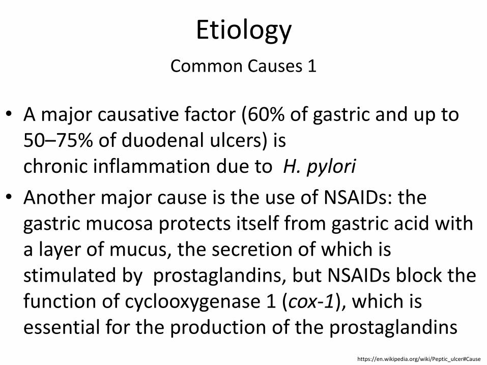
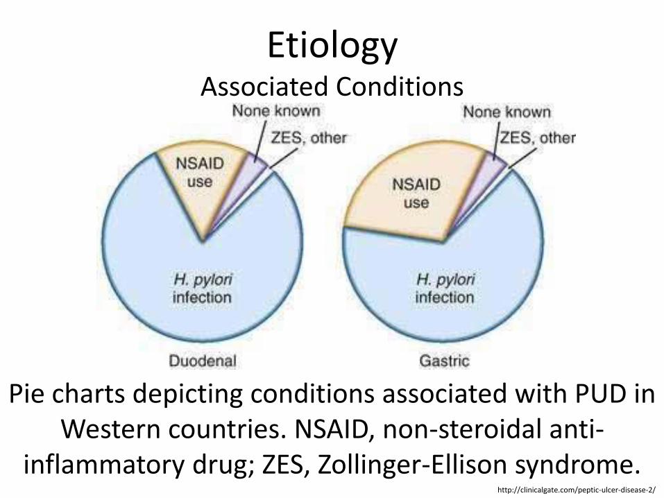
• A major causative factor (60% of gastric and up to 50–75% of duodenal ulcers) is chronic inflammation due to H. pylori
• Another major cause is the use of NSAIDs: the gastric mucosa protects itself from gastric acid with a layer of mucus, the secretion of which is stimulated by prostaglandins, but NSAIDs block the function of cyclooxygenase 1 (cox-1), which is essential for the production of the prostaglandins
https://en.wikipedia.org/wiki/Peptic_ulcer#Cause
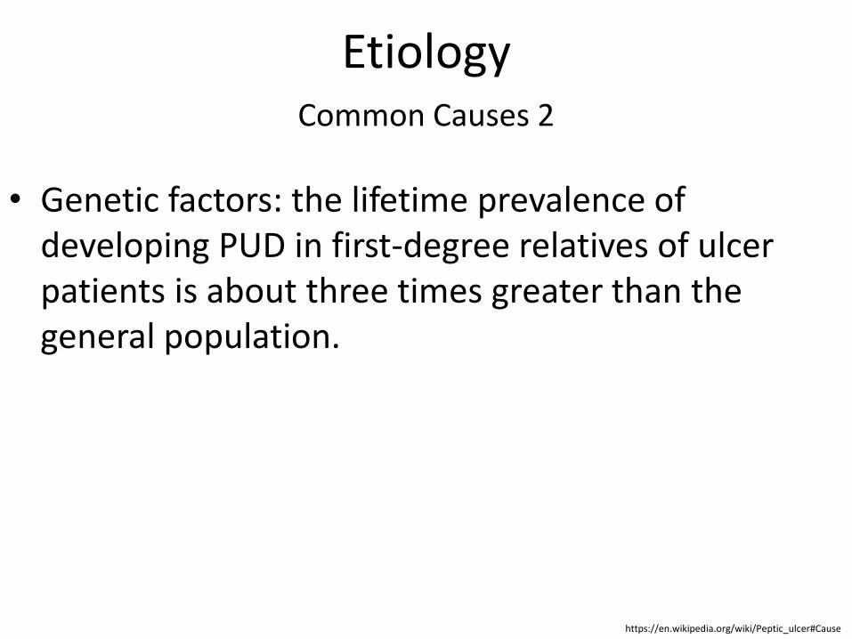
Etiology Common Causes 2
• Genetic factors: the lifetime prevalence of developing PUD in first-degree relatives of ulcer patients is about three times greater than the general population.
https://en.wikipedia.org/wiki/Peptic_ulcer#Cause
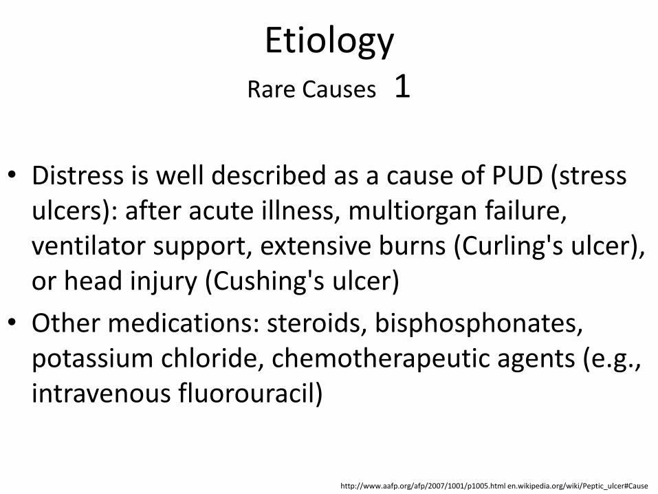
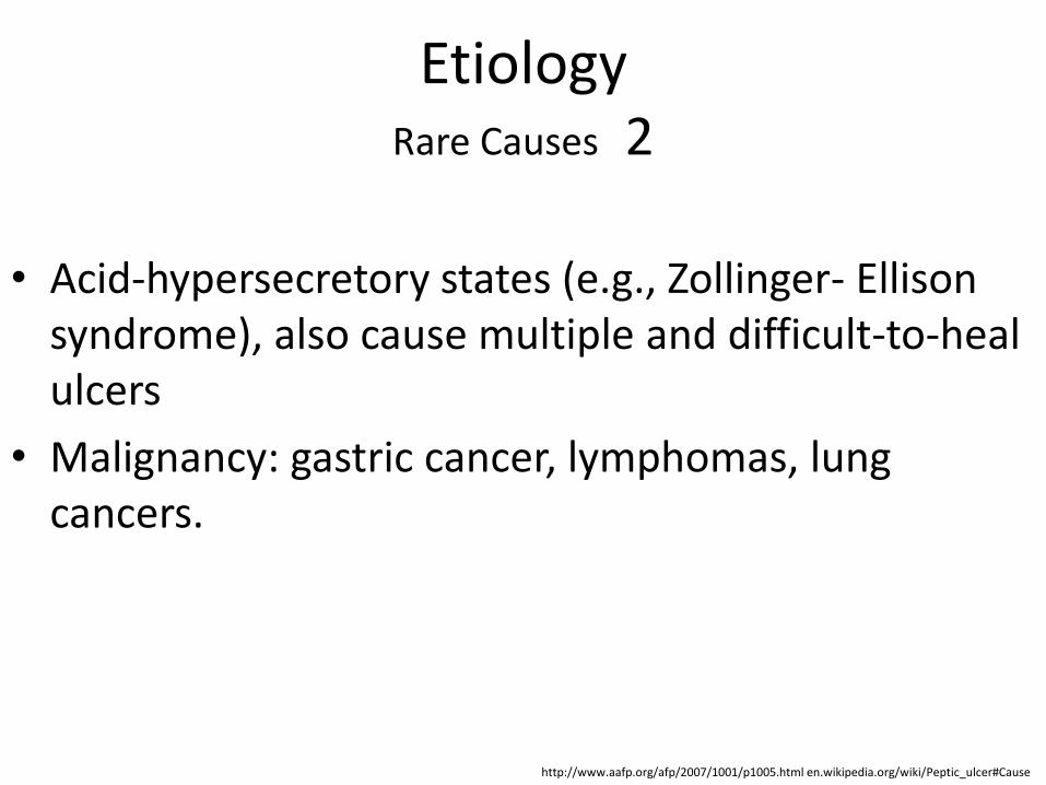
Etiology Rare Causes 1
• Distress is well described as a cause of PUD (stress ulcers): after acute illness, multiorgan failure, ventilator support, extensive burns (Curling's ulcer), or head injury (Cushing's ulcer)
A 52-year-old Caucasian male presents to your office complaining of black, tarry stool. Which of the following possible causes of this patient's presentation is LEAST associated with the development of carcinoma?
The correct answer is 5. In contrast to gastric ulcers, duodenal ulcers are not associated with the development of carcinoma. Incorrect Answers: 1: Barrett's esophagus is metaplasia of the esophagus that frequently progresses to adenocarcinoma., 2: H. pylori infection is a common cause of gastric ulcers and raises a patient's risk of gastric carcinoma., 3: Adenomatous colon polyps are precursors of colorectal cancer., 4: Gastric ulcers are commonly caused by H. pylori, which raises a patient's risk of gastric carcinoma.
https://www.mommd.com/usmle1to10.shtml
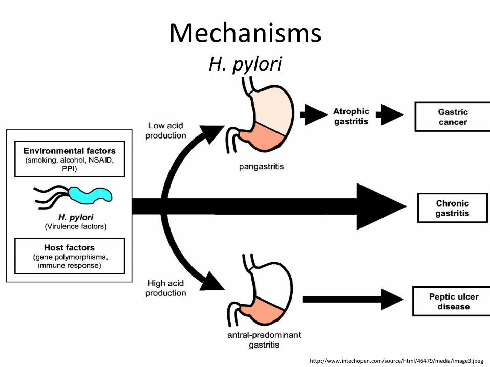
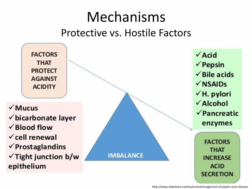
Mechanisms H. pylori 1
• Although H. pylori is present in the gastroduodenal mucosa in most patients with duodenal ulcers, only a minority (10 to 15 percent) of patients with H. pylori infection develop ЗГВ
• H. pylori bacteria adhere to the gastric mucosa; the presence of an outer inflammatory protein and a functional cytotoxin-associated gene island in the bacterial chromosome increases virulence and probably ulcerogenic potential
http://www.aafp.org/afp/2007/1001/p1005.html
Mechanisms H. pylori 2
• Patients with H. pylori infection have increased resting and meal-stimulated gastrin levels and decreased gastric mucus production and duodenal mucosal bicarbonate secretion, all of which favor ulcer formation
• Eradication of H. pylori greatly reduces the incidence of ulcer recurrence from 60% to 5%.
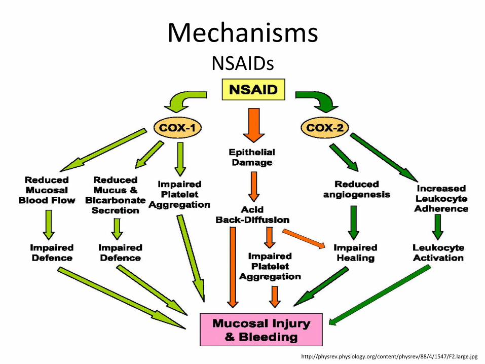
• Topical effects of NSAIDs cause submucosal erosions
• By inhibiting cyclooxygenase, they inhibit the formation of prostaglandins and their protective cyclooxygenase-2–mediated effects (i.e., enhancing gastric mucosal protection by stimulating mucus and bicarbonate secretion and epithelial cell proliferation and increasing mucosal blood flow)
http://www.aafp.org/afp/2007/1001/p1005.html
Mechanisms NSAIDs 2
• Coexisting H. pylori infection increases the likelihood and intensity of NSAID-induced damage
• The annual risk of a life-threatening ulcer-related complication is (1 – 4)% in patients who use NSAIDs long-term, with older patients at the highest risk
• NSAID use is responsible for approximately one half of perforated ulcers.
• The classic triad of Zollinger-Ellison syndrome involves peptic ulcers in unusual locations (i.e., the jejunum), massive gastric acid hypersecretion, and a gastrinproducing islet cell tumor of the pancreas (gastrinoma)
• Gastrinoma in the pancreas appears in approximately 50% of patients
• Another 20% of patients have it in the duodenum and others have it in the stomach, peripancreatic lymph nodes, liver, ovary, or small-bowel mesentery
• Zollinger-Ellison syndrome accounts for only 0.1% of all duodenal ulcer disease.
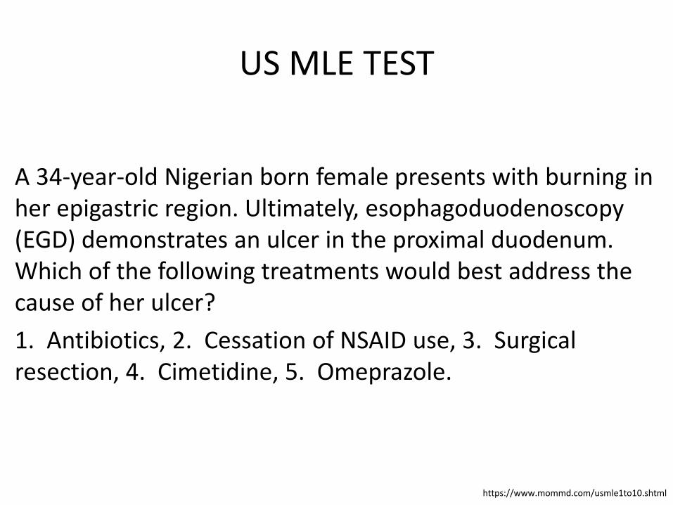
A 34-year-old Nigerian born female presents with burning in her epigastric region. Ultimately, esophagoduodenoscopy (EGD) demonstrates an ulcer in the proximal duodenum. Which of the following treatments would best address the cause of her ulcer?
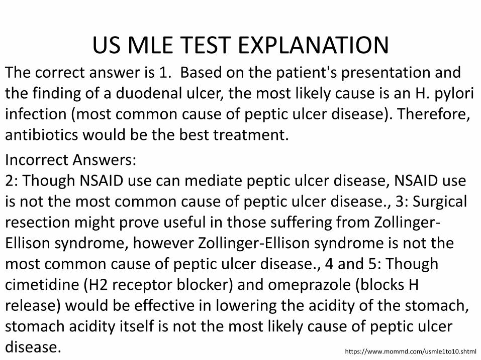
US MLE TEST EXPLANATION The correct answer is 1. Based on the patient's presentation and the finding of a duodenal ulcer, the most likely cause is an H. pylori infection (most common cause of peptic ulcer disease). Therefore, antibiotics would be the best treatment.
Incorrect Answers: 2: Though NSAID use can mediate peptic ulcer disease, NSAID use is not the most common cause of peptic ulcer disease., 3: Surgical resection might prove useful in those suffering from Zollinger-Ellison syndrome, however Zollinger-Ellison syndrome is not the most common cause of peptic ulcer disease., 4 and 5: Though cimetidine (H2 receptor blocker) and omeprazole (blocks H release) would be effective in lowering the acidity of the stomach, stomach acidity itself is not the most likely cause of peptic ulcer disease.
https://www.mommd.com/usmle1to10.shtml
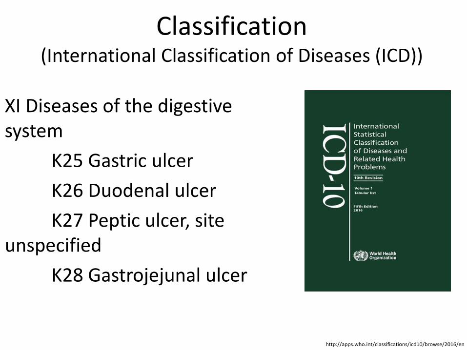
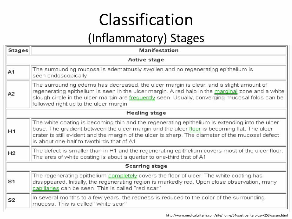
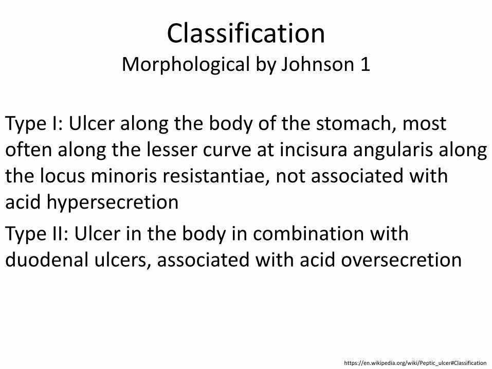
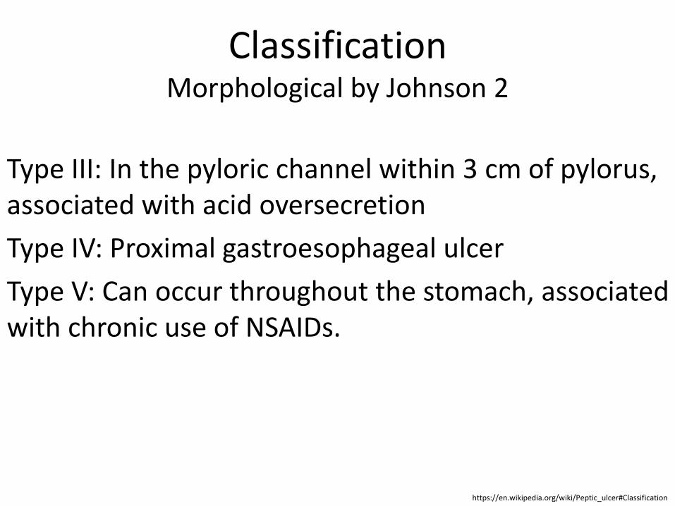
Classification (International Classification of Diseases (ICD))
Type I: Ulcer along the body of the stomach, most often along the lesser curve at incisura angularis along the locus minoris resistantiae, not associated with acid hypersecretion
Type II: Ulcer in the body in combination with duodenal ulcers, associated with acid oversecretion
• Uncomplicated PUD: epigastric and right upper quadrant tenderness, guaiac-positive stool resulting from occult blood loss, melena resulting from acute or subacute gastrointestinal bleeding, succussion splash resulting from partial or complete gastric outlet obstruction
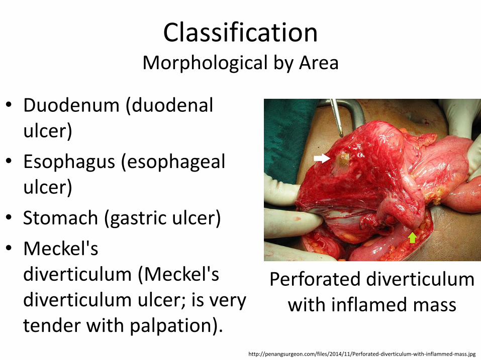
• Patients with perforated PUD usually present with a sudden onset of severe, sharp abdominal pain; as even slight movement can tremendously worsen their pain, these patients assume a fetal position; abdominal examination discloses generalized tenderness, rebound tenderness, guarding, and rigidity
• Patients with perforated PUD may also demonstrate signs and symptoms of septic shock, such as tachycardia, hypotension, and anuria; these indicators of shock may be absent in elderly or immunocompromised patients or in those with diabetes.
US MLE TEST A 45-year-old male patient with a history of recurrent nephrolithiasis and chronic lower back pain presents to the ER with severe, sudden-onset, upper abdominal pain. The patient is febrile, hypotensive, and tachycardic, and is rushed to the OR for exploratory laporotomy. Surgery reveals that the patient has a perforated gastric ulcer. Despite appropriate therapy, the patient expires, and subsequent autopsy reveals multiple ulcers in the stomach, duodenum, and jejunum. The patient had been complaining of abdominal pain and diarrhea for several months but had only been taking ibuprofen for his lower back pain for the past 3 weeks. What is the most likely cause of the patient's presentation?
1. A gastrin-secreting tumor of the pancreas, 2. A vasoactive-intestinal-peptide (VIP) secreting tumor of the pancreas, 3. Cytomegalovirus infection, 4. H. pylori infection, 5. Chronic NSAID use.
https://www.mommd.com/usmle1to10.shtml
US MLE TEST EXPLANATION
The correct answer is 1. This patient with multiple, severe ulcers from his stomach to his small bowel most likely had Zollinger-Ellison syndrome (ZES), which is caused by a gastrin-secreting tumor. Incorrect Answers: 2: VIP-secreting tumors may occur in MEN1, and often present with nausea, diarrhea, and abdominal pain. However, VIP suppresses gastric acid production, and peptic ulcers are not typically present., 3: Cytomegalovirus is associated with peptic ulcer formation, but does not typically cause multiple or jejunal ulcers., 4: H. pylori is one of the leading causes of peptic ulcers, but does not typically cause ulcers reaching into the jejunum., 5: Chronic NSAID use typically causes gastritis. Though it can cause peptic ulcer disease, it would not explain the severity of this patient's disease.
• Obstruction (persist or recur despite endoscopic balloon dilation)
• Penetration
• Perforation
https://www.ncbi.nlm.nih.gov/pubmed/22095016
Complications 2 • Peritonitis
• Gastric malignancy
Endoscopy is the treatment of choice for bleeding ulcers.
Transcatheter arterial embolization is the treatment of choice for duodenal ulcers re-bleeding after
therapeutic endoscopy or surgery.
https://www.ncbi.nlm.nih.gov/pubmed/22095016
Diagnosis 1 • The diagnosis is mainly established based on the
characteristic symptoms
• Confirmation of the diagnosis is made with the help of tests such as endoscopies or barium contrast x-rays; the tests are typically ordered if the symptoms do not resolve after a few weeks of treatment, or when they first appear in a person who is over age 45 or who has other symptoms such as weight loss, because stomach cancer can cause similar symptoms
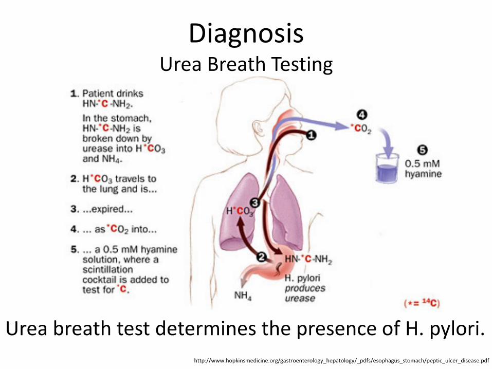
Diagnosis 2 • The diagnosis of Helicobacter pylori can be made by
urea breath test; direct culture from an esophagogastroduodenoscopy (EGD)biopsy specimen; direct detection of urease activity in a biopsy specimen (rapid urease test); measurement of antibody levels in blood; stool antigen test; histological examination and staining of an EGD biopsy.
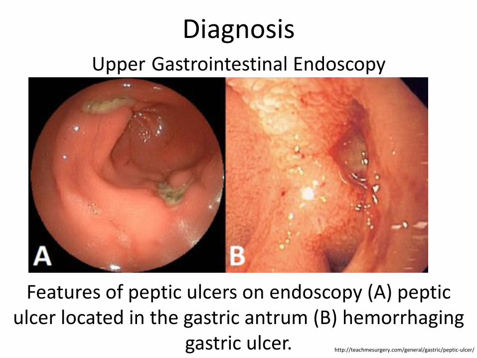
• Most gastric ulcers tend to occur at the junction of the fundus and antrum, along the lesser curvature; benign ulcers tend to have a smooth, regular, rounded edge with a flat smooth base and surrounding mucosa that shows radiating folds; malignant ulcers usually have irregular heaped-up or overhanging margins; the ulcerated mass often protrudes into the lumen, and the folds surrounding the ulcer crater are often nodular and irregular
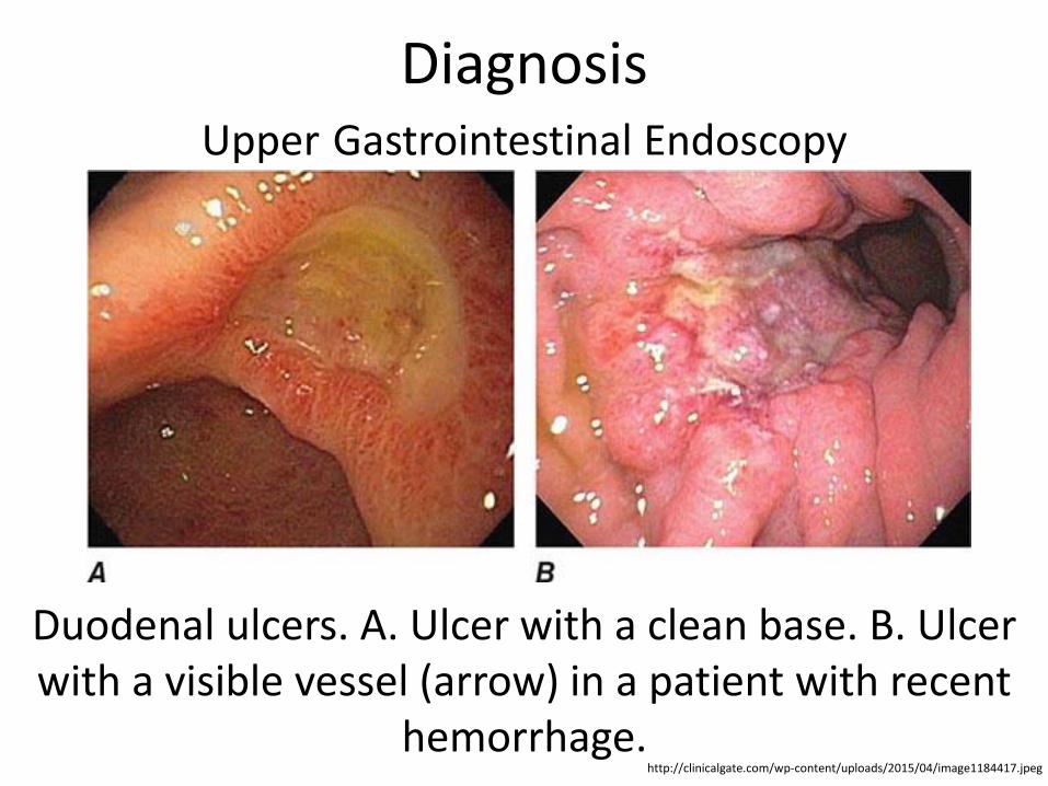
Duodenal ulcers. A. Ulcer with a clean base. B. Ulcer with a visible vessel (arrow) in a patient with recent
hemorrhage.
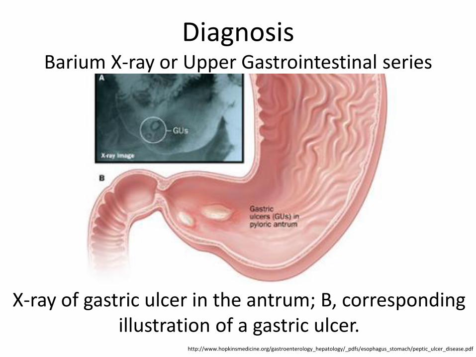
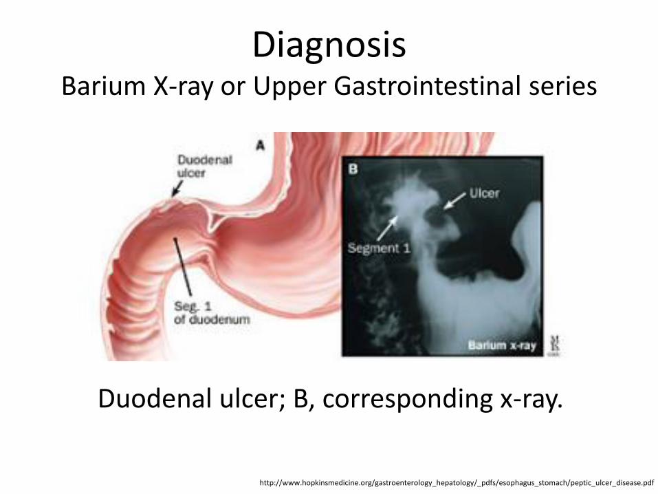
Diagnosis Barium X-ray or Upper Gastrointestinal series 1
• Barium x-ray or upper GI series is a widely available and accepted method to establish a diagnosis of peptic ulcer in the stomach or duodenum
• Though less invasive than endoscopy, the barium x-ray is limited by being less sensitive and accurate at defining mucosal disease, or distinguishing benign from malignant ulcer disease
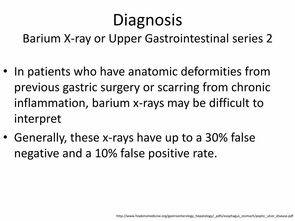
Diagnosis Barium X-ray or Upper Gastrointestinal series 2
• In patients who have anatomic deformities from previous gastric surgery or scarring from chronic inflammation, barium x-rays may be difficult to interpret
• Generally, these x-rays have up to a 30% false negative and a 10% false positive rate.
Urea breath test determines the presence of H. pylori. http://www.hopkinsmedicine.org/gastroenterology_hepatology/_pdfs/esophagus_stomach/peptic_ulcer_disease.pdf
Differential Diagnosis 1 • Acute cholangitis
• Acute coronary syndrome
• Acute and chronic gastritis
• Cholecystitis and biliary colic (in emergency medicine)
• Diverticulitis
• Emergent treatment of gastroenteritis
• Esophageal rupture and tears in emergency medicine
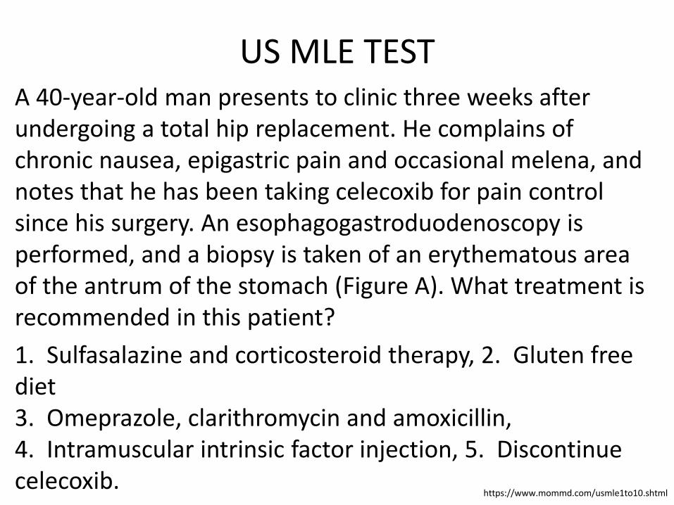
US MLE TEST A 40-year-old man presents to clinic three weeks after undergoing a total hip replacement. He complains of chronic nausea, epigastric pain and occasional melena, and notes that he has been taking celecoxib for pain control since his surgery. An esophagogastroduodenoscopy is performed, and a biopsy is taken of an erythematous area of the antrum of the stomach (Figure A). What treatment is recommended in this patient?
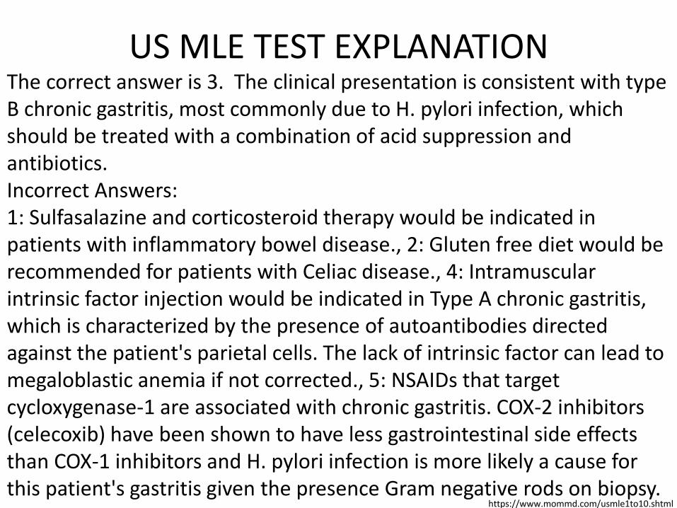
US MLE TEST EXPLANATION The correct answer is 3. The clinical presentation is consistent with type B chronic gastritis, most commonly due to H. pylori infection, which should be treated with a combination of acid suppression and antibiotics. Incorrect Answers: 1: Sulfasalazine and corticosteroid therapy would be indicated in patients with inflammatory bowel disease., 2: Gluten free diet would be recommended for patients with Celiac disease., 4: Intramuscular intrinsic factor injection would be indicated in Type A chronic gastritis, which is characterized by the presence of autoantibodies directed against the patient's parietal cells. The lack of intrinsic factor can lead to megaloblastic anemia if not corrected., 5: NSAIDs that target cycloxygenase-1 are associated with chronic gastritis. COX-2 inhibitors (celecoxib) have been shown to have less gastrointestinal side effects than COX-1 inhibitors and H. pylori infection is more likely a cause for this patient's gastritis given the presence Gram negative rods on biopsy.
https://www.mommd.com/usmle1to10.shtml
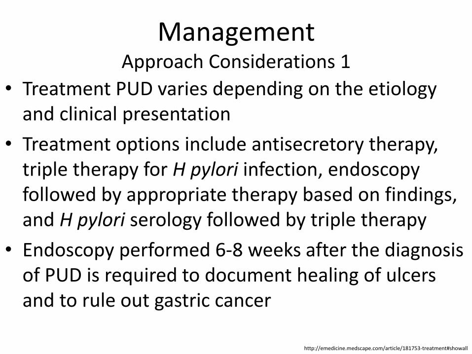
Management Approach Considerations 1
• Treatment PUD varies depending on the etiology and clinical presentation
• Treatment options include antisecretory therapy, triple therapy for H pylori infection, endoscopy followed by appropriate therapy based on findings, and H pylori serology followed by triple therapy
• Endoscopy performed 6-8 weeks after the diagnosis of PUD is required to document healing of ulcers and to rule out gastric cancer
• Documentation of H pylori cure with a noninvasive test, such as the urea breath test or fecal antigen test, is appropriate in patients with complicated ulcers.
• Perform endoscopy early in patients older than 45-50 years and in patients with associated so-called alarm symptoms, such as dysphagia, recurrent vomiting, weight loss, or bleeding.
• If an ulcer is seen (endoscopy) and the patient is infected with H. pylori, treatment for the infection is started, followed by 4 to 8 weeks of treatment with a proton pump inhibitor
• If an ulcer is seen but H. pylori is not present, patients are usually treated with PPI for 8 weeks
• If no ulcer is seen and the patient is not infected with H. pylori, the first treatment attempt will usually be with PPI
• If the PPI dose is not effective, occasionally doubling the dose will relieve symptoms
• Those who do not respond to treatment, or whose symptoms return relatively quickly, will often need an upper endoscopy
• Using antibiotics when there is no clear evidence of ulcers will lead to unnecessary antibiotic prescriptions and increase the risk for side effects.
• Reported cure rates for H. pylori range from 70 - 90% after antibiotic treatment
• The standard treatment regimen uses two antibiotics (clarithromycin, amoxicillin) and a PPI (omeprazole, lansoprazole, esomeprazole, and rabeprazole)
• PPIs are important for all types of peptic ulcers, and are a critical partner in antibiotic regimens; they reduce acidity in the intestinal tract, and increase the ability of antibiotics to destroy H. pylori
• Active ulcers associated with NSAID use are treated with an appropriate course of PPI therapy and the cessation of NSAIDs
• For patients with a known history of ulcer and in whom NSAID use is unavoidable, the lowest possible dose and duration of the NSAID and co-therapy with a PPI or misoprostol are recommended.
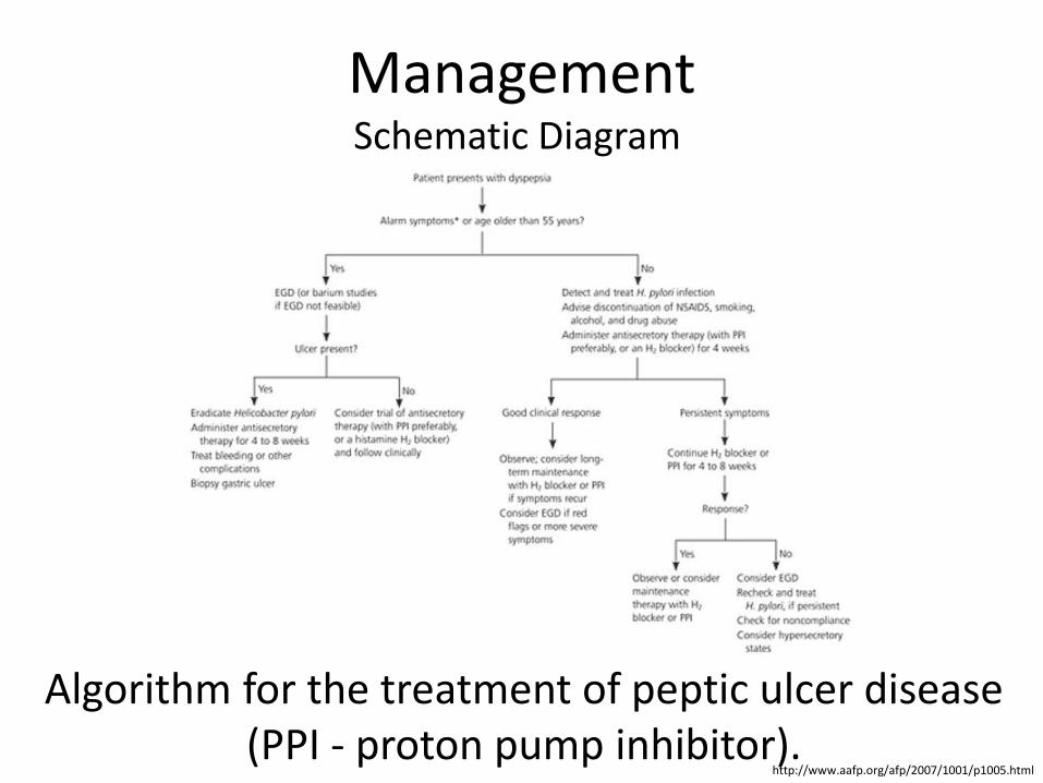
Algorithm for the treatment of peptic ulcer disease (PPI - proton pump inhibitor).
Management Refractory Ulcers 1
• Refractory PUD (i.e., disease that fails to heal after eight to 12 weeks of therapy) may be caused by persistent or resistant H. pylori infection, continued NSAID use, giant ulcers requiring longer healing time, cancer, tolerance of or resistance to medications, or hypersecretory states
• Therapy for refractory PUD involves treatment of the underlying cause and prolonged administration of standard doses of a proton pump inhibitor
http://www.aafp.org/afp/2007/1001/p1005.html
Management Refractory Ulcers 2
• Up to 25% of patients with gastric ulcers who continue to take NSAIDs may require PPI therapy for longer than eight weeks.
http://www.aafp.org/afp/2007/1001/p1005.html
Management Surgery 1
• Surgery is indicated in patients who are intolerant of medications or do not comply with medication regimes, and those at high risk of complications (e.g., patients dependent on steroids or NSAIDs, those with giant ulcer, those with ulcers that fail to heal with treatment)
• Surgery should also be considered for patients who have a relapse during maintenance treatment or who have had multiple courses of medications
http://www.aafp.org/afp/2007/1001/p1005.html
Management Surgery 2
• Surgical options for duodenal ulcers include truncal vagotomy and drainage (pyloroplasty or gastrojejunostomy), selective vagotomy (preserving the hepatic and celiac branches of the vagus) and drainage, highly selective vagotomy, or partial gastrectomy
• The indications for urgent surgery include failure to achieve hemostasis endoscopically, recurrent bleeding despite endoscopic attempts at achieving hemostasis, and perforation.
http://www.aafp.org/afp/2007/1001/p1005.html
US MLE TEST
A 41-year-old female complains of frequent diarrhea and abdominal pain between meals. Endoscopy reveals a duodenal ulcer distal to the duodenal bulb. CT scan of the abdomen demonstrates a pancreatic mass, and subsequent tissue biopsy of the pancreas reveals a malignant islet cell tumor. Which of the following hormones is likely to be markedly elevated in this patient?
The correct answer is 1. The patient is suffering from Zollinger-Ellison (ZE) syndrome due to a pancreatic gastrinoma. Excess gastrin secretion leads to excess gastric acid production in the stomach, often resulting in peptic ulcer disease.
Incorrect Answers: 2: Cholecystokinin regulates gallbladder contraction, gastric emptying, and pancreatic secretion. It is secreted by I cells in the duodenum and jejunum in response to fatty acids and amino acids., 3: Secretin is secreted by duodenal S cells. It decreases gastric acid secretion and increases pancreatic bicarbonate secretion., 4: Vasoactive intestinal peptide increases intestinal water and electrolyte secretion and prompts relaxation of intestinal smooth muscle., 5: Motilin is secreted by the small intestine and is an important regulator of peristalsis.
https://www.mommd.com/usmle1to10.shtml
Prognosis 1
• Typically, duodenal ulcers heal in 4 weeks and gastric ulcers in 8 weeks with PPI therapy
• For patients with peptic ulcers caused by H pylori, the prognosis after H pylori eradication is good: less than 20% will experience recurrence, and this is lower for duodenal ulcers than for gastric ulcers
• H pylori eradication is also beneficial in those with complicated ulcer disease


![Role of dietary polyphenols in the management of peptic ulcer · 2017. 4. 26. · Peptic ulcer disease is a multifactorial and complex disease involving gastric and duodenal ulcers[1,2].](https://static.documents.pub/doc/80x56/602db182f8cf5c32fc5bf263/role-of-dietary-polyphenols-in-the-management-of-peptic-ulcer-2017-4-26-peptic.jpg)


